Bilateral Retinoblastoma Joseph Junewick, MD FACR
|
|
|
- Stephany Ferguson
- 5 years ago
- Views:
Transcription
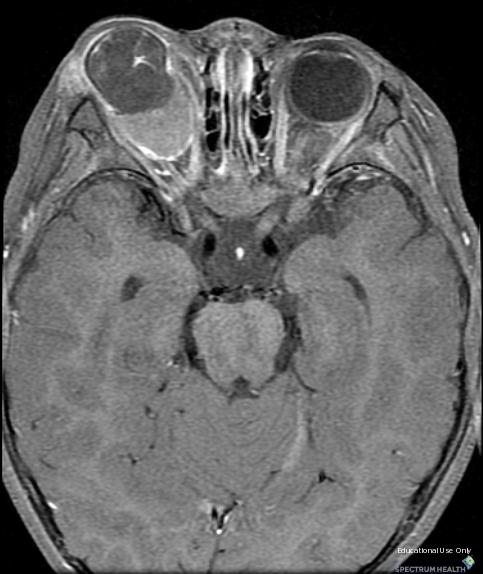
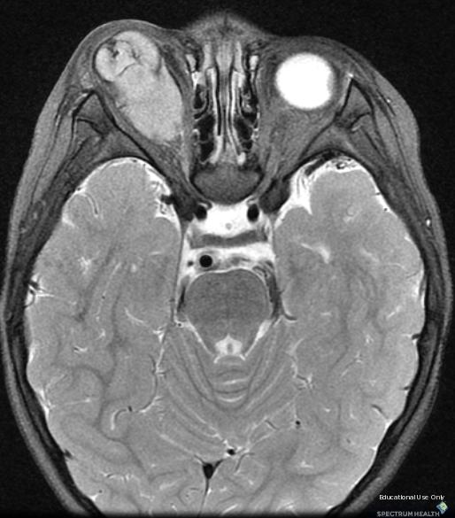
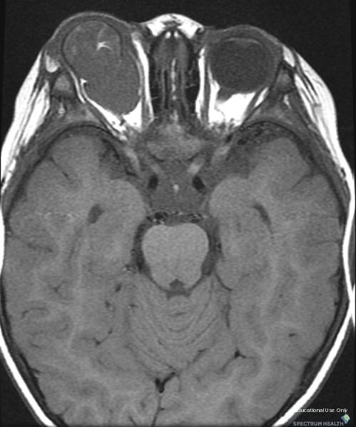
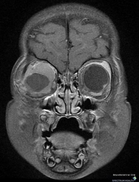
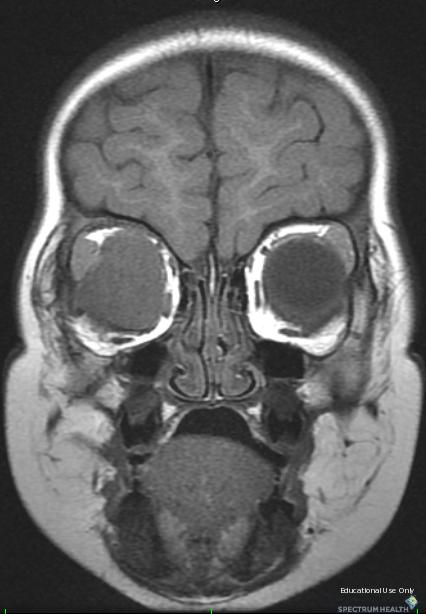
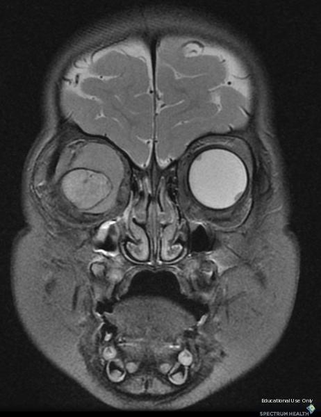
1 Bilateral Retinoblastoma Joseph Junewick, MD FACR 06/11/2010 History 17 month old adopted female with proptosis. Diagnosis Bilateral Retinoblastoma Discussion Retinoblastoma is the most common pediatric intraocular neoplasm. It is a highly malignant tumor of the primitive neural retina. Modern cure rates are greater than 90%; extraocular extension occurs in less than 10% of patients and is associated with a considerably higher mortality rate. Retinoblastoma spreads by direct extension into the orbit along scleral emissary vessels, invasion of the optic nerve, and dispersion through the cerebrospinal fluid. The growth pattern of retinoblastoma can be subdivided into three types: endophytic, exophytic, and combined endophytic and exophytic. Endophytic tumors arise from inner layers of the retina and grow into the vitreous. Exophytic tumors start in the outer layers and growth is in the subretinal space. Beside enucleation, conservative treatment methods, such as thermochemotherapy, radioactive plaque therapy, cryotherapy, laser photocoagulation, external-beam radiation therapy, and tumor reduction chemotherapy, can be applied. The treatment of retinoblastoma depends on several parameters: tumor volume and localization, intraocular tumor extension, extraocular stage of disease, and laterality (side) of tumor. Magnetic resonance imaging is preferred for staging and should include intraocular (choroid, sclera, prelaminar optic nerve), extraocular (postlaminar optic nerve or orbital invasion) and intracranial (pineal and leptomeningeal metastases) evaluation for tumor spread. The reported predictors for metastatic retinoblastoma are invasion of the optic nerve, invasion of the choroid, and orbital involvement. Anterior chamber involvement is associated with an increased risk for metastases. Optic nerve invasion is reported to be present more often in patients with exophytic retinoblastoma, tumor thickness of 15 mm or larger, and vitreous hemorrhage. CT should not be routinely performed in hereditary retinoblastoma since cumulative radiation effects increases the risk for the development of second primary tumors. Findings MR-Axial and coronal T1, fat-suppressed T2 and fat-suppressed postgadolinium T1 images reveal bilateral intraocular masses. Intraorbital extension on the right is predominantly retrobulbar resulting in proptosis. The right oculus is small and associated with retinal detachment. Reference degraaf P, Barkoff F, Moll AC, Imhof SM, Knol DL, van der Valk P, Castelijns JA. Retinoblastoma: MR imaging parameters in detection of tumor extent. Radiology (2005); 235:
2
3
4
5
6
7
8 Sponsored By Disclaimer This teaching site is partially funded by an educational grant from GE Healthcare and Advanced Radiology Services, PC. The material on this site is independently controlled by Advanced Radiology Services, PC, and GE Healthcare and Spectrum Health have no influence over the content of this site Content Download Agreement The cases and images on this website are owned by Spectrum Health. Permission is granted (for nonprofit educational purposes) to download and print materials to distribute for the purpose of facilitating the education of health professionals. The authors retain all rights to the material and users are requested to acknowledge the source of the material. Site Disclaimer This site is developed to reach healthcare professionals and medical students. Nothing this site should be considered medical advice. Only your own doctor can help you make decisions about your medical care. If you have a specific medical question or are seeking medical care, please contact your physician. The information in this website is provided for general medical education purposes only and is not meant to substitute for the independent medical judgment of a physician relative to diagnostic and treatment options of a specific medical condition. The viewpoints expressed in these cases are those of the authors. They do not represent an endorsement. In no event will Advanced Radiology Associates, PC, Spectrum Health Hospitals (Helen Devos Children's Hospital) or GE Healthcare be liable for any decision made or action taken in reliance upon the information provided through this website.
Paraspinal Venous Malformation Joseph Junewick, MD FACR
 Paraspinal Venous Malformation Joseph Junewick, MD FACR 06/04/2010 History 2 year old with history of fall. Rule out spinal injury. Diagnosis Paraspinal Venous Malformation Additional Clinical CT of the
Vein of Galen Malformation Joseph Junewick, MD FACR
 Vein of Galen Malformation Joseph Junewick, MD FACR 04/14/2018 History Midline cystic intracranial mass on prenatal ultrasound. Diagnosis Vein of Galen Malformation Discussion In normal fetal development,
Tuberculous Meningitis Joseph Junewick, MD FACR
 Tuberculous Meningitis Joseph Junewick, MD FACR 08/11/2010 History 14 month old with fever and increasing lethargy. Diagnosis Tuberculous Meningitis Additional Clinical Grandmother with active tuberculosis.
Chiari III Joseph Junewick, MD FACR
 Chiari III Joseph Junewick, MD FACR 07/02/2010 History Newborn with suboccipital mass. Diagnosis Chiari III Additional Clinical Surgery-Skin covered suboccipital cystic mass confined by the dura. Pathology-Leptomeningeal
Pleural Empyema Joseph Junewick, MD FACR
 Pleural Empyema Joseph Junewick, MD FACR 03/19/2010 History Teenager with persistent fever and cough. Pneumonia diagnosed 1 week ago. Diagnosis Pleural Empyema Additional Clinical Surgery-Clear fluid with
Pituitary Macroadenoma Joseph Junewick, MD FACR
 Pituitary Macroadenoma Joseph Junewick, MD FACR 08/13/2010 History 12 year old female with headache and visual disturbance. Diagnosis Pituitary Macroadenoma Additional Clinical Markedly elevated growth
Radiation Pneumonitis Joseph Junewick, MD FACR
 Radiation Pneumonitis Joseph Junewick, MD FACR 03/19/2010 History 16 year old with history of relapsed stage IV-A Hodgkin disease. Prior pulmonary involvement was irradiated. Diagnosis Radiation Pneumonitis
Spinal LCH Joseph Junewick, MD FACR
 Spinal LCH Joseph Junewick, MD FACR 05/16/2009 History 16 year old female with multiply recurrent Langerhans Cell Histiocytosis now with severe left sided neck pain. Diagnosis Langerhans Cell Histiocytosis
Atlanto-occipital Dislocation Joseph Junewick, MD FACR
 Atlanto-occipital Dislocation Joseph Junewick, MD FACR 09/23/2009 History 12 year old male restrained back seat passenger in a car hit by a snowplow. Diagnosis Atlanto-occipital Dislocation Discussion
Presacral Neuroblastoma Joseph Junewick, MD FACR
 Presacral Neuroblastoma Joseph Junewick, MD FACR 01/12/2010 History 16 month old male with irritability. Diagnosis Presacral Neuroblastoma Additional Clinical Initial US to evaluate for intussusception
Ulcerative Colitis Joseph Junewick, MD FACR
 Ulcerative Colitis Joseph Junewick, MD FACR 06/04/2010 History 16 year old male with hematochezia and anemia. Diagnosis Ulcerative Colitis Additional Clinical History of ulcerative colitis. Discussion
Term Hypoxic Ischemic Injury Joseph Junewick, MD FACR
 Term Hypoxic Ischemic Injury Joseph Junewick, MD FACR 08/11/2010 History Term infant with perinatal distress and attempted forceps delivery. Diagnosis Term Hypoxic Ischemic Injury Discussion Encephalopathy
Transverse Dural Sinus Thrombosis Joseph Junewick, MD FACR
 Transverse Dural Sinus Thrombosis Joseph Junewick, MD FACR 03/19/2010 History Child with headache and otomastoiditis. Diagnosis Dural venous thrombosis secondary to mastoiditis Discussion The cerebral
Chance Fracture Joseph Junewick, MD FACR
 Chance Fracture Joseph Junewick, MD FACR 08/02/2010 History Restrained teenager involved in motor vehicle accident. Diagnosis Chance Fracture (Hyperflexion-Distraction Injury) Discussion Chance-type spinal
Diskitis Joseph Junewick, MD FACR
 Diskitis Joseph Junewick, MD FACR 09/20/2010 History 2 year old with fever, back pain and elevated sedimentation rate. Diagnosis Diskitis Discussion Diskitis is an inflammatory process of the intervertebral
Neuroblastoma Joseph Junewick, MD FACR
 Neuroblastoma Joseph Junewick, MD FACR 03/18/2011 History 15 month old with anemia. Diagnosis Neuroblastoma Discussion Neuroblastic tumors derive from primordial neural crest cells destined for sympathetic
Retroperitoneal Teratoma Heather Borders, MD
 Retroperitoneal Teratoma Heather Borders, MD 03/04/2012 History Newborn with congenitally diagnosed mass. No other clinical symptoms. Diagnosis Retroperitoneal Teratoma; Immature teratoma, grade 1, with
Scrofula Joseph Junewick, MD FACR
 Scrofula Joseph Junewick, MD FACR 06/20/2012 History 4 year old male with refractory cervical adenopathy Diagnosis Scrofula Additional Clinical Positive PPD skin test. Discussion Scrofula refers to tuberculous
Testicular Microlithiasis related to McCune-Albright Syndrome Joseph Junewick, MD FACR
 Testicular Microlithiasis related to McCune-Albright Syndrome Joseph Junewick, MD FACR 04/25/2010 History 12 year old with McCune-Albright syndrome. Diagnosis Testicular Microlithiasis related to Mcune-Albright
Fallopian tube torsion and paratubal cyst Heather Borders, MD
 Fallopian tube torsion and paratubal cyst Heather Borders, MD 01/24/2012 History 13 year old female with one week of pelvic pain Diagnosis Fallopian tube torsion with paratubal cyst Additional Clinical
Thymic Involvement in Chronic Granulomatous Disease of Childhood
 Thymic Involvement in Chronic Granulomatous Disease of Childhood Joseph Junewick, MD FACR 07/16/2010 History 3 year old male with multifocal osteomyelitis. Diagnosis Thymic Involvement in Chronic Granulomatous
Posterior Slipped Capital Femoral Epiphysis Joseph Junewick, MD FACR
 Posterior Slipped Capital Femoral Epiphysis Joseph Junewick, MD FACR 08/11/2010 History 6 year old male with intermittent hip pain for several months, acutely worsened after climbing the sand dunes. Diagnosis
Gastrointestinal Hemangiomatosis Joseph Junewick, MD FACR
 Gastrointestinal Hemangiomatosis Joseph Junewick, MD FACR 03/06/2010 History 3 month old with protuberant abdomen and anemia. Diagnosis Gastrointestinal Hemangiomatosis Discussion Gastrointestinal hemangiomatosis
Early detection of Retinoblastoma in children. Max Mantik
 Early detection of Retinoblastoma in children Max Mantik Introduction The most common primary intraocular malignancy of childhood 10 to 15 % of cancers that occur within the first year of life Typical
A RESOURCE MANUAL MANAGEMENT RETINOBLASTOMA LOW & MIDDLE RESOURCE SETTINGS
 A RESOURCE MANUAL FOR THE MANAGEMENT OF RETINOBLASTOMA IN LOW & MIDDLE RESOURCE SETTINGS UPDATED SEPTEMBER 2017 1 CONTENTS PAGE INTRODUCTION 3 SERVICE LEVEL for Rb MANAGEMENT 4 SCREENING 5 EARLY DIAGNOSIS
The Egyptian Journal of Hospital Medicine (October 2018) Vol. 73 (9), Page
 Vol. 73 (9), Page") The Egyptian Journal of Hospital Medicine (October 2018) Vol. 73 (9), Page 7412-7417 Mohammad Ahmad Wahdan 1, Abd Allah El Hussainy Shaleel 1, Hossam El Dein Ahmed El Zomor 2, Hossam El Din Hassan El Sayed
The Egyptian Journal of Hospital Medicine (October 2018) Vol. 73 (9), Page 7412-7417 Mohammad Ahmad Wahdan 1, Abd Allah El Hussainy Shaleel 1, Hossam El Dein Ahmed El Zomor 2, Hossam El Din Hassan El Sayed
Retinoblastoma: A Review of Current Treatment Strategies
 Retinoblastoma: A Review of Current Treatment Strategies ABSTRACT: Since the last review of retinoblastoma therapies in the 15 years ago, there has been a significant shift in the approach to treating
Tiffany L. Kruger, D.O. Children s Hospital of Michigan Wayne State University/Kresge Eye Institute
 Pediatric Cases Nt Not To Be Missed Tiffany L. Kruger, D.O. Pediatric Ophthalmology Fellow Children s Hospital of Michigan Wayne State University/Kresge Eye Institute Case Presentation CC: Left eye turns
Pediatric Ocular Sonography
 Pediatric Ocular Sonography Cicero J Torres A Silva, MD Associate Professor of Radiology 2016 SPR Pediatric Ultrasound Course Yale University School of Medicine None Disclosures Objectives of Presentation
Retinoblastoma. Protocol applies to retinoblastoma only.
 Retinoblastoma Protocol applies to retinoblastoma only. Protocol revision date: January 2005 Based on AJCC/UICC TNM, 6 th edition Procedures Cytology (No Accompanying Checklist) Biopsy (No Accompanying
Financial Disclosures
 Retinoblastoma Management: Update Jesse L. Berry, MD Associate Director, Ocular Oncology Service Associate Program Director USC/CHLA, Keck School of Medicine Financial Disclosures Research Support: Bright
CATARACT IN RETINOBLASTOMA
 CATARACT IN RETINOBLASTOMA P R O F. D R A D E L A L E I E L D I N, M D R E S E A E R C H I N S T I T U T E O F O P H T H A L M O L O G Y H E A D O F P E D I A T R I C O P H T H A L M O L O G Y A N D O
Outline. Brief history and principles of ophthalmic ultrasound. Types of ocular ultrasound. Examination techniques. Types of Ultrasound
 Ultrasound and Intraocular Tumors 2015 Ophthalmic Photographers' Society Mid-Year Program Cagri G. Besirli MD, PhD Kellogg Eye Center University of Michigan Outline Brief history and principles of ophthalmic
Financial Disclosures. The Eye in Neoplastic Disease. Course Goal. We wish to acknowledge and thank: Tumor Definition
 The Eye in Neoplastic Disease Carlo J. Pelino, OD, FAAO Joseph J. Pizzimenti, OD, FAAO cpelino@salus.edu pizzimen@uiwtx.edu Financial Disclosures! Speakers have no relevant financial relationships to declare.
Michael P. Blair, MD Retina Consultants, Ltd Libertyville/Des Plaines, Illinois Clinical Associate University of Chicago 17 October 2015
 Michael P. Blair, MD Retina Consultants, Ltd Libertyville/Des Plaines, Illinois Clinical Associate University of Chicago 17 October 2015 So What Parts of the Eye Retina are Affected by VHL Neural tissue
NEW YORK UNIVERSITY SCHOOL OF MEDICINE DEPARTMENT OF OPHTHALMOLOGY EDUCATIONAL OBJECTIVES AND GOALS
 NEW YORK UNIVERSITY SCHOOL OF MEDICINE DEPARTMENT OF OPHTHALMOLOGY EDUCATIONAL OBJECTIVES AND GOALS Revision Date: 6/30/06 Distribution Date: 7/6/06 The Department of Ophthalmology at the NYU Medical Center
Retinoblastoma. Retinoblastoma
 Retinoblastoma Authors: Ayda G. Nambayan, DSN, RN, St. Jude Children s Research Hospital Erin Gafford, Pediatric Oncology Education Student, St. Jude Children s Research Hospital; Nursing Student, School
What is so special about Retinoblastoma?
 Definition Retinoblastoma is a primary malignant neoplasm of the retina that arises from immature retinal cells. It is the most common primary intraocular malignancy of childhood. What is so special about
INDIAN COUNCIL OF MEDICAL RESEARCH INDIAN RETINOBLASTOMA GROUP
 NATIONAL RETINOBLASTOMA REGISTRY INDIAN COUNCIL OF MEDICAL RESEARCH INDIAN RETINOBLASTOMA GROUP Centre Code 01. Dr. Rajendra Parasad Centre for Ophthalmic Sciences 02. L.V. Prasad Eye Institute, Hyderabad
Retinoblastoma. At-A-Glance
 5 2 Retinoblastoma At-A-Glance SUMMARY OF CHANGES Clinical Classification The definitions of T1 T4 were modified The definitions for M1 were modified Pathologic Classification Minor modifications were
EVIDENCE BASED MANAGEMENT FOR Retinoblastoma
 CLINICAL EVALUATION & STAGING EVIDENCE BASED MANAGEMENT FOR Retinoblastoma Symptoms & Signs : White eye reflex, squint, diminished vision, red eye, proptosis. History - Family history of retinoblastoma
Uveal Melanoma. Protocol applies to malignant melanoma of the uvea.
 Uveal Melanoma Protocol applies to malignant melanoma of the uvea. Protocol revision date: January 2005 Based on AJCC/UICC TNM, 6 th edition Procedures Cytology (No Accompanying Checklist) Biopsy (No Accompanying
From a suspicious cystic pineal gland to pineoblastoma in a patient with familial unilateral retinoblastoma
 From a suspicious cystic pineal gland to pineoblastoma in a patient with familial unilateral retinoblastoma Marcus C de Jong, Annette C Moll, Sophia Göricke, Paul van der Valk, Wijnanda A Kors, Jonas A
Advances in Ocular Imaging
 Wide angle fundus imaging and Fuorescein angiography in evaluation and management of intraocular tumors Ihab Saad Othman, MD, FRCS Professor of Ophthalmology Cairo University Cairo, Egypt Advances in Ocular
PEDIATRIC ORBITAL TUMORS RADIOTHERAPY PLANNING
 PEDIATRIC ORBITAL TUMORS RADIOTHERAPY PLANNING ANATOMY ANATOMY CONT ANATOMY CONT. ANATOMY CONT. EYE OF A CHILD Normal tissue tolerance doses (in conventional #) TD 5/5 TD 50/5 Endpoint Gy Gy Optic nerve
Vision I. Steven McLoon Department of Neuroscience University of Minnesota
 Vision I Steven McLoon Department of Neuroscience University of Minnesota 1 Eye Cornea Sclera Conjunctiva 2 Eye The conjunctiva lines the inner surface of the eyelids and outer surface of the sclera. 3
UNDERSTAND MORE ABOUT UVEITIS UVEITIS
 UNDERSTAND MORE ABOUT UVEITIS UVEITIS Uveitis What is uveitis? Uveitis is inflammation of the uvea, the middle layer of your eye. The eye is shaped much like a tennis ball, with three different layers
Incidence. Clinical Manifestations
 Incidence Retinoblastoma is the most frequent neoplasm of the eye in childhood occurring in about 1 in 14,000-18,000 live births. Thus, an estimated 300 children develop retinoblastoma each year in the
Diffuse infiltrating retinoblastoma
 Brit. 1. Ophthal. (I 971) 55, 6oo Diffuse infiltrating retinoblastoma GWYN MORGAN Department of Pathology, Institute of Ophthalmology, University of London The term "diffuse infiltrating retinoblastoma"
Gene Expression Profiling has been proposed as a method of risk stratification for uveal melanoma.
 Last Review Status/Date: September 2014 Description Page: 1 of 5 Gene Expression Profiling has been proposed as a method of risk stratification for uveal melanoma. Background Uveal melanoma Uveal melanoma,
Intraocular tumors. Zsuzsa Récsán
 Intraocular tumors Zsuzsa Récsán Definition Uveal melanoma Primary acquired malignant neoplasm of uveal melanocytes Epidemiology: most common primary mal tu in adults Much more common in lighter-skinned
Clues of a Ruptured Globe
 Definition any eye that has sustained a full thickness traumatic disruption of the cornea or sclera Overwhelmingly, rupture accidents occur in young men, small children and the elderly Corneal laceration
Choroidal Melanoma: from diagnosis to treatment
 Choroidal Melanoma: from diagnosis to treatment Poster No.: R-025 Congress: RANZCR-AOCR 2012 Type: Educational Exhibit Authors: C. Mandel, N. Bergen, C. Phillips Keywords: Eyes, Oncology, Neuroradiology
Brain Tumors. What is a brain tumor?
 Scan for mobile link. Brain Tumors A brain tumor is a collection of abnormal cells that grows in or around the brain. It poses a risk to the healthy brain by either invading or destroying normal brain
Retinal Detachment PATIENT EDUCATION
 Retinal Detachment PATIENT EDUCATION What is Retinal Detachment (RD)? Retina is the light-sensitive layer at the back of the eye that converts light images into nerve impulses that are relayed to the brain
Systemic and ocular follow-up after conservative management of an intraocular tumor
 Systemic and ocular follow-up after conservative management of an intraocular tumor 7 th Thessaloniki international Vitreo Retinal Summer School,26.6-1.7.2017 L. Zografos MD Jules Gonin Eye Hospital Periodic
Transvitreal Fine Needle Aspiration Biopsy of Choroidal Melanoma via Pars Plana Vitrectomy
 Surgical Technique Is pars plana vitrectomy a safe method for performing fine needle aspiration biopsy of choroidal melanoma? What are the rates of complications? Clinical Characteristics Do tumor thickness
MANAGEMENT OF RETINOBLASTOMA
 MANAGEMENT OF RETINOBLASTOMA INTRODUCTION Most common intraocular malignancy of childhood arising from embryonic neural retinal cell. Unifocal/ Multifocal. Unilateral (70%)/ Bilateral (30%). Sporadic (94%)/
Carlo Mosci. Ocular Oncology Service Galliera Hospital Genova Italy (www.galliera.it)
") RADIATION INDUCED Carlo Mosci Ocular Oncology Service Galliera Hospital Genova Italy (www.galliera.it) "Working Day - Radiation Side Effects" carlo.mosci@galliera.it RADIATION INDUCED Different treatment
RADIATION INDUCED Carlo Mosci Ocular Oncology Service Galliera Hospital Genova Italy (www.galliera.it) "Working Day - Radiation Side Effects" carlo.mosci@galliera.it RADIATION INDUCED Different treatment
optic disc neovascularisation
 British Journal of Ophthalmology, 1979, 63, 412-417 A comparative study of argon laser and krypton laser in the treatment of diabetic optic disc neovascularisation W. E. SCHULENBURG, A. M. HAMILTON, AND
Retinoblastoma in Nepal: case report and review
 Retinoblastoma in Nepal: case report and review Stephen V Lau 1, Ben Limbu 2 Abstract Retinoblastoma often sparks interest because the underlying cancer gene mutation was the first to be identified and
Characteristic Ultrasonographic Findings of Choroidal Tumors
 Characteristic Ultrasonographic Findings of Choroidal Tumors Tsung-Jen Wang, Chang-Hao Yang, Shu-Lang Liao, Tzyy-Chang Ho, Jen-Shang Huang, Chang-Ping Lin, Chung-May Yang, Muh-Shy Chen and Luke Long-Kuang
Recei in f reatmentfforfretino lastoma atfst.fjudefchildren'sfresearchfhospital
 W it r nnietinoblastoma? e n ttnnrr AfGuidefforfParentsfoffChildren tinob nin nairrnro P ea n rrer Recei in f reatmentfforfretino lastoma atfst.fjudefchildren'sfresearchfhospital Welcome We created this
MEDICAL POLICY SUBJECT: TRANSPUPILLARY THERMOTHERAPY. POLICY NUMBER: CATEGORY: Technology Assessment
 MEDICAL POLICY SUBJECT: TRANSPUPILLARY EDITED DATE: 08/20/15, 08/18/16, 08/17/17 PAGE: 1 OF: 6 If a product excludes coverage for a service, it is not covered, and medical policy criteria do not apply.
Probe Selection A high frequency (7-12 MHz) linear array transducer should be used to visualize superficial structures (Image 1).
 linear array transducer should be used to visualize superficial structures (Image 1).") ! Teresa S. Wu, MD, FACEP Director, Emergency Ultrasound Program & Fellowships Co-Director, Women s Imaging Fellowship Maricopa Medical Center Associate Professor, Emergency Medicine Director, Simulation
! Teresa S. Wu, MD, FACEP Director, Emergency Ultrasound Program & Fellowships Co-Director, Women s Imaging Fellowship Maricopa Medical Center Associate Professor, Emergency Medicine Director, Simulation
Retinoblastoma in Great Britain : incidence,
 British Journal of Ophthalmology, 1988, 72, 576-583 Retinoblastoma in Great Britain 1969-80: incidence, treatment, and survival B M SANDERS,' G J DRAPER,' AND J E KINGSTON2 From the 'Childhood Cancer Research
Retinoblastoma. all information provided by:
 basic information & treatment plans of Retinoblastoma all information provided by: Ocular Oncology Service Wills Eye Hospital Philadelphia, PA 840 WALNUT STREET SUITE 1440, PHILADELPHIA, PA 19107 WWW.FIGHTEYECANCER.COM
OUR EYES & HOW WE SEE
 OUR EYES & HOW WE SEE UNDERSTAND MORE ABOUT OUR EYES & HOW WE SEE Our Eyes & How We See The eye is our visual gateway to the world. Within it, an array of delicate components labour away to give us the
RETINOBLASTOMA. Executive Summary
 RETINOBLASTOMA Executive Summary Retinoblastoma is the most frequent neoplasm of the eye in childhood, and represents 3% of all childhood malignancies. It is a cancer of the very young; two-thirds are
COMMUNICATIONS PHOTOCOAGULATION OF THE RETINA* OPHTHALMOSCOPIC AND HISTOLOGICAL FINDINGS. photocoagulation of the rabbit's retina.
 Brit. J. Ophthal. (1963) 47, 577. COMMUNICATIONS PHOTOCOAGULATION OF THE RETINA* OPHTHALMOSCOPIC AND HISTOLOGICAL FINDINGS BY A. LAVYEL Haifa, Israel SINCE the introduction of the photocoagulator by Meyer-Schwickerath
Test Bank for Medical Surgical Nursing An Integrated Approach 3rd Edition by White
 Test Bank for Medical Surgical Nursing An Integrated Approach 3rd Edition by White Link full download : http://testbankair.com/download/test-bank-for-medical-surgical-nursing-anintegrated-approach-3rd-edition-by-white/
Leukocoria. Khalid Al Husseiny. Lecturer of ophthalmology Kaser Al Ainy, Cairo University 3 rd vitreoretinal school.
 Leukocoria Khalid Al Husseiny Lecturer of ophthalmology Kaser Al Ainy, Cairo University 3 rd vitreoretinal school Side Questions Do u see leukocoria cases? What are u doing when u see retinoblastoma? What
Dr. Lim, maybe we should start by you telling us a little about yourself and what exactly you do.
 Support for Yale Cancer Answers comes from AstraZeneca, dedicated to providing innovative treatment options for people living with cancer. Learn more at astrazeneca-us.com Welcome to Yale Cancer Answers
CM EARTICLE Retinoblastoma
 IPictorial Essay Singapore Med J 2012;53(2) 128 CM EARTICLE Retinoblastoma Mehta M1, MS, DNB, Sethi Sl, MS, Pushker N1, MD, Kashyap S2, MD, Sen S2, MD, Bajaj MS1, MD, Ghose 51, MD, MNAMS ABSTRACT Retinoblastoma
Retinoblastoma: clinical picture and grouping at the time of first presentation
 Original Article Retinoblastoma: clinical picture and grouping at the time of first presentation Correspondence: Dr. Jamshed Ahmed, House No. 700, First Floor, PIB Colony, Karachi - Pakistan. E-mail: jamshi_62@yahoo.com
Relevant Financial Disclosures
 DO THE EXTRAOCULAR MUSCLES CAUSE GLAUCOMA AND ANTERIOR ISCHEMIC OPTIC NEUROPATHY? Joseph L. Demer, MD, PhD Arthur L. Rosenbaum Chair of Pediatric Ophthalmology Professor of Neurology Relevant Financial
Glaucoma Clinical Update. Barry Emara MD FRCS(C) Giovanni Caboto Club October 3, 2012
 Giovanni Caboto Club October 3, 2012") Glaucoma Clinical Update Barry Emara MD FRCS(C) Giovanni Caboto Club October 3, 2012 Objectives Understand the different categories of glaucoma Recognize the symptoms and signs of open angle and angle-closure
Glaucoma Clinical Update Barry Emara MD FRCS(C) Giovanni Caboto Club October 3, 2012 Objectives Understand the different categories of glaucoma Recognize the symptoms and signs of open angle and angle-closure
Adenocarcinorna of the Ciliary Body A Report of 2 Cases in Dogs
 Path. vet. 5: 122-126 (1968) From the Ophthalmic Pathology Laboratory, Department of Ophthalmology, New York University School of Medicine, New York Adenocarcinorna of the Ciliary Body A Report of 2 Cases
Retinoblastoma associated orbital cellulitis
 Br J Ophthalmol 1998;82:517 521 517 King Khaled Eye Specialist Hospital, Riyadh, Kingdom of Saudi Arabia P B Mullaney Z A Karcioglu A M Huaman S Al-Mesfer Correspondence to: The Librarian, The Medical
Ocular warning signs in GP practice: Paediatric Eye Pointers
 Ocular warning signs in GP practice: Paediatric Eye Pointers Dr Benjamin Chang MB, BCh, BAO, MMedSci, FRCS(Irel), FRCS(Edin), FRCOphth(Lond) Senior Consultant Ophthalmology and Visual Sciences Khoo Teck
MR Imaging of Orbital and Ocular Disease
 259 MR Imaging of Orbital and Ocular Disease David F. Sobel 1 William Kelly 1 Bent O. Kjos1 Devron Char 2 Michael Brant-Zawadzki1 David Norman 1 Magnetic resonance (MR) images of 27 patients with ocular
Postenucleation adjuvant chemotherapy with vincristine, etoposide, and carboplatin for the treatment of high-risk retinoblastoma.
 Thomas Jefferson University Jefferson Digital Commons Wills Eye Institute Papers Wills Eye Institute 11-1-2011 Postenucleation adjuvant chemotherapy with vincristine, etoposide, and carboplatin for the
Conservative treatment strategies can be successful in the
 ORIGINAL RESEARCH P. de Graaf P. van der Valk A.C. Moll S.M. Imhof A.Y.N. Schoutenvan Meeteren D.L. Knol J.A. Castelijns Contrast-Enhancement of the Anterior Eye Segment in Patients with Retinoblastoma:
Acknowledgements. Outline. Who were von Hippel and Lindau? Eugen von Hippel German Ophthalmologist
 Ophthalmic Therapies & Standard of Care Acknowledgements Eric Jonasch, MD & Surena Matin, MD Collaborators Franco DeMonte, MD Marcy Johnson Ian McCutcheon, MD Chaan Ng, MD Nancy Perrier, MD Dawid Schellingerhout,
2. The clinician will know how to manage common pediatric ocular diseases
 Ida Chung, OD, MSHE, FCOVD, FAAO Western University College of Optometry Associate Professor/Assistant Dean of Learning 309 E. Second Street, Pomona, CA 91766 Office: 909 938 4140 Email: ichung@westernu.edu
Pseudohypopyon in Retinoblastoma. Choroidal Nevus. Masquerade Syndromes. Vision pathways. Flat with uniform color
 Primary Intraocular Tumors Thomas F. Freddo, O.D., Ph.D., F.A.A.O. Professor and Former Director School of Optometry University of Waterloo Masquerade Syndromes
CLINICAL SCIENCES. with thermotherapy or cryotherapy is an important
 CLINICAL SCIENCES Macular Retinoblastoma Managed With Chemoreduction Analysis of Tumor Control With or Without Adjuvant Thermotherapy in 68 Tumors Carol L. Shields, MD; Arman Mashayekhi, MD; Jacqueline
Protocol for the Examination of Specimens From Patients With Retinoblastoma
 Protocol for the Examination of Specimens From Patients With Retinoblastoma Version: Protocol Posting Date: June 2017 Includes ptnm requirements from the 8 th Edition, AJCC Staging Manual For accreditation
The Orbit. The Orbit OCULAR ANATOMY AND DISSECTION 9/25/2014. The eye is a 23 mm organ...how difficult can this be? Openings in the orbit
 The eye is a 23 mm organ...how difficult can this be? OCULAR ANATOMY AND DISSECTION JEFFREY M. GAMBLE, OD COLUMBIA EYE CONSULTANTS OPTOMETRY & UNIVERSITY OF MISSOURI DEPARTMENT OF OPHTHALMOLOGY CLINICAL
Proton Radiation Therapy of Ocular Melanoma at PSI
 Proton Radiation Therapy of Ocular Melanoma at PSI G. Goitein*, A. Schalenbourg, J. Verwey*, A. Bolsi*, C. Ares*, L. Chamot, E. Hug*, L. Zografos *Paul Scherrer Institut, 5232 Villigen PSI; Hôpital Ophtalmique,
CLINICAL PEARLS IN OCULAR ONCOLOGY
 CLINICAL PEARLS IN OCULAR ONCOLOGY IRIS NEVUS - Two kinds circumscribed and diffuse - Photodocumentation important to monitor growth - Risk Factors for iris nevus growth to melanoma (ABCDEF) A Age (young),
Recent Developments in Retinoblastoma
 Recent Advances ISSN 0972-0200 Recent Developments in Retinoblastoma Raksha Rao, Santosh G Honavar National Retinoblastoma Foundation, Ocular Oncology Service, Centre for Sight, Banjara Hills, Hyderabad,
Optic Nerve Disorders: Structure and Function and Causes
 Optic Nerve Disorders: Structure and Function and Causes Using Visual Fields, OCT and B-scan Ultrasound to Diagnose and Follow Optic Nerve Visual Losses Ohio Ophthalmological Society and Ophthalmic Tech
Around The Globe in 60 Minutes
 Around The Globe in 60 Minutes Around the GLOBE in Sixty Minutes Basic Ocular Anatomy, Examination, and Diagnostic Techniques Introduction Focusing on canine and feline ocular anatomy and basic examination
Coagulative necrosis in a malignant melanoma of the choroid at the macula with extensive subretinal hemorrhage
 Coagulative necrosis in a malignant melanoma of the choroid at the macula with extensive subretinal hemorrhage Robert D. Yee, Robert Y. Foos, and Bradley R. Straatsma The authors present a case report
Surgical Anatomy Ear and Eye. Presenters: Dr. Jim Hurrell and Dr. Dennis McCurnin
 Surgical Anatomy Ear and Eye Presenters: Dr. Jim Hurrell and Dr. Dennis McCurnin A Warm Welcome from My Faculty TEAM and Me!!! 2 The Pledge of Allegiance 3 The Senses 4 Hearing 3 Layers of Ear EXTERNAL
Retina Center of Oklahoma Sam S. Dahr, M.D. Adult Intraocular Tumors
 Adult Intraocular Tumors Sam S. Dahr, M.D. Retina Center of Oklahoma www.retinacenteroklahoma.com www.rcoklahoma.com Table of Contents Posterior uveal malignant melanoma Uveal metastasis Uveal melanoma
Keep Imaging Simple: An Introduction To Neuroimaging
 Keep Imaging Simple: An Introduction To Neuroimaging Meghan Elkins, OD, FAAO Please silence all mobile devices and remove items from chairs so others can sit. Unauthorized recording of this session is
A Handbook for Families. Retinoblastoma ONCOLOGY SERIES
 A Handbook for Families Retinoblastoma ONCOLOGY SERIES A Handbook for Families Retinoblastoma ONCOLOGY SERIES RETINOBLASTOMA A HANDBOOK FOR FAMILIES Written by: Mindy J. Lipson, MSN RN BC APRN With contributions