Nonepithelial Tumors
|
|
|
- Gervais Poole
- 6 years ago
- Views:
Transcription
1 Nonepithelial Tumors
.")
2 General Definitions Mesenchymal Tumors These tumors consist of tissues that originate in the middle germ layer or mesoderm, primarily from the pluripotential supporting tissue of the embryo (mesenchyma). This definition applies to tumors of connective tissue, supporting tissue, and muscle.

3 General Definitions Soft Tissue Tumors This is a collective term for nonepithelial tumors that arise exclusively from cells of nonskeletal tissue including peripheral nerve tissue.
.")
4 Benign Nonepithelial Tumors General morphology: These tumors exhibit striking similarity to their physiologic tissue of origin. For this reason, the names of these neoplasms include the tissue of origin with the suffix -oma (for example, a tumor of fatty tissue is a lipoma). Clinical presentation: These tumors grow slowly and can become very large. Malignant Nonepithelial Tumors General morphology: Because of fleshy appearance of their cut surface, these tumors are known as sarcomas (from Greek sarx, flesh). Clinical presentation: These tumors grow rapidly with extensive tissue destruction and produce primarily hematogenous metastases.
5 Fibrous Tumors Fibroma Occurrence: Ubiquitous and common. Definition and morphology: Benign, nodular, fibrous tumor. Depending on their collagen content, fibromas are referred to as hard or soft. Fibrosarcoma Occurrence: Rare, found primarily in the lower extremities. Definition: Malignant, metastasizing fibrous tumor. Histologic findings include spindle-shaped and polymorphic tumor cells in a herring bone pattern. Immunohistochemical findings include tumor cells with a cytoskeleton containing vimentin. The prognosis is generally unfavorable and depends on the tumor stage and extent of its spread.
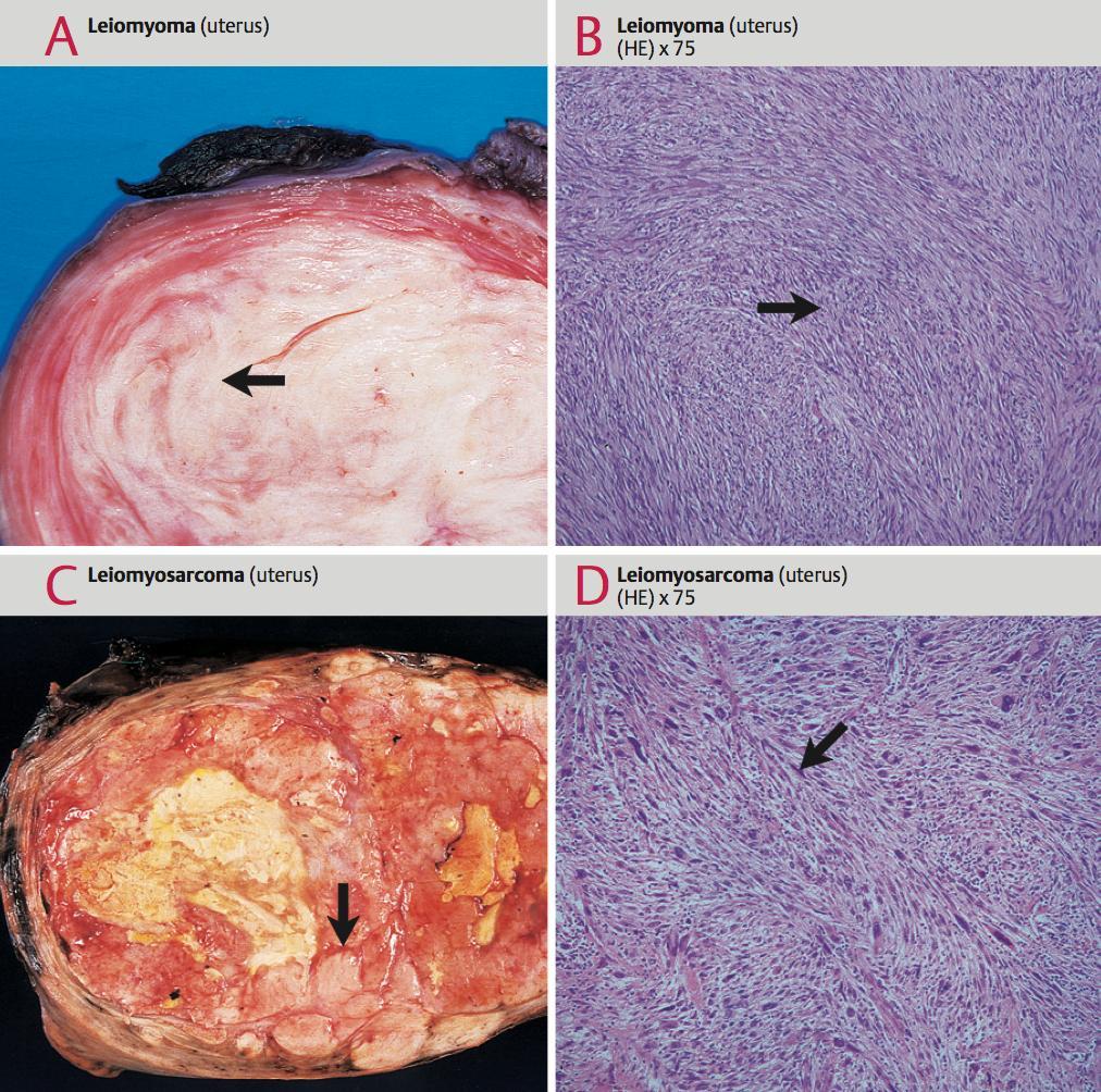
6 Muscle Tumors Leiomyomas Occurrence: These common tumors usually occur in groups in the uterus, intestinal tract, and vascular walls. Definition: A benign spherical tumor composed of mature smooth muscle cells. Histologic findings include a swirling pattern of interwoven tumor cells with cigar-shaped nuclei. Each tumor cell (smooth muscle cell) has a sock of basement membrane wrapped around it. Regressive changes such as hyalinization, calcification, and/or ossification may be present. However, no necrosis will occur. Leiomyosarcoma Occurrence: This rare tumor occurs in the retroperitoneum and, rarely, in the uterus. Definition: A malignant nodular tumor com- posed of immature smooth muscle cells. Histologic findings generally resemble a hypercellular leiomyoma often with only slight cellular polymorphism but with mitoses. Necroses are usually present as well. Immunohistochemical findings include tumor cells with a cytoskeleton containing actin. The prognosis depends on the tumor stage.
7 Muscle Tumors Rhabdomyoma Occurrence: This very rare tumor occurs primarily in the heart. Definition: Benign tumor of mature striated muscle cells. Histologic findings include spider cells (polygonal spider-like tumor cells with vacuolar cytoplasm (containing glycogen), some of which exhibit striations. Rhabdomyosarcoma Occurrence: This rare tumor occurs in the head and neck region and genitals in infants and in the extremities in adults. Definition: A group of highly malignant tumors of embryonic striated muscle tissue. Histologic findings include stellate and spindle- shaped polymorphic tumor cells with striations in cytoplasmic tails ( tadpole cells ). Regressive change include necroses. Immunohistochemical findings include tumor cells with a cytoskeleton containing desmin and myosin. The prognosis is very poor.
8
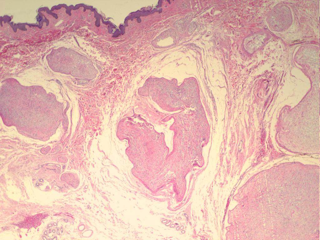
9 Tumors of Fatty Tissue Lipoma Occurrence: This tumor is found in subcutaneous and submucosal tissue, Rarely, it may also occur at intramuscular sites, and these tumors tend to recur. It is the most common benign mesenchymal tumor. Definition: Benign solid tumor of mature tumor cells (adipocytes) that store fat in a single vacuole. Histologic findings perfectly mimic fatty tissue but with the connective tissue organizing it into the lobular structure typical of fatty tissue. Liposarcoma Occurrence: This tumor is found in the thigh, back, and retroperitoneum. Definition: Malignant soft yellowish tumor of fat cell precursors (preadipocytes). Histologic findings: Fat is stored in multiple vacuoles in the cytoplasm and nucleus of the tumor cell, creating a typical scalloped nucleus. Regressive changes include necroses and myxoid degeneration of the stroma.


10 Tumors of Fatty Tissue Lipoma Liposarcoma
11 Cartilage Tumors Chondroma Occurrence: The tumor occurs within the bones of the hands and feet. An osteochondroma is a neoplasm arising as cartilagecapped bony outgrowth on a skeletal bone. Definition and morphology: A benign tumor of mature chondrocytes that perfectly mimics hyaline cartilage. Chondrosarcoma Occurrence: The tumor arises in the pelvis or in long cortical bones such as the humerus, tibia, and femur. Definition and morphology: This is a slow-growing malignant tumor arising from chondrocyte precursors, some of which develop into bizarre giant cells, and exhibiting some of the characteristics of hyaline cartilage. Regressive changes include necrosis, bleeding, and osteolysis.
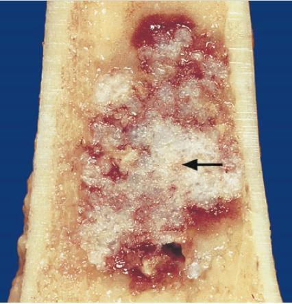
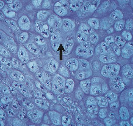
12 Chondrosarcoma
13 Bone Tumors Osteoma Occurrence: The tumor arises primarily in bones that develop from the desmocranium. Definition and morphology: This benign tumor arising from osteocytes perfectly mimics cancellous lamellar bone with a fibrous fatty medulla. It exhibits slowly expansive growth. Osteosarcoma Occurrence: This tumor is found primarily in long bones in adolescents. It is the most common malignant tumor of the skeletal system. Definition: A highly malignant sarcoma of plur- ipotential osteocyte precursors. Histologic findings include sarcomatous stromal tissue with polymorphic cells with an uncalcified tumor osteoid and disorganized tumorous cancellous tissue. One of the following types of osteosarcoma will occur: Proliferation of osteoblasts successively leads to bone remodeling and an osteoplastic osteosarcoma. Proliferation of osteoclasts successively leads to dis-solution of bone and an osteolytic osteosarcoma.
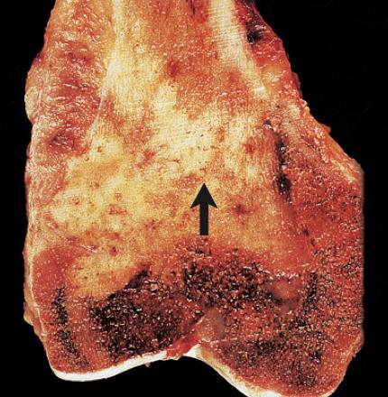
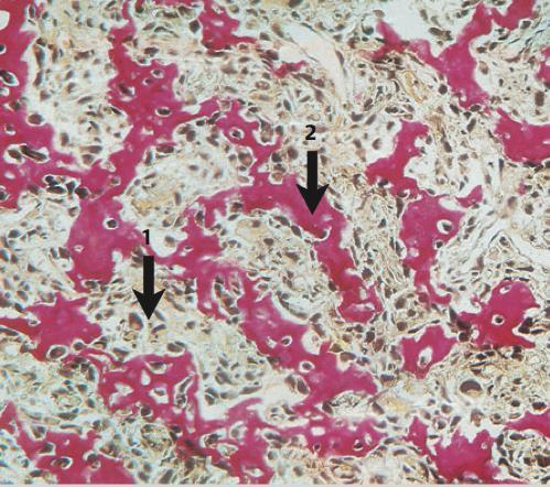
14 Osteosarcoma
15 Tumors from nervous tissue
16 Features of Brain Tumors Disontogenetic nature Absolute clinical malignancy Infiltrative growth pattern even in morphologically benign tumors Metastasizing by CSF pathways, very rare outside of cranial cavity Formation of typical histological structures


17 Specific to neural tumors rhythmic structure True rossette False perivascular rosette Homer Wright rosette Palisades
18 Ependymoma. True rosette
19 Ependymoma. Perivascular rossette.

20 Homer-Wright rosette in medulloblastoma
21

22 Palisades in schwannoma

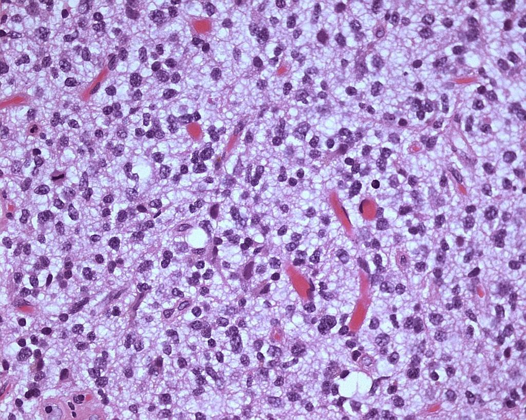
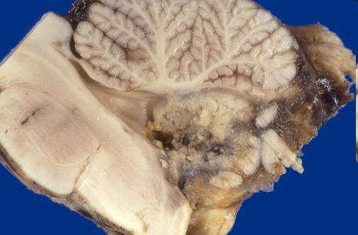
23 Glioblastoma. Foci of necrosis with formation of pseudopalisades
24 Degree determination of tumor malignancy in central nervous system Histological degree determination of malignancy (Grade) is based on identification and combination of the following major pathological signs: 1. nuclear atypism 2. mitosis 3. Vascular (endothelial) proliferation 4. necrosis
25 NEUROEPITHELIAL TUMORS Astrocytal tumors Oligodendroglial Ependymal Tumors of choroid plexus Neuronal Pineal Embryonic
26 ASTROCYTAL TUMORS Astrocytomas - the most common CNS tumors in infants The main histological types: Pilocytic (children) G1 Fibrillar astrocytoma (adults) G2 Protoplasmic astrocytoma G2 Anaplastic astrocytoma G3 Glioblastoma multiforme - undifferentiated glioma, astrocytoma with remnants of G4

27 Astrocytoma with multiple cysts

28 Cerebellum pilocytic astrocytoma in the form of a large cyst

29 Astrocytoma. Diffuse infiltrative type of growth
")
30 Pilocytic (juvenile) astrocytoma
31 Protoplasmic astrocytoma

32 Glioblastoma with foci of necrosis and hemorrhage, intracranial metastases in the opposite hemisphere of brain

33 Glioblastoma. Foci of necrosis with the formation psevdopalisade, expressed cellular atypia, proliferation of vascular endothelium
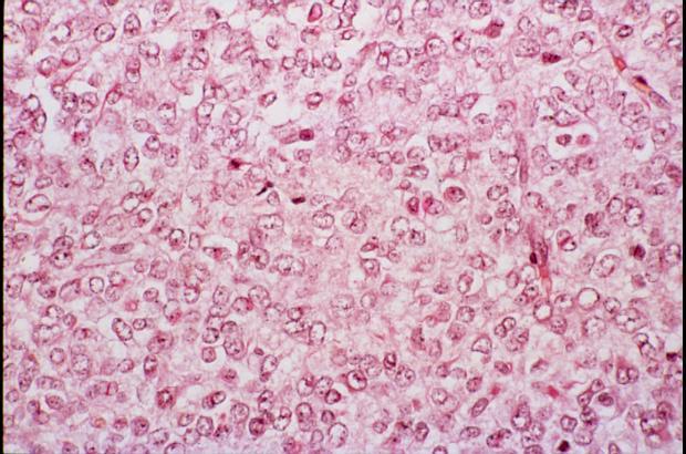
34 Oligodendroglioma 5-15% of gliomas Localization in frontal and lateral parts of cerebral hemispheres Are more common among middle-aged Could be mixed with astrocytomas variants histological structure Grow slowly Rarely metastasize Variants: oligodendroglioma (G2), anaplastic oligodendroglioma (G3) Morphology: Ill-defined tumor of small, densely packed tumor cells (exhibiting a dark nucleus in bright cytoplasm) that creates a honeycomb pattern. Signs of regression include bleeding, cysts, and calcification.
35 Oligodendroglioma

36 Oligodendroglioma of brain with arcade blood vessels and calcification
37 Anaplastic oligodendroglioma
38 Ependymoma More common in infants Localization: children in 4-th ventricle, adults usually in spinal cord Arise from the ependymal lining the ventricles and liquor conducting system Micro: tumor cells form a true rosette, Homer Wright rosette, and perivascular pseudorosette

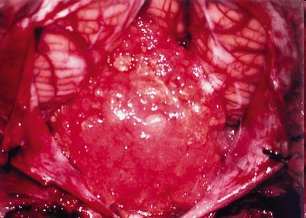
39 Ependymoma of 4-th cerebrum ventricle

40 Ependymoma. Perivascular rosette.

41 Ependymoma. True rosette
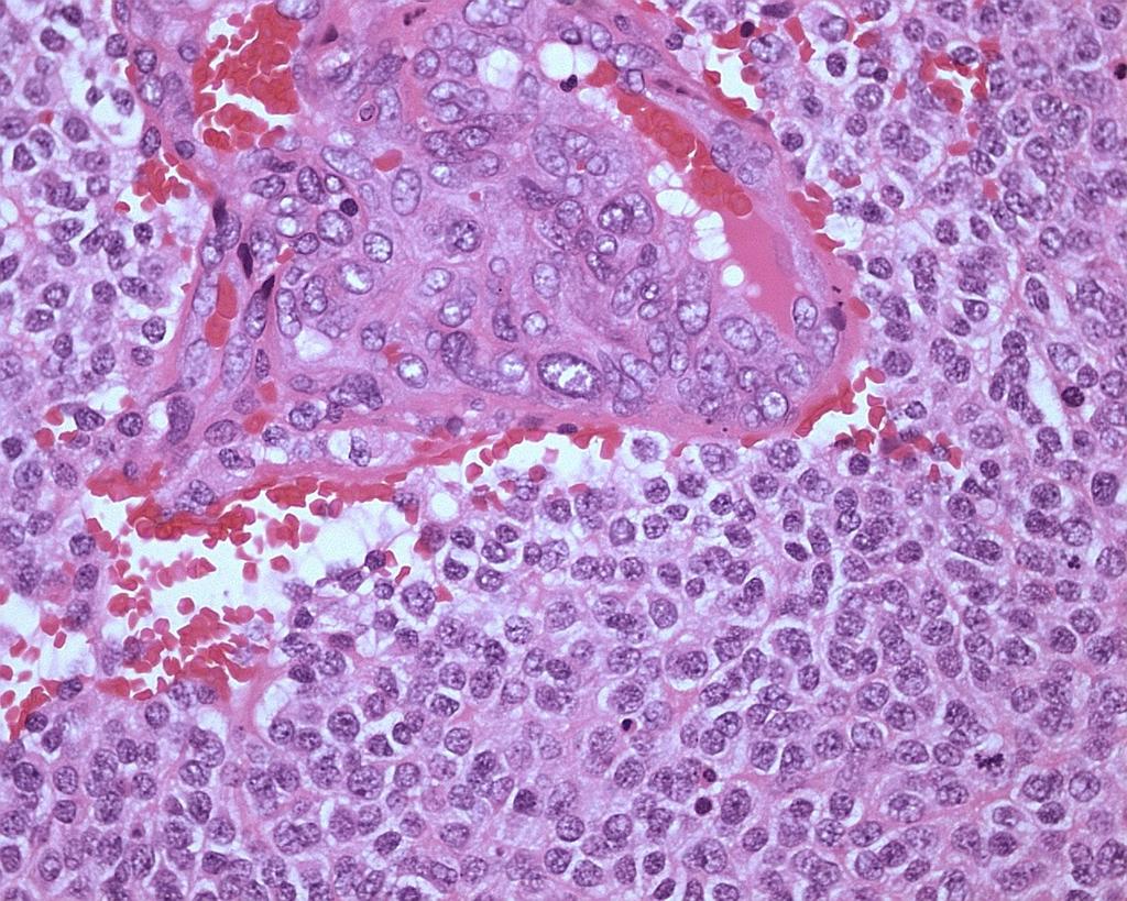
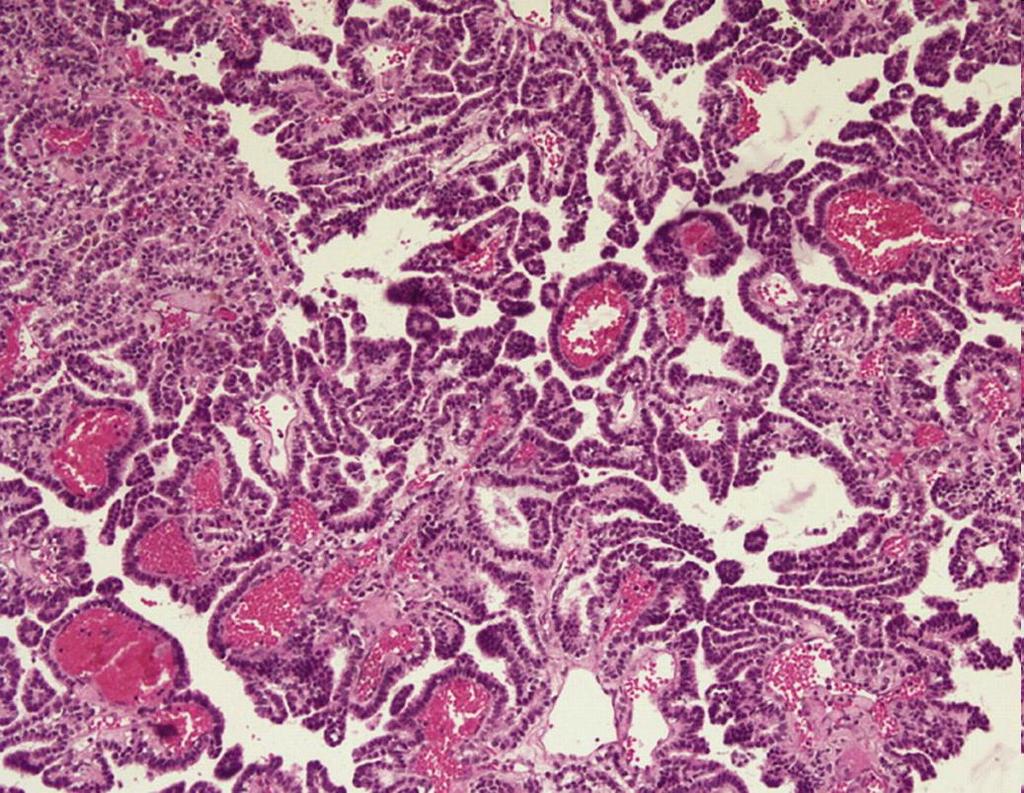
42 Choroid plexus tumors Rare (0.5% Op. CNS), usually in children in the lateral ventricles in adults IV ventricle. Grow slowly, gradually break outflow of liquor, causing hydrocephalus. Horioidpapilloma and chorioidcarcinoma The terminology does not match the histogenesis! The name given to the tumor in relation to characteristic of histological features - a tumor composed of papillary structures (papillae).
43
44 Embryonal tumors Medullary epithelioma Neuroblastoma (sympathicoblastoma?) Ganglioneuroblastoma Ependymoblastoma Primitive neuroectodermal tumors (PNET) PNET of cerebellum - Medulloblastoma
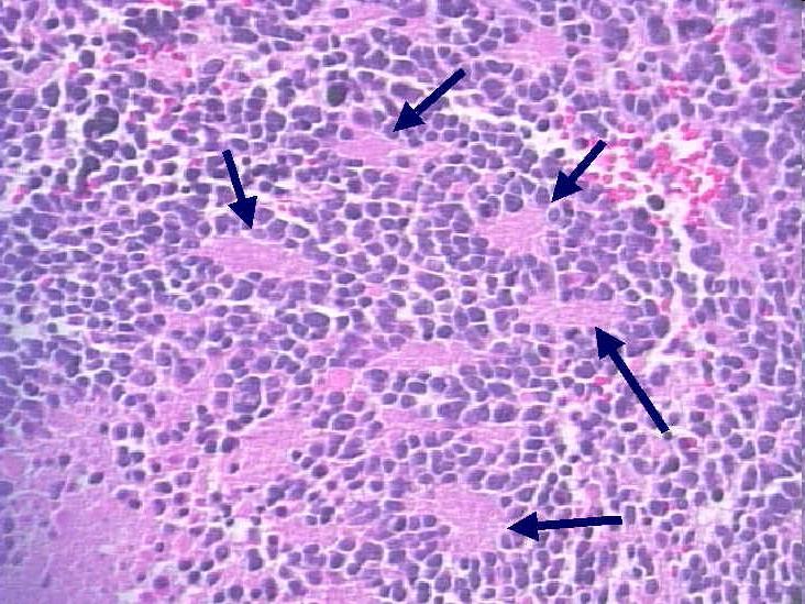
45 Medulloblastoma The most common PNET Second frequency at children CNS tumor Localization cerebellum Grade 4 Characteristically ingrowth IV ventricle and as a consequence development of hydrocephalus Micro: tumor is formed by small undifferentiated cells with hyperchromatic nuclei, cells form a Homer-Wright rosettes
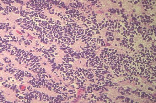
46 Homer-Wright rosettes in medulloblastoma
47 Meningioma The tumor arises from cells of brain arachnoid membrane Usually occurs in adults Macro: looks like a node associated with the meninges Variety: meningothelial, psammomatous, fibroblastic, microcystic, angiomatous Typical histological structures: tumor cells form vortexlike structures, could be foci of dystrophic calcification in the form of psammom

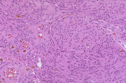
48 Psammoma bodies Meningothelial meningioma
49 Peripheral nervous system tumors Schwannoma (neurilemmoma/ neurinoma) Neurofibroma and neurofibromatosis
50 Schwannoma A benign tumor arising from Schwann cells form nerve membranes Usually occurs in middle age Two variants of of structure - Antoni A (cells form a true palisades), Antoni B (no palisades)

51 Neurilemmoma of acoustic nerve

52 Schwannoma, Antoni type A Schwannoma, Antoni type B
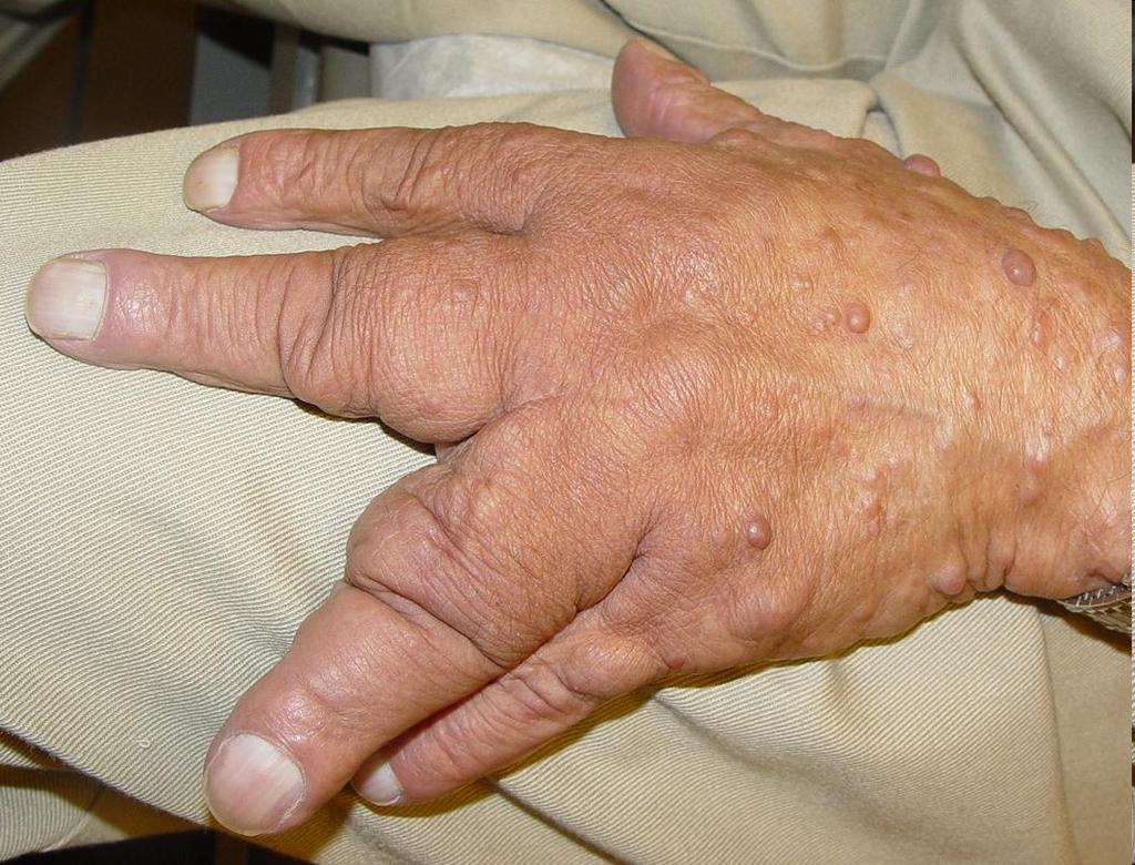
53 Neurofibroma Occurrence: A solitary lesion may be present, or the tumor may occur as multiple lesions in type 1 neurofibromatosis (Recklinghausen disease) Definition and morphology: Benign encapsulated Schwanncell tumor arising from craniospinal and peripheral nerves. The cut surface of the tumor is whitish-yellow. Histologic findings include spindle-shaped tumor cells in a loose, undulating, fibrous mesh.
54
 - chromosome 17.")


55 Neurofibromatosis (type 1) Mutation of neurofibromin gene (NF1) - chromosome 17. Clinically: multiple neurofibromas, 6 and more skin spots such as "coffee au lait", pigmented retina hamartomas, sided schwannomas of auditory nerve, optic nerve and other peripheral nerves and the combination with congential malformations (megacolon), Wilms' tumor, multiple lipomas, etc.
56

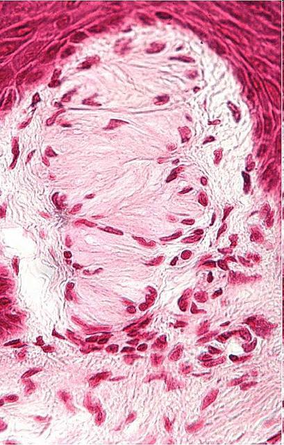
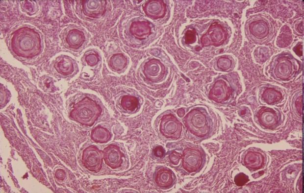
57 Vater-Pacini corpuscles
General: Brain tumors are lesions that have mass effect distorting the normal tissue and often result in increased intracranial pressure.
 1 Lecture Objectives Know the histologic features of the most common tumors of the CNS. Know the differences in behavior of the different tumor types. Be aware of the treatment modalities in the various
Tumors of the Central Nervous System
 Tumors of the Central Nervous System 1 Financial Disclosures I have NO SIGNIFICANT FINANCIAL, GENERAL, OR OBLIGATION INTERESTS TO REPORT Introduction General: Brain tumors are lesions that have mass effect
CNS TUMORS. D r. Ali Eltayb ( U. of Omdurman. I ). M. Path (U. of Alexandria)
. M. Path (U. of Alexandria)") CNS TUMORS D r. Ali Eltayb ( U. of Omdurman. I ). M. Path (U. of Alexandria) CNS TUMORS The annual incidence of intracranial tumors of the CNS ISmore than intraspinal tumors May be Primary or Secondary
CNS TUMORS D r. Ali Eltayb ( U. of Omdurman. I ). M. Path (U. of Alexandria) CNS TUMORS The annual incidence of intracranial tumors of the CNS ISmore than intraspinal tumors May be Primary or Secondary
Tumors of the Nervous System
 Tumors of the Nervous System Peter Canoll MD. PhD. What I want to cover What are the most common types of brain tumors? Who gets them? How do they present? What do they look like? How do they behave? 1
CNS pathology Third year medical students. Dr Heyam Awad 2018 Lecture 12: CNS tumours 2/3
 CNS pathology Third year medical students Dr Heyam Awad 2018 Lecture 12: CNS tumours 2/3 Pilocytic astrocytoma Relatively benign ( WHO grade 1) Occurs in children and young adults Mostly: in the cerebellum
Peter Canoll MD. PhD.
 Tumors of the Nervous System Peter Canoll MD. PhD. What I want to cover What are the most common types of brain tumors? Who gets them? How do they ypresent? What do they look like? How do they behave?
5/10. Pathology Soft tissue tumors. Farah Bhani. Mohammed Alorjani
 5/10 Pathology Soft tissue tumors Mohammed Alorjani Farah Bhani Slides are included in this sheet. Objectives: Soft tissue tumors 1. Describe soft tissue tumors. 2. Understand the classification of soft
Neoplasms. Nomenclature. Cellular atypia/anaplasia. Benign (locally malignant or rarely metastasizing) Benign v/s malignant neoplasm.
 Benign v/s malignant neoplasm.") Neoplasms-3 1 - Leiomyoma 2 - Lipoma 3-4 - Chondrosarcoma Non-epithelial tumors Neoplasms Benign (locally malignant or rarely metastasizing) borderline/intermediate malignant 5 - Meningioma 6 Glioblastoma
Neoplasms-3 1 - Leiomyoma 2 - Lipoma 3-4 - Chondrosarcoma Non-epithelial tumors Neoplasms Benign (locally malignant or rarely metastasizing) borderline/intermediate malignant 5 - Meningioma 6 Glioblastoma
Malignant Peripheral Nerve Sheath Tumor
 C H A P T E R 120 Malignant Peripheral Nerve Sheath Tumor Currently, malignant peripheral nerve sheath tumor (MPNST) is the most commonly used generic name for the neoplasms known in the past as neurosarcoma,
Brain Tumors. Medulloblastoma. Pilocytic astrocytoma: Ahmed Koriesh, MD. Pathological finding
 NeuroPathology Page 8 Brain Tumors Pathological finding Pseudorosette Rosenthal fibers Rosettes Wet Keratin Psammoma bodies Fried egg Tumor Ependymoma, SEGA Pilocytic astrocytoma Medulloblastoma Craniopharyngioma
TUMORS of nervous system
 TUMORS of nervous system By: Shifaa Alqa qa Done By : Ola Hijjawi CNS tumors : The annual incidence of CNS tumors ranges from 10 to 17 per 100,000 persons for intracranial tumors and 1 to 2 per 100,000
Site Specific Coding Rules MALIGNANT CENTRAL NERVOUS SYSTEM TUMORS
 Multiple Primary and Histology Site Specific Coding Rules MALIGNANT CENTRAL NERVOUS SYSTEM TUMORS 1 Prerequisites 2 Completion of Multiple Primary and Histology General Coding Rules 3 There are many ways
Tumors of Adipose Tissue Tumors Epidemiology Clinical Features. Morphology. Mature Adipocytes Separated by delicate fibrous septa
 Tumors of Adipose Tissue Lipoma Liposarcoma Most commonly happens in female The most common soft tissue tumor o Originates from matured Adipocytes Most commonly happes at the 4 th and 5 th decade of life
Brain tumors: tumor types
 Brain tumors: tumor types Tumor types There are more than 120 types of brain tumors. Today, most medical institutions use the World Health Organization (WHO) classification system to identify brain tumors.
2. Subependymal giant cell astrocytoma:
 I. Astrocytomas: A. Diffusely infiltrating ( astrocytoma, anaplastic astrocytoma, GBM) B. Localised (pilocytic astrocytoma, pleomorphic xanthoastrocytoma, SGCA) *Grading: Diffuse: 1. Astrocytoma WHO grade
* I have no disclosures or any
 Howard Rosenthal, M.D. Associate Professor of Orthopedic Surgery University of Kansas Sarcoma Center I have no disclosures or any conflicts related to the content of this presentation. Objectives 1. Describe
Histopathological Study and Categorisation of Brain Tumors
 Histopathological Study and Categorisation of Brain Tumors Ruchira Wadhwa 1*, Purvi Patel 2, Hansa Goswami 3 1 Third Year Resident, 2 Assistant Professor, 3 Professor and Head, Department of Pathology,
Neoplasia literally means "new growth.
 NEOPLASIA Neoplasia literally means "new growth. A neoplasm, defined as "an abnormal mass of tissue the growth of which exceeds and is uncoordinated with that of the normal tissues and persists in the
number Done by Corrected by Doctor Maha Shomaf
 number 16 Done by Waseem Abo-Obeida Corrected by Zeina Assaf Doctor Maha Shomaf MALIGNANT NEOPLASMS The four fundamental features by which benign and malignant tumors can be distinguished are: 1- differentiation
Grading of Bone Tumors
 Grading of Bone Tumors Joon Hyuk Choi, M.D. Department of Pathology College of Medicine, Yeungnam University Introduction to grading system of bone tumor used at Mayo Clinic WHO Histologic Classification
A neoplasm is defined as "an abnormal tissue proliferation, which exceeds that of adjacent normal tissue. This proliferation continues even after
 NEOPLASIA Neoplasia is a very important topic in pathology because neoplasms are both common and serious diseases. A neoplasm literally means a new growth, and this term is used interchangeably with a
Musculoskeletal Sarcomas
 Musculoskeletal Sarcomas Robert C. Orth, M.D., Ph.D. Edward B. Singleton Department of Pediatric Radiology Texas Children s Hospital Page 0 xxx00.#####.ppt 9/23/2012 9:01:18 AM No disclosures Page 1 xxx00.#####.ppt
Von Recklinghausen s Disease with a Giant Lipoma
 Von Recklinghausen s Disease with a Giant Lipoma Daiki Iwana¹( ) Kazutaka Izawa¹ Mitsuhiro Kawamura¹ Takaharu Nabeshima¹ Hideki Yoshikawa² ¹Department of Orthopaedic Surgery, Toneyama National Hospital,
IN THE NAME OF GOD Dr. Kheirandish Oral and maxillofacial pathology
 IN THE NAME OF GOD Dr. Kheirandish Oral and maxillofacial pathology ORAL FOCAL MUCINOSIS Uncommon Tumorlike Cutaneous myxoid cyst Overproduction of hyaluronic acid by firoblasts Young adults Female Gingiva
3/27/2017. Disclosure of Relevant Financial Relationships
 Ophthalmic Pathology Evening Specialty Conference USCAP 2017 5 th March, 2017 Mukul K. Divatia, MD Assistant Professor Department of Pathology & Genomic Medicine Weill Cornell Medical College Houston Methodist
Neoplasia 2018 Lecture 2. Dr Heyam Awad MD, FRCPath
 Neoplasia 2018 Lecture 2 Dr Heyam Awad MD, FRCPath ILOS 1. List the differences between benign and malignant tumors. 2. Recognize the histological features of malignancy. 3. Define dysplasia and understand
Methoden / Methods inc. ICCC-3 105
 Methoden / Methods inc. ICCC-3 105 Internationale Klassifikation der Krebserkrankungen bei Kindern (ICCC-3) Zuordnung von ICD-O-3-Codes für Morphologie und Topographie zu diagnostischen Kategorien International
Neoplasia part I. Dr. Mohsen Dashti. Clinical Medicine & Pathology nd Lecture
 Neoplasia part I By Dr. Mohsen Dashti Clinical Medicine & Pathology 316 2 nd Lecture Lecture outline Review of structure & function. Basic definitions. Classification of neoplasms. Morphologic features.
Soft Tissue Sarcomas: Questions and Answers
 Soft Tissue Sarcomas: Questions and Answers 1. What is soft tissue? The term soft tissue refers to tissues that connect, support, or surround other structures and organs of the body. Soft tissue includes
NEOPLASIA-I CANCER. Nam Deuk Kim, Ph.D.
 NEOPLASIA-I CANCER Nam Deuk Kim, Ph.D. 1 2 Tumor in the hieroglyphics of the Edwin Smith papyrus (1,600 B.C., Breasted s translation 1930) 3 War on Cancer (National Cancer Act, 1971) 4 Cancer Acts in Korea
Accuracy of intra-operative rapid diagnosis by Squash smear in CNS lesions An early institutional experience. KK Bansal,
 Accuracy of intra-operative rapid diagnosis by Squash smear in CNS lesions An early institutional experience. KK Bansal, Monika Bansal, Sanjeev Kishore, Anuradha K, Meena H, Dushyant G. Department of Neurosurgery
Special slide seminar
 Special slide seminar Tomáš Rozkoš The Fingerland Department of Pathology Charles University Medical Faculty and Faculty Hospital in Hradec Králové Czech Republic Case history, 33 years old resistance
Spindle Cell Lesions Of The Breast. Emad Rakha Professor of Breast Pathology and Consultant Pathologist
 Spindle Cell Lesions Of The Breast Emad Rakha Professor of Breast Pathology and Consultant Pathologist * SCLs comprise a wide spectrum of diseases, ranging from reactive processes to aggressive malignant
Part 1. Slides 1-38, Rita Alaggio Soft tissue tumors Trondheim 14. mars 2013
 Part 1 Slides 1-38, Rita Alaggio Soft tissue tumors Trondheim 14. mars 2013 Pediatric Pathology Soft Tissue Tumors AN UPDATE Rita Alaggio Azienda Ospedaliera Università di Padova Soft Tissue Tumors More
Pathologic Analysis of CNS Surgical Specimens
 2015 Kenneth M. Earle Memorial Neuropathology Review Pathologic Analysis of CNS Surgical Specimens Peter C. Burger, MD Interdisciplinary Quality Control Familiarity with entities Use of diagnostic algorithm
Primary bone tumors > metastases from other sites Primary bone tumors widely range -from benign to malignant. Classified according to the normal cell
 Primary bone tumors > metastases from other sites Primary bone tumors widely range -from benign to malignant. Classified according to the normal cell counterpart and line of differentiation. Among the
Classification (1) Classification (3) Classification (2) Spindle cell lesions. Spindle cell lesions of bladder (Mills et al.
 Classification (3) Classification (2) Spindle cell lesions. Spindle cell lesions of bladder (Mills et al.") Non-epithelial tumours and nonepithelial tumour-like lesions of the bladder Dr Jonathan H Shanks The Christie NHS Foundation Trust, Manchester, UK Classification (1) Myofibroblastic proliferations and
Non-epithelial tumours and nonepithelial tumour-like lesions of the bladder Dr Jonathan H Shanks The Christie NHS Foundation Trust, Manchester, UK Classification (1) Myofibroblastic proliferations and
Rhabdomyomas and Rhabdomyosarcomas (RMS) David M. Parham, MD Chief of Anatomic Pathology
 David M. Parham, MD Chief of Anatomic Pathology") Rhabdomyomas and Rhabdomyosarcomas (RMS) David M. Parham, MD Chief of Anatomic Pathology Tumors of skeletal muscle: Rhabdomyomas and rhabdomyosarcomas Embryonal muscle 2 3 4 5 6 7 8 Rhabdomyoma Benign
Rhabdomyomas and Rhabdomyosarcomas (RMS) David M. Parham, MD Chief of Anatomic Pathology Tumors of skeletal muscle: Rhabdomyomas and rhabdomyosarcomas Embryonal muscle 2 3 4 5 6 7 8 Rhabdomyoma Benign
USCAP Neuropathology. Case No. 3 Elisabeth J. Rushing, MD Armed Forces Institute of Pathology Washington, DC
 USCAP Neuropathology Case No. 3 Elisabeth J. Rushing, MD Armed Forces Institute of Pathology Washington, DC Clinical history The patient is a 9 year-old boy who has had seizures since age 2, at which time
The Relevance of Cytologic Atypia in Cutaneous Neural Tumors
 The Relevance of Cytologic Atypia in Cutaneous Neural Tumors Recent Findings - New Developments New Problems Zsolt B. Argenyi, M.D. Professor of Pathology & Dermatology Director of Dermatopathology Department
Classification of spontaneous brain tumors in rats
 Classification of spontaneous brain tumors in rats Central Nervous System Neoplasms in the Rat (Solleveld HA, et al., 1991) 1. Tumors of Neuroepithelial Tissue A. Astrocytic and oligodendroglial tumors
Supra- and infratentorial brain tumors from childhood to maternity
 Supra- and infratentorial brain tumors from childhood to maternity What to expect? I am going to show you the characteristic imaging findings of following tumors: Thierry A.G.M. Huisman, MD, FICIS, EQNR
Newer soft tissue entities
 Newer soft tissue entities Examples among fibroblastic tumors Turku, May 6, 2010 Markku Miettinen, M.D. AFIP, Washington, DC Fibroblastic neoplasms Solitary fibrous tumor /Hemangiopericytoma Low-grade
SPECIAL SLIDE SEMINAR CASE 3
 SPECIAL SLIDE SEMINAR CASE 3 Tihana Džombeta, MD Leo Pažanin, MD, PhD Department of Pathology, School of Medicine, University of Zagreb Department of Pathology, Clinical Hospital Centre Sestre milosrdnice
Introduction to Musculoskeletal Tumors. James C. Wittig, MD Orthopedic Oncologist Sarcoma Surgeon
 Introduction to Musculoskeletal Tumors James C. Wittig, MD Orthopedic Oncologist Sarcoma Surgeon www.tumorsurgery.org Definitions Primary Bone / Soft tissue tumors Mesenchymally derived tumors (Mesodermal)
Spinal Neoplasms. First Things First!! Localize the Lesion!! Ependymomas. Common Intramedullary Lesions
 Acta Radiológica Portuguesa, Vol.XXIII, nº 90, pág. 101-114, Abr.-Jun., 2011 Spinal Neoplasms Bruno A Policeni University of Iowa Hospitals and Clinics Assistant Professor of Radiology Disclosure of Commercial
H Haloes cautions, 57 neurocytomas, perinuclear, 56 Headache blue cell tumors, 147 cautions, 135, 147, 152 clinical history, 132, 144, 148
 Index A ADC. See Apparent diffusion coefficient Adult. See also Supratentorial mass, adult cerebral tumor, 1 headache and ataxia cysts, mural nodules, 118 sporadic tumors, 118 headaches and visual changes,
Epithelial tumors. Dr. F.F. Khuzin, PhD Dr. M.O. Mavlikeev
 Epithelial tumors Dr. F.F. Khuzin, PhD Dr. M.O. Mavlikeev Epithelial tumors Tumors from the epithelium are the most frequent among tumors. There are 2 group features of these tumors: The presence in most
Normal endometrium: A, proliferative. B, secretory.
 Normal endometrium: A, proliferative. B, secretory. Nội mạc tử cung Nội mạc tử cung Cyclic changes in endometrium.. Approximate relationship of useful microscopic changes. Arias-Stella reaction in endometrial
Essential Dermatopathology: Neoplastic American Academy of Dermatology Annual Meeting NEURAL AND SMOOTH MUSCLE NEOPLASMS
 Essential Dermatopathology: Neoplastic American Academy of Dermatology Annual Meeting NEURAL AND SMOOTH MUSCLE NEOPLASMS Kevin P. White M.D. Oregon Health and Science University Associate Professor of
Supplementary Information
 Rise in Glioblastoma Multiforme incidence in England 1995 2015 suggests an adverse environmental or lifestyle factor Alasdair Philips, Denis L Henshaw, Graham Lamburn, Michael J O Carroll Supplementary
An Overview of Genital Stromal Tumors
 An Overview of Genital Stromal Tumors By Konstantinos Linos MD, FCAP, FASDP Bone, Soft Tissue and Dermatopathology Assistant Professor of Pathology Dartmouth-Hitchcock Medical Center Geisel School of Medicine
2018 ICD-O-3 Updates in Table Format with Annotation for Reference
 Status Histology Description (this may be preferred term or a synonym) Report Comments New term 8010 3 Urachal carcinoma (C65.9, C66.9, C67._, C68._) New term 8013 3 Combined large cell neuroendocrine
Note: The cause of testicular neoplasms remains unknown
 - In the 15- to 34-year-old age group, they are the most common tumors of men. - Tumors of the testis are a heterogeneous group of neoplasms that include: I. Germ cell tumors : 95%; all are malignant.
CODING TUMOUR MORPHOLOGY. Otto Visser
 CODING TUMOUR MORPHOLOGY Otto Visser INTRODUCTION The morphology describes the tissue of the tumour closest to normal tissue Well differentiated tumours are closest to normal Undifferentiated tumours show
أملس عضلي غرن = Leiomyosarcoma. Leiomyosarcoma 1 / 5
 Leiomyosarcoma 1 / 5 EPIDEMIOLOGY Exact incidence is unknown, but older studies suggest that leiomyosarcomas comprise approximately 3 percent of soft-tissue sarcomas. Superficial leiomyosarcoma occurs
Diplomate of the American Board of Pathology in Anatomic and Clinical Pathology
 A 33-year-old male with a left lower leg mass. Contributed by Shaoxiong Chen, MD, PhD Assistant Professor Indiana University School of Medicine/ IU Health Partners Department of Pathology and Laboratory
Atypical Palisaded Myofibroblastoma of Lymph Node: Report of a rare case.
 ISPUB.COM The Internet Journal of Pathology Volume 10 Number 1 Atypical Palisaded Myofibroblastoma of Lymph Node: Report of a rare case. V Kinnera, R Nandyala, M Yootla, K Mandyam Citation V Kinnera, R
Effective January 1, 2018 ICD O 3 codes, behaviors and terms are site specific
 Effective January 1, 2018 codes, behaviors and terms are site specific /N 8551/3 Acinar adenocarcinoma (C34. _) Lung primaries diagnosed prior to 1/1/2018 use code 8550/3 For prostate (all years) see 8140/3
INFECTIONS OF THE NERVOUS SYSTEM
 NEUROPATHOLOGY INFECTIONS OF THE NERVOUS SYSTEM An infectious agent must use one of several routes of entry to reach the CNS & cause a disease. 1. Hematogenous spread via the arterial blood supply is the
Effective January 1, 2018 ICD O 3 codes, behaviors and terms are site specific
 Effective January 1, 2018 codes, behaviors and terms are site specific Status /N 8010/3 Urachal carcinoma (C65.9, C66.9, C67. _, C68._) 8013/3 Combined large cell neuroendocrine carcinoma (C34. _, C37.9)
Imaging the Spinal Cord & Intradural Disease
 Department of Radiology University of California San Diego Imaging the Spinal Cord & Intradural Disease John R. Hesselink, M.D. Spinal Cord Diseases Tumors Syringohydromyelia Trauma Ischemia / Infarction
Infections. Meningitis- leptomenings (usually bakterial)
") Neuropathology Neuropathology Cerebral Edema, raised intracranial pressure, Herniatio Malformations, Developmental Diseases Perinatal Brain Injury Trauma Cerebrovascular Diseases Infections Transmissible
Neuropathology Neuropathology Cerebral Edema, raised intracranial pressure, Herniatio Malformations, Developmental Diseases Perinatal Brain Injury Trauma Cerebrovascular Diseases Infections Transmissible
1/9/2013 EXTRAMEDULLARY TUMORS OF THE PEDIATRIC SPINE. Introduction. Classification for Extramedullary Tumors
 EXTRAMEDULLARY TUMORS OF THE PEDIATRIC SPINE Eugene Wang 1/20/12 Dent Neurologic Institute Introduction 2/3 of all intraspinal tumors of childhood are extramedullary 50% Extradural 10-15% Intradural Back
DUSTURBANCES OF GROWTH. MLS Basic histological diagnosis MLS HIST 422 Semester 8- batch 7 L8 Uz: Musa
 DUSTURBANCES OF GROWTH MLS Basic histological diagnosis MLS HIST 422 Semester 8- batch 7 L8 Uz: Musa Agnesia: means complete absence of an organ (Kidney). Aplasia: s defined in general as "defective development
MVST BOD & NST PART IB Thurs. 2 nd & Fri. 3 rd March 2017 Pathology Practical Class 23
 MVST BOD & NST PART IB Thurs. 2 nd & Fri. 3 rd March 2017 Pathology Practical Class 23 Neoplasia I Neoplasia I: Benign and malignant neoplasms in glandular epithelium and mesenchyme 1.0. Aims 1. To understand
Pleomorphic Xanthoastrocytoma
 Pleomorphic Xanthoastrocytoma Christine E. Fuller Keywords Pleomorphic xanthoastrocytoma; Pleomorphic xanthoastrocytoma with anaplastic features 2.1 OVERVIEW Pleomorphic xanthoastrocytoma (PXA) is an uncommon
the urinary system pathology Dr. Fairoz A Eltorgman
 the urinary system pathology Dr. Fairoz A Eltorgman Tumors of the renal pelvis & kidney Benign tumors of the renal pelvis: Hemangioma Leiomyoma Malignant tumors: Transitional cell carcinoma Squamous cell
Neurocutaneous Syndromes. Phakomatoses
 Neurocutaneous Syndromes Phakomatoses Financial Disclosures I have NO SIGNIFICANT FINANCIAL, GENERAL, OR OBLIGATION INTERESTS TO REPORT Neurocutaneous Syndomes Definition Entities Diagnosis/ Presentation
Immunohistochemistry in Bone and Soft Tissue Tumors. Sahar Rassi Zankoul, MD
 Immunohistochemistry in Bone and Soft Tissue Tumors Sahar Rassi Zankoul, MD Introduction Bone tumors represent a wide variety of tumors of various origins and malignant potentials. These different tumor
CYSTIC TUMORS OF THE KIDNEY JOHN N. EBLE, M.D. CYSTIC NEPHROMA
 Page 1 CYSTIC TUMORS OF THE KIDNEY JOHN N. EBLE, M.D. Department of Pathology & Laboratory Medicine Phone (317) 274-4806 Medical Science A-128 FAX: (317) 278-2018 635 Barnhill Drive jeble @iupui.edu Indianapolis,
SURGICAL MANAGEMENT OF BRAIN TUMORS
 SURGICAL MANAGEMENT OF BRAIN TUMORS JASSIN M. JOURIA, MD DR. JASSIN M. JOURIA IS A MEDICAL DOCTOR, PROFESSOR OF ACADEMIC MEDICINE, AND MEDICAL AUTHOR. HE GRADUATED FROM ROSS UNIVERSITY SCHOOL OF MEDICINE
Neoplasms of the BRAIN and CNS
 Neoplasms of the BRAIN and CNS 2015-21016 FCDS Educational Webcast Series Steven Peace, BS, CTR October 15, 2015 2015 Focus Anatomy SSS 2000 MPH Rules AJCC TNM Presentation Outline Overview Reportable
Cells and Tissues 3PART D. PowerPoint Lecture Slide Presentation by Patty Bostwick-Taylor, Florence-Darlington Technical College
 PowerPoint Lecture Slide Presentation by Patty Bostwick-Taylor, Florence-Darlington Technical College Cells and Tissues 3PART D Connective Tissue Found everywhere in the body Includes the most abundant
Year 2003 Paper two: Questions supplied by Tricia
 question 43 A 42-year-old man presents with a two-year history of increasing right facial numbness. He has a history of intermittent unsteadiness, mild hearing loss and vertigo but has otherwise been well.
2 Berkeley Street, Suite 403, Toronto, Ontario M5A 2W3 Visit us at: Tel: Fax:
 E-Path A.I. Engine Knowledge Base Enhancements Version 1.0.0.29 April 1, 2018 The major enhancements in the E-Path Knowledge Base from versions 1.0.0.28 through 1.0.0.29 are as follows: 1. Addition/modification
Q&A. Fabulous Prizes. Collecting Cancer Data:CNS 2/7/12. NAACCR Webinar Series Collecting Cancer Data Central Nervous System
 Collecting Cancer Data Central Nervous System NAACCR 2012 2013 Webinar Series 2/7/2013 Q&A Please submit all questions concerning webinar content through the Q&A panel. Reminder: If you have participants
Pediatric Spine Tumors (and other masses)
") Pediatric Spine Tumors (and other masses) Francisco A Perez, MD, PhD Assistant Professor Neuroradiology and Pediatric Radiology Seattle Children s Hospital University of Washington, Seattle Commercial
Pediatric Spine Tumors (and other masses) Francisco A Perez, MD, PhD Assistant Professor Neuroradiology and Pediatric Radiology Seattle Children s Hospital University of Washington, Seattle Commercial
ONCOLOGY. Csaba Bödör. Department of Pathology and Experimental Cancer Research november 19., ÁOK, III.
 ONCOLOGY Csaba Bödör Department of Pathology and Experimental Cancer Research 2018. november 19., ÁOK, III. bodor.csaba1@med.semmelweis-univ.hu ONCOLOGY Characteristics of Benign and Malignant Neoplasms
Case 8 Soft tissue swelling
 Case 8 Soft tissue swelling 26-year-old female presented with a swelling on the back of the left knee joint since the last 6 months and chronic pain in the calf and foot since the last 2 months. Pain in
Collecting Cancer Data: Central Nervous System Prizes! Question of the Month! Tip of the Month! Q&A
 Collecting Cancer Data: Central Nervous NAACCR 2008 2009 Webinar Series April 2, 2009 Prizes! Question of the Month! The participant that submits the best question of the session will receive a fbl fabulous
Endometrial Stromal Tumors
 Endometrial Stromal Tumors WHO Categories: Endometrial Stromal Nodule (ESN) Endometrial Stromal Sarcoma, low grade (LGESS) Endometrial Stromal Sarcoma, high grade (HGESS) Undifferentiated Uterine Sarcoma
TUMOR,NEOPLASM. Pathology Department, Zhejiang University School of Medicine,
 TUMOR,NEOPLASM Pathology Department, Zhejiang University School of Medicine, 马丽琴,maliqin198@zju.edu.cn The points in this chapter What is a neoplasm (conception) Morphology of neoplasm Macroscopy of Neoplasm
Lab Animal Tissue. LEARNING OBJECTIVES: To understand the relationship between the structure and function of different animal tissues
 Name: Bio A.P. PURPOSE: HYPOTHESIS: NONE Lab Animal Tissue BACKGROUND: In animals, groups of closely related cells specialized to perform the same function are called tissues. There are four general classes
BAH1 - Primary Glioblastoma
 BAH1 - Primary Glioblastoma R frontal tumour for frozen section. No known primary. Contrast enhancing lesion. Cholecystectomy. FROZEN SECTION REPORT Right frontal tumour: The specimen consists of multiple
Body Tissues Pearson Education, Inc.
 Body Tissues Tissues Groups of cells with similar structure and function Four primary types: Epithelial tissue (epithelium).1 Connective tissue.2 Muscle tissue.3 Nervous tissue.4 Epithelial Tissues Locations:
SUPPLEMENTARY INFORMATION
 VOLUME: 1 ARTICLE NUMBER: 0027 In the format provided by the authors and unedited. Rapid intraoperative histology of unprocessed surgical specimens via fibre-laser-based stimulated Raman scattering microscopy
Understanding general brain tumor pathology, Part I: The basics. Craig Horbinski, M.D., Ph.D. Department of Pathology University of Kentucky
 Understanding general brain tumor pathology, Part I: The basics Craig Horbinski, M.D., Ph.D. Department of Pathology University of Kentucky plan of attack what IS a pathologist, anyway? what s so special
I vened an international group of neuropathologists to
 Revision of the World Health Organization Classification of Brain Tumors for Childhood Brain Tumors LUCY BALIAN RORKE, MD,' FLOYD H. GILLES, MD,t RICHARD L. DAVIS, MD,* AND LAURENCE E. BECKER, MDS COMMITTEE
Mayo Medical Laboratories
 Mayo Medical Laboratories Virtual Lectures 2014 MFMER 2016 MFMER slide-1 Virtual Lectures Planning Committee Disclosure Summary As a provider accredited by ACCME, College of Medicine, Mayo Clinic (Mayo
Bone Tumours - a synopsis. Dr Zena Slim SpR in Histopathology QAH 2009
 Bone Tumours - a synopsis Dr Zena Slim SpR in Histopathology QAH 2009 Aims General approach to diagnosis Common entities.and not so common ones. Mini quiz Challenge of bone tumour diagnosis Bone tumours
Disclosures. An update on ancillary techniques in the diagnosis of soft tissue tumors. Ancillary techniques. Introduction
 Disclosures An update on ancillary techniques in the diagnosis of soft tissue tumors. I have nothing to disclose. Andrew Horvai, MD, PhD Clinical Professor, Pathology Introduction Ancillary techniques
CLIC Sargent Eligibility Criteria
 1 Eligibility Criteria DOCUMENT GOVERNANCE: Eligibility criteria Produced by J. Hawkins & Grants Team Sponsored by Dara de Burca Version Approval by Executive Team 10 th June 2014 Board of Trustees 3 rd
Uterine Mesenchymal Tumors from a Gynaecological Point of View: A Mini-Review
 EC Gynaecology Special Issue - 2017 Uterine Mesenchymal Tumors from a Gynaecological Point of View: A Mini-Review Mini Review Dr. Huseyin Aydogmus, Dr. Servet Gencdal, Dr. Nihan Gencdal and Dr. Serpil
Childhood Brain and Spinal Cord Tumors Treatment Overview (PDQ )
") 1 di 14 27/11/2016 17.42 NCBI Bookshelf. A service of the National Library of Medicine, National Institutes of Health. PDQ Cancer Information Summaries [Internet]. Bethesda (MD): National Cancer Institute
1 di 14 27/11/2016 17.42 NCBI Bookshelf. A service of the National Library of Medicine, National Institutes of Health. PDQ Cancer Information Summaries [Internet]. Bethesda (MD): National Cancer Institute
Cancer Fundamentals. Julie Randolph-Habecker, Ph.D. Director, Experimental Histopathology Shared Resource
 Cancer Fundamentals Julie Randolph-Habecker, Ph.D. Director, Experimental Histopathology Shared Resource Cancer Overview Leading cause of death in US 1.2 million diagnosed each year More common after age
Astroblastoma: Radiologic-Pathologic Correlation and Distinction from Ependymoma
 AJNR Am J Neuroradiol 23:243 247, February 2002 Case Report Astroblastoma: Radiologic-Pathologic Correlation and Distinction from Ependymoma John D. Port, Daniel J. Brat, Peter C. Burger, and Martin G.
Case 2. Dr. Sathima Natarajan M.D. Kaiser Permanente Medical Center Sunset
 Case 2 Dr. Sathima Natarajan M.D. Kaiser Permanente Medical Center Sunset History 24 year old male presented with a 3 day history of right flank pain, sharp in nature Denies fever, chills, hematuria or
Bone Tumors Clues and Cues
 William Herring, M.D. 2002 Bone Tumors Clues and Cues In Slide Show mode, advance the slides by pressing the spacebar All Photos Retain the Copyright of their Authors Clues by Appearance of Lesion Patterns
Pediatric Brain Tumors Pre, Intra & Post Op Evaluation and Management. Timothy M. George, MD, FACS, FAAP
 Pediatric Brain Tumors Pre, Intra & Post Op Evaluation and Management Timothy M. George, MD, FACS, FAAP PEDIATRIC BRAIN TUMORS BACKGROUND: Incidence: Third most common pediatric tumor type (leukemia, neuroblastoma,