Spinal Neoplasms. First Things First!! Localize the Lesion!! Ependymomas. Common Intramedullary Lesions
|
|
|
- Terence Fleming
- 5 years ago
- Views:
Transcription


1 Acta Radiológica Portuguesa, Vol.XXIII, nº 90, pág , Abr.-Jun., 2011 Spinal Neoplasms Bruno A Policeni University of Iowa Hospitals and Clinics Assistant Professor of Radiology Disclosure of Commercial Interest Neither I nor my immediate family members have a financial relationship with a commercial organization that may have a direct or indirect interest in the content THERE IS NO SPECIFIC IMAGING PATTERN THAT RELIABLY PERMITS DIFFERENTIATION BETWEEN EPENDYMOMAS AND ASTROCYTOMAS First Things First!! Localize the Lesion!! Intramedullary Extramedullary Extradural Intradual Ependymomas Most common intramedullary tumor in adults- 60% Mean age 43 years; females predominate (slightly) Arise from ependymal cells lining central canal Slow-growing; canal expansion is typical 56% cervical; 28% thoracic; 16% lumbar 75% are isointense on T1WI; variable signal on T2WI Cystic degeneration and hemorrhage are common Hemosiderin deposition-common at periphery Heterogeneous enhancement in 65% Common Intramedullary Lesions Non-neoplastic: Acute trauma (contusion, edema ) Syringohydromyelia Syrinx-cavity in cord NOT lined by ependyma Hydromyelia-dilatation of the central canal lined by ependyma Demyelinating disease (MS) Neoplastic Ependymoma Astrocytoma ARP 101


2 Astrocytomas Second most common IM tumor overall; most common tumor in children Cervical=thoracic; M=F; mean age 21 years Typically pilocytic and diffuse fibrillary types (low grade); anaplastic astrocytomas and GBMs are rare May extend to involve the entire cord Cyst formation is common; Syrinx- above or below T1: Iso- to hypointense; T2: Hyperintense Hemorrhage is LESS COMMON than in ependymomas Virtually 100% enhance 102 ARP
 Non-neoplastic Lesions Vascular")


3 Uncommon Rare Non-neoplastic Lesions Acute cord ischemia/stroke Myelitis (Post viral ADEM, etc) Neoplastic Lesions Hemangioblastoma Astrocytoma (anaplastic and GBMs) Non-neoplastic Lesions Vascular lesions (cavernomas, AVM, etc) Infections (sarcoid, TB, Lyme disease ) Neoplastic Lesions Metastases Lipoma Subependymoma Oligodendroglioma Ganglioma Paraganglioma Hemangioblastoma Uncommon; 1%-5% of cord tumors Peak age between 30 and 40 years One third associated with VHL syndrome 85% intramedullary or combined intramedullary/ extramedullary-intradural 50% thoracic; 40% cervical; 80% are solitary Isointense on T1WI; hyperintense on T2WI; strong enhancement; may see flow voids, etc. On angio, highly vascular mass; dense prolonged tumor stain, prominent draining veins Intramedullary Metastases Intramedullary metastases are rare 1%-3% of all intramedullary tumors No specific imaging characteristics to clearly distinguish from other intramedullary lesions Breast and lung most common Also lymphoma, leukemia, and malignant melanoma ARP 103


4 Non-neoplastic Intramedullary Pathology Subpial Lipomas Originate from fat cells in subpial region Conus region seems most common Congenital malformation with proliferating fat cells Very rare Excessive weight gain and pregnancy may aggravate 104 ARP


.")


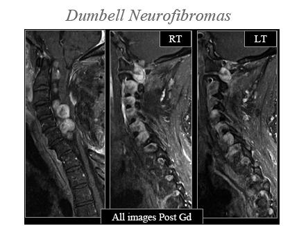
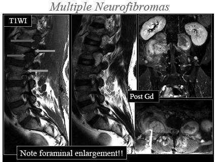
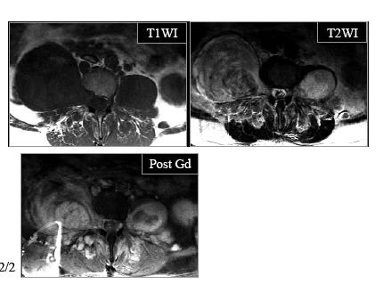
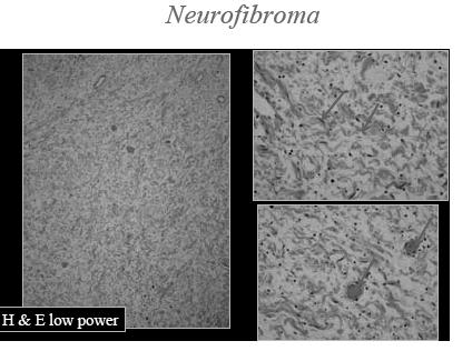
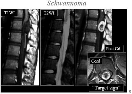
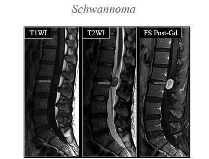
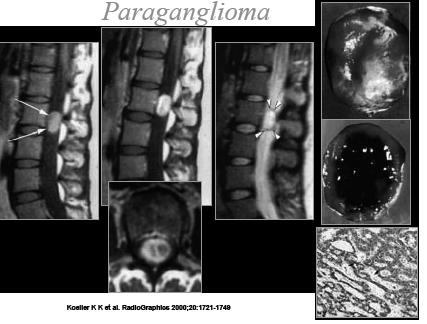
5 Extramedullary-Intradural Lesions Common Neurogenic Neoplasms Neurofibromas Schwannomas Meningiomas Myxopapillary Ependymomas Arachnoiditis Uncommon Lipomas Arachnoid Cysts Epidermoids/ Dermoids Drop metastases AVM Infection Paragangliomas Nerve Sheath Tumors Most common extramedullary-intradural tumors (70%-75%). 15% extradural; 15% dumbell Schwannomas more common than neurofibromas unless patient has NF Neurofibromas: M=F; years. No true capsule Localized, diffuse, or plexiform Schwannomas: M=F; years. Encapsulated 40% cystic changes; 10% hemorrhage; Neural foraminal enlargement; pedicle thinning Isointense on T1WI; Hyperintense on T2WI; Enhance Multiple in neurofibromatosis ARP 105
6 106 ARP


 > Lumbar (4%) More")


7 Meningiomas Second most common EMID spinal tumor (25%) 90% intradural; 10% extradural or dumbell Females > males at 4:1; Primarily 5th-6th decade Thoracic (80%) > Cervical (16%) > Lumbar (4%) More often anterior in cervical region Below C7, more common posterior to cord Usually single unless NF 2 Iso on T1WI; iso or hyperintense on T2WI; hypointense if Ca++ Marked homogeneous enhancement ARP 107


8 Myxopapillary Ependymoma 90% of filum tumors Occur exclusively in conus and filum terminale from ependymal cells in filum Males:females = 2:1; peak between 30 and 40 years Slow growing and may fill entire lumbar spinal canal Vertebral scalloping; canal enlargement Highly vascular hemorrhage is common Iso on T1WI; hyper on T2WI; hypointense margin if hemosiderin Intense enhancement in 100% 108 ARP


9 Other Extramedullary-Intradural Neoplastic Lesions Epidermoid Cysts Less than 1% of spinal tumors Congenital-60%; Aquired-40% Strong association with lumbar puncture in neonatal period (implantation epidermoid) Iso or slightly hyperintense on T1WI; hyper on T2WI Mild rim enhancement DWI can DDx from arachnoid cysts and other lesions Dermoid Cysts Congenital (100%) Symptomatic before age 20; M=F 80% in lumbosacral or cauda Hypointense areas-? Water content from sweat gland secretions Fat hyperintensity on T1WI May cause chemical meningitis if rupture with cholesteol crystals discharged into CSF Drop Metastases CNS Primary Tumors Astrocytomas Medulloblastomas Pineal cell tumors Ependymomas Germ cell tumors Non-CNS Primary Tumors Breast Lung Lymphoma Melanoma Pituitary ARP 109

; Ewing sarcoma and neuroblastoma")

 Lymphoma")


10 Extradural Metastases Most common extradural malignant tumor in adults Spine metastases occur in 15-40% of disseminated cancers Breast, lung, prostate (adults); Ewing sarcoma and neuroblastoma (children) Complications Epidural mass Pathologic fracture Cord compression Epidural Metastases Herniated discs, etc Degenerative lesions (osteophytes, ligament infolding ) Lymphoma Infection (discitis ) Extradural Lesions Common Uncommon Epidural Abscess Meningeal cysts Vertebral tumors-benign Vertebral tumors-malignant Paget s disease Epidural hematoma Epidural lipomatosis Extramedullary hematopoesis Angiolipomas 110 ARP
 Mean age = 50 years (range = 9-77 years) Multiple in 25-30% When discovered incidentally, male = female When")
 Hemangiomas (cont) Plain Films:")
 Treatment for agg.")

11 Primary Vertebral Neoplasms-Benign Hemangioma Aneurysmal bone cyst Osteoblastoma Osteochondroma Eosinophilic granuloma Giant cell tumor Hemangiomas Common benign hamartomatous lesions (10% of adults) Mean age = 50 years (range = 9-77 years) Multiple in 25-30% When discovered incidentally, male = female When symptomatic (neurologic deficit, pain ) F > M Features of compressive/aggressive hemangiomas: Location between T3 and T9 Entire vertebral body involved +/- extension to neural arch Cortex expansion with indistinct margins; soft tissue mass Irregular honeycombing (corduroy appearance) Hemangiomas (cont) Plain Films: Well-defined vertical striations or honeycombed appearance CT: Stippled, polka dot appearance (course trabeculae) MR (typical): T1WI: Hyperintense (due to fat) T2WI: Hyperintense (probably due to cellular elements within the tumor/blood) Treatment for agg. lesions: Sclerotherapy, vertebroplasty, surgery, post-op radiation therapy Aneurysmal Bone Cyst 1-2% of primary bone tumors 10-30% in spine/sacrum Arise in neural arch 80% < 20 years of age Non-malignant hamartomatous lesion-progressively destructive Most common presenting sx = pain, esp. at night (72%) Eggshell cortex; F/F levels from hemorrhage sedimentation Rx: Embolization +/- excision ARP 111


12 Giant Cell Tumors Giant Cell Tumor The spine is the 4th most common location, but only 5% of all giant cells occur here. The sacrum is most common, followed by cervical, thoracic, and lumbar spine. These tumors are more common in the vertebral BODIES. Other tumors such as ABC, osteoblastoma, and osteoid osteoma are more common in the posterior elements. Is there a history of radiation? DDx: Metastasis, plasmacytoma, GCT, chordoma 5% occur in the spine, sacrum most common Involve vertebral BODIES rather than posterior elements 112 ARP

 > L > T] Rare in African-Americans Spine: M:F = 2:1; peak in 5th-6th")

; M>F Most common presenting sx = pain Osteolytic expansion /")

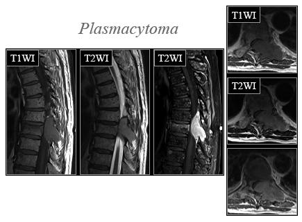
13 Primary Vertebral Neoplasm-Malignant Chordomas Plasmacytomas Ewing s sarcoma Hodgkin s disease Malignant lymphoma Chondrosarcoma Osteogenic sarcoma Chordomas Slow-growing, rare malignant tumors arising from notochordal remnants Sacrococcygeal 50%; Clivus 35%-40%; Vertebral body 10%-15% [C (20-50%) > L > T] Rare in African-Americans Spine: M:F = 2:1; peak in 5th-6th decades Plain films/ct: Destructive lesion; primarily lytic with scattered areas of sclerosis/intratumoral Ca++ MR: Hypointense on T1WI Hyperintense on T2WI Enhancement is typical Distant mets: 5-40% (lung, liver, lymph nodes, bone) Plasmacytoma Mean age = 55 years (younger than patients with MM); M>F Most common presenting sx = pain Osteolytic expansion / compression; easily mimics metastases or benign compression fracture Hypointense on T1WI w/ curvilinear low-signal areas / infoldings caused by endplate fxs Hyperintense on T2WI Mild-mod diffuse enhancement ARP 113


14 114 ARP
Essentials of Clinical MR, 2 nd edition. 51. Primary Neoplasms
 51. Primary Neoplasms As with spinal central canal neoplasms in other regions, those of the lumbar spine may be classified as extradural, intradural extramedullary, and medullary. If an extradural lesion
Spine. Neuroradiology. Spine. Spine Pathology. Distribution of fractures. Radiological algorithm. Role of radiology 18/11/2015
 Spine Neuroradiology Spine Prof.Dr.Nail Bulakbaşı X Ray: AP/L/Oblique Vertebra & disc spaces CT & CTA Vertebra, discs, vessels MRI & MRA Vertebra, disc, vessels, meninges Spinal cord & nerves Myelography
Imaging the Spinal Cord & Intradural Disease
 Department of Radiology University of California San Diego Imaging the Spinal Cord & Intradural Disease John R. Hesselink, M.D. Spinal Cord Diseases Tumors Syringohydromyelia Trauma Ischemia / Infarction
Pediatric Spine Tumors (and other masses)
") Pediatric Spine Tumors (and other masses) Francisco A Perez, MD, PhD Assistant Professor Neuroradiology and Pediatric Radiology Seattle Children s Hospital University of Washington, Seattle Commercial
Pediatric Spine Tumors (and other masses) Francisco A Perez, MD, PhD Assistant Professor Neuroradiology and Pediatric Radiology Seattle Children s Hospital University of Washington, Seattle Commercial
Introduction to Neuroimaging spine. John J. McCormick MD
 Introduction to Neuroimaging spine John J. McCormick MD Neuroanatomy Netter drawings Radiographic Anatomy Cervical Spine Cervical Spine Oblique View Cervical Spine Dens View Thoracic Spine Lumbar Spine
1/9/2013 EXTRAMEDULLARY TUMORS OF THE PEDIATRIC SPINE. Introduction. Classification for Extramedullary Tumors
 EXTRAMEDULLARY TUMORS OF THE PEDIATRIC SPINE Eugene Wang 1/20/12 Dent Neurologic Institute Introduction 2/3 of all intraspinal tumors of childhood are extramedullary 50% Extradural 10-15% Intradural Back
A Journey Down The Canal
 A Journey Down The Canal Radiological Assessment of Spinal Cord Masses John Berry-Candelario HMS III Gillian Lieberman, MD BIDMC Objectives Patient review Anatomy of the spine Imaging techniques Classification
Spinal cord tumours Luc van den Hauwe et al.
 overview spinal cord tumours L. van den Hauwe 1,2, D. Balériaux 3, J.W. Van Goethem 2, C. Venstermans 2, F. De Belder 2, P.M. Parizel 2 introduction imaging spinal tumour classification spinal cord tumours
Ependymoma of the spine
 Ependymoma of the spine Tenny Zhang, MS-3 Harvard Medical School 1 Case presentation: history and exam HPI: A 30-year-old man with no significant past medical history presents with one week of bilateral
RINGS N THINGS: Imaging Patterns in Differential Diagnosis. Anne G. Osborn, M.D.
 RINGS N THINGS: Imaging Patterns in Differential Diagnosis Anne G. Osborn, M.D. ExpDDxs: Intra-axial (Parenchymal) Lesions Ring-enhancing lesions, solitary 1 Ring-enhancing lesion crossing corpus callosum
NEURORADIOLOGY. Part III. Angela Csomor University of Szeged Department of Radiology
 NEURORADIOLOGY Part III Angela Csomor University of Szeged Department of Radiology DISEASES OF SPINE AND SPINAL CORD I. Non-tumourous diseases developmental anomalies vascular disorders inflammatory processes
Kathleen R. Fink, MD Virginia Mason Medical Center. 6 th Nordic Emergency Radiology Course 2017
 Kathleen R. Fink, MD Virginia Mason Medical Center 6 th Nordic Emergency Radiology Course 2017 Disclosure My spouse receives research salary support from: Guerbet Outline Acute neck and back pain Acute
Vertebral and Paravertebral Diseases
 Department of Radiology University of California San Diego Vertebral and Paravertebral Diseases John R. Hesselink, M.D. Vertebral / Paravertebral Disease (Extradural) Metastatic disease Primary bone tumors
Bone Tumors Clues and Cues
 William Herring, M.D. 2002 Bone Tumors Clues and Cues In Slide Show mode, advance the slides by pressing the spacebar All Photos Retain the Copyright of their Authors Clues by Appearance of Lesion Patterns
PITUITARY PARASELLAR LESIONS. Kim Learned, MD
 PITUITARY PARASELLAR LESIONS Kim Learned, MD DIFFERENTIALS Pituitary Sella Clivus, Sphenoid Sinus Suprasellar Optic chiasm, Hypothalamus, Circle of Willis Parasellar Cavernous Sinus Case 1 17 YEAR-OLD
Neuroimaging. spine / spinal cord
 Neuroimaging spine / spinal cord Spine & spinal cord imaging methodology Plain x-ray of spine Computed tomography CT - traditional ( normal CT) - reconstructions - myelo-ct Magnetic resonance MR - standard
AMERICAN ACADEMY OF NEUROLOGY SPINE FELLOWSHIP CORE CURRICULUM
 AMERICAN ACADEMY OF NEUROLOGY SPINE FELLOWSHIP CORE CURRICULUM Introduction Spine conditions affect virtually everyone at some time during their life. Surveys indicate a yearly prevalence of spine-related
Brain Tumors. Medulloblastoma. Pilocytic astrocytoma: Ahmed Koriesh, MD. Pathological finding
 NeuroPathology Page 8 Brain Tumors Pathological finding Pseudorosette Rosenthal fibers Rosettes Wet Keratin Psammoma bodies Fried egg Tumor Ependymoma, SEGA Pilocytic astrocytoma Medulloblastoma Craniopharyngioma
IMAGING OF A CASE OF SPINAL MENINGIOMA- A CASE REPORT
 IMAGING OF A CASE OF SPINAL MENINGIOMA- A CASE REPORT Ramneet Wadi 1, Anil Kumar Shukla 2, Seetha Pramila V. V 3, Sabyasachi Basu 4, Sonam Sanjay 5 1Postgraduate Student, Department of Radiodiagnosis,
MARK D. MURPHEY MD, FACR. Physician-in-Chief, AIRP. Chief, Musculoskeletal Imaging
 ALPHABET SOUP AND CYSTIC LESIONS OF THE BONE MARK D. MURPHEY MD, FACR Physician-in-Chief, AIRP Chief, Musculoskeletal Imaging ALPHABET SOUP AND CYSTIC LESIONS OF THE BONE Giant cell tumor (GCT) Unicameral
Pediatric Spinal Anomalies
 Department of Radiology University of California San Diego Pediatric Spinal Anomalies John R. Hesselink, M.D. Spine Embryogenesis 1. Primitive streak 2. Proliferation of cells at primitive pit (Hensen's
ESSENTIALS OF PLAIN FILM INTERPRETATION: SPINE DR ASIF SAIFUDDIN
 ESSENTIALS OF PLAIN FILM INTERPRETATION: SPINE DR ASIF SAIFUDDIN Consultant Musculoskeletal Radiologist Royal National Orthopaedic Hospital Stanmore,UK. INTRODUCTION 2 INTRODUCTION 3 INTRODUCTION Spinal
4/14/2017. Unknown Case #1 Intramedullary Lesion
 4/14/2017 Intradural, Intramedullary Tumor or Mimic Intradural, Extramedullary Tumor or Mimic Extradural Tumor or Mimic Unknown Case #1 Intramedullary Lesion Unknown Case #1 -POST - MAG AXIAL Cranial Caudal
General: Brain tumors are lesions that have mass effect distorting the normal tissue and often result in increased intracranial pressure.
 1 Lecture Objectives Know the histologic features of the most common tumors of the CNS. Know the differences in behavior of the different tumor types. Be aware of the treatment modalities in the various
Tumors of the Nervous System
 Tumors of the Nervous System Peter Canoll MD. PhD. What I want to cover What are the most common types of brain tumors? Who gets them? How do they present? What do they look like? How do they behave? 1
ORIGINAL ARTICLE. Abstract. Aim. Materials and methods. Introduction. Results
 Is anatomical distribution helpful for differentiating TB spondylitis from neoplastic causes of extradural spinal cord compression in children? A pilot study Reena George, MD, MMed Rad, FRCR (UK) Savvas
Intradural spinal tumours and their mimics: a review of radiographic features
 Department of Radiology, Royal Melbourne Hospital, Parkville, Victoria, Australia Correspondence to Dr Sara Wein, Radiology Department, Royal Melbourne Hospital, Grattan Street, Parkville, VIC 3050, Australia;
Tumors of the Central Nervous System
 Tumors of the Central Nervous System 1 Financial Disclosures I have NO SIGNIFICANT FINANCIAL, GENERAL, OR OBLIGATION INTERESTS TO REPORT Introduction General: Brain tumors are lesions that have mass effect
CT & MRI Evaluation of Brain Tumour & Tumour like Conditions
 CT & MRI Evaluation of Brain Tumour & Tumour like Conditions Dr. Anjana Trivedi 1, Dr. Jay Thakkar 2, Dr. Maulik Jethva 3, Dr. Ishita Virda 4 1 M.D. Radiology, Professor and Head, P.D.U. Medical College
IMAGING OF SPINE TUMORS
 IMAGING OF SPINE TUMORS Laszlo Mechtler, MD, FAAN, FASN SUMMARY Historic classification of spine tumors based on computed tomography myelopgraphy. (A) Normal, (B) extradural extramedullary, (C) intradural
Imaging of Hearing Loss
 Contemporary Imaging of Sensorineural Hearing Loss Imaging of Hearing Loss Discussion Outline (SNHL) Imaging Approaches Anatomic Relationships Lesions: SNHL KL Salzman, MD University of Utah School of
11 May Disclosure. + Outline: Acute Spine Emergencies
 Kathleen R. Fink, MD University of Washington 5 th Nordic Emergency Radiology Course May 21, 2015 Disclosure My spouse receives research salary support from: Bracco BayerHealthcare Guerbet K Fink Nordic
Case Studies in the Skull Base
 Case Studies in the Skull Base Amy C Tsai, MD Neuroradiology Fellow Department of Radiology and Imaging Sciences University of Utah Health Sciences Center Salt Lake City, Utah, USA No disclosures related
Role of Magnetic Resonance Imaging in the Evaluation of Compressive Myelopathy in Rohilkhand Region, India
 Mohit Agarwal et al Original article 10.5005/jp-journals-10050-10091 Role of Magnetic Resonance Imaging in the Evaluation of Compressive Myelopathy in Rohilkhand Region, India 1 Mohit Agarwal, 2 Pramod
CNS TUMORS. D r. Ali Eltayb ( U. of Omdurman. I ). M. Path (U. of Alexandria)
. M. Path (U. of Alexandria)") CNS TUMORS D r. Ali Eltayb ( U. of Omdurman. I ). M. Path (U. of Alexandria) CNS TUMORS The annual incidence of intracranial tumors of the CNS ISmore than intraspinal tumors May be Primary or Secondary
CNS TUMORS D r. Ali Eltayb ( U. of Omdurman. I ). M. Path (U. of Alexandria) CNS TUMORS The annual incidence of intracranial tumors of the CNS ISmore than intraspinal tumors May be Primary or Secondary
Brain tumors: tumor types
 Brain tumors: tumor types Tumor types There are more than 120 types of brain tumors. Today, most medical institutions use the World Health Organization (WHO) classification system to identify brain tumors.
SPINAL MAGNETIC RESONANCE IMAGING INTERPRETATION
 CLINICAL VIGNETTE 2017; 3:2 SPINAL MAGNETIC RESONANCE IMAGING INTERPRETATION Editor-in-Chief: Idowu, Olufemi E. Neurological surgery Division, Department of Surgery, LASUCOM/LASUTH, Ikeja, Lagos, Nigeria.
Spinal Vascular Lesions
 Spinal Vascular Lesions Spinal Vascular Lesions Spinal cord infarction Hemangioblastoma Cavernous malformation Vascular malformations (Type 1-4) Spinal artery aneurysm Troy Hutchins, MD Assistant Professor
Tumors and tumorlike lesions of the craniovertebral junction
 Tumors and tumorlike lesions of the craniovertebral junction Poster No.: C-2632 Congress: ECR 2010 Type: Educational Exhibit Topic: Neuro Authors: S.-C. Hung, Y. L. Chen, W.-Y. Guo, G.-M. Yang ; Taoyuan/
Imaging of Sacral Masses: Self-Assessment Module
 1.5 CME AJR Integrative Imaging LIFELONG LEARNING FOR RADIOLOGY Imaging of Sacral Masses: Self-Assessment Module Alice S. Ha 1, Felix S. Chew ABSTRACT The educational objectives for this self-assessment
Metastasis. 57 year old with progressive Headache and Right Sided Visual Loss
 Metastasis 1% of sellar/parasellar masses Usually occurs with known primary Can involve third ventricle, hypothalamus, infundibular stalk May be both supra-, intrasellar 57 year old with progressive Headache
SHORT OVERVIEW OF SPINAL CORD TUMORS
 SHORT OVERVIEW OF SPINAL CORD TUMORS 1 INTRODUCTION RARE HETEROGENEOUS GROUP OF TUMORS. 15%OFALLPRIMARYCNSNEOPLASMSARISEINTHESC. INCIDENCE HIGHER IN MALES THAN FEMALES AGE 10TO40YRS MOST PRIMARIES ARE
Tumors and pseudotumors of the spine: a review of the main aspects in computed tomography and magnetic resonance imaging.
 Tumors and pseudotumors of the spine: a review of the main aspects in computed tomography and magnetic resonance imaging. Poster No.: C-1851 Congress: ECR 2012 Type: Educational Exhibit Authors: A. A.
102 Results RESULTS. Age Mean=S.D Range 42= years -84 years Number % <30 years years >50 years
 102 Results RESULTS A total of 50 cases were studied 39 males and 11females.Their age ranged between 16 years and 84 years (mean 42years). T1 and T2WI were acquired for all cases in sagittal and axial
The Radiology Assistant : Bone tumor - ill defined osteolytic tumors and tumor-like lesions
 Bone tumor - ill defined osteolytic tumors and tumor-like lesions Henk Jan van der Woude and Robin Smithuis Radiology department of the Onze Lieve Vrouwe Gasthuis, Amsterdam and the Rijnland hospital,
JMSCR Vol 04 Issue 04 Page April 2016
 www.jmscr.igmpublication.org Impact Factor 5.244 Index Copernicus Value: 5.88 ISSN (e)-2347-176x ISSN (p) 2455-0450 DOI: http://dx.doi.org/10.18535/jmscr/v4i4.13 Malignant Nerve Sheath Tumor with Secondary
Peter Canoll MD. PhD.
 Tumors of the Nervous System Peter Canoll MD. PhD. What I want to cover What are the most common types of brain tumors? Who gets them? How do they ypresent? What do they look like? How do they behave?
Primary bone tumors > metastases from other sites Primary bone tumors widely range -from benign to malignant. Classified according to the normal cell
 Primary bone tumors > metastases from other sites Primary bone tumors widely range -from benign to malignant. Classified according to the normal cell counterpart and line of differentiation. Among the
Clinico-pathological study of intradural extramedullary spinal tumors
 International Journal of Research in Medical Sciences Venugopal G et al. Int J Res Med Sci. 2015 Oct;3(10):2795-2799 www.msjonline.org pissn 2320-6071 eissn 2320-6012 Research Article DOI: http://dx.doi.org/10.18203/2320-6012.ijrms20150685
Pathologic Analysis of CNS Surgical Specimens
 2015 Kenneth M. Earle Memorial Neuropathology Review Pathologic Analysis of CNS Surgical Specimens Peter C. Burger, MD Interdisciplinary Quality Control Familiarity with entities Use of diagnostic algorithm
MRI Findings of Giant Cell Tumors of the Spine
 MRI of Spinal Tumors Musculoskeletal Imaging Clinical Observations Jong Won Kwon 1 Hye Won Chung 1,2 Eun Yoon Cho 3 Sung Hwan Hong 4 Sang-Hee Choi 1 Young Cheol Yoon 1 Sang Kyu Yi 1 Kwon JW, Chung HW,
NEURORADIOLOGY DIL part 5
 NEURORADIOLOGY DIL part 5 Masses and tumors K. Agyem MD, G. Hall MD, D. Palathinkal MD, Alexandre Menard March/April 2015 OVERVIEW Introduction to Neuroimaging - DIL part 1 Basic Brain Anatomy - DIL part
APPROPRIATE USE GUIDELINES
 APPROPRIATE USE GUIDELINES Appropriateness of Advanced Imaging Procedures (MRI, CT, Bone Scan/PET) in Patients with Neck Pain CDI QUALITY INSTITUTE: PROVIDER LED ENTITY (PLE) Updated June, 2017 Contents
Dumbbell Shaped Thoracic Spine Cavernous Hemangioma: A Case Report and Review of the Literature
 ISPUB.COM The Internet Journal of Neurosurgery Volume 3 Number 1 Dumbbell Shaped Thoracic Spine Cavernous Hemangioma: A Case Report and Review of the Literature J Gonzalez-Cruz, A Nanda Citation J Gonzalez-Cruz,
Optic Pathway Gliomas, Germinomas, Spinal Cord Tumours. Colin Kennedy March 2015
 Optic Pathway Gliomas, Germinomas, Spinal Cord Tumours Colin Kennedy March 2015 Glioma of the optic chiasm. T1-weighted MRI with gadolinium enhancement, showing intense irregular uptake of contrast. The
Pediatric Retroperitoneal Masses Radiologic-Pathologic Correlation
 Acta Radiológica Portuguesa, Vol.XVIII, nº 70, pág. 61-70, Abr.-Jun., 2006 Pediatric Retroperitoneal Masses Radiologic-Pathologic Correlation Marilyn J. Siegel Mallinckrodt Institute of Radiology, Washington
Sacral bone Tumor Imaging
 Sacral bone Tumor Imaging Poster No.: C-1651 Congress: ECR 2016 Type: Educational Exhibit Authors: S. Zaouali, M. Ben Ammar, A. DAGHFOUS, M. Matri, L. REZGUI; tunis/tn Keywords: Bones, Oncology, Neuroradiology
In-Training Examination for Diagnostic Radiology Residents Rationales
 28th Annual In-Training Examination for Diagnostic Radiology Residents Rationales Sponsored by: Commission on Education Committee on Residency Training in Diagnostic Radiology February 3, 2005 The American
The surgical treatment of metastatic disease of the spine
 The surgical treatment of metastatic disease of the spine Péter Banczerowski National Institute of Neurosurgery, Budapest Spine tumours 15% of the primary tumours of the CNS affect the spine The spine
Pediatric back pain and diagnostic strategies
 ANDREA ROSSI, MD Department of Pediatric Neuroradiology G. Gaslini Children s Research Hospital Genoa Italy Pediatric back pain and diagnostic strategies Pediatric back pain: an underestimated problem
Imaging Of Cystic Paravertebral Masses:
 Imaging Of Cystic Paravertebral Masses: Differential Diagnosis and Key Discriminators John P. Lichtenberger III, MD, Maj, USAF, MC Brent McCarragher, MD, CPT, USA John R. Dryden, MD, LT, USN P. Gabriel
Intradural and extradural dorsal spinal pediatric lesions
 Intradural and extradural dorsal spinal pediatric lesions Poster No.: C-2520 Congress: ECR 2015 Type: Educational Exhibit Authors: L. Riaza, M. Pérez Rubiralta, C. Perez Balagueró, M. Rebollo, 1 2 4 3
Adult - Cerebrovascular. Adult - Cranio-Cervical Junction. Adult - Epilepsy. Adult - Hydrocephalus
 list for SET and IMG Neurosurgery Adult - Cerebrovascular Aneurysm - Clipping: Anterior circulation Aneurysm - Clipping: Posterior circulation AVM excision Carotid endarterectomy Carotid trapping Cavernoma
SACRAL LESIONS: Differential diagnosis for the general radiologist
 SACRAL LESIONS: Differential diagnosis for the general radiologist Poster No.: C-2147 Congress: ECR 2017 Type: Educational Exhibit Authors: M. R. Campos Arenas, M. C. Sánchez-Porro, J. Garrido Rull ; 1
Extramedullary Spinal SOL Outcome of Surgery
 TAJ June 2009; Volume 22 Number 1 ISSN 1019-8555 The Journal of Teachers Association RMC, Rajshahi Original Article Extramedullary Spinal SOL Outcome of Surgery M Zahed Hossain 1, Monzurul Hoque 2 Abstract
Spine and spinal cord
 NEURORADIOLOGY Spine and spinal cord Erika Vörös University of Szeged Department of Radiology SZEGED DISEASES OF SPINE AND SPINAL CORD I. Non-tumourous diseases developmental anomalies vascular disorders
Patients Treated with Leksell Gamma Knife
 Patients Treated with Leksell Gamma Knife 1968-2016 TREATMENTS REPORTED 2016 BY REGION AND INDICATION INDICATION Asia excl. Europe Latin Middle East & Africa North Grand Total Benign Tumors 12283 9778
ISSN X (Print) Original Research Article. DOI: /sjams
 Original Research Article. DOI: /sjams") DOI: 10.21276/sjams Scholars Journal of Applied Medical Sciences (SJAMS) Sch. J. App. Med. Sci., 27; 5(8A):32-31 Scholars Academic and Scientific Publisher (An International Publisher for Academic and
DOI: 10.21276/sjams Scholars Journal of Applied Medical Sciences (SJAMS) Sch. J. App. Med. Sci., 27; 5(8A):32-31 Scholars Academic and Scientific Publisher (An International Publisher for Academic and
Evaluation of Neck Mass. Disclosure. Learning Objectives 3/24/2014. Karen T. Pitman MD, FACS Banner MDACC, Gilbert AZ. Nothing to disclose
 Evaluation of Neck Mass Karen T. Pitman MD, FACS Banner MDACC, Gilbert AZ Nothing to disclose Disclosure Learning Objectives 1. Describe a systematic method to evaluate a patient with a neck mass 2. Select
Contents. Basic Ultrasound Principles and Terminology. Ultrasound Nodule Characteristics
 Contents Basic Ultrasound Principles and Terminology Basic Ultrasound Principles... 1 Ultrasound System... 2 Linear Transducer for Superficial Images and Ultrasound-Guided FNA... 3 Scanning Planes... 4
Role of MRI in the Evaluation of Compressive Myelopathy
 IOSR Journal of Dental and Medical Sciences (IOSR-JDMS) e-issn: 2279-0853, p-issn: 2279-0861.Volume 15, Issue 4 Ver. XIII (Apr. 2016), PP 21-26 www.iosrjournals.org Role of MRI in the Evaluation of Compressive
Imaging Findings of Sacral Tumors 1
 Imaging Findings of Sacral Tumors 1 Seung Ho Kim, M.., Sung Hwan Hong, M.., Ja-Young hoi, M.., Sung Hye Koh, M.., Hye Won hung, M.., Jung-h hoi, M.., Heung Sik Kang, M.. The various pathologic conditions
Pelvic tumor in childhood Classification, imaging approach and radiological findings
 Pelvic tumor in childhood Classification, imaging approach and radiological findings M. Mearadji International Foundation for Pediatric Imaging Aid Rotterdam, The Netherlands Solid pelvic masses in childhood
PATHOLOGY, EPIDEMIOLOGY. EXTRADURAL SPINAL TUMORS, VERTEBRAL TUMORS Onc56 (1)
") EXTRADURAL SPINAL TUMORS, VERTEBRAL TUMORS Onc56 (1) Extradural Spinal Tumors, Vertebral Tumors Last updated: September 5, 2017 PATHOLOGY, EPIDEMIOLOGY... 1 Metastases... 1 Primary Vertebral Tumors...
EXTRADURAL SPINAL TUMORS, VERTEBRAL TUMORS Onc56 (1) Extradural Spinal Tumors, Vertebral Tumors Last updated: September 5, 2017 PATHOLOGY, EPIDEMIOLOGY... 1 Metastases... 1 Primary Vertebral Tumors...
Cross sectional imaging of Intracranial cystic lesions Abdel Razek A
 Cross sectional imaging of Intracranial cystic lesions Abdel Razek A Department of Radiology. Mansoura Faculty of Medicine, Mansoura. Egypt. arazek@mans.edu.eg Introduction Intracranial cystic lesions
Contrast Guidelines for Common CT/CTA & MRI/MRA
 Contrast Guidelines for Common /A & /MRA Body Imaging Gastrointestinal CLINICAL GUIDELINES EXAM DESCRIPTION /A CPT CODES EXAM DESCRIPTION /MRA CPT CODES Abdominal mass Abdomen & Pelvis w 74177 Abdomen
Magnetic resonance evaluation of intra dural spinal tumors with histopathology correlation
 International Journal of Research in Medical Sciences Rao UM et al. Int J Res Med Sci. 2015 Nov;3(11):3051-3057 www.msjonline.org pissn 2320-6071 eissn 2320-6012 Research Article DOI: http://dx.doi.org/10.18203/2320-6012.ijrms20151002
Magnetic resonance imaging of intramedullary spinal cord lesions
 Magnetic resonance imaging of intramedullary spinal cord lesions Poster No.: C-1762 Congress: ECR 2014 Type: Educational Exhibit Authors: M. Abdelkafi, H. Derbel, H. Abid, S. Haddar, B. Souissi, N. 1 1
Neuroimaging Core Curriculum
 Neuroimaging Core Curriculum Program Content The purpose of the training program is to prepare the physician for the independent practice of neuroimaging. Neuroimaging is the subspecialty of Neurology
Objectives. Comprehension of the common spine disorder
 Objectives Comprehension of the common spine disorder Disc degeneration/hernia Spinal stenosis Common spinal deformity (Spondylolisthesis, Scoliosis) Osteoporotic fracture Destructive spinal lesions Anatomy
Neckmasses in infancy and childhood: Clinical and radiological classification and imaging approaches M. Mearadji
 Neckmasses in infancy and childhood: Clinical and radiological classification and imaging approaches M. Mearadji International Foundation for Pediatric Imaging Aid Introduction Neck masses are a frequent
Benign brain lesions
 Benign brain lesions Diagnostic and Interventional Radiology Hung-Wen Kao Department of Radiology, Tri-Service General Hospital, National Defense Medical Center Computed tomography Hounsfield unit (HU)
Pathology with CT Sectional Anatomy Correlation, Part I. Tom Haller, RT (R)(CT)(MR)
(CT)(MR)") Pathology with CT Sectional Anatomy Correlation, Part I Tom Haller, RT (R)(CT)(MR) Objectives Upon completion of the course, the participant will be able to: 1. State pathologies that commonly require
Pathology with CT Sectional Anatomy Correlation, Part I Tom Haller, RT (R)(CT)(MR) Objectives Upon completion of the course, the participant will be able to: 1. State pathologies that commonly require
MAGNETIC RESONANCE IMAGING OF SPINAL TUMORS
 MAGNETIC RESONANCE IMAGING OF SPINAL TUMORS A study using a 0.3 T vertical magnetic field MING HUA LI - HEXL- Lund 992 Organization LUND UNIVERSITY Department of Radiology University Hospital S-22 85 Lund,
CTA/MRA of Pediatric Hepatic Masses Radiology-Pathology Correlation
 Acta Radiológica Portuguesa, Vol.XVIII, nº70, pág. 41-50, Abr.-Jun., 2006 CTA/MRA of Pediatric Hepatic Masses Radiology-Pathology Correlation Marilyn J. Siegel Mallinckrodt Institute of Radiology, Washington
Spinal meningioma imaging
 Spinal meningioma imaging Poster No.: C-0448 Congress: ECR 2018 Type: Educational Exhibit Authors: M. Smoljan, D. Zadravec ; Zagreb/HR, Zageb/HR Keywords: Neoplasia, Imaging sequences, Education, MR, CT,
AMSER Rad-Path Case of the Month January 2019
 AMSER Rad-Path Case of the Month January 2019 Intradural Spinal Tumor Ashley Graziano OMS IV, Lake Erie College of Osteopathic Medicine Dr. Matthew Hartman M.D., Allegheny Health Network Dr. David Oliver-Smith
A Comprehensive Review of Intraspinal tumors: Diagnostic, classification and radio-pathologic correlation.
 A Comprehensive Review of Intraspinal tumors: Diagnostic, classification and radio-pathologic correlation. Award: Magna Cum Laude Poster No.: C-2112 Congress: ECR 2013 Type: Educational Exhibit Authors:
Index. aneurysm, 92 carotid occlusion, 94 ICA stenosis, 95 intracranial, 92 MCA, 94
 A ADC. See Apparent diffusion coefficient (ADC) Aneurysm cerebral artery aneurysm, 93 CT scan, 93 gadolinium, 93 Angiography, 13 Anoxic brain injury, 25 Apparent diffusion coefficient (ADC), 7 Arachnoid
Accuracy of intraoperative frozen section diagnosis in spinal cord lesions
 Accuracy of intraoperative frozen section diagnosis in spinal cord lesions Department of Orthopedic Surgery Niigata University Medical and Dental Hospital Toru Hirano, Kei Watanabe, Keiichi Katsumi, Masayuki
A Comprehensive Review of Intraspinal tumors: Diagnostic, classification and radio-pathologic correlation.
 A Comprehensive Review of Intraspinal tumors: Diagnostic, classification and radio-pathologic correlation. Award: Magna Cum Laude Poster No.: C-2112 Congress: ECR 2013 Type: Educational Exhibit Authors:
Hidayatullah Hamidi. MD Consultant Radiologist. Lumbar Spine MR Imaging Interpretation
 Hidayatullah Hamidi. MD Consultant Radiologist Lumbar Spine MR Imaging Interpretation 13/12/2018 Presenter Hidayatullah Hamidi Consultant Radiologist, Radiology PGME program director, FMIC, Kabul, Afghanistan
Primary Bone Tumors: Spine Surgery Live -Video Techniques Mobile Spine
 Primary Bone Tumors: Spine Surgery Live -Video Techniques Mobile Spine Christopher Ames MD Professor of Neurosurgery and Orthopedic Surgery Director of Spine Tumor And Deformity Surgery UCSF Department
Giant cystic intradural extramedullary pilocytic astrocytoma of Cauda equina
 1 di 7 13/03/2014 09.02 J Neurosci Rural Pract. 2013 Oct-Dec; 4(4): 453 456. doi: 10.4103/0976-3147.120217 PMCID: PMC3858770 Giant cystic intradural extramedullary pilocytic astrocytoma of Cauda equina
Imaging in neurofibromatosis type 1: An original research article with focus on spinal lesions
 Original Research Article Imaging in neurofibromatosis type 1: An original research article with focus on spinal lesions Kalpesh Patel 1*, Siddharth Zala 2, C. Raychaudhuri 3 1 Assistant Professor, 2 1
Mediastinal Tumors: Imaging
 Mediastinal Tumors: Imaging References Imaging in Oncology, Husband and Reznek Computed Tomography and Magnetic Resonance of the thorax, Naidich, Zerhouni, Siegelman, Mediastinal compartments Anterior:
Spinal Imaging. Bearbeitet von Herwig Imhof. 1. Auflage Taschenbuch. 312 S. Paperback ISBN Format (B x L): 12,5 x 19 cm
: 12,5 x 19 cm") Spinal Imaging Bearbeitet von Herwig Imhof 1. Auflage 2007. Taschenbuch. 312 S. Paperback ISBN 978 3 13 144071 6 Format (B x L): 12,5 x 19 cm Weitere Fachgebiete > Medizin > Sonstige Medizinische Fachgebiete
Spinal Imaging Bearbeitet von Herwig Imhof 1. Auflage 2007. Taschenbuch. 312 S. Paperback ISBN 978 3 13 144071 6 Format (B x L): 12,5 x 19 cm Weitere Fachgebiete > Medizin > Sonstige Medizinische Fachgebiete
Pediatric CNS Tumors. Disclosures. Acknowledgements. Introduction. Introduction. Posterior Fossa Tumors. Whitney Finke, MD
 Pediatric CNS Tumors Disclosures Whitney Finke, MD Neuroradiology Fellow PGY-6 University of Utah Health Sciences Center Salt Lake City, Utah None Acknowledgements Introduction Nicholas A. Koontz, MD Luke
Advances In Orbital Neuropathology
 Advances In Orbital Neuropathology Charles G. Eberhart, MD PhD Associate Professor of Pathology, Ophthalmology and Oncology Johns Hopkins University School of Medicine Overview Non-neoplastic lesions Microphthalmos/pseudoglioma
A Patient s Guide to Spinal Tumors
 A Patient s Guide to Spinal Tumors 763 Larkfield Road 2nd Floor Commack, NY 11725 Phone: (631) 462-2225 Fax: (631) 462-2240 DISCLAIMER: The information in this booklet is compiled from a variety of sources.