PET-CT for radiotherapy planning in lung cancer: current recommendations and future directions
|
|
|
- Prosper Bond
- 6 years ago
- Views:
Transcription


1 PET-CT for radiotherapy planning in lung cancer: current recommendations and future directions Gerry Hanna Centre for Cancer Research and Cell Biology Queen s University of
2 Talk Outline Key principles underlying PET/CT acquisition PET/CT for staging in NSCLC Background of PET/CT for RTP in NSCLC How to acqiure a PET/CT for RTP? Displaying a PET/CT for RTP Guidance on PET/CT based TVD 4D PET/CT
3 KEY PRINCIPLES OF PET

4 Glucose is key to PET
5 Positron Emission Tomography (PET) An imaging process based on the decay of a nucleus by positron emission CH 2 OH O OH OH OH F 18
6 Positron Emission Tomography (PET) An imaging process based on the decay of a nucleus by positron emission γ (511 KeV) e + e - Annihilation γ (511 KeV) CH 2 OH O OH OH OH F 18
7 Positron Emission Tomography (PET) An imaging process based on the decay of a nucleus by positron emission γ (511 KeV) e + e - CH 2 OH O OH OH OH F 18 γ (511 KeV) Annihilation Detector Ring e kev 511 kev β + Coincidence Unit
 e + e - CH 2 OH O OH OH OH F 18 γ (511 KeV) Annihilation e - 511 kev 511 kev β + Coincidence")
8 Positron Emission Tomography (PET) An imaging process based on the decay of a nucleus by positron emission γ (511 KeV) e + e - CH 2 OH O OH OH OH F 18 γ (511 KeV) Annihilation e kev 511 kev β + Coincidence Unit
 e + e - CH 2 OH O OH OH OH F 18 γ (511 KeV) Annihilation e - 511 kev 511 kev β + Coincidence")
9 Positron Emission Tomography (PET) An imaging process based on the decay of a nucleus by positron emission γ (511 KeV) e + e - CH 2 OH O OH OH OH F 18 γ (511 KeV) Annihilation e kev 511 kev β + Coincidence Unit
10 Metabolic Tracers Specific molecules That are involved in a metabolic pathway of interest That accumulate in the presence of a specific disease That are chemically feasible for stable labeling That are applicable for human use Example tracers Cell metabolism Glucose - F18 DNA synthesis Thymidine - F18 Tracer Molecule Cell membrane synthesis Choline - C11 Octreotide receptor expression DOTA-NOC - Ga68 Tissue hypoxia Misonidazole - F18 Label
11 Radionuclides and Radiopharmaceuticals in use Half-life (min) Max β+ energy (MeV) Max Range (mm) Radionuclide Radiopharmaceutical Clinical Use 11 C N O F Nitrogen N Ammonia Blood Flow (cardiology) Oxygen O Water Blood Flow Carbon C Methionine Amino Acid Metabolism Fluorine F - FDG Fluoro-deoxyglucose 18 F MISO Fluoromisonidazole 18 F FLT Fluoro-deoxythymidine Glucose Metabolism Hypoxia Tumour proliferation
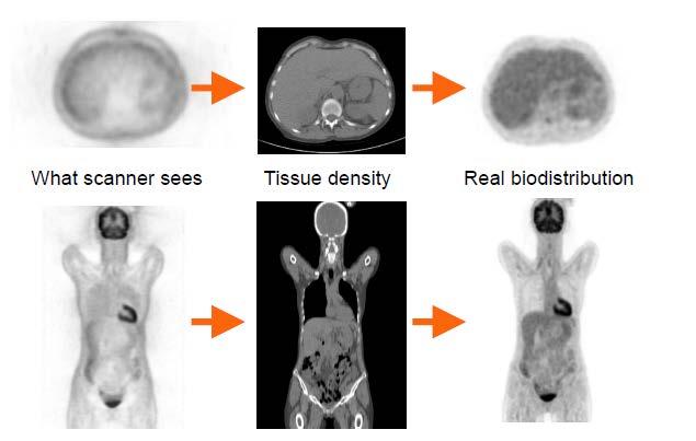
12 Attenuation Correction
13

14 PET in Oncology Functional imaging technique : Many tumour cells have increased glycolysis rate, leading to increased FDG uptake PET-CT images from radiotherapy planning patient Current and possible uses diagnosis of primary tumours staging evaluation of response to treatment target volumes for radiotherapy Evaluation of radiation damage
15 PET/CT FOR STAGING IN NSCLC
16 ROC Curve for LN staging in NSCLC From Birim O, et al. Ann Thorac Surg 2005;79:375 81
17 ROC Curve for LN staging in NSCLC Staging modality is poor From Birim O, et al. Ann Thorac Surg 2005;79:375 81
18 ROC Curve for LN staging in NSCLC Staging modality is excellent From Birim O, et al. Ann Thorac Surg 2005;79:375 81
19 ROC Curve for LN staging in NSCLC Only 5-10% of Malignant LN are being missed by PET From Birim O, et al. Ann Thorac Surg 2005;79:375 81
20 PET for Patient Selection for Radical Therapy MacManus MP, Int J Radiat Oncol Biol Phys 2002;52:351
21 PET/CT for RT planning in NSCLC

22 Why use PET/CT in RT Planning in NSCLC? CT current gold standard for GTV definition in the radical treatment of NSCLC with radiotherapy. Despite technical improvements in RT delivery, increased use of systemic therapies and more accurate staging, survival remains poor. > 50% local failure despite radical local therapy PET/CT is more accurate than CT alone in the staging of NSCLC.
23 Problems with using CT alone for GTV Definition Giraud P et al. Radiotherapy and Oncology 2002;62:27 36
24 Intra-observer Variability Ciernik et al, Red 2003


25 Intra-observer Variability Ciernik et al, Red 2003 Upstaging with PET Bradley et al Red 2004
26 PET/CT in RTP Atelectasis Significant potential benefit by reducing RT volumes However: False positive uptake in postobstructive inflammation Histological correlation of PET findings with pathology are lacking

27 PET/CT in RTP Atelectasis Significant potential benefit by reducing RT volumes However: False positive uptake in postobstructive inflammation Histological correlation of PET findings with pathology are lacking Nestle U, et al. Int J Radiat Oncol Biol Phys 1999;44:
28 HOW TO ACQIURE A PET/CT FOR RTP
29 PET acquisition for RTP - Options 1. Staging PET/CT A. Visually correlated with the RTP CT scan: used only as a diagnostic aid to identify areas of disease location during treatment planning session (visual correlation) B. Registered to a RTP CT scan (Registration and Patient Positioning Issues) 1. Dedicated RTP PET/CT A. Combined RTP / staging with whole body PET/CT scan as single scan B. Dedicated RTP scan after a staging PET/CT
30 Recommendations about registering a PET/CT to a RTP CT PET/CT images should be registered using a rigid registration (for 4DCT register to the average intensity projection (Ave-IP) scan) Registration should focus on bony anatomy which is not affected by respiratory motion (e.g. spinal column) It is advised that this approach should not be used routinely for gated treatments

31 Lung board set-up for RTP PET/CT Requirements Flat bed couch insert Laser lights alignment system QA of image registration Fixed slots on the scanner Appropriate staff i. Nuclear medicine technical officer ii. Therapy radiographer iii. Medical Physics staff Arms positioned above the head, T-bar grip and arm supports. Small bore - restricts the positioning of the arms and prevents tilting of the lung board


32 Patient set-up on RTP PET/CT
33 Protocol for RTP PET Scan Cold session (pre-fdg injection) patient positioning, initial marking and set-up Hot session (post injection and 45 minute uptake period) re-position patient, attach radio-opaque markers Images acquired using routine diagnostic PET/CT scan protocol Permanent marks made on patient (post scan) Jarritt P, Hounsell A, Carson KJ, et al. Br Journal of Radiol 2005;Suppl28:33-40.
34 PET/CT BASED TARGET VOLUME DELINEATION
35
36 DISPLAYING A PET/CT FOR RTP


37 Setting the PET display for contouring Avoid using multi-coloured PET displays

38 Setting the PET display for contouring Thresholding is key to PET based TVD


39 Setting the PET display for contouring Need to use a standardized SUV level intensity using the liver as a reference Liver should have hetereogenous grainy display
40 Setting the PET display for contouring Use a standardised display Use no more that two colours in any colour wash display Navigate to the liver Adjust the brightness and contrast levels in the PET window in a way that you can still see the shape of anatomical structures e.g. the skin, The liver should contain almost no white pixels
41 WHAT TO DELINEATE?
42 Reproducibility of Tumour contouring in NSCLC Several studies have shown that tumour contouring by expert physicians on CT is not reproducible Physician variability is the biggest pitfall Peter Mac study showed that use of a rigorous protocol improved reproducibility Reproducibility is better for PET/CT but inter and intra clinician variability still exists
43 Image processing and display Image Processing - Most centre use similar image processing protocols for a diagnostic/staging PET/CT as for a RTP PET/CT (attenuation correction, image reconstruction etc ) RTP PET display - Ensure that when images are exported to RT planning software they appear the same (need to undertake phantom studies)
44 Contouring methods Human/Visual Subject to variability of edge definition Variation in interpretation Uses human knowledge, intelligence and experience Uses all available information Is actually the final arbiter when patients are treated Automated Entirely reproducible for a given dataset and technology Gives widely different results depending on algorithm chosen Ideal algorithm does not exist Uses only PET information Not intelligent

45 PET and respiratory movement Expiration
46 PET and respiratory movement Inspiration
47 PET and respiratory movement PET averaged
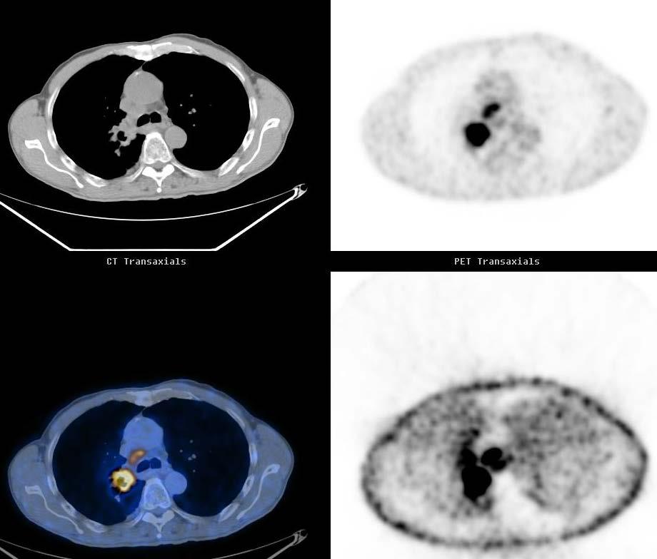
48 Using CT alone for RT Planning Where is the Cancer?
49 Using PET/CT for RT Planning Where is the edge of the Cancer?

50 Using PET/CT for RT Planning Where is the edge of the Cancer?
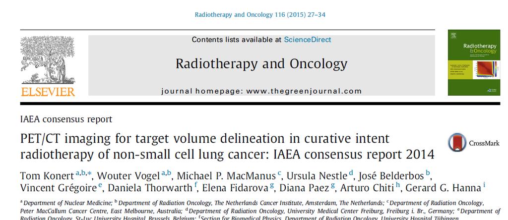
51 PET/CT for delineation of atelectasis Konert T, et al. Radiother Oncol 2015;116(1):27-34.


52 Defining the edge of the GTV/ITV If CT margin extends beyond PET - contour the CT unless clearly atelectasis If PET extends beyond CT edge of tumour or is obscured by atelectasis -More difficult!


53 Defining the edge of the GTV

54 Peer Review
55

56 More Contour Examples Tumor beside the liver Konert T, et al. Radiother Oncol 2015;116(1):27-34.

57 More Contour Examples CT edge versus PET edge Konert T, et al. Radiother Oncol 2015;116(1):27-34.
58 PET/CT RTP Margins to use When delineating on PET structure incorporates a respiratory motion Consensus statement refers to this as a respiratory expanded GTV (regtv) Suggest regtv CTV 6mm or 8mm Suggest CTV PTV at least 5mm (dependent on local set-up error) Konert T, et al. Radiother Oncol 2015;116(1):27-34.
59 HOW TO INCORPORATE GATING?
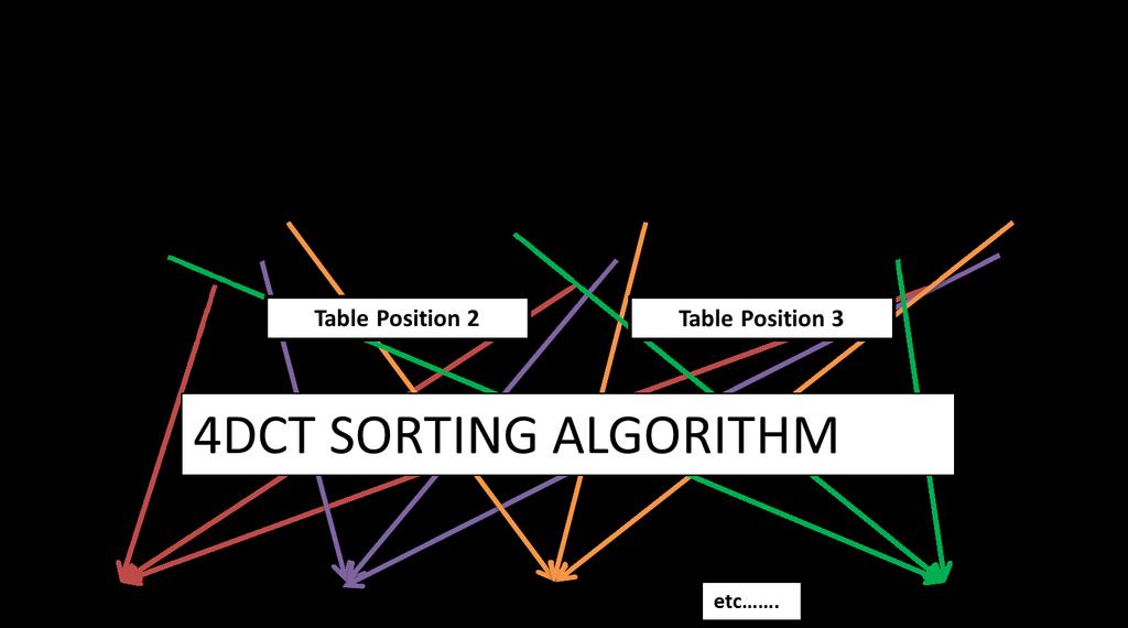
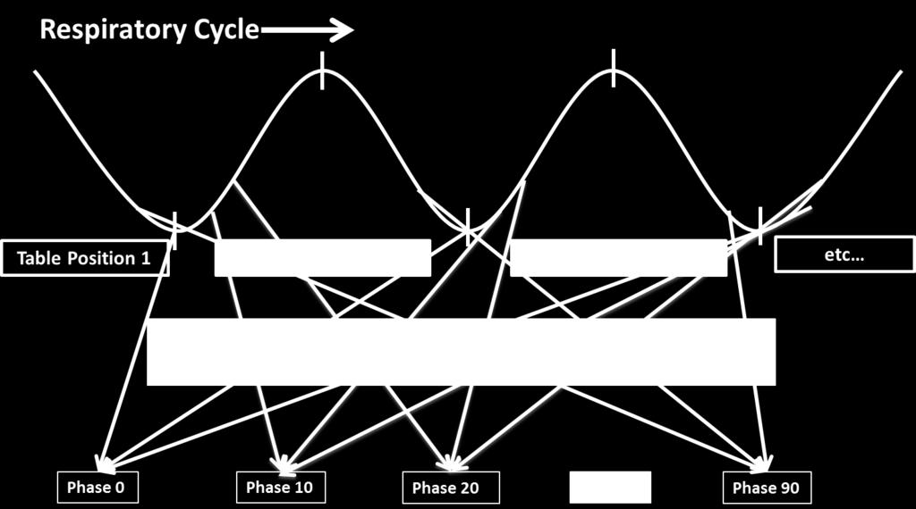
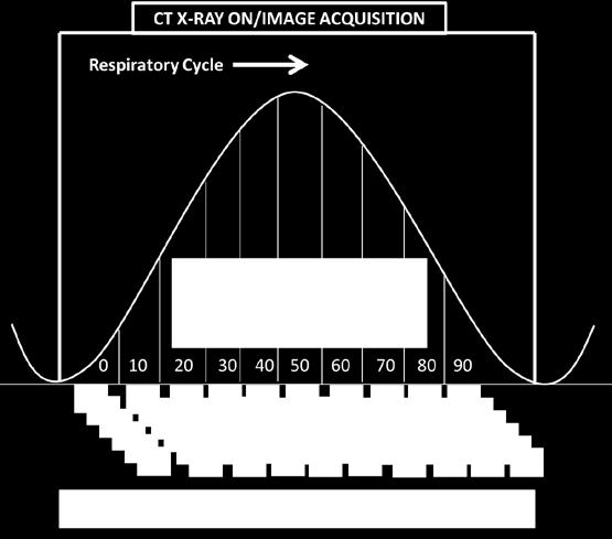
60 How to incorporate gating? 4DCT now standard technique to account for tumour motion 4DCT gives reliable information about tumour morphology and movement Where 4DCT acquisition is used alongside PET/CT the GTV edge should be based on the 4DCT The PET being used to discriminate tumor and non-tumor sites to adapt the GTV where appropriate
61 4D PET/CT
62
63 Phase based PET attenuation correction CT in each Phase PET in each Phase Phase by Phase attenuated PET
:576-85")
64 Attenuation Correction Hamill et al, Med. Phys. 2008;35(2):576-85
65 3D vs 4D PET/CT 3D PET/CT Disadvantages Respiratory Averaging Possibly inaccurate SUV Advantages No Noise Relatively Fast Acquisition 4D PET/CT Technically difficult Noisy patterns Prolonged Acquisition times Accurate tumour volume definition Detection of Small Lesions Better quantification of SUV (SUV MAX ) Dose Painting / Better characterisation of heterogeneity within tumour
 using 3D PET. Aristophanous, et al.")
66 Detection of Small Lesions with 4D PET/CT Lesion detected at hilar area in 4D PET; Not visible (blurred) using 3D PET. Aristophanous, et al. Int J Radiat Oncol Biol Phys 2012;82(1):e99-105

67 3DPET vs 4D PET vs 4DCT Callaghan J, et al. Int J Radiat Oncol Biol Phys 2013;86(4):749-54


68 PET/CT for RTP in NSCLC PET/CT has establish role in staging PET/CT must be used to inform TVD in NSCLC RTP A dedicated PET/CT is preferable QA of transfer of the PET images to the RTP system is essential IAEA Consensus Statement 2014 provides guidance on PET/CT delineation approach 4D PET/CT may be provide more accurate tracer quantification
IAEA RTC. PET/CT and Planning of Radiation Therapy 20/08/2014. Sarajevo (Bosnia & Hercegovina) Tuesday, June :40-12:20 a.
 Tuesday, June :40-12:20 a.") IAEA RTC PET/CT and Planning of Radiation Therapy Sarajevo (Bosnia & Hercegovina) Tuesday, June 17 2014 11:40-12:20 a.m María José García Velloso Servicio de Medicina Nuclear Clínica Universidad de Navarra
IAEA RTC PET/CT and Planning of Radiation Therapy Sarajevo (Bosnia & Hercegovina) Tuesday, June 17 2014 11:40-12:20 a.m María José García Velloso Servicio de Medicina Nuclear Clínica Universidad de Navarra
4D PET: promises and limitations
 4D PET: promises and limitations Tinsu Pan, Ph.D. M.D. Anderson Cancer Center The University of Texas Background Outlines Gating techniques: Deep inspiration breath hold 4D PET/CT Non-gating techniques
Radiation treatment planning in lung cancer
 Radiation treatment planning in lung cancer Georg Dietmar 1,2 1 Div. Medical Rad. Phys., Dept. of Radiation Oncology / Medical Univ. Vienna & AKH Wien 2 Christian Doppler Laboratory for Medical Radiation
Title: TC simulation versus TC/PET simulation for radiotherapy in lung cancer: volumes comparison in two cases.
 Title: TC simulation versus TC/PET simulation for radiotherapy in lung cancer: volumes comparison in two cases. Authors: Franzone, P.; 1* Muni, A; 2 Cazzulo, E.; 3 Berretta, L.; 1 Pozzi, G. 1 ; Todisco,
Defining Target Volumes and Organs at Risk: a common language
 Defining Target Volumes and Organs at Risk: a common language Eduardo Rosenblatt Section Head Applied Radiation Biology and Radiotherapy (ARBR) Section Division of Human Health IAEA Objective: To introduce
PET in Radiation Therapy. Outline. Tumor Segmentation in PET and in Multimodality Images for Radiation Therapy. 1. Tumor segmentation in PET
 Tumor Segmentation in PET and in Multimodality Images for Radiation Therapy Wei Lu, Ph.D. Department of Radiation Oncology Mallinckrodt Institute of Radiology Washington University in St. Louis Outline
POSITRON EMISSION TOMOGRAPHY PHANTOM STUDIES FOR RADIATION THERAPY TARGET DELINEATION MICHAEL VINTSON LAWRENCE
 POSITRON EMISSION TOMOGRAPHY PHANTOM STUDIES FOR RADIATION THERAPY TARGET DELINEATION BY MICHAEL VINTSON LAWRENCE A Dissertation Submitted to the Graduate Faculty of WAKE FOREST UNIVERSITY GRADUATE SCHOOL
REVISITING ICRU VOLUME DEFINITIONS. Eduardo Rosenblatt Vienna, Austria
 REVISITING ICRU VOLUME DEFINITIONS Eduardo Rosenblatt Vienna, Austria Objective: To introduce target volumes and organ at risk concepts as defined by ICRU. 3D-CRT is the standard There was a need for a
45 Hr PET Registry Review Course
 45 HR PET/CT REGISTRY REVIEW COURSE Course Control Document Timothy K. Marshel, MBA, R.T. (R), (N)(CT)(MR)(NCT)(PET)(CNMT) The PET/CT Training Institute, Inc. SNMMI-TS 028600-028632 45hr CEH s Voice Credits
Mathieu Hatt, Dimitris Visvikis. To cite this version: HAL Id: inserm
 Defining radiotherapy target volumes using 18F-fluoro-deoxy-glucose positron emission tomography/computed tomography: still a Pandora s box?: in regard to Devic et al. (Int J Radiat Oncol Biol Phys 2010).
Integrated PET/CT systems State of the art and Clinical Applications
 Integrated PET/CT systems State of the art and Clinical Applications V. Bettinardi Nuclear Medicine Dep. Scientific Institute San Raffaele Hospital Milan Italy The announcement of a new Diagnostic Imaging
21 st Century Radiotherapy: State-of-the-art and predicting the future. Clinical applications of PET. PET Imaging
 21 st Century Radiotherapy: State-of-the-art and predicting the future Clinical applications of PET Pat Price Ralston Paterson Professor of Radiation Oncology Christie Hospital Manchester UK Mayneord-Phillips
Utility of 18 F-FDG PET/CT in metabolic response assessment after CyberKnife radiosurgery for early stage non-small cell lung cancer
 Utility of F-FDG PET/CT in metabolic response assessment after CyberKnife radiosurgery for early stage non-small cell lung cancer Ngoc Ha Le 1*, Hong Son Mai 1, Van Nguyen Le 2, Quang Bieu Bui 2 1 Department
Principles of nuclear metabolic imaging. Prof. Dr. Alex Maes AZ Groeninge Kortrijk and KULeuven Belgium
 Principles of nuclear metabolic imaging Prof. Dr. Alex Maes AZ Groeninge Kortrijk and KULeuven Belgium I. Molecular imaging probes A. Introduction - Chemical disturbances will precede anatomical abnormalities
Metabolic volume measurement (physics and methods)
") Metabolic volume measurement (physics and methods) Ronald Boellaard Department of Nuclear Medicine & PET Research, VU University Medical Center, Amsterdam r.boellaard@vumc.nl Metabolic volume vs tumor
Metabolic volume measurement (physics and methods) Ronald Boellaard Department of Nuclear Medicine & PET Research, VU University Medical Center, Amsterdam r.boellaard@vumc.nl Metabolic volume vs tumor
Pitfalls and Remedies in PET/CT imaging for RT planning
 Pitfalls and Remedies in PET/CT imaging for RT planning Tinsu Pan, Ph.D. M.D. Anderson Cancer Center The University of Texas Outlines Background Average CT (< 1 msv) to reduce mis-alignment of PET and
Evaluation of Whole-Field and Split-Field Intensity Modulation Radiation Therapy (IMRT) Techniques in Head and Neck Cancer
 Techniques in Head and Neck Cancer") 1 Charles Poole April Case Study April 30, 2012 Evaluation of Whole-Field and Split-Field Intensity Modulation Radiation Therapy (IMRT) Techniques in Head and Neck Cancer Abstract: Introduction: This study
1 Charles Poole April Case Study April 30, 2012 Evaluation of Whole-Field and Split-Field Intensity Modulation Radiation Therapy (IMRT) Techniques in Head and Neck Cancer Abstract: Introduction: This study
Typical PET Image. Elevated uptake of FDG (related to metabolism) Lung cancer example: But where exactly is it located?
 Lung cancer example: But where exactly is it located?") Typical PET Image Elevated uptake of FDG (related to metabolism) Lung cancer example: But where exactly is it located? PET/CT Oncology Imaging Anatometabolic fusion images are useful in the management
Typical PET Image Elevated uptake of FDG (related to metabolism) Lung cancer example: But where exactly is it located? PET/CT Oncology Imaging Anatometabolic fusion images are useful in the management
Outline. Positron Emission Tomography for Oncologic Imaging and Treatment. Radionuclide. Physics of PET and PET-CT
 Positron Emission Tomography for Oncologic Imaging and Treatment J. Daniel Bourland, PhD Department of Radiation Oncology Bioanatomic Imaging and Treatment Program Wake Forest University School of Medicine
Image Fusion, Contouring, and Margins in SRS
 Image Fusion, Contouring, and Margins in SRS Sarah Geneser, Ph.D. Department of Radiation Oncology University of California, San Francisco Overview Review SRS uncertainties due to: image registration contouring
The Role of PET / CT in Lung Cancer Staging
 July 2004 The Role of PET / CT in Lung Cancer Staging Vlad Vinarsky, Harvard Medical School Year IV Patient AM HPI: 81 yo F p/w hemoptysis x 1 month LLL lesion on CXR, not responsive to Abx 35 pack-year
Improving prediction of radiotherapy response and optimizing target definition by using FDG-PET for lung cancer patients
 Investigations and research Improving prediction of radiotherapy response and optimizing target definition by using FDG-PET for lung cancer patients R.J.H.M. Steenbakkers G.R. Borst M. van Herk H. Bartelink
PHYSICS 2: HSC COURSE 2 nd edition (Andriessen et al) CHAPTER 20 Radioactivity as a diagnostic tool (pages 394-5)
 CHAPTER 20 Radioactivity as a diagnostic tool (pages 394-5)") PHYSICS 2: HSC COURSE 2 nd edition (Andriessen et al) CHAPTER 20 Radioactivity as a diagnostic tool (pages 394-5) 1. (a) A radioisotope is an isotope that is unstable and will emit particles from the nucleus
PHYSICS 2: HSC COURSE 2 nd edition (Andriessen et al) CHAPTER 20 Radioactivity as a diagnostic tool (pages 394-5) 1. (a) A radioisotope is an isotope that is unstable and will emit particles from the nucleus
Radiotherapy Planning (Contouring Lung Cancer for Radiotherapy dose prescription) Dr Raj K Shrimali
 Dr Raj K Shrimali") Radiotherapy Planning (Contouring Lung Cancer for Radiotherapy dose prescription) Dr Raj K Shrimali Let us keep this simple and stick to some basic rules Patient positioning Must be reproducible Must be
Radiotherapy Planning (Contouring Lung Cancer for Radiotherapy dose prescription) Dr Raj K Shrimali Let us keep this simple and stick to some basic rules Patient positioning Must be reproducible Must be
biij Initial experience in treating lung cancer with helical tomotherapy
 Available online at http://www.biij.org/2007/1/e2 doi: 10.2349/biij.3.1.e2 biij Biomedical Imaging and Intervention Journal CASE REPORT Initial experience in treating lung cancer with helical tomotherapy
Ruolo dell imaging nella pianificazione del trattamento
 Simposio AIRO-SIRM: Diagnostica per immagini morfologica e funzionale nella stadiazione, terapia e follow-up dei sarcomi delle parti molli Ruolo dell imaging nella pianificazione del trattamento Marco
An audit of radiation dose of 4D CT in a radiotherapy department
 An audit of radiation dose of 4D CT in a radiotherapy department Poster No.: R-0097 Congress: Type: Authors: Keywords: DOI: 2014 CSM Scientific Exhibit T. Hubbard, J. Callahan, J. Cramb, R. Budd, T. Kron;
Use of imaging systems for patient modeling - PET and SPECT
 Use of imaging systems for patient modeling - PET and SPECT Sasa Mutic Department of Radiation Oncology Siteman Cancer Center Mallinckrodt Institute of Radiology Washington University School of Medicine
MRI Applications in Radiation Oncology:
 MRI Applications in Radiation Oncology: Physician s Perspective Jeff Olsen, MD Department of Radiation Oncology Washington University, St. Louis, MO Disclosures Washington University has research and service
Outline. Positron Emission Tomography for Oncologic Imaging and Treatment. Positron Annihilation. Physics of PET and PET-CT
 Positron Emission Tomography for Oncologic Imaging and Treatment J. Daniel Bourland, PhD Department of Radiation Oncology Bioanatomic Imaging and Treatment Program Wake Forest University School of Medicine
ADVANCES IN RADIATION TECHNOLOGIES IN THE TREATMENT OF CANCER
 ADVANCES IN RADIATION TECHNOLOGIES IN THE TREATMENT OF CANCER Bro. Dr. Collie Miller IARC/WHO Based on trends in the incidence of cancer, the International Agency for Research on Cancer (IARC) and WHO
8/10/2016. PET/CT for Tumor Response. Staging and restaging Early treatment response evaluation Guiding biopsy
 PET/CT for Tumor Response Evaluation August 4, 2016 Wei Lu, PhD Department of Medical Physics www.mskcc.org Department of Radiation Oncology www.umaryland.edu FDG PET/CT for Cancer Imaging Staging and
Appendix 1: Regional Lymph Node Stations for Staging Esophageal Cancer
 Appendix 1: Regional Lymph Node Stations for Staging Esophageal Cancer Locoregional (N stage) disease was redefined in the seventh edition of the AJCC Cancer Staging Manual as any periesophageal lymph
This house believes that the use of Functional Imaging for treatment planning of head and neck tumors needs to be carefully considered.
 This house believes that the use of Functional Imaging for treatment planning of head and neck tumors needs to be carefully considered. IMRT for Head and Neck Tumors Vincent GREGOIRE, M.D., Ph.D., Hon.
How effective is current clinical trial QA
 How effective is current clinical trial QA Elizabeth Miles UK National RadioTherapy Trials QA (RTTQA) group Mount Vernon Cancer Centre United Kingdom ESTRO 35 29 April 3 May Turin, Italy Contents QA processes
Image Guided Stereotactic Radiotherapy of the Lung
 Image Guided Stereotactic Radiotherapy of the Lung Jamie Marie Harris, MS DABR Avera McKennan Radiation Oncology September 25, 2015 Stereotactic Body Radiotherapy - Clinical Dose/Fractionation - Normal
Linac or Non-Linac Demystifying And Decoding The Physics Of SBRT/SABR
 Linac or Non-Linac Demystifying And Decoding The Physics Of SBRT/SABR PhD, FAAPM, FACR, FASTRO Department of Radiation Oncology Indiana University School of Medicine Indianapolis, IN, USA Indra J. Das,
FDG PET/CT imaging for tumor staging and definition of tumor volumes in radiation treatment planning in non small cell lung cancer
 ONCOLOGY LETTERS 7: 1015-1020, 2014 FDG PET/CT imaging for tumor staging and definition of tumor volumes in radiation treatment planning in non small cell lung cancer YUANDA ZHENG 1, XIAOJIANG SUN 1, JIAN
Nuclear Medicine and PET. D. J. McMahon rev cewood
 Nuclear Medicine and PET D. J. McMahon 150504 rev cewood 2018-02-15 Key Points Nuclear Medicine and PET: Imaging: Understand how Nuc Med & PET differ from Radiography & CT by the source of radiation. Be
Herlev radiation oncology team explains what MRI can bring
 Publication for the Philips MRI Community Issue 46 2012/2 Herlev radiation oncology team explains what MRI can bring The radiotherapy unit at Herlev University Hospital investigates use of MRI for radiotherapy
Identifying Image Artifacts, Their Causes and How to Fix Them: PET. Brad J Kemp, PhD Mayo Clinic, Rochester, MN
 Identifying Image Artifacts, Their Causes and How to Fix Them: PET Brad J Kemp, PhD Mayo Clinic, Rochester, MN Case 1 Can we scan with a defective block detector? Daily Quality Assurance Results Singles
Development of an Expert Consensus Target Delineation Atlas for Thymic Cancers: Initial Quantitative Analysis of an Expert Survey.
 Development of an Expert Consensus Target Delineation Atlas for Thymic Cancers: Initial Quantitative Analysis of an Expert Survey. Charles R. Thomas, Jr., MD, Jayashree Kalpathy-Cramer, PhD, Jehee Choi,
Endobronchial Ultrasound in the Diagnosis & Staging of Lung Cancer
 Endobronchial Ultrasound in the Diagnosis & Staging of Lung Cancer Dr Richard Booton PhD FRCP Lead Lung Cancer Clinician, Consultant Respiratory Physician & Speciality Director Manchester University NHS
GE Healthcare. Rad Rx. White Paper
 GE Healthcare Rad Rx White Paper Introduction This publication is part of a series of white papers aimed at communicating the importance of each component in the image chain of a PET/CT study. From data
Allan Price NHS Lothian, Edinburgh, UK
 Allan Price NHS Lothian, Edinburgh, UK Radiotherapy Dose Volume Timing Technique PCI Surgery Systemic agents 1 Study Dose Time Induction CT Ann Arbor 65.1-75.6 Gy Duke 73.6-80 Gy RTOG 77.4 Gy 74 Gy 6.5-7.5
Reproducibility of Uptake Estimates in FDG PET: a Monte Carlo study
 Reproducibility of Uptake Estimates in FDG PET: a Monte Carlo study Juliette Feuardent, Marine Soret, Irène Buvat 1 Abstract Tumor glucose metabolism measurements from Fluoro-deoxyglucose (FDG) Positron
Use of molecular and functional imaging for treatment planning The Good, The Bad and The Ugly
 Use of molecular and functional imaging for treatment planning The Good, The Bad and The Ugly Robert Jeraj, PhD Associate Professor of Medical Physics, Human Oncology, Radiology and Biomedical Engineering
Positron Emission Tomography in Lung Cancer
 May 19, 2003 Positron Emission Tomography in Lung Cancer Andrew Wang, HMS III Patient DD 53 y/o gentleman presented with worsening dyspnea on exertion for the past two months 30 pack-year smoking Hx and
4D PET-CT GUIDED RADIATION THERAPY*
 JBR BTR, 2013, 96: 155-159. 4D PET-CT GUIDED RADIATION THERAPY* X. Geets 1 Tremendous technological progress in the field of imaging and computation have been revolutionizing radiotherapy of non-small
Pitfalls in SBRT Treatment Planning for a Moving Target
 Pitfalls in SBRT Treatment Planning for a Moving Target Cynthia F. Chuang, Ph.D. Department of Radiation Oncology University of California-San Francisco I have no conflicts of interests to disclose In
Evaluation of Lung Cancer Response: Current Practice and Advances
 Evaluation of Lung Cancer Response: Current Practice and Advances Jeremy J. Erasmus I have no financial relationships, arrangements or affiliations and this presentation will not include discussion of
Utilisation du PET-FDG pour la définition des volumes cibles en radiothérapie des tumeurs de la sphère cervico-maxillo-faciale: mythe et réalité
 Utilisation du PET-FDG pour la définition des volumes cibles en radiothérapie des tumeurs de la sphère cervico-maxillo-faciale: mythe et réalité Vincent GREGOIRE, M.D., Ph.D. Head and Neck Oncology Program,
Johannes C. Athanasios Dimopoulos
 BrachyNext Symposium Miami Beach, USA, May 30 31, 2014 Imaging Modalities: Current Challenges and Future Directions Johannes C. Athanasios Dimopoulos Imaging Modalities: Current Challenges and Future Directions
PET is Underutilized in Oncology. Brent D. Murphy, MS, DABR
 PET is Underutilized in Oncology Brent D. Murphy, MS, DABR 1 President, Radiological Technologies University Nationally Accredited, FAFSA, GI/VA Loan, F-1 Student Visa Offers Degrees in Medical Dosimetry,
FDG-PET value in deep endometriosis
 Gynecol Surg (2011) 8:305 309 DOI 10.1007/s10397-010-0652-6 ORIGINAL ARTICLE FDG-PET value in deep endometriosis A. Setubal & S. Maia & C. Lowenthal & Z. Sidiropoulou Received: 3 December 2010 / Accepted:
Positron emission tomography/computer tomography in the evaluation of head and neck cancer treatment
 Positron emission tomography/computer tomography in the evaluation of head and neck cancer treatment Severina Šedienė 1, Ilona Kulakienė 1, Viktoras Rudžianskas 2 1 Lithuanian University of Health Sciences,
Basics of nuclear medicine
 Basics of nuclear medicine Prof. dr. Davor Eterović Prof. dr. Vinko Marković Radioisotopes are used both in diagnostics and in therapy Diagnostics gamma emitters are used since gamma rays can penetrate
PSMA PET SCANNING AND THERANOSTICS IN PROSTATE CANCER KEVIN TRACEY, MD, FRCPC PRECISION DIAGNSOTIC IMAGING REGIONAL PET/CT CENTRE
 PSMA PET SCANNING AND THERANOSTICS IN PROSTATE CANCER KEVIN TRACEY, MD, FRCPC PRECISION DIAGNSOTIC IMAGING REGIONAL PET/CT CENTRE DISCLOSURES/CONFLICTS NONE OBJECTIVES Understand current diagnostic role
Institute of Oncology & Radiobiology. Havana, Cuba. INOR
 Institute of Oncology & Radiobiology. Havana, Cuba. INOR 1 Transition from 2-D 2 D to 3-D 3 D conformal radiotherapy in high grade gliomas: : our experience in Cuba Chon. I, MD - Chi. D, MD - Alert.J,
The Use of PET Scanning in Urologic Oncology
 The Use of PET Scanning in Urologic Oncology Dr Nicholas C. Buchan Uro-oncology Fellow 1 2 Aims To understand the basic concepts underlying PET scanning. Understand the emerging role of PET Scanning for
Clinical evaluation of respiration-induced attenuation uncertainties in pulmonary 3D PET/CT
 Kruis et al. EJNMMI Physics (2015) 2:4 DOI 10.1186/s40658-014-0107-7 ORIGINAL RESEARCH Open Access Clinical evaluation of respiration-induced attenuation uncertainties in pulmonary 3D PET/CT Matthijs F
An Introduction to PET Imaging in Oncology
 January 2002 An Introduction to PET Imaging in Oncology Janet McLaren, Harvard Medical School Year III Basics of PET Principle of Physiologic Imaging: Allows in vivo visualization of structures by their
4D Radiotherapy in early ca Lung. Prof. Manoj Gupta Dept of Radiotherapy & oncology I.G.Medical College Shimla
 4D Radiotherapy in early ca Lung Prof. Manoj Gupta Dept of Radiotherapy & oncology I.G.Medical College Shimla Presentation focus on ---- Limitation of Conventional RT Why Interest in early lung cancer
MOLECULAR IMAGING IN RADIATION ONCOLOGY
 MOLECULAR IMAGING IN RADIATION ONCOLOGY Dr Dodul Mondal (Hons) MD, DNB, MNAMS Radiation Oncologist INTRODUCTION Concept of radiotherapy planning has evolved from simple anatomy based planning to anatomic
PET CT for Staging Lung Cancer
 PET CT for Staging Lung Cancer Rohit Kochhar Consultant Radiologist Disclosures Neither I nor my immediate family members have financial relationships with commercial organizations that may have a direct
Deformation Effect on SUV max Changes in Thoracic Tumors Using 4-D PET/CT Scan
 Deformation Effect on SUV max Changes in Thoracic Tumors Using 4-D PET/CT Scan Tzung-Chi Huang 1 *, Yao-Ching Wang 2 1 Department of Biomedical Imaging and Radiological Science, China Medical University,
Protocol of Radiotherapy for Small Cell Lung Cancer
 107 年 12 月修訂 Protocol of Radiotherapy for Small Cell Lung Cancer Indication of radiotherapy Limited stage: AJCC (8th edition) stage I-III (T any, N any, M0) that can be safely treated with definitive RT
8/10/2016. PET/CT Radiomics for Tumor. Anatomic Tumor Response Assessment in CT or MRI. Metabolic Tumor Response Assessment in FDG-PET
 PET/CT Radiomics for Tumor Response Evaluation August 1, 2016 Wei Lu, PhD Department of Medical Physics www.mskcc.org Department of Radiation Oncology www.umaryland.edu Anatomic Tumor Response Assessment
FDG PET/CT STAGING OF LUNG CANCER. Dr Shakher Ramdave
 FDG PET/CT STAGING OF LUNG CANCER Dr Shakher Ramdave FDG PET/CT STAGING OF LUNG CANCER FDG PET/CT is used in all patients with lung cancer who are considered for curative treatment to exclude occult disease.
PET Guidance of Therapy for BNCT and in vivo B-10 imaging
 INFN LNL Legnaro 17-19 Novembre 2009 Principles of Positron Emission Tomography and Radiopharmaceuticals PET Guidance of Therapy for BNCT and in vivo B-10 imaging Luca Menichetti, Ph.D C.N.R. Institute
Title Radiotherapy Treatment Planning in Nasopharyngeal Carcinoma.
 A New Brain Positron Emission Tomog Title Semiconductor Detectors for Target Radiotherapy Treatment Planning in Nasopharyngeal Carcinoma. Katoh, Norio; Yasuda, Koichi; Shiga Author(s) Masakazu; Onimaru,
Molecular Imaging and Cancer
 Molecular Imaging and Cancer Cancer causes one in every four deaths in the United States, second only to heart disease. According to the U.S. Department of Health and Human Services, more than 512,000
Automated Tumour Delineation Using Joint PET/CT Information
 Automated Tumour Delineation Using Joint PET/CT Information Vaclav Potesil 1,2, Xiaolei Huang 1,3, Xiang Sean Zhou 1, 1 Siemens Medical Solutions USA, Computer Aided Diagnosis and Therapy Solutions; 2
International Conference on Clinical PET and Molecular Nuclear Medicine (IPET 2011)
") IAEA-CN-185 International Conference on Clinical PET and Molecular Nuclear Medicine (IPET 2011) Trends in Clinical PET and Radiopharmaceutical Development 8 11 November 2011 Vienna, Austria Announcement
IAEA-CN-185 International Conference on Clinical PET and Molecular Nuclear Medicine (IPET 2011) Trends in Clinical PET and Radiopharmaceutical Development 8 11 November 2011 Vienna, Austria Announcement
Automatic Definition of Planning Target Volume in Computer-Assisted Radiotherapy
 Automatic Definition of Planning Target Volume in Computer-Assisted Radiotherapy Angelo Zizzari Department of Cybernetics, School of Systems Engineering The University of Reading, Whiteknights, PO Box
Biases affecting tumor uptake measurements in FDG-PET
 Biases affecting tumor uptake measurements in FDG-PET M. Soret, C. Riddell, S. Hapdey, and I. Buvat Abstract-- The influence of tumor diameter, tumor-tobackground activity ratio, attenuation, spatial resolution,
Using PET/CT in Prostate Cancer
 Using PET/CT in Prostate Cancer Legal Disclaimer These materials were prepared in good faith by MITA as a service to the profession and are believed to be reliable based on current scientific literature.
Colorectal Cancer and FDG PET/CT
 Hybrid imaging in colorectal & esophageal cancer Emmanuel Deshayes IAEA WorkShop, November 2017 Colorectal Cancer and FDG PET/CT 1 Clinical background Cancer of the colon and rectum is one of the most
Which Planning CT Should be Used for Lung SBRT? Ping Xia, Ph.D. Head of Medical Physics in Radiation Oncology Cleveland Clinic
 Which Planning CT Should be Used for Lung SBRT? Ping Xia, Ph.D. Head of Medical Physics in Radiation Oncology Cleveland Clinic Outline Image quality and image dose Free breathing CT, 4DCT, and synthetic
Positron Emission Tomography Computed Tomography (PET/CT)
") Positron Emission Tomography Computed Tomography (PET/CT) What is Positron Emission Tomography Computed Tomography (PET/CT) Scanning? What are some common uses of the procedure? How should I prepare for
Positron Emission Tomography Computed Tomography (PET/CT) What is Positron Emission Tomography Computed Tomography (PET/CT) Scanning? What are some common uses of the procedure? How should I prepare for
Austin Radiological Association Ga-68 NETSPOT (Ga-68 dotatate)
") Austin Radiological Association Ga-68 NETSPOT (Ga-68 dotatate) Overview Ga-68 dotatate binds to somatostatin receptors, with highest affinity for subtype 2 receptors (sstr2). It binds to cells that express
Austin Radiological Association Ga-68 NETSPOT (Ga-68 dotatate) Overview Ga-68 dotatate binds to somatostatin receptors, with highest affinity for subtype 2 receptors (sstr2). It binds to cells that express
IN VIVO IMAGING Proton Beam Range Verification With PET/CT
 IN VIVO IMAGING Proton Beam Range Verification With PET/CT Antje-Christin Knopf 1/3 K Parodi 2, H Paganetti 1, T Bortfeld 1 Siemens Medical Solutions Supports This Project 1 Department of Radiation Oncology,
Might Adaptive Radiotherapy in NSCLC be feasible in clinical practice?
 Might Adaptive Radiotherapy in NSCLC be feasible in clinical practice? E.Molfese, P.Matteucci, A.Iurato, L.E.Trodella, A.Sicilia, B.Floreno, S.Ramella, L.Trodella Radioterapia Oncologica, Università Campus
PET/MR:Techniques, Indications and Applications
 PET/MR:Techniques, Indications and Applications Franz Wolfgang Hirsch Professor and Head of the Department of Pediatric Radiology University Hospital Leipzig / Germany Children s Hospital University Leipzig
FDOPA, C11Choline, C11 Methionine. Dr K.G.Kallur
 FDOPA, C11Choline, C11 Methionine Dr K.G.Kallur Why? 11C Methionine scan Had undergone resection Earlier. Post op recurrent hypercalcemia C11 Methionine Unable to see in Sestamibi scan Brain Tumor After
UNIVERSITY OF WISCONSIN-LA CROSSE Graduate Studies
 UNIVERSITY OF WISCONSIN-LA CROSSE Graduate Studies A SINGLE INSTITUTION S EXPERIENCE IN DEVELOPING A PURPOSEFUL AND EFFICIENT OFF-LINE TECHNIQUE FOR ADAPTIVE RADIOTHERAPY IN A CLINICAL ENVIRONMENT A Research
Motion gating and tracking techniques: overview and recent developments
 Motion gating and tracking techniques: overview and recent developments Gig S Mageras, PhD, FAAPM Department of Medical Physics Memorial Sloan Kettering Cancer Center, New York MSK/gsm 15-Jun-2018 1 Disclosure
Presentation Outline. Patient Setup Imaging in RT: Getting the Most Bang for your Buck. Main Errors in RT. Hypothetical Patient Examples
 Patient Setup Imaging in RT: Getting the Most Bang for your Buck Olivier Morin, PhD UC SF Comprehensive Cancer Center San Francisco, CA Presentation Outline Errors in RT. Radiographic films in RT. On-board
Nuclear medicine in oncology. 1. Diagnosis 2. Therapy
 Nuclear medicine in oncology 1. Diagnosis 2. Therapy Diagnosis - Conventional methods - Nonspecific radiopharmaceuticals cumulating in tumours - Specific radiopharmaceuticals (receptor- and immunoscintigraphy)
肺癌放射治療新進展 Recent Advance in Radiation Oncology in Lung Cancer 許峰銘成佳憲國立台灣大學醫學院附設醫院腫瘤醫學部
 肺癌放射治療新進展 Recent Advance in Radiation Oncology in Lung Cancer 許峰銘成佳憲國立台灣大學醫學院附設醫院腫瘤醫學部 Outline Current status of radiation oncology in lung cancer Focused on stage III non-small cell lung cancer Radiation
Intensity-Modulated and Image- Guided Radiation Treatment. Outline. Conformal Radiation Treatment
 Intensity-Modulated and Image- Guided Radiation Treatment J. Daniel Bourland, PhD Professor Departments of Radiation Oncology, Physics, and Biomedical Engineering Wake Forest University School of Medicine
The Proper Use of PET/CT in Tumoring Imaging
 The Proper Use of PET/CT in Tumoring Imaging Mijin Yun, M.D. Jong Doo Lee, M.D. Department of Radiology / Division of Nuclear Medicine Yonsei University College of Medicine, Severance Hospital E mail :
Introduction Pediatric malignancies Changing trends & Radiation burden Radiation exposure from PET/CT Image gently PET & CT modification - PET/CT
 Introduction Pediatric malignancies Changing trends & Radiation burden Radiation exposure from PET/CT Image gently PET & CT modification - PET/CT protocols Tips Leukaemia / lymphoma: ~ 35% acute lymphoblastic
EMISSION GUIDED RADIATION THERAPY: A FEASIBILITY STUDY
 EMISSION GUIDED RADIATION THERAPY: A FEASIBILITY STUDY A Thesis Presented to The Academic Faculty by Qiyong Fan In Partial Fulfillment of the Requirements for the Degree Master of Science in Medical Physics
Mobile PET Center Project
 Journal of Physics: Conference Series PAPER OPEN ACCESS Mobile PET Center Project To cite this article: O Ryzhikova et al 2017 J. Phys.: Conf. Ser. 784 012051 View the article online for updates and enhancements.
Option D: Medicinal Chemistry
 Option D: Medicinal Chemistry Basics - unstable radioactive nuclei emit radiation in the form of smaller particles alpha, beta, positron, proton, neutron, & gamma are all used in nuclear medicine unstable
Stereotaxy. Outlines. Establishing SBRT Program: Physics & Dosimetry. SBRT - Simulation. Body Localizer. Sim. Sim. Sim. Stereotaxy?
 Establishing SBRT Program: Physics & Dosimetry Lu Wang, Ph.D. Radiation Oncology Department Fox Chase Cancer Center Outlines Illustrate the difference between SBRT vs. CRT Introduce the major procedures
Dr Sneha Shah Tata Memorial Hospital, Mumbai.
 Dr Sneha Shah Tata Memorial Hospital, Mumbai. Topics covered Lymphomas including Burkitts Pediatric solid tumors (non CNS) Musculoskeletal Ewings & osteosarcoma. Neuroblastomas Nasopharyngeal carcinomas
Cardiac Imaging Tests
 Cardiac Imaging Tests http://www.medpagetoday.com/upload/2010/11/15/23347.jpg Standard imaging tests include echocardiography, chest x-ray, CT, MRI, and various radionuclide techniques. Standard CT and