MOLECULAR IMAGING IN RADIATION ONCOLOGY
|
|
|
- Daisy Goodman
- 5 years ago
- Views:
Transcription
1 MOLECULAR IMAGING IN RADIATION ONCOLOGY Dr Dodul Mondal (Hons) MD, DNB, MNAMS Radiation Oncologist


2 INTRODUCTION Concept of radiotherapy planning has evolved from simple anatomy based planning to anatomic image based planning to highly precise modern day radiotherapy planning using more precise functional, molecular and genetic aspect of cancer
3 Molecular Imaging Originated from the concepts of radiopharmacology to better understand the fundamental molecular pathways inside organisms Non-invasive Enormous potential after the description of the human genome Images particular targets or pathways with specific probes or biomarkers Paradigm shift of cancer management: organ treatment to treatment specific molecular abnormality
4 Molecular imaging cont.. Development of novel biomarkers and imaging of gene expression earlier and more precise diagnosis, treatment Quantitative tests and greater degree of objectivity Principle of mass spectrometry
5 BASICS OF FEW IMAGING TECHNIQUES
6 IMPROVEMENTS IN IMAGING Spectacular advance in our knowledge of cancer at the molecular level Cross fertilization of multiple disciplines Past: Anatomic Present: Bilogic/Mechanistic Wide spectrum of information Hence BIOLOGICAL Metabolic Functional Biologic Functional Others: Molecular Genotypic Phenotypic
7 Computed tomography Tomos (slice) and Graphein (to write) EMI scan/cat (Computed Axial Tomography) scan/ CT scan: First utilized in 1971 for brain scan by Sir Hounsefield. Two dimensional X-ray images taken around a single axis of rotation at multiple sections using multiple detectors Digital reconstruction generate a three-dimensional image Essentially anatomic imaging Uses principle of photoelectric effect, however at high KV, Compton effect becomes predominant
8 Computed Tomography cont.. Windowing - CT data manipulated demonstrate various bodily structures based on their ability to block X-ray Axial images reconstructed to get three dimensional images
9 Magnetic Resonance Imaging (MRI) Utilizes magnetic relaxation properties of cancer and non cancer tissues Provides information about resonance of populations of nuclei Their resonance is influenced by several physical and biological properties of interest Local tissue chemical content Temperature Water diffusion Blood flow Tissue elasticity Provides anatomic, physiological, biochemical and molecular information
10 MAGNETIC RESONANCE SPECTROSCOPY MR spectrometer is in-built in all MRI machines. Routinely it ignores spectrum of resonances that come from a single voxel and clubs them together and hence we see a single signal either white or black or intermediate gray. With proper software and hardware modifications it can provide information within a region. Hence MRS is shown as a spectrum from a single voxel or else a grid of voxels. Spectra are collected from spinning nuclei Routinely from hydrogen nuclei ( water is the most abundant molecule)
11 ROLE OF MRS Brain can distinguish high grade gliomas from low grade gliomas Primary vs. metastatic lesion (pediatric tumors) Radiation necrosis vs. recurrence Serves as predictor of survival or progression. Choline to citrate ratio- prostate cancer, gliomas Results from enhanced phospholipid cell membrane turnover associated with tumor proliferation, increased cellularity and growth Creatine, lactate- Adverse prognosis Increased metastatic potential Radio resistance

12 NMR spectroscopy of 31P can provide information about energy status. Membrane precursor phosphocholine and phosphoethanolamine- changes in response to radio/chemotherapy- hence have early predictor of tumor response.
13 CONTRAST ENHANCED MRI MRI Contrast- shorten T1( bright signal) Shorten T2 ( dark signal) Ferumoxtran-10- an iron based T2 agent that is collected in normal lymph nodes but excluded from metastatic nodes- shorten T2 signal (Not approved in India) Multihance- Hepatocyte specific T1 contrast agent (Approved in india): For characterizing hepatic tumors-can differentiate between FNH and HCC Newer contrast agents- action specific- receptor specific
14 NEWER ADVANCES: PET-MR Currently, no clinical indication for combined PET/MR has been established With PET/MR it might finally be possible, in the future, to gather the information necessary to perform radiotherapy with dose painting and to establish truly predictive imaging markers
15 PET SCAN
16 Positron Emission Tomography (PET) Functional imaging modality showing: Blood flow Glucose metabolism Receptor density Basic Principle Injection of a radioactive tracer to image chemical/biological processes. Radioactive tracer decays by Positron Emission. When the tracer is introduced into the body, it is sitespecific uptake can be traced by means of the labelled atom
 and are always emitted in opposite direction.")
17 p = n + β + +υ A nucleus with too low a neutron-to-proton ratio converts a proton to a neutron, emitting a positron (β + ) and a neutrino (υ) to carry off the excess energy Photons emitted must have a specific energy(511 kev) and are always emitted in opposite direction. Positron Decay

18 Dependent on the altered metabolic characteristics of tumor cells compared to its surroundings Uncontrolled proliferation- Hallmark Most widely used in oncologic practice is FDG- glucose analog
19 TRACERS FOR PET
20 PET in radiotherapy planning BASIS FOR ROLE 1. Distinguishes metabolically active tissue from scar. 2. Can detect functional/metabolic activity of cells 3. Quantification of metabolic activity of cells 4. Capability to detect signal intensity changes rather than lesion size. 5. Independent from anatomy and organ relationship. Hence is able to detect abnormal metabolic activity in tumor recurrence in patients post surgery and post RT where architecture is distorted. 6. Ability to assess different specific tissue functions due to functional specificity of developed pharmaceuticals.
21 ROLE OF PET 1. Diagnosis and staging. 2. Definition of extent of disease staging and restaging 3. Identification and localization of disease foci in patients with unknown primary. 4. Assessment of response to therapy and its monitoring. 5. Identification of relapse and recurrence versus other imaging non-specific changes and increased tumor markers. 6. Biopsy site guide. 7. Predictor of response and survival based on SUV. 8. Most importantly, radiotherapy planning and guidance.
22 PET FOR RT PLANNING 30-40% of RT plans for cancer patients are changed when PET scan findings are featured into plan. Scanning for radiotherapy- simulation scans 1. Couch- flat table 2. Precise positioning 3. Precise immobilization 4. Laser used to guide marking 5. 3 fiducial markers: to establish reference slice; reference point pseudoisocentre 6. From pseudoisocentre precise target is identified as necessary and true isocentre is defined with respect to it 7. Consistent and optimum spacing required
23 PET FOR RT PLANNING 8. Fasting for 4-6 hrs to enhance tracer uptake by tumor. 9. Refrain strenuous exercises 48 hrs before FDG administrationto avoid physiological uptake in recovering muscles. 10. Asked to wear warm clothing, particularly around shoulders and neck to avoid uptake in brown adipose tissue of neck and upper torso. 11.Discourage patients from moving or speaking during min of FDG uptake. 12.Before scanning patients are asked to urinate. 13.Sedatives, anti-cholinergics, anti-emetics as required.


24 PET FOR RT PLANNING 15. Once data is acquired it is sent to RTP software. 16. RTP software must validate the DICOM compatibility of CT or PET. Positioning of the patient exactly in the middle of the longitudinal laser beam Metal pins defining the isocentre

25
26 Impact on staging Effect on radiotherapy T stage N stage M stage Upstaging (Larger extension of disease) Downstaging (Less extension of disease) Upstaging (Detection of new site of lymph node) Downstaging(Omission of lymph node diagnosed as malignant on CT/MRI) Enlargement of radiotherapy fields Change of indication from curative to palliative Field reduction reducing normal tissue exposure, possible dose escalation Change of indication from palliative to curative Enlargement of radiotherapy fields to avoid geographical miss Change of indication from curative to palliative Field reduction reducing normal tissue exposure, possible dose escalation Change of indication from palliative to curative Detection of metastasis Change of indication from curative to DR DODUL palliative MONDAL
27 Rational use of PET in radiation treatment planning depends on qualities of PET. Sensitivity Specificity Positive predictive value Negative predictive value Accuracy
28 PET Based Contouring CT positive but PET negative and vice versa? Who Needs To contour? Optimal PET volume for radiation therapy?
29 Optimal PET volume for radiation therapy? CT Tumor margin sharp, PET Fuzzy Philosophy should be PET finds it CT defines it Exceptions PET defines tumor edge : Neck/Pelvic mass that blends in with tumor surrounding soft tissue Lung mass with accompanying atelectasis Manual segmentation Automatic segmentation based on SUV
30 Who Needs To contour? Radiation oncologist vs. Nuclear medicine expert Physiological variation and uptake: post-surgery sites irradiated sites areas of inflammation SUV variability Patient LBW activity of injected isotope BSA High background activity as in brain Collaboration with nuclear medicine expert As experience grows requirement will be far less frequent
31 positive on CT but negative on PET and vice versa? 1. No consensus lack of experience and long-term data. 2. Any obvious tumor seen with CT that does not show FDG uptake within it should be still be included. 3. PET lesion to be included in GTV it should either correspond to Underlying CT abnormality Lymph node Convincing intensity within a common site for disease, that cannot be explained by a benign process or artifact
32 Role of PET in planning for NSCLC Accurate staging Selection of appropriate treatment- radical vs. palliative Monitor response to therapy Define local recurrence Aid for dose escalation-clearer def of GTV Determine sites of nodal involvement In patients with atelectatic lung reduce treatment fields. GTV should be, in a majority of cases, equivalent to extent of hot spot depicted by PET complemented with information given by CT Target volumes were reduced slightly more frequently, but volumes were also enlarged with no clear or consistent pattern among patients.
33 PET for Mediastinum: Important when elective mediastinal radiation is not considered. In the setting of neoadjuvant therapy. Useful when nodal sites are marginal on CT scan Dose Escalation No data on its impact on survival Important as a part of response adaptive therapy as a method of identifying the response of different populations of cancer cells to treatment. Hence allows treatment optimization-change in fractionation, concurrent chemo. PET intensity as a marker of biological behavior 2yrsurvival SUV < 5-91%;/ SUV<7-83%; / SUV < 10 52%. PET to stage after neoadjuvant therapy Early changes in PET to assess response to treatment
34


 showing")
 and the final GTV")
35 Transaxial fused PET/CT images of a patient with nasopharygeal cancer with lymph node metastases. Note the small lymph nodes of 4 mm defined by PET (Arrow) Transaxial CT images (same as above) showing the GTV delineated on PET/CT (GTV PET, blue lines) and the final GTV (red lines)
, while PET shows a few FDG-avid regions in the abdomen (delineated using a blue curve).")
36 CT PET-CT PET CT WITH DOSEPLAN PET/CT-based radiotherapy planning of a paediatric tumour (sarcoma). The identification of the tumour is difficult on the CT scan (left), while PET shows a few FDG-avid regions in the abdomen (delineated using a blue curve). These regions, and a margin deemed sufficient, are included in the subsequent radiation therapy plan showing the dose distribution in temperature scale/colourwash (right)

37 Pathological contrast enhancement in MRI imaging in a patient with suspected recurrence of glioblastoma multiforme Fused images of FET-PET and MRI for better anatomical correlation


38


39
40 Future of PET-CT based planning: respiratory motion management PET-CT based respiratory motion management system available, but not widely used Two general philosophies of breathing adaptation: Breath-hold PET scanning (eliminate motion by acquiring images in one single breathing phase (e.g. end-inspiration or end-expiration) 4DPET(capture the tumour in all phases of the breathing cycle (similar to the 4DCT techniques)
 Images are free (or almost free) of motion artefacts and minimises the probability of a mismatch between PET and CT Mismatch between CT and")
41 Breath-hold PET scanning Eliminate motion artefacts Patients asked to hold their breath Whole-body PET scan is not feasible in the breath-hold Often limited to a single field of view (e.g. over the thorax) Rest of the patient s anatomy is imaged in normal respiration. Respiratory guidance (such as visual and/or audio coaching) Images are free (or almost free) of motion artefacts and minimises the probability of a mismatch between PET and CT Mismatch between CT and PETimages during a hybrid PET/CT examination. The CT lesion can be seen close to the liver (top image), while the PETpositive focus appears to have moved in the cranial direction
")
42 Breath-hold PET scanning Transverse reconstructions of conventional (normal breathing) PET/CT followed by a single field of view, deep inspiration breathhold PET/CT in a Hodgkin lymphoma patient. Note the changes in tumour appearance (more defined, homogeneous uptake in the breath-hold image)
 May need to increase the activity injected (for")
43 4DPET Uses adapted version of the cardiac gating mode available on PET/CT SUV max, SUVmean and tumour volume can be recovered in spite of considerable tumour motion (over 2 cm) 4DPET recovers the true SUV of the tumour even in the presence of breathing motion Few caveats: Examination time is prolonged (about 20 min for a single bed position) May need to increase the activity injected (for example, by 30% Similar to 4DCT, 4DPET depends on a regular breathing patternto reduce artefacts The same lung tumour imaged with conventional PET and 4D PET. Notice how the uptake area appears blurred owing to breathing artefacts in the conventional image

44 PET-CT based planning: Is Everything good? Conclusion: Since the literature is very limited standard implementation of FDG-PET/CT into the tumour delineation process for radiation treatment seems unjustified and needs further clinical validation first.
45 hypoxia imaging
46 hypoxia imaging Tumour hypoxia has been shown to strongly affect individual outcome of radiotherapy (RT) treatment Reduced tumour perfusion shown to correlate with therapy failure Most clinical data for head and neck cancer (HNC) and gynecological cancers Higher local control rates might be reached, especially for head-and-neck cancer (HNC) patients, by individually adapting the dose to hypoxic tumour regions (Dose Painting)
47 Why hypoxia in tumors? Reasons for hypoxia in tumors High O 2 consumption rate. Low vascular density. Inefficient orientation of blood vessels. Intense variations in red blood cell flux, resulting in regions of cycling hypoxia. A limited arteriolar supply low vascular po2 in regions distant from the arteriolar source (longitudinal O2 gradient). Stiffening of hypoxic red blood cells Increased blood viscosity. Sluggish blood flow Large-diameter shunts that divert blood away from the tumor bed regions of low po2.
48 Biology of Tumor Hypoxia Hypoxic Region Gene/Protein Regulation Blood Vessel Selection of Apoptosis Resistance Increased Genomic Instability Chemo/Radio-therapy Resistance O 2 / Drug Concentration Increased Glycolysis Increased Angiogenesis
49 Hypoxia: Poor clinical outcomes Enhanced resistance to chemotherapy and radiotherapy Selection of apoptosis-resistant clones (p53) Facilitation of tumor invasion and metastasis Increased expression of drug-resistance genes Reduced expression of DNA-repair genes Increased genomic instability

50 Molecular pathways in hypoxic cells DR DODUL Denko, MONDAL N.C., Nature Reviews Cancer 8, (September 2008)
51 Hypoxia PET Tracer Commonest non-invasive hypoxia imaging performed with PET Commonest tracer: [18F]-fluoromisonidazole (FMISO) Others: [18F]-fluoroazomycin (FAZA) Cu-ATSM EF3 EF5 [32] FMISO, FAZA, Cu-ATSM validated in clinical studies
52 18 F-MISO PET scan Late 1980 s Misonidazole first proposed by Chapman for hypoxic imaging Misonidazole covalently binds to viable hypoxic tumor cells preferentially; higher concentrations in areas of hypoxia Radiolabelled fluoromisonidazole as an imaging agent for tumor hypoxia 1 st by Rasey et al in 1989 Int J Radiat Oncol Biol Phys NoSurgicav;17(5):
53 Tried in human tumors, esp H&N, Lung, bladder Single pretreatment evaluation adequate to predict hypoxic areas,even in fractionated courses of treatment FDG-PET cannot reliably detect hypoxic areas from normoxic ones, F-MISO correlates better FDG--a marker of tumor hypoxia? A comparison with [18F]fluoromisonidazole and po2-polarography in metastatic head and neck cancer.zimny M. et al. Department of Nuclear Medicine, University Hospital Aachen, Aachen, Germany

54 Conflicting evidence regarding whether F- MISO PET correlates to the actual tumor hypoxia as measured by needle electrodes. And the prognostic significance of MISO-PET? Next approach - to define hypoxic areas of the tumor in order to escalate doses to part of the tumor


55
 is only theoretically possible as")
56 Dose painting by contours(dpc) is feasible while Dose painting by numbers (DPBN) is only theoretically possible as of now


57 Co-registered CT and FMISO-PET images in transaxial, sagittal, and coronal projections for BOT example patient
 is covered by 80-Gy isodose.")
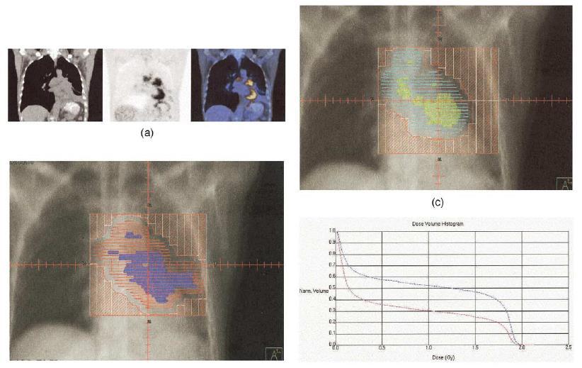
58 Isodose display on axial slices for SIB IMRT plan showing conformal 70-Gy dose around primary PTV (red) and 60-Gy dose around affected nodes (pink and blue). Hypoxic GTV (green) is covered by 80-Gy isodose. Parotid glands (orange and lilac) are avoided by high isodose lines Conclusion: FMISO-PET will prove useful for selecting patients for the most appropriate treatment. Its applications include: (1) Identification and localization of significant hypoxia (2) Delineation of hypoxic sub-volumes within the GTV for boost radiation (3) Selection of appropriate systemic agents to complement the boost therapy
59 FAZA 18 F-labeled PET hypoxia-tracer 18 F azomycin arabinoside (FAZA) Superior pharmacokinetics than F-MISO: faster clearance from normal tissues Significantly higher tumor to background (TB) ratio than F- MISO PET= better resolution
 axial; (b) coronal; (c) intensity-modulated radiotherapy dose painting). The hypoxic area (biological target volume) is treated with 2.")
60 Integrated 18 F-azomycin arabinoside (FAZA)-PET/CT in a patient with larynx squamous cell carcinoma. The PET/CT image fusion ((a) axial; (b) coronal; (c) intensity-modulated radiotherapy dose painting). The hypoxic area (biological target volume) is treated with 2.4 Gy/fraction; the gross tumour volume, based on radiological and clinical data is treated with 2 Gy/fraction


61 Color-washed images illustrated regions of heterogeneous 60Cu-ATSM intensity within the gross tumor representing the presence of tumor hypoxia.


62
63 Fractionation and Hypoxia Fractionated course of radiotherapy is more effective in a given hypoxic tumor than a single fraction Minimum time between 2 fractions of radiotherapy should be 4 hours Hyperfractionation/ accelerated radiotherapy also tackles the issue of accelerated repopulation of tumor cells, esp in H&N cancer where most of the studies of hypoxic modification have also been carried out Delivering hypofractionated radiation to the hypoxic component may give better outcome
64 Hypoxia imaging: Unresolved issues Hypoxia-a dynamic phenomenon within tumor Limited spatial resolution Influence of changes in the oxygenation status before and during treatment? Dose levels required to effectively eliminate these radioresistant subpopulations?
65 OTHER MOLECULAR IMAGING

66 Tumor Proliferation Radiolabelled deoxy-uridines: rapid degradation of these compounds in vivo FLT- 18F-3 deoxy 3 flourothymidine.- 2 studies have shown significant correlation with Ki-67 labeling index
67 Other targets EGFR Cyclin D Molecular risk profiling Search for fingerprinting of malignant phenotypes, sensitive to a specific type of modified RT(accelerated /hyperfractionated). Many new Theragnostic imaging modalities are likely to be identified. Theragnostic imaging for radiation oncology is use of molecular and biological imaging to prescribe the distribution of radiation in four dimensions- 3 dimensions of space plus time.

68 Results: TCD50 after EBRT was significantly decreased by EGFRtargeted RIT in FaDu (RT responder) but not in UT-SCC-5 (RT nonresponder). Conclusion: EGFR-targeted EBRIT can improve permanent local tumor control compared to EBRT alone. PET imaging of bioavailability of labeled cetuximab appears to be a suitable predictor for response to EBRIT.
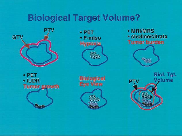
69 BIOLOGICAL TARGET VOLUME/ BIOLOGICAL EYE VIEW Dose uniformity within PTV in external beam-matter of tradition and convention. IMRT-Non uniform dose, Dose painting/sculpting but how? Answer is: Biological imaging Biological tumor volume Derived from biological images and their use may guide customized dose delivery to various parts of treatment volume.
70

71


72 Bioluminescence Bioluminescence is the production and emission of light by a living organism Bioluminescence is emitted when chemical energy is converted to light In presence of activated Cre recombinase, luciferase activity, and by proxy, HPV oncogenes were induced to 11-fold higher levels. Tamoxifen treatment resulted in oral tumor development with increased bioluminescent activity Decreased bioluminescence after treatment with rapamycin or image-guided radiotherapy Novel system enables to rapidly visualize HPV-positive tumor growth and new interventions using clinically relevant drugs and radiotherapy techniques
73 CONCLUSION Molecular imaging-a powerful tool in cancer management PET scan is most widely accepted and utilized Anatomic imaging and molecular imaging- complementary to each other Molecular imaging-perhaps the future of radiation oncology decision making and planning
74 Thank You
PET-CT for radiotherapy planning in lung cancer: current recommendations and future directions
 PET-CT for radiotherapy planning in lung cancer: current recommendations and future directions Gerry Hanna Centre for Cancer Research and Cell Biology Queen s University of Belfast @gerryhanna Talk Outline
This house believes that the use of Functional Imaging for treatment planning of head and neck tumors needs to be carefully considered.
 This house believes that the use of Functional Imaging for treatment planning of head and neck tumors needs to be carefully considered. IMRT for Head and Neck Tumors Vincent GREGOIRE, M.D., Ph.D., Hon.
IMRT - the physician s eye-view. Cinzia Iotti Department of Radiation Oncology S.Maria Nuova Hospital Reggio Emilia
 IMRT - the physician s eye-view Cinzia Iotti Department of Radiation Oncology S.Maria Nuova Hospital Reggio Emilia The goals of cancer therapy Local control Survival Functional status Quality of life Causes
Positron emission tomography/computer tomography in the evaluation of head and neck cancer treatment
 Positron emission tomography/computer tomography in the evaluation of head and neck cancer treatment Severina Šedienė 1, Ilona Kulakienė 1, Viktoras Rudžianskas 2 1 Lithuanian University of Health Sciences,
Molecular Imaging and Cancer
 Molecular Imaging and Cancer Cancer causes one in every four deaths in the United States, second only to heart disease. According to the U.S. Department of Health and Human Services, more than 512,000
Utility of 18 F-FDG PET/CT in metabolic response assessment after CyberKnife radiosurgery for early stage non-small cell lung cancer
 Utility of F-FDG PET/CT in metabolic response assessment after CyberKnife radiosurgery for early stage non-small cell lung cancer Ngoc Ha Le 1*, Hong Son Mai 1, Van Nguyen Le 2, Quang Bieu Bui 2 1 Department
PET Guidance of Therapy for BNCT and in vivo B-10 imaging
 INFN LNL Legnaro 17-19 Novembre 2009 Principles of Positron Emission Tomography and Radiopharmaceuticals PET Guidance of Therapy for BNCT and in vivo B-10 imaging Luca Menichetti, Ph.D C.N.R. Institute
First, how does radiation work?
 Hello, I am Prajnan Das, Faculty Member in the Department of Radiation Oncology at The University of Texas MD Anderson Cancer Center. We are going to talk today about some of the basic principles regarding
IAEA RTC. PET/CT and Planning of Radiation Therapy 20/08/2014. Sarajevo (Bosnia & Hercegovina) Tuesday, June :40-12:20 a.
 Tuesday, June :40-12:20 a.") IAEA RTC PET/CT and Planning of Radiation Therapy Sarajevo (Bosnia & Hercegovina) Tuesday, June 17 2014 11:40-12:20 a.m María José García Velloso Servicio de Medicina Nuclear Clínica Universidad de Navarra
IAEA RTC PET/CT and Planning of Radiation Therapy Sarajevo (Bosnia & Hercegovina) Tuesday, June 17 2014 11:40-12:20 a.m María José García Velloso Servicio de Medicina Nuclear Clínica Universidad de Navarra
Evaluation of Whole-Field and Split-Field Intensity Modulation Radiation Therapy (IMRT) Techniques in Head and Neck Cancer
 Techniques in Head and Neck Cancer") 1 Charles Poole April Case Study April 30, 2012 Evaluation of Whole-Field and Split-Field Intensity Modulation Radiation Therapy (IMRT) Techniques in Head and Neck Cancer Abstract: Introduction: This study
1 Charles Poole April Case Study April 30, 2012 Evaluation of Whole-Field and Split-Field Intensity Modulation Radiation Therapy (IMRT) Techniques in Head and Neck Cancer Abstract: Introduction: This study
HALF. Who gets radiotherapy? Who gets radiotherapy? Half of all cancer patients get radiotherapy. By 1899 X rays were being used for cancer therapy
 The Physical and Biological Basis of By 1899 X rays were being used for cancer therapy David J. Brenner, PhD, DSc Center for Radiological Research Department of Radiation Oncology Columbia University Medical
Use of imaging systems for patient modeling - PET and SPECT
 Use of imaging systems for patient modeling - PET and SPECT Sasa Mutic Department of Radiation Oncology Siteman Cancer Center Mallinckrodt Institute of Radiology Washington University School of Medicine
Brain Tumors. What is a brain tumor?
 Scan for mobile link. Brain Tumors A brain tumor is a collection of abnormal cells that grows in or around the brain. It poses a risk to the healthy brain by either invading or destroying normal brain
Role of radiotherapy in the treatment of lymphoma in Lena Specht MD DMSc Professor of Oncology Rigshospitalet, University of Copenhagen Denmark
 Role of radiotherapy in the treatment of lymphoma in 2017 Lena Specht MD DMSc Professor of Oncology Rigshospitalet, University of Copenhagen Denmark Disclosures Member of Advisory Board and Principal Investigator,
PET/CT Frequently Asked Questions
 PET/CT Frequently Asked Questions General Q: Is FDG PET specific for cancer? A: No, it is a marker of metabolism. In general, any disease that causes increased metabolism can result in increased FDG uptake
Herlev radiation oncology team explains what MRI can bring
 Publication for the Philips MRI Community Issue 46 2012/2 Herlev radiation oncology team explains what MRI can bring The radiotherapy unit at Herlev University Hospital investigates use of MRI for radiotherapy
8/10/2016. PET/CT for Tumor Response. Staging and restaging Early treatment response evaluation Guiding biopsy
 PET/CT for Tumor Response Evaluation August 4, 2016 Wei Lu, PhD Department of Medical Physics www.mskcc.org Department of Radiation Oncology www.umaryland.edu FDG PET/CT for Cancer Imaging Staging and
Dr Sneha Shah Tata Memorial Hospital, Mumbai.
 Dr Sneha Shah Tata Memorial Hospital, Mumbai. Topics covered Lymphomas including Burkitts Pediatric solid tumors (non CNS) Musculoskeletal Ewings & osteosarcoma. Neuroblastomas Nasopharyngeal carcinomas
MRI Based treatment planning for with focus on prostate cancer. Xinglei Shen, MD Department of Radiation Oncology KUMC
 MRI Based treatment planning for with focus on prostate cancer Xinglei Shen, MD Department of Radiation Oncology KUMC Overview How magnetic resonance imaging works (very simple version) Indications for
ADVANCES IN RADIATION TECHNOLOGIES IN THE TREATMENT OF CANCER
 ADVANCES IN RADIATION TECHNOLOGIES IN THE TREATMENT OF CANCER Bro. Dr. Collie Miller IARC/WHO Based on trends in the incidence of cancer, the International Agency for Research on Cancer (IARC) and WHO
Appendix 1: Regional Lymph Node Stations for Staging Esophageal Cancer
 Appendix 1: Regional Lymph Node Stations for Staging Esophageal Cancer Locoregional (N stage) disease was redefined in the seventh edition of the AJCC Cancer Staging Manual as any periesophageal lymph
Changing Paradigms in Radiotherapy
 Changing Paradigms in Radiotherapy Marco van Vulpen, MD, PhD Mouldroomdag-2015 Towards the elimination of invasion 1 NIH opinion on the future of oncology Twenty-five years from now,i hope that we won
Use of molecular and functional imaging for treatment planning The Good, The Bad and The Ugly
 Use of molecular and functional imaging for treatment planning The Good, The Bad and The Ugly Robert Jeraj, PhD Associate Professor of Medical Physics, Human Oncology, Radiology and Biomedical Engineering
Nuclear Medicine and PET. D. J. McMahon rev cewood
 Nuclear Medicine and PET D. J. McMahon 150504 rev cewood 2018-02-15 Key Points Nuclear Medicine and PET: Imaging: Understand how Nuc Med & PET differ from Radiography & CT by the source of radiation. Be
COMENIUS-Project: SM&CLIL Radiation & Medicine
 Medical imaging refers to the techniques and processes used to create images of the human body (or parts thereof) for clinical purposes. Thanks to modern mathematics and computer technology, medical imaging
Efficient SIB-IMRT planning of head & neck patients with Pinnacle 3 -DMPO
 Investigations and research Efficient SIB-IMRT planning of head & neck patients with Pinnacle 3 -DMPO M. Kunze-Busch P. van Kollenburg Department of Radiation Oncology, Radboud University Nijmegen Medical
Bone PET/MRI : Diagnostic yield in bone metastases and malignant primitive bone tumors
 Bone PET/MRI : Diagnostic yield in bone metastases and malignant primitive bone tumors Lars Stegger, Benjamin Noto Department of Nuclear Medicine University Hospital Münster, Germany Content From PET to
Indications of PET/CT in oncology
 Monday, August 27, 2012 Session 1, 10:00-10:40 Indications of PET/CT in oncology Helle Westergren Hendel MD, PhD, assistant professor Bacelor in Leadership & Health Ecomomics Head of Clinical PET, Herlev
Linac or Non-Linac Demystifying And Decoding The Physics Of SBRT/SABR
 Linac or Non-Linac Demystifying And Decoding The Physics Of SBRT/SABR PhD, FAAPM, FACR, FASTRO Department of Radiation Oncology Indiana University School of Medicine Indianapolis, IN, USA Indra J. Das,
Option D: Medicinal Chemistry
 Option D: Medicinal Chemistry Basics - unstable radioactive nuclei emit radiation in the form of smaller particles alpha, beta, positron, proton, neutron, & gamma are all used in nuclear medicine unstable
Target Delineation in Gliomas. Prof PK Julka Department of Radiotherapy and Oncology AIIMS, New Delhi
 Target Delineation in Gliomas Prof PK Julka Department of Radiotherapy and Oncology AIIMS, New Delhi 1 What is a glioma? A primary brain tumour that originated from a cell of the nervous system 2 Recommendations:
PET CT for Staging Lung Cancer
 PET CT for Staging Lung Cancer Rohit Kochhar Consultant Radiologist Disclosures Neither I nor my immediate family members have financial relationships with commercial organizations that may have a direct
Principles of nuclear metabolic imaging. Prof. Dr. Alex Maes AZ Groeninge Kortrijk and KULeuven Belgium
 Principles of nuclear metabolic imaging Prof. Dr. Alex Maes AZ Groeninge Kortrijk and KULeuven Belgium I. Molecular imaging probes A. Introduction - Chemical disturbances will precede anatomical abnormalities
MRI Applications in Radiation Oncology:
 MRI Applications in Radiation Oncology: Physician s Perspective Jeff Olsen, MD Department of Radiation Oncology Washington University, St. Louis, MO Disclosures Washington University has research and service
Brain Tumor Treatment
 Scan for mobile link. Brain Tumor Treatment Brain Tumors Overview A brain tumor is a group of abnormal cells that grows in or around the brain. Tumors can directly destroy healthy brain cells. They can
Ruolo dell imaging nella pianificazione del trattamento
 Simposio AIRO-SIRM: Diagnostica per immagini morfologica e funzionale nella stadiazione, terapia e follow-up dei sarcomi delle parti molli Ruolo dell imaging nella pianificazione del trattamento Marco
Breast Cancer. What is breast cancer?
 Scan for mobile link. Breast Cancer Breast cancer is a malignant tumor in or around breast tissue. It usually begins as a lump or calcium deposit that develops from abnormal cell growth. Most breast lumps
Chapter 10. Summary, conclusions and future perspectives
 Chapter 10 Summary, conclusions and future perspectives 10.1 SUMMARY In this thesis, a new tumor imaging tracer in nuclear medicine is studied. This 123 tracer, L-3-[ I]Iodo-alpha-methyl-tyrosine (IMT),
Oral cavity cancer Post-operative treatment
 Oral cavity cancer Post-operative treatment Dr. Christos CHRISTOPOULOS Radiation Oncologist Centre Hospitalier Universitaire (C.H.U.) de Limoges, France Important issues RT -techniques Patient selection
LYMPHATIC DRAINAGE IN THE HEAD & NECK
 LYMPHATIC DRAINAGE IN THE HEAD & NECK Like other parts of the body, the head and neck contains lymph nodes (commonly called glands). Which form part of the overall Lymphatic Drainage system of the body.
PET imaging of cancer metabolism is commonly performed with F18
 PCRI Insights, August 2012, Vol. 15: No. 3 Carbon-11-Acetate PET/CT Imaging in Prostate Cancer Fabio Almeida, M.D. Medical Director, Arizona Molecular Imaging Center - Phoenix PET imaging of cancer metabolism
FDG PET/CT STAGING OF LUNG CANCER. Dr Shakher Ramdave
 FDG PET/CT STAGING OF LUNG CANCER Dr Shakher Ramdave FDG PET/CT STAGING OF LUNG CANCER FDG PET/CT is used in all patients with lung cancer who are considered for curative treatment to exclude occult disease.
State of the Art Radiotherapy for Pediatric Tumors. Suzanne L. Wolden, MD Memorial Sloan-Kettering Cancer Center
 State of the Art Radiotherapy for Pediatric Tumors Suzanne L. Wolden, MD Memorial Sloan-Kettering Cancer Center Introduction Progress and success in pediatric oncology Examples of low-tech and high-tech
THE EFFECT OF USING PET-CT FUSION ON TARGET VOLUME DELINEATION AND DOSE TO ORGANS AT RISK IN 3D RADIOTHERAPY PLANNING OF PATIENTS WITH NSSLC
 THE EFFECT OF USING PET-CT FUSION ON TARGET VOLUME DELINEATION AND DOSE TO ORGANS AT RISK IN 3D RADIOTHERAPY PLANNING OF PATIENTS WITH NSSLC Hana Al-Mahasneh,M.D*., Mohammad Khalaf Al-Fraessan, M.R.N,
Image Fusion, Contouring, and Margins in SRS
 Image Fusion, Contouring, and Margins in SRS Sarah Geneser, Ph.D. Department of Radiation Oncology University of California, San Francisco Overview Review SRS uncertainties due to: image registration contouring
Defining Target Volumes and Organs at Risk: a common language
 Defining Target Volumes and Organs at Risk: a common language Eduardo Rosenblatt Section Head Applied Radiation Biology and Radiotherapy (ARBR) Section Division of Human Health IAEA Objective: To introduce
Protocol of Radiotherapy for Small Cell Lung Cancer
 107 年 12 月修訂 Protocol of Radiotherapy for Small Cell Lung Cancer Indication of radiotherapy Limited stage: AJCC (8th edition) stage I-III (T any, N any, M0) that can be safely treated with definitive RT
Radiotherapy Planning (Contouring Lung Cancer for Radiotherapy dose prescription) Dr Raj K Shrimali
 Dr Raj K Shrimali") Radiotherapy Planning (Contouring Lung Cancer for Radiotherapy dose prescription) Dr Raj K Shrimali Let us keep this simple and stick to some basic rules Patient positioning Must be reproducible Must be
Radiotherapy Planning (Contouring Lung Cancer for Radiotherapy dose prescription) Dr Raj K Shrimali Let us keep this simple and stick to some basic rules Patient positioning Must be reproducible Must be
UNIVERSITY OF WISCONSIN-LA CROSSE Graduate Studies
 UNIVERSITY OF WISCONSIN-LA CROSSE Graduate Studies A SINGLE INSTITUTION S EXPERIENCE IN DEVELOPING A PURPOSEFUL AND EFFICIENT OFF-LINE TECHNIQUE FOR ADAPTIVE RADIOTHERAPY IN A CLINICAL ENVIRONMENT A Research
Evaluation of Lung Cancer Response: Current Practice and Advances
 Evaluation of Lung Cancer Response: Current Practice and Advances Jeremy J. Erasmus I have no financial relationships, arrangements or affiliations and this presentation will not include discussion of
4D PET: promises and limitations
 4D PET: promises and limitations Tinsu Pan, Ph.D. M.D. Anderson Cancer Center The University of Texas Background Outlines Gating techniques: Deep inspiration breath hold 4D PET/CT Non-gating techniques
Radiation treatment planning in lung cancer
 Radiation treatment planning in lung cancer Georg Dietmar 1,2 1 Div. Medical Rad. Phys., Dept. of Radiation Oncology / Medical Univ. Vienna & AKH Wien 2 Christian Doppler Laboratory for Medical Radiation
What Radiologists do?
 Multimodality Imaging in Oncology 2018 March 5 th 9th Diagnostic Imaging in Oncology What Radiologists do? Chikako Suzuki, MD, PhD Department of Diagnostic Radiology, KS Solna Department of Molecular Medicine
TOPICS. Primary Radiation Therapy. Targeted Therapy in Oncology. Principles of Radiation Therapy. Principles of Radiation Therapy
 Peter B. Schiff, M.D., Ph.D. Department of Radiation Oncology Columbia University College of Physicians & Surgeons May 4, 2007 Targeted Therapy in Oncology Surgical Oncology Minimal invasive techniques
Radiotherapy physics & Equipments
 Radiotherapy physics & Equipments RAD 481 Lecture s Title: An Overview of Radiation Therapy for Health Care Professionals Dr. Mohammed Emam Vision :IMC aspires to be a leader in applied medical sciences,
The Paul Evans Memorial Lecture Functional radiotherapy targeting using focused dose escalation. Roberto Alonzi Mount Vernon Cancer Centre
 The Paul Evans Memorial Lecture Functional radiotherapy targeting using focused dose escalation Roberto Alonzi Mount Vernon Cancer Centre Overview Introduction and rationale for focused dose escalation
Imaging in gastric cancer
 Imaging in gastric cancer Gastric cancer remains a deadly disease because of late diagnosis. Adenocarcinoma represents 90% of malignant tumors. Diagnosis is based on endoscopic examination with biopsies.
Molecular Imaging and Breast Cancer
 Molecular Imaging and Breast Cancer Breast cancer forms in tissues of the breast usually in the ducts, tubes that carry milk to the nipple, and lobules, the glands that make milk. It occurs in both men
The Use of PET Scanning in Urologic Oncology
 The Use of PET Scanning in Urologic Oncology Dr Nicholas C. Buchan Uro-oncology Fellow 1 2 Aims To understand the basic concepts underlying PET scanning. Understand the emerging role of PET Scanning for
FDOPA, C11Choline, C11 Methionine. Dr K.G.Kallur
 FDOPA, C11Choline, C11 Methionine Dr K.G.Kallur Why? 11C Methionine scan Had undergone resection Earlier. Post op recurrent hypercalcemia C11 Methionine Unable to see in Sestamibi scan Brain Tumor After
Page 1. Helical (Spiral) Tomotherapy. UW Helical Tomotherapy Unit. Helical (Spiral) Tomotherapy. MVCT of an Anesthetized Dog with a Sinus Tumor
 Tomotherapy. UW Helical Tomotherapy Unit. Helical (Spiral) Tomotherapy. MVCT of an Anesthetized Dog with a Sinus Tumor") Helical (Spiral) Tomotherapy Novel Clinical Applications of IMRT Linac Ring Gantry CT Detector X-Ray Fan Beam Binary Multileaf Collimator Binary MLC Leaves James S Welsh, MS, MD Department of Human Oncology
Helical (Spiral) Tomotherapy Novel Clinical Applications of IMRT Linac Ring Gantry CT Detector X-Ray Fan Beam Binary Multileaf Collimator Binary MLC Leaves James S Welsh, MS, MD Department of Human Oncology
Pitfalls and Remedies in PET/CT imaging for RT planning
 Pitfalls and Remedies in PET/CT imaging for RT planning Tinsu Pan, Ph.D. M.D. Anderson Cancer Center The University of Texas Outlines Background Average CT (< 1 msv) to reduce mis-alignment of PET and
Chapters from Clinical Oncology
 Chapters from Clinical Oncology Lecture notes University of Szeged Faculty of Medicine Department of Oncotherapy 2012. 1 RADIOTHERAPY Technical aspects Dr. Elemér Szil Introduction There are three possibilities
肺癌放射治療新進展 Recent Advance in Radiation Oncology in Lung Cancer 許峰銘成佳憲國立台灣大學醫學院附設醫院腫瘤醫學部
 肺癌放射治療新進展 Recent Advance in Radiation Oncology in Lung Cancer 許峰銘成佳憲國立台灣大學醫學院附設醫院腫瘤醫學部 Outline Current status of radiation oncology in lung cancer Focused on stage III non-small cell lung cancer Radiation
Title: TC simulation versus TC/PET simulation for radiotherapy in lung cancer: volumes comparison in two cases.
 Title: TC simulation versus TC/PET simulation for radiotherapy in lung cancer: volumes comparison in two cases. Authors: Franzone, P.; 1* Muni, A; 2 Cazzulo, E.; 3 Berretta, L.; 1 Pozzi, G. 1 ; Todisco,
Radiotherapy What are our options and what is on the horizon. Dr Kevin So Specialist Radiation Oncologist Epworth Radiation Oncology
 Radiotherapy What are our options and what is on the horizon Dr Kevin So Specialist Radiation Oncologist Epworth Radiation Oncology Outline Advances in radiotherapy technique Oligo - disease Advancements
Subject: Image-Guided Radiation Therapy
 04-77260-19 Original Effective Date: 02/15/10 Reviewed: 01/25/18 Revised: 01/01/19 Subject: Image-Guided Radiation Therapy THIS MEDICAL COVERAGE GUIDELINE IS NOT AN AUTHORIZATION, CERTIFICATION, EXPLANATION
A VMAT PLANNING SOLUTION FOR NECK CANCER PATIENTS USING THE PINNACLE 3 PLANNING SYSTEM *
 Romanian Reports in Physics, Vol. 66, No. 2, P. 401 410, 2014 A VMAT PLANNING SOLUTION FOR NECK CANCER PATIENTS USING THE PINNACLE 3 PLANNING SYSTEM * M. D. SUDITU 1,2, D. ADAM 1,2, R. POPA 1,2, V. CIOCALTEI
21 st Century Radiotherapy: State-of-the-art and predicting the future. Clinical applications of PET. PET Imaging
 21 st Century Radiotherapy: State-of-the-art and predicting the future Clinical applications of PET Pat Price Ralston Paterson Professor of Radiation Oncology Christie Hospital Manchester UK Mayneord-Phillips
A Comparison of IMRT and VMAT Technique for the Treatment of Rectal Cancer
 A Comparison of IMRT and VMAT Technique for the Treatment of Rectal Cancer Tony Kin Ming Lam Radiation Planner Dr Patricia Lindsay, Radiation Physicist Dr John Kim, Radiation Oncologist Dr Kim Ann Ung,
REVISITING ICRU VOLUME DEFINITIONS. Eduardo Rosenblatt Vienna, Austria
 REVISITING ICRU VOLUME DEFINITIONS Eduardo Rosenblatt Vienna, Austria Objective: To introduce target volumes and organ at risk concepts as defined by ICRU. 3D-CRT is the standard There was a need for a
Future upcoming technologies and what audit needs to address
 Future upcoming technologies and what audit needs to address Dr R.I MacKay History of audit Absolute dose - Simple phantom standard dose measurement Point doses in beams - Phantoms of relatively simple
Sarcoma and Radiation Therapy. Gabrielle M Kane MB BCh EdD FRCPC Muir Professorship in Radiation Oncology University of Washington
 Sarcoma and Radiation Therapy Gabrielle M Kane MB BCh EdD FRCPC Muir Professorship in Radiation Oncology University of Washington Objective: Helping you make informed decisions Introduction Process Radiation
TUMOR HYPOXIA BENCH TO BEDSIDE
 TUMOR HYPOXIA BENCH TO BEDSIDE Marianne Nordsmark, Lise Saksø Mortensen, Morten Busk, Michael Horsman, Markus Alber, Niels Bassler, Jørgen Petersen and Jens Overgaard. Dept. Oncology, Aarhus University
Innovations in Radiation Therapy, including SBRT, IMRT, and Proton Beam Therapy. Sue S. Yom, M.D., Ph.D.
 Innovations in Radiation Therapy, including SBRT, IMRT, and Proton Beam Therapy Sue S. Yom, M.D., Ph.D. Disclosures Genentech: advisory, research support ImClone: research support Plexxikon: research support
A Patient s Guide to SRS
 A Patient s Guide to SRS Stereotactic Radiosurgery 230 Nebraska St. Sioux City, IA 51101 NOTES 230 Nebraska St. Sioux City, IA 51101 Contents page Introduction 1 SRS and how it works 2 The technology involved
Functional aspects of anatomical imaging techniques
 Functional aspects of anatomical imaging techniques Nilendu Purandare Associate Professor & Consultant Radiologist Tata Memorial Centre Functional/metabolic/molecular imaging (radioisotope scanning) PET
POSITRON EMISSION TOMOGRAPHY (PET)
") Status Active Medical and Behavioral Health Policy Section: Radiology Policy Number: V-27 Effective Date: 08/27/2014 Blue Cross and Blue Shield of Minnesota medical policies do not imply that members should
Status Active Medical and Behavioral Health Policy Section: Radiology Policy Number: V-27 Effective Date: 08/27/2014 Blue Cross and Blue Shield of Minnesota medical policies do not imply that members should
Breast Cancer. What is breast cancer?
 Scan for mobile link. Breast Cancer Breast cancer is a malignant tumor in or around breast tissue. It usually begins as a lump or calcium deposit that develops from abnormal cell growth. Most breast lumps
The Role of PET / CT in Lung Cancer Staging
 July 2004 The Role of PET / CT in Lung Cancer Staging Vlad Vinarsky, Harvard Medical School Year IV Patient AM HPI: 81 yo F p/w hemoptysis x 1 month LLL lesion on CXR, not responsive to Abx 35 pack-year
PET Assessment of Tumor Hypoxia
 PET Assessment of Tumor Hypoxia Farrokh Dehdashti, M.D. Mallinckrodt Institute of Radiology Washington University St. Louis, Missouri 9/30/10 This work was supported by National Institute of Health R21
Molecular Imaging in the Development of Cancer Therapeutics. Johannes Czernin
 Molecular Imaging in the Development of Cancer Therapeutics Johannes Czernin Ahmanson Biological Imaging Division University of California Los Angeles Cancer Statistics Cancer Type 5-year Survival Rate
PET-MRI in malignant bone tumours. Lars Stegger Department of Nuclear Medicine University Hospital Münster, Germany
 PET-MRI in malignant bone tumours Lars Stegger Department of Nuclear Medicine University Hospital Münster, Germany Content From PET to PET/MRI General considerations Bone metastases Primary bone tumours
General Nuclear Medicine
 General Nuclear Medicine What is General Nuclear Medicine? What are some common uses of the procedure? How should I prepare? What does the equipment look like? How does the procedure work? How is the procedure
Stereotactic radiotherapy
 Stereotactic radiotherapy Influence of patient positioning and fixation on treatment planning - clinical results Frank Zimmermann Institut für Radioonkologie Universitätsspital Basel Petersgraben 4 CH
Flattening Filter Free beam
 Dose rate effect in external radiotherapy: biology and clinic Marta Scorsetti, M.D. Radiotherapy and Radiosurgery Dep., Istituto Clinico Humanitas, Milan, Italy Brescia October 8th/9th, 2015 Flattening
Colorectal Cancer and FDG PET/CT
 Hybrid imaging in colorectal & esophageal cancer Emmanuel Deshayes IAEA WorkShop, November 2017 Colorectal Cancer and FDG PET/CT 1 Clinical background Cancer of the colon and rectum is one of the most
Improving prediction of radiotherapy response and optimizing target definition by using FDG-PET for lung cancer patients
 Investigations and research Improving prediction of radiotherapy response and optimizing target definition by using FDG-PET for lung cancer patients R.J.H.M. Steenbakkers G.R. Borst M. van Herk H. Bartelink
An introduction to medical imaging and radiotherapy: Current status and future directions I
 An introduction to medical imaging and radiotherapy: Current status and future directions I Dr Colin Baker Head of Radiotherapy Physics Royal Berkshire NHS Foundation Trust Overview Lecture 1 Principles
Molecular Imaging Guided Therapy: The Perfect Storm. David M Schuster, MD Emory University Department of Radiology Atlanta, GA
 Molecular Imaging Guided Therapy: The Perfect Storm David M Schuster, MD Emory University Department of Radiology Atlanta, GA Talk can be found at radiology.emory.edu Let s start with a case 74 year
Radiotherapy Physics and Equipment
 Radiological Sciences Department Radiotherapy Physics and Equipment RAD 481 Lecture s Title: Introduction Dr. Mohammed EMAM Ph.D., Paris-Sud 11 University Vision :IMC aspires to be a leader in applied
I. Equipments for external beam radiotherapy
 I. Equipments for external beam radiotherapy 5 linear accelerators (LINACs): Varian TrueBeam 6, 10 & 18 MV photons, 6-18 MeV electrons, image-guided (IGRT) and intensity modulated radiotherapy (IMRT),
FOR CMS (MEDICARE) MEMBERS ONLY NATIONAL COVERAGE DETERMINATION (NCD) FOR MAGNETIC RESONANCE IMAGING:
 MEMBERS ONLY NATIONAL COVERAGE DETERMINATION (NCD) FOR MAGNETIC RESONANCE IMAGING:") National Imaging Associates, Inc. Clinical guidelines BONE MARROW MRI Original Date: July 2008 Page 1 of 5 CPT Codes: 77084 Last Review Date: September 2014 NCD 220.2 MRI Last Effective Date: July 2011
National Imaging Associates, Inc. Clinical guidelines BONE MARROW MRI Original Date: July 2008 Page 1 of 5 CPT Codes: 77084 Last Review Date: September 2014 NCD 220.2 MRI Last Effective Date: July 2011
Typical PET Image. Elevated uptake of FDG (related to metabolism) Lung cancer example: But where exactly is it located?
 Lung cancer example: But where exactly is it located?") Typical PET Image Elevated uptake of FDG (related to metabolism) Lung cancer example: But where exactly is it located? PET/CT Oncology Imaging Anatometabolic fusion images are useful in the management
Typical PET Image Elevated uptake of FDG (related to metabolism) Lung cancer example: But where exactly is it located? PET/CT Oncology Imaging Anatometabolic fusion images are useful in the management
Clinical Implications Of Dose Summation And Adaptation
 Clinical Implications Of Dose Summation And Adaptation Patrick Kupelian, M.D. Professor and Vice Chair University of California Los Angeles Department of Radiation Oncology pkupelian@mednet.ucla.edu August
Lung Cancer Imaging. Terence Z. Wong, MD,PhD. Department of Radiology Duke University Medical Center Durham, NC 9/9/09
 Lung Cancer Imaging Terence Z. Wong, MD,PhD Department of Radiology Duke University Medical Center Durham, NC 9/9/09 Acknowledgements Edward F. Patz, Jr., MD Jenny Hoang, MD Ellen L. Jones, MD, PhD Lung
An Introduction to PET Imaging in Oncology
 January 2002 An Introduction to PET Imaging in Oncology Janet McLaren, Harvard Medical School Year III Basics of PET Principle of Physiologic Imaging: Allows in vivo visualization of structures by their
Therapeutic ratio - An Overview. Past Present Future Prof Ramesh S Bilimaga
 Therapeutic ratio - An Overview Past Present Future Prof Ramesh S Bilimaga Radiation Oncology Discipline of human medicine concerned with the generation, conservation and dissemination of knowledge concerning
PHYSICS 2: HSC COURSE 2 nd edition (Andriessen et al) CHAPTER 20 Radioactivity as a diagnostic tool (pages 394-5)
 CHAPTER 20 Radioactivity as a diagnostic tool (pages 394-5)") PHYSICS 2: HSC COURSE 2 nd edition (Andriessen et al) CHAPTER 20 Radioactivity as a diagnostic tool (pages 394-5) 1. (a) A radioisotope is an isotope that is unstable and will emit particles from the nucleus
PHYSICS 2: HSC COURSE 2 nd edition (Andriessen et al) CHAPTER 20 Radioactivity as a diagnostic tool (pages 394-5) 1. (a) A radioisotope is an isotope that is unstable and will emit particles from the nucleus