CD30 expression utilization for the accuracy of classical Hodgkin s lymphoma staging
|
|
|
- Violet Wheeler
- 6 years ago
- Views:
Transcription
1 Romanian Journal of Morphology and Embryology 2006, 47(2): ORIGINAL PAPER CD30 expression utilization for the accuracy of classical Hodgkin s lymphoma staging CORINA FLANGEA 1), ELENA POTENCZ 2), RODICA MIHĂESCU 3), A. ANGHEL 1), S. GÎJU 4), MARILENA MOTOC 1), C. DOGARU 1) 1) Biochemistry Department 2) Pathology Department 3) Hematology Department Victor Babeş University of Medicine and Pharmacy, Timişoara 4) Central Laboratory, County Hospital no. 1, Timişoara Abstract Introduction. The presence of Reed Sternberg malignant cells is absolutely necessary for Hodgkin s lymphoma diagnostic, but it is not always sufficient because can be observed Reed Sternberg-like cells in other malignant and benignant diseases, too. The CD30 expression at Hodgkin and Reed Sternberg level can give us supplementary information in differential diagnostic and can be used as progressive disease factor. Material and methods. Our study was composed from 63 histopathological diagnosticated with Hodgkin s lymphoma and hospitalized in Hematology Department of County Hospital Timişoara. CD30 expression was immunohistochemical semiquantitative evaluated using clone BerH2 as primary antibody and APAAP New Fuchsin as visualization system. Results and discussions. The increasing of CD30 expression occurs in the same time with advanced stages and the disease progression (p 0.001). For I and II stages CD30 expression does not overcome (-/+) category while the III and IV stages, all the are situated in (+/-) and (+) categories. No connection can be noticed between histological type and CD30 expression (p 1). We consider that using this staining, although less used in Romania, must be done in all Hodgkin s lymphoma and Hodgkin s lymphoma-like. We say that because the main cause of relapses is represented by inadequate clinical staging and diagnostic. Conclusions. In our study, the increasing of CD30 expression is associated with advanced disease stage. We recommend reinvestigating and restaging all that was included into an incipient stages and they have a CD30 expression situated in (+/-) and (+) intervals because some lymph nodes could be overlooked. Keywords: Hodgkin s lymphoma, CD30, tumoral progression. Introduction The Hodgkin s lymphoma diagnostic is mainly a histological diagnostic based by Reed Sternberg multinucleated cells recognizing surrounded by an inflammatory reaction composed from lymphocytes, granulocytes, plasma cells and fibroblasts. Even if the Reed Sternberg cell presence is necessary for Hodgkin s lymphoma diagnostic, it is not specific, occurred in some benignant disease (e.g. in infectious mononucleosis), in some malignant lymphomas (e.g. in anaplastic large cell lymphoma) and in some carcinomas. The cellular inflammatory reaction aspect is important in diagnostic establish, too, that representing the host answering at tumoral invasion [1 3]. CD30 surface antigen was identified for the first time in Hodgkin and Reed Sternberg cells from Hodgkin s lymphoma. It was demonstrated that antibody it is not so specific to Reed Sternberg cells that believed, it noticed into a non-hodgkin s lymphomas, especially in anaplastic large cell lymphoma. In Hodgkin s lymphoma, CD30 expression on Reed Sternberg cells surface is associate with size and invasively of tumor, thus representing a possible indicator of disease aggressively independent by age, race and symptoms [4 7]. The aim of our study is the utilization of CD30 monoclonal antibody (clone Ber-H2) for accuracy of Hodgkin s lymphoma staging. On the other hand, this expression has a very important contribution to Hodgkin s lymphoma diagnostic. Material and methods We had 63 patients hospitalized into the Hematology Department of the County Hospital Timişoara, between January 2000 and June After the histopathological examination into the Pathologic Anatomy Laboratory, these patients were diagnosed as suffering from classical Hodgkin s lymphoma. All the lymph nodes were formalin fixed (10%) and paraffin embedded, sectioned at 4 µm. They were offered kindly by Professor Elena Lazăr. Usual staining was performed with Hematoxylin Eosin and PAS reaction. We used DAKO reagents for immunohistochemical reactions. The visualization was performed by APAAP system using New Fuchsin as chromogen. For antigen retrieving we used Target Retrieval Solution (DAKO). The dilution for primary monoclonal antibody (clone BerH2) was 1/50. Levamisol adding is absolutely necessary for endogen alkaline phosphatase inactivating.
2 114 CD30 samples were appreciated semi-quantitative as follows: expression (+) all the malignant cells positive; (+/-) positive malignant cells 50 95%; (-/+) positive cells under 50%; (-) all the malignant cells negative. Results From the whole 64 patients, 42 (66%) were females and 22 (34%) were males. The age repartition is: years, 15 (24%); years, 13 (21%); years, nine (14%); years, 11 (17%); years, seven (11%) and over 60 years, eight (13%). We can observe that 45% (almost a half) from whole patients are young persons under 30 years. From the clinical point of view, we performed the staging according to Costwolds classification. The majority of the patients, 36 (56%), were diagnosed in the stage III, 12 (19%) in the stage II, 12 (19%) in the stage IV, and only three (6%) in the stage I. Histopathological diagnostic was performed using REAL/WHO classification. Using usual staining the lymph node architecture, we observed also the malignant cells morphology and characteristic of the reactive cellular background. Classical Hodgkin s lymphoma exhibited a diffuse pattern of growth, some atrophic or residual lymphoid follicles. Reed Sternberg cells were clearly visible in all. The cellular background was composed of small lymphocytes and a variable proportion of histiocytes, plasma cells and eosinophils. We could not correlate tissue eosinophilia abundance with clinical status because majority MC CHL and NS CHL revealed rich eosinophils and plasma cells infiltrate. Also, we could not identify Hodgkin and Reed Sternberg cells in bone marrow aspirate. From the whole patients, 32 (50%) were mixed cellularity (MC CHL), 12 (19%) were nodular sclerosis (NS CHL), 19 (29%) were lymphocyte depletion (LD CHL); lymphocyte rich classic Hodgkin s lymphoma was not met. Differential diagnostic with anaplastic large cell lymphoma was performed on histopathological aspect and CD30 expression. In Hodgkin s lymphoma was occurs a cytoplasmatic and Golgi area intense staining and in anaplastic large cell lymphoma (ALCL), the Reed Sternberg-like cells was observed a cytoplasmatic diffuse staining (Figures 1 and 2). Some of the Hodgkin s and Reed Sternberg-like cells display an intense cytoplasmatic and Golgi area staining but differential diagnostic can be easy performed due to specific aspect and expression pattern of both lymphomas. In addition, the typically cohesive pattern of ALCL tumor cells can be better evidenced using CD30 expression. All these anaplastic large cell lymphoma were negative for CD15. CD30 expression function to clinical stage was Corina Flangea et al. contented by us in (+) interval for main of the with stage IV. The data from Table 1 show a CD30 expression increasing according to disease progression. Thus, for stage I, the CD30 expression does not overcome (-/+) interval, majority as (-) and only a single case being in (-/+) interval. For stage II, the CD30 expression extended to (+) interval in a low percentage (16%), predominating into (-/+) and (+/-) intervals. For stages III and IV, the CD30 expression occurred only in (+/-) and (+) intervals, a single case with stage III being in (-/+) interval (Table 1). Table 1 CD30 expression functions to the disease stage Clinical stage (+) CD30 expression (+/-) (-/+) (-) Stage I 1 2 Stage II Stage III Stage IV 7 5 Statistical analysis showed a significant distribution: degrees of freedom: 9, χ 2 = ; p All from (+) and (+/-) interval displayed an intense Hodgkin and Reed Sternberg s staining, especially at Golgi area level. Cases from (-/+) interval have few malignant cells from with intense CD30 expression but majority have a weak expression. All from (-) interval display a weak CD30 expression in Hodgkin and Reed Sternberg s cells. From the histological point of view, we can observe that in CHL have not significant differences of one histological type in CD30 expression. MC CHL displayed a highest percent in (+/-) interval followed by (+) interval. NS CHL was predominated in (+) interval and LD CHL in (+/-) interval. Thus, we could not observe any association between the histological type and the CD30 expression (Table 2). Table 2 CD30 expression functions to the histological type Histological type (+) CD30 expression (+/-) (-/+) (-) MC CHL LD CHL NS CHL test showed a distribution that is not statistic significant: degrees of freedom: 6, χ 2 = (for significance at the 0.05 level, χ 2 should be greater than or equal to ); p 1. CD30 expression is displayed in the Figures 3 6. χ 2 Discussions In the Vermeer MH et al. [8] study neither classical Hodgkin s disease does belong to (-) category and were only MC CHL and NS CHL from histological point of view.


")
")
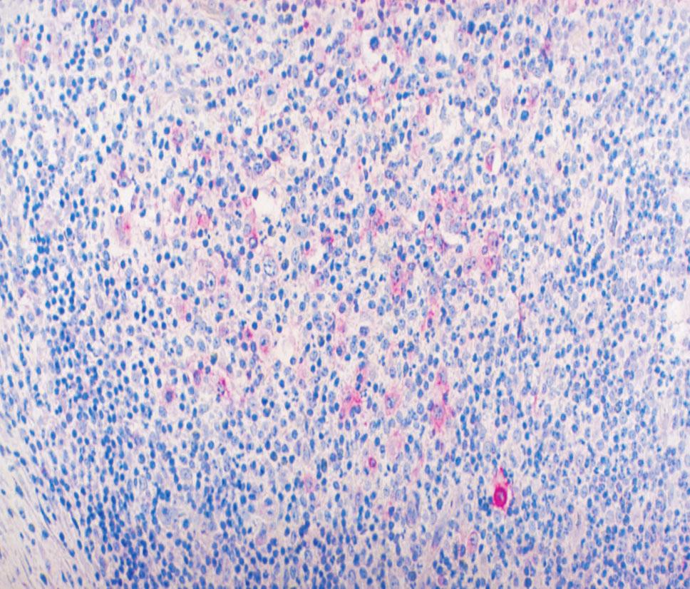
3 CD30 expression utilization for the accuracy of classical Hodgkin's lymphoma staging 115 Figure 1 ALCL, CD30 expression intense positive at Reed Sternberg-like malignant cells level (APAAP New Fuchsin technique, 400; Figure 2 ALCL, CD30 expression intense positive at Reed Sternberg-like malignant cells level (APAAP New Fuchsin technique, 400; Figure 3 LD CHL, with a (+) CD30 expression (APAAP New Fuchsin technique, 200; Figure 4 LD CHL, with a (+) CD30 expression (APAAP New Fuchsin technique, 400; Figure 5 MC CHL, with a (+/-) CD30 expression (APAAP New Fuchsin technique, 400; Figure 6 MC CHL, with a (-/+) CD30 expression (APAAP New Fuchsin technique, 200;
4 116 Watanabe K et al. [9] studied 51 classical Hodgkin s lymphoma, and they could not do any association in their study between histological type and CD30 expression because majority of are MC CHL. Regarding histological type, no connection we can be noticed between histological type and CD30 expression because we have not a significant predominance of one of them into one of the intervals. Wasielewski S et al. [10] reported almost all studied with NS-CHD as CD30 positive (99% from ) and they could not observe any correlation between this expression and disease evolution. In our study, the CD30 expression increasing is associated with an advanced disease stage (stage III or IV). Some authors [11, 12] explain the CD30 expression increasing in the same time with disease progression by capability of Hodgkin and Reed Sternberg cells to inhibit activate T cells proliferation by CD30 interaction. The result of this interaction is an inefficient antitumoral immunity which promotes tumoral cells development and surviving. Berge RL et al. [13] conclude that the percentage of activated cytotoxic T-lymphocytes present in tumor biopsies of CHL patients is a strong prognostic marker for unfavorable clinical outcome, independent of histological diagnosis or clinical characteristics. Other authors [14 16] identified c-flip supraexpression (an apoptosis inhibitor mediated by FAS transmembranar receptor) in Hodgkin and Reed Sternberg cells which have an intense CD30 expression. In their opinion, c-flip has a protective effect against apoptosis for these tumoral cells. On the other hand, interaction between CD30 and its ligand (CD30L) seems to be crucial in proliferation process of Hodgkin and Reed Sternberg cells [4]. CD30 binding to CD30L can induced malignant cell proliferation as well as initiate an antiapoptotic signal in these cells [17]. Also, we identified in many, mummified Hodgkin cell with an intense CD30 expression. These cells were described by Lorenzen J et al. [18] but they could not observe CD30 expression in malignant cells with classical signs of apoptosis. We proposed for the patients which have a CD30 expression situated in (+/-) and (+) intervals and they are considered in incipient stages, should be reexamined and restaged because some lymph nodes from less accessible imagistic methods areas could be missed. Also, we recommend histopathological reexamination of them besides using other immunohistochemical markers for a differential diagnostic for the which we could not observe the CD30 expression but that have bulky lymph nodes above and beneath the diaphragm, with other organs invasion. In our opinion, the CD30 expression must be investigate in all Hodgkin s disease for evaluate the disease progression. We say that because the main cause of relapses is inadequate staging, independent by clinical hematologist experience. Although the co-expression of CD30 and CD15 is typical for CHL, it is also present in a subset of Corina Flangea et al. peripheral T-cell lymphomas, including ALCL. Gorczyca W et al. study [5] found nine of 56 (16%) in their series showed a strong expression of CD15 and this subset of ALCL may be misdiagnosed as CHL, especially in small specimens. They recommend using of a broad of immunophenotypic panel in with limited material and/or those overlapping histological patterns which will best discriminate between CHL and ALCL in atypical large cell lymphoid neoplasms [5]. Society of Hematopathology and European Association of Hematopathologists recommends term unclassifiable Hodgkin s lymphoma when could not be differentiated a Hodgkin s lymphoma by anaplastic large cell lymphoma [19]. Because this expression was performed before the beginning of the treatment, our results values are independent by treatment schemes and depend only by tumor and by tumor effects against organism. In addition, 45% from our patients are young person under 30 years. That is why especially in this group, must be done a complex immunohistochemical examination for tumor aggressiveness before therapy starting. Conclusions The increasing of CD30 expression occurs in the same time with the disease progression. Due to the fact that the increasing of CD30 expression is associated with advanced disease stage, the that was included into an incipient stages and they have an CD30 expression situated in (+/-) and (+) intervals should be reinvestigated and restaged. We recommend that because some lymph nodes could be overlooked and therapeutically attitude is different in advanced stages comparative with incipient stages. References [1] WEINSHEL E. L., PETERSON B. A., Hodgkin s disease, CA Cancer J Clin, 1993, 43: [2] HANSMANN M. L., WILLENBROCK K., Die WHO Klassifikation des Hodgkin-Lymphomas und ihre molekularpathologische Relevanz, Pathologe, 2002, 23: [3] FOSS H. D., MARAFIOTI T., STEIN H., Hodgkin-Lymphome. Klassifikation und Pathogenese, Pathologe, 2000, 21: [4] PILERI S. A., ASCANI S., LEONCINI L. et al., Hodgkin s lymphoma: the pathologist s viewpoint, J Clin Pathol, 2002, 55: [5] GORCZYCA W., TSANG P., LIU Z. et al., CD30 positive T-cell lymphomas co-expressing CD15: an immunohistochemical analysis, Int J Oncol, 2003, 22: [6] MIKATA I. A., Hodgkin s disease: past, present and future, J Clin Experim Hematopathol, 2001, 41(1): [7] MUSCHEN M., RAJEWSKY K., BRAUNINGER A. et al., Rare occurrence of classical Hodgkin s disease as a T-cell lymphoma, J Exp Med, 2000, 191(2): [8] VERMEER M. H., DUKERS D. F., TEN BERGE R. L. et al., Differential expression of thymus and activation regulated chemokine and its receptor CCR4 in nodal and cutaneous anaplastic large-cell lymphomas and Hodgkin s disease, Mod Pathol, 2002, 15(8): [9] WATANABE K., YAMASHITA Y., NAKAYAMA A. et al., Varied B-cell immunophenotypes of Hodgkin/Reed Sternberg cells in classic Hodgkin s disease, Histopathol, 2000, 36:
5 CD30 expression utilization for the accuracy of classical Hodgkin s lymphoma staging 117 [10] WASIELEWSKI S., FRANKLIN J., FISCHER R. et al., Nodular sclerosing Hodgkin disease: new grading predicts prognosis in intermediate and advanced stages, Blood, 2003, 101(10): [11] SU C. C., CHIU H. H., CHANG C. C. et al., CD30 is involved in inhibition of T-cell proliferation by Hodgkin s Reed Sternberg cells, Cancer Res, 2004, 64: [12] LIN P., MEDEIROS L. J., WILDER R. B. et al., The activation profile of tumor-associated reactive T-cells differs in the nodular and diffuse patterns of lymphocyte predominant Hodgkin s disease, Histopathol, 2004, 44: [13] BERGE R. L., OUDEJANS J. J., DUKERS D. F. et al., Percentage of activated cytotoxic T-lymphocytes in anaplastic large cell lymphoma and Hodgkin s disease: an independent biological prognostic marker, Leukemia, 2001, 15: [14] MAGGIO E. M., VAN DER BERG A., DE JONG D. et al., Low frequency of FAS mutations in Reed Sternberg cells of Hodgkin s lymphoma, Am J Pathol, 2003, 162(1): [15] MATHAS S., LIETZ A., ANAGNOSTOPOULOS I. et al., C-FLIP mediates resistance of Hodgkin/Reed Sternberg cells to death receptor-induced apoptosis, J Experim Med, 2004, 199(8): [16] THOMAS R. K., RE D., WOLF J., DIEHL V., Part I: Hodgkin s lymphoma molecular biology of Hodgkin and Reed Sternberg cells, Lancet Oncol, 2004, 5: [17] WASIELEWSKI R., SETH S., FRANKLIN J. et al., Tissue eosinophilia correlates strongly with poor prognosis in nodular sclerosing Hodgkin s disease allowing for known prognostic factors, Blood, 2000, 95(4): [18] LORENZEN J., THIELE J., FISCHER R., The mummified Hodgkin cell: cell death in Hodgkin s disease, J Pathol, 1997, 182: [19] HARRIS N. L., JAFFE E. S., DIEBOLD J. et al., The World Health Organization Classification of Hematological Malignancies Report of the Clinical Advisory Committee Meeting, Airlie House, Virginia, November 1997, Mod Pathol, 2000, 13(2): Mailing address Corina Flangea, Assistant Professor, MD, PhD, Department of Biochemistry, Victor Babeş University of Medicine and Pharmacy Timişoara, 2 Eftimie Murgu Square, Timişoara, Romania; Phone: , flangeacorina@yahoo.com Received: June 27 th, 2006 Accepted: August 25 th, 2006
HODGKIN LYMPHOMA DR. ALEJANDRA ZARATE OSORNO HOSPITAL ESPAÑOL DE MEXICO
 HODGKIN LYMPHOMA DR. ALEJANDRA ZARATE OSORNO HOSPITAL ESPAÑOL DE MEXICO HODGKIN LYMPHOMA CLASSIFICATION Lukes & Butler Rye WHO-2016 Linphocytic and/or histiocytic Nodular & diffuse Nodular Sclerosis Lymphocyte
Incidence. Bimodal age incidence 15-40, >55 years Childhood form (0-14) more common in developing countries M:F=1.5:1; in all subtypes except NS
 more common in developing countries M:F=1.5:1; in all subtypes except NS") Hodgkin Lymphoma Hodgkin Lymphoma 30% of all lymphomas Absolute incidence unchanged Arise in lymph node, cervical region Neoplastic tissues usually contain a small number of tumor cells Incidence Bimodal
Hodgkin Lymphoma Hodgkin Lymphoma 30% of all lymphomas Absolute incidence unchanged Arise in lymph node, cervical region Neoplastic tissues usually contain a small number of tumor cells Incidence Bimodal
Case Report PAX5-Negative Classical Hodgkin Lymphoma: A Case Report of a Rare Entity and Review of the Literature
 Hindawi Case Reports in Hematology Volume 2017, Article ID 7531729, 4 pages https://doi.org/10.1155/2017/7531729 Case Report PAX5-Negative Classical Hodgkin Lymphoma: A Case Report of a Rare Entity and
During past decades, because of the lack of knowledge
 Staging and Classification of Lymphoma Ping Lu, MD In 2004, new cases of non-hodgkin s in the United States were estimated at 54,370, representing 4% of all cancers and resulting 4% of all cancer deaths,
Lymphoma: What You Need to Know. Richard van der Jagt MD, FRCPC
 Lymphoma: What You Need to Know Richard van der Jagt MD, FRCPC Overview Concepts, classification, biology Epidemiology Clinical presentation Diagnosis Staging Three important types of lymphoma Conceptualizing
Primary Cutaneous CD30-Positive T-cell Lymphoproliferative Disorders
 Primary Cutaneous CD30-Positive T-cell Lymphoproliferative Disorders Definition A spectrum of related conditions originating from transformed or activated CD30-positive T-lymphocytes May coexist in individual
Mimics of Lymphoma in Routine Biopsies. Mixed follicular and paracortical hyperplasia. Types of Lymphoid Hyperplasia
 Mimics of Lymphoma in Routine Biopsies Patrick Treseler, MD, PhD Professor of Pathology University of California San Francisco Types of Lymphoid Hyperplasia Follicular hyperplasia (B-cells) Paracortical
Protocol for the Examination of Specimens From Patients With Hodgkin Lymphoma*
 Protocol for the Examination of Specimens From Patients With Hodgkin Lymphoma* Version: Hodgkin 3.1.0.1 Protocol Posting Date: October 2013 This protocol is NOT required for accreditation purposes *This
Update in Lymphoma Imaging
 Update in Lymphoma Imaging Victorine V. Muse, MD Lymphoma Update in Lymphoma Imaging Victorine V Muse, MD Heterogeneous group of lymphoid neoplasms divided into two broad histological categories Hodgkin
Mimics of Lymphoma in Routine Biopsies. I have nothing to disclose regarding the information to be reported in this talk.
 Mimics of Lymphoma in Routine Biopsies Patrick Treseler, MD, PhD Professor of Pathology University of California San Francisco I have nothing to disclose regarding the information to be reported in this
Immunopathology of Lymphoma
 Immunopathology of Lymphoma Noraidah Masir MBBCh, M.Med (Pathology), D.Phil. Department of Pathology Faculty of Medicine Universiti Kebangsaan Malaysia Lymphoma classification has been challenging to pathologists.
Case 3. Ann T. Moriarty,MD
 Case 3 Ann T. Moriarty,MD Case 3 59 year old male with asymptomatic cervical lymphadenopathy. These images are from a fine needle biopsy of a left cervical lymph node. Image 1 Papanicolaou Stained smear,100x.
, , 2011 HODGKIN LYMPHOMA
 European Federation of Cytology Societies 4tu Annual Tutorial in Cytopathology Trieste, June 6-10, 2011 HODGKIN LYMPHOMA Classification The World Health Organization Classification of Lymphomas (2001)
Leukaemia Section Short Communication
 Atlas of Genetics and Cytogenetics in Oncology and Haematology OPEN ACCESS JOURNAL INIST-CNRS Leukaemia Section Short Communication Classification of Hodgkin lymphoma over years Antonino Carbone, Annunziata
Immunohistochemical determinations in evaluating the prognostic in patient with urinary bladder tumors
 Romanian Journal of Morphology and Embryology 2006, 47(2):175 179 ORIGINAL PAPER Immunohistochemical determinations in evaluating the prognostic in patient with urinary bladder tumors E. TRAŞCĂ 1), R.
DETERMINATION OF A LYMPHOID PROCESS
 Chapter 2 Applications of Touch Preparation Cytology to Intraoperative Consultations: Lymph Nodes and Extranodal Tissues for Evaluation of Hematolymphoid Disorders INTRODUCTION As discussed in Chap. 1,
Anaplastic Large Cell Lymphoma (of T cell lineage)
") Anaplastic Large Cell Lymphoma (of T cell lineage) Definition T-cell lymphoma comprised of large cells with abundant cytoplasm and pleomorphic, often horseshoe-shaped nuclei CD30+ Most express cytotoxic
Anaplastic Large Cell Lymphoma (of T cell lineage) Definition T-cell lymphoma comprised of large cells with abundant cytoplasm and pleomorphic, often horseshoe-shaped nuclei CD30+ Most express cytotoxic
T cell lymphoma diagnostics and differential diagnosis to Hodgkin lymphoma
 T cell lymphoma diagnostics and differential diagnosis to Hodgkin lymphoma Sylvia Hartmann Dr. Senckenberg Institute of Pathology Goethe University Frankfurt Overview Borderline ALCL classical HL Borderline
Mantle Cell Lymphoma
 HEMATOPATHOLOGY Original Article Mantle Cell Lymphoma Morphologic Findings in Bone Marrow Involvement JAY WASMAN, MD, 1 NANCY S. ROSENTHAL, MD,' AND DIANE C. FARHI, MD 2 Although mantle cell lymphoma (MCL),
Nodular lymphocyte predominant Hodgkin lymphoma. Lymphoma Tumor Board. January 5, 2018
 Nodular lymphocyte predominant Hodgkin lymphoma Lymphoma Tumor Board January 5, 2018 Etiology Subtypes of Classical Hodgkin Lymphoma (chl)* Nodular sclerosing HL Most common subtype Composed of large tumor
Non-Hodgkin lymphomas (NHLs) Hodgkin lymphoma )HL)
 Hodgkin lymphoma )HL)") Non-Hodgkin lymphomas (NHLs) Hodgkin lymphoma )HL) Lymphoid Neoplasms: 1- non-hodgkin lymphomas (NHLs) 2- Hodgkin lymphoma 3- plasma cell neoplasms Non-Hodgkin lymphomas (NHLs) Acute Lymphoblastic Leukemia/Lymphoma
Non-Hodgkin lymphomas (NHLs) Hodgkin lymphoma )HL) Lymphoid Neoplasms: 1- non-hodgkin lymphomas (NHLs) 2- Hodgkin lymphoma 3- plasma cell neoplasms Non-Hodgkin lymphomas (NHLs) Acute Lymphoblastic Leukemia/Lymphoma
Differential diagnosis of hematolymphoid tumors composed of medium-sized cells. Brian Skinnider B.C. Cancer Agency, Vancouver General Hospital
 Differential diagnosis of hematolymphoid tumors composed of medium-sized cells Brian Skinnider B.C. Cancer Agency, Vancouver General Hospital Lymphoma classification Lymphoma diagnosis starts with morphologic
Lymphocyte Predominant Hodgkin s Lymphoma. Case Presentation. How would you treat the patient?
 Lymphocyte Predominant Hodgkin s Lymphoma Wei Ai, MD, PhD Assistant Clinical Professor University of California, San Francisco January 2010 Case Presentation 32 yo male, diagnosed with stage IIIA lymphocyte
Anatomic and pathological aspects in the pathology of malignant gastric tumors
 Romanian Journal of Morphology and Embryology 2006, 47(2):163 168 ORIGINAL PAPER Anatomic and pathological aspects in the pathology of malignant gastric tumors IZABELLA SARGAN 1), A. MOTOC 1), MONICA-ADRIANA
A Report of a Rare Case of Anaplastic Large Cell Lymphoma of the Oral Cavity
 AJMS Al Ameen J Med Sci (2 0 1 2 )5 (1 ):9 8-1 0 2 (A US National Library of Medicine enlisted journal) I S S N 0 9 7 4-1 1 4 3 C O D E N : A A J M B G CASE REPORT A Report of a Rare Case of Anaplastic
Follicular Lymphoma: the WHO
 Follicular Lymphoma: the WHO and the WHERE? Yuri Fedoriw, MD Associate Professor of Pathology and Laboratory Medicine Director of Hematopathology University of North Carolina Chapel Hill, NC Disclosure
LEUKAEMIA and LYMPHOMA. Dr Mubarak Abdelrahman Assistant Professor Jazan University
 LEUKAEMIA and LYMPHOMA Dr Mubarak Abdelrahman Assistant Professor Jazan University OBJECTIVES Identify etiology and epidemiology for leukemia and lymphoma. Discuss common types of leukemia. Distinguish
Plasma cell myeloma (multiple myeloma)
") Plasma cell myeloma (multiple myeloma) Common lymphoid neoplasm, present at old age (70 years average) Remember: plasma cells are terminally differentiated B-lymphocytes that produces antibodies. B-cells
Plasma cell myeloma (multiple myeloma) Common lymphoid neoplasm, present at old age (70 years average) Remember: plasma cells are terminally differentiated B-lymphocytes that produces antibodies. B-cells
Contents. vii. Preface... Acknowledgments... v xiii
 Contents Preface... Acknowledgments... v xiii SECTION I 1. Introduction... 3 Knowledge-Based Diagnosis... 4 Systematic Examination of the Lymph Node... 7 Cell Type Identification... 9 Cell Size and Cellularity...
NEW IHC A n t i b o d i e s
 NEW IHC Antibodies TABLE OF CONTENTS NEW IHC ANTIBODIES from Cell Marque CITED1 (5H6).... 1 Claudin 7 (5D10F3).... 1 GATA1 (4F5).... 1 Transgelin (2A10C2).... 1 NEW IHC ANTIBODIES using RabMAb Technology
Lách
 Lách Lách Lách Lách Splenogonadal fusion. Splenic tissue is attached to testicular tissue. Pseudocyst (false or secondary cyst). A, Outer aspect. Pseudocyst (false or secondary cyst). B, Inner surface.
Spectrum of Hodgkin s Disease in Children and Adults: Impact of Combined Morphologic and Phenotypic approach for exclusion of Look-alikes
 Spectrum of Hodgkin s Disease in Children and Adults: Impact of Combined Morphologic and Phenotypic approach for exclusion of Look-alikes Pages with reference to book, From 211 To 214 Tanya Siddiqui,Shahid
The Lymphomas. An overview..
 The Lymphomas An overview.. Peter Anglin MD, FRCPC, MBA Stronach Regional Cancer Centre Newmarket, ON The lymphomas are an important part of the history of medicine 1666 Magpighi publishes first recorded
Morphometric Characterization of Small Cell Lymphocytic Lymphoma
 ARS Medica Tomitana - 2014; 4(79): 179-183 10.1515/arsm-2015-0002 Chisoi Anca 1, Aşchie Mariana 2, Poinăreanu I. 2 Morphometric Characterization of Small Cell Lymphocytic Lymphoma 1 Spitalul Clinic Judetean
Pearls and pitfalls in interpretation of lymphoid lesions in needle biopsies
 Pearls and pitfalls in interpretation of lymphoid lesions in needle biopsies Megan S. Lim MD PhD University of Pennsylvania October 8, 2018 Objectives To understand how the trend toward less invasive lymph
Protocol for the Examination of Specimens From Patients With Hodgkin Lymphoma
 Protocol for the Examination of Specimens From Patients With Hodgkin Lymphoma Protocol applies to Hodgkin lymphoma involving any site. # Based on AJCC/UICC TNM, 7 th Edition Protocol web posting date:
Case Report Follicular lymphoma mimicking marginal zone lymphoma in lymph node: a case report
 Int J Clin Exp Pathol 2014;7(10):7076-7081 www.ijcep.com /ISSN:1936-2625/IJCEP0001940 Case Report Follicular lymphoma mimicking marginal zone lymphoma in lymph node: a case report Ikuo Matsuda 1, Yoshifumi
Bone Marrow. Procedures Blood Film Aspirate, Cell Block Trephine Biopsy, Touch Imprint
 Bone Marrow Protocol applies to acute leukemias, myelodysplastic syndromes, myeloproliferative disorders, chronic lymphoproliferative disorders, malignant lymphomas, plasma cell dyscrasias, histiocytic
A Unique Case of Nasal NK/T Cell Lymphoma with Frequent Remission and Relapse Showing Different Histological Features During 12 Years of Follow Up
 J Clin Exp Hematopathol Vol. 50, No. 1, May 2010 Case Study A Unique Case of Nasal NK/T Cell Lymphoma with Frequent Remission and Relapse Showing Different Histological Features During 12 Years of Follow
Unknown Case 6. Ann T. Moriarty, MD
 Unknown Case 6 Ann T. Moriarty, MD Unknown Case 6 61 year old male with an enlarged cervical lymph node. He has a history of lung carcinoma, renal cell carcinoma and lymphoma. Case 6 Image 1: Fine needle
Differential cell counts in the histiocytic variant of lymphocytic predominance subtype of Hodgkin's
 Journal of Clinical Pathology, 1978, 31, 1234-1238 Differential cell counts in the histiocytic variant of lymphocytic predominance subtype of Hodgkin's disease FIONA I. SUTHERLAND, J. B. MAcGILLIVRAY,
Case Report A case of EBV positive diffuse large B-cell lymphoma of the adolescent
 Int J Clin Exp Med 2014;7(1):307-311 www.ijcem.com /ISSN:1940-5901/IJCEM1311029 Case Report A case of EBV positive diffuse large B-cell lymphoma of the adolescent Qilin Ao 2, Ying Wang 1, Sanpeng Xu 2,
HEMATOPATHOLOGY (SHANDS HOSPITAL AT THE UNIVERSITY OF FLORIDA): Rotation Director: Ying Li, M.D., Ph.D., Assistant Professor
: Rotation Director: Ying Li, M.D., Ph.D., Assistant Professor") HEMATOPATHOLOGY (SHANDS HOSPITAL AT THE UNIVERSITY OF FLORIDA): Rotation Director: Ying Li, M.D., Ph.D., Assistant Professor I. Description of the rotation: During this rotation, the resident will gain
HEMATOPATHOLOGY (SHANDS HOSPITAL AT THE UNIVERSITY OF FLORIDA): Rotation Director: Ying Li, M.D., Ph.D., Assistant Professor I. Description of the rotation: During this rotation, the resident will gain
react with these antibodies. Reports from different recent review Crocker and Burnett suggested that
 JClin Pathol 1989;42:1096-1 100 Laboratory techniques Comparative quality assessment in immunocytochemistry: pilot study of CD15 staining in paraffin wax embedded tissue in Hodgkin's disease CAROLE A ANGEL,
CD5 Positive Follicular Lymphomas- A Diagnostic Dilemma in a Resource Restricted Laboratory Setting
 Original Article DOI: 10.21276/APALM.1364 CD5 Positive Follicular Lymphomas- A Diagnostic Dilemma in a Resource Restricted Laboratory Setting Sakthi Sankari S 1 *, Arjunan A 2, Bhuvaneswari M.G. 2, Sindhuja
MECHANISMS OF HUMAN DISEASE: LABORATORY SESSIONS LYMPHOMA. April 16, 2008
 MECHANISMS OF HUMAN DISEASE: LABORATORY SESSIONS LYMPHOMA April 16, 2008 FACULTY COPY GOAL: Learn the appearance of normal peripheral blood elements and lymph nodes. Recognize abnormal peripheral blood
Usefulness of K-i (CD-30) Marker in Hodgkin s Disease
 Marker in Hodgkin s Disease") Usefulness of K-i (CD-30) Marker in Hodgkin s Disease K.A. Shakoor,A. Saleh,M.S. Khanzada ( Department of Pathology, BMSI, Jinnah Postgraduate Medical Centre, Karachi. ) Abstract Objective:To identify
Usefulness of K-i (CD-30) Marker in Hodgkin s Disease K.A. Shakoor,A. Saleh,M.S. Khanzada ( Department of Pathology, BMSI, Jinnah Postgraduate Medical Centre, Karachi. ) Abstract Objective:To identify
Hemophagocytic Lymphohistiocytosis Secondary to T cell/histiocyte-rich Large B-cell Lymphoma
 Hemophagocytic Lymphohistiocytosis Secondary to T cell/histiocyte-rich Large B-cell Lymphoma Katherine Devitt, M.D., Benjamin Chen, M.D., Ph.D., Hongbo Yu, M.D., Ph.D., Bruce Woda, M.D. 1 1 Department
Many of the hematolymphoid disorders are derived
 REVIEW ARTICLE Practical Immunohistochemistry in Hematopathology: A Review of Useful Antibodies for Diagnosis Ji Lu, MD and Karen L. Chang, MD Abstract: This review article offers some useful panels of
Gastric Carcinoma with Lymphoid Stroma: Association with Epstein Virus Genome demonstrated by PCR
 Gastric Carcinoma with Lymphoid Stroma: Association with Epstein Virus Genome demonstrated by PCR Pages with reference to book, From 305 To 307 Irshad N. Soomro,Samina Noorali,Syed Abdul Aziz,Suhail Muzaffar,Shahid
3.1 Introduction. It is emphasised that not all tests are necessarily required in every case. 3.2 Taxonomic structure
 CHAPTER 3 CLASSIFICATION 3.1 Introduction Accurate diagnosis underpins lymphoma management. Historically, competing lymphoma classifications have been a source of frustration to pathologists, clinicians
Presentation material is for education purposes only. All rights reserved URMC Radiology Page 1 of 98
 Presentation material is for education purposes only. All rights reserved. 2011 URMC Radiology Page 1 of 98 Radiology / Pathology Conference February 2011 Brooke Koltz, Cytopathology Resident Presentation
Exploring the Borderlands between Diffuse Large B-cell Lymphoma and Classical Hodgkin s Lymphoma
 Exploring the Borderlands between Diffuse Large B-cell Lymphoma and Classical Hodgkin s Lymphoma Elaine S. Jaffe National Cancer Institute Bethesda, MD, USA On the Pathological Changes In Hodgkin s Disease
88-year-old Female with Lymphadenopathy. Faizi Ali, MD
 88-year-old Female with Lymphadenopathy Faizi Ali, MD Clinical History A 88-year-old caucasian female presented to our hospital with the complaints of nausea, vomiting,diarrhea, shortness of breath and
Classification of Hematologic Malignancies. Patricia Aoun MD MPH
 Classification of Hematologic Malignancies Patricia Aoun MD MPH Objectives Know the basic principles of the current classification system for hematopoietic and lymphoid malignancies Understand the differences
Lymphoid Neoplasms Associated With IgM Paraprotein A Study of 382 Patients
 Hematopathology / LYMPHOMAS WITH IGM PARAPROTEIN Lymphoid Neoplasms Associated With IgM Paraprotein A Study of 382 Patients Pei Lin, MD, 1 Suyang Hao, MD, 1* Beverly C. Handy, MD, 2 Carlos E. Bueso-Ramos,
Lymphoma/CLL 101: Know your Subtype. Dr. David Macdonald Hematologist, The Ottawa Hospital
 Lymphoma/CLL 101: Know your Subtype Dr. David Macdonald Hematologist, The Ottawa Hospital Function of the Lymph System Lymph Node Lymphocytes B-cells develop in the bone marrow and influence the immune
Update on Thyroid FNA The Bethesda System. Shikha Bose M.D. Associate Professor Cedars Sinai Medical Center
 Update on Thyroid FNA The Bethesda System Shikha Bose M.D. Associate Professor Cedars Sinai Medical Center Thyroid Nodules Frequent occurrence Palpable: 4-7% of adults Ultrasound: 10-31% Majority benign
Defined lymphoma entities in the current WHO classification
 Defined lymphoma entities in the current WHO classification Luca Mazzucchelli Istituto cantonale di patologia, Locarno Bellinzona, January 29-31, 2016 Evolution of lymphoma classification Rappaport Lukes
Hepatic Lymphoma Diagnosis An Algorithmic Approach
 Hepatic Lymphoma Diagnosis An Algorithmic Approach Ryan M. Gill, M.D., Ph.D. University of California, San Francisco PLEASE TURN OFF YOUR CELL PHONES Disclosure of Relevant Financial Relationships USCAP
Thomas Hodgkin and Hodgkin lymphoma
 J Hematopathol (2014) 7:123 138 DOI 10.1007/s12308-014-0214-3 REVIEW ARTICLE Thomas Hodgkin and Hodgkin lymphoma Judith A. Ferry Received: 26 June 2014 /Accepted: 31 July 2014 /Published online: 12 August
Medical Science. Clinicopathological Profile of Pediatric Classical Hodgkin Lymphoma in Egypt and Role of CD20 Expression ABSTRACT.
 Clinicopathological Profile of Pediatric Classical Hodgkin Lymphoma in Egypt and Role of CD20 Expression Medical Science KEYWORDS : Classical Hodgkin Lymphoma CD20 * Asmaa Salama Lubna O. Abdel-Salam MahaMosaad
Burkitt lymphoma. Sporadic Endemic in Africa associated with EBV Translocations involving MYC gene on chromosome 8
 Heme 8 Burkitt lymphoma Sporadic Endemic in Africa associated with EBV Translocations involving MYC gene on chromosome 8 Most common is t(8;14) Believed to be the fastest growing tumor in humans!!!! Morphology
A 53 year-old woman with a lung mass, right hilar mass and mediastinal adenopathy.
 November 2015 Case of the Month A 53 year-old woman with a lung mass, right hilar mass and mediastinal adenopathy. Contributed by: Rasha Salama, M.D., IU Department of Pathology and Laboratory Medicine
Hodgkin Lymphomas: An Update
 Hodgkin Lymphomas: An Update Roberto N. Miranda, M.D. Professor UT MD Anderson Cancer Center November 10 th, 2018 Disclosures Scientific Advisory Board, Allergan Inc, 2018 Hodgkin Lymphomas Classical Hodgkin
ECP meeting, Lisbon, september 2012 Slide seminar New and old challenges in the diagnosis of peripheral T-cell lymphomas
 ECP meeting, Lisbon, september 2012 Slide seminar New and old challenges in the diagnosis of peripheral T-cell lymphomas Philippe Gaulard, Dept of Pathology, INSERM U955, Hôpital Henri Mondor, 94010 -
Uncommon pattern in soft tissues epithelioid sarcoma
 Romanian Journal of Morphology and Embryology 2005, 46(3):229 233 Uncommon pattern in soft tissues epithelioid sarcoma CARMEN ARDELEANU 1, 2), MARIA COMĂNESCU 3), VIOLETA COMĂNESCU 4), F. ANDREI 1) 1)
VENTANA hematopathology solutions Comprehensive aids for detecting and subtyping
 VENTANA hematopathology solutions Comprehensive aids for detecting and subtyping 1 12/4/2015 9:47:24 AM 2 Hematopathology diagnostic solutions Contents VENTANA hematopathology assays 3 Detecting and subtyping
Lymphoma and Pseudolymphoma
 Lymphoma and Pseudolymphoma Laura B. Pincus, MD Co-Director, Cutaneous Lymphoma Clinic Associate Professor Dermatology and Pathology University of California, San Francisco I HAVE NO RELEVANT RELATIONSHIPS
Lymphocyte-Depleted Classical Hodgkin s Lymphoma: A Comprehensive Analysis From the German Hodgkin Study Group
 Published Ahead of Print on September 12, 211 as 1.12/JCO.211.36.473 The latest version is at http://jco.ascopubs.org/cgi/doi/1.12/jco.211.36.473 JOURNAL OF CLINICAL ONCOLOGY O R I G I N A L R E P O R
Approach to Core Biopsy Specimens
 BDIAP 108th Symposium on Haematopathology Joint Meeting of the BDIAP and BLPG at-bristol, Anchor Road, Harbourside, Bristol BS1 5DB 15th - 17th May 2014 Approach to Core Biopsy Specimens Dr Stefan Dojcinov
UNUSUAL OCULAR HODGKINʼS-LIKE LYMPHOMA IN A DOG
 Bulgarian Journal of Veterinary Medicine, 2019 ONLINE FIRST ISSN 1311-1477; DOI: 10.15547/bjvm.2241 Case report UNUSUL OCULR HODGKINʼS-LIKE LYMPHOM IN DOG Summary M. KHORDDMEHR 1, S. JROLMSJED 2, J. SHRFI-HELN
Characterization and significance of MUC1 and c-myc expression in elderly patients with papillary thyroid carcinoma
 Characterization and significance of MUC1 and c-myc expression in elderly patients with papillary thyroid carcinoma Y.-J. Hu 1, X.-Y. Luo 2, Y. Yang 3, C.-Y. Chen 1, Z.-Y. Zhang 4 and X. Guo 1 1 Department
Diffuse variant of lymphocyte-predominant Hodgkin lymphoma: a diagnostic challenge
 J Hematopathol (2013) 6:145 150 DOI 10.1007/s12308-012-0162-8 CASE REPORT Diffuse variant of lymphocyte-predominant Hodgkin lymphoma: a diagnostic challenge Yuanming Zhang & Ihsane Ouansafi & Wayne Tam
FOLLICULARITY in LYMPHOMA
 FOLLICULARITY in LYMPHOMA Reactive Follicular Hyperplasia Follicular Hyperplasia irregular follicles Follicular Hyperplasia dark and light zones Light Zone Dark Zone Follicular hyperplasia MIB1 Follicular
VENTANA hematopathology solutions. Deliver diagnostic confidence
 VENTANA hematopathology solutions Deliver diagnostic confidence 2 Hematopathology diagnostic solutions Contents VENTANA hematopathology assays 3 Detecting and subtyping hematological cancers 4 The importance
7 Omar Abu Reesh. Dr. Ahmad Mansour Dr. Ahmad Mansour
 7 Omar Abu Reesh Dr. Ahmad Mansour Dr. Ahmad Mansour -Leukemia: neoplastic leukocytes circulating in the peripheral bloodstream. -Lymphoma: a neoplastic process in the lymph nodes, spleen or other lymphatic
Nuclear morphometric study of Non- Hodgkin's Lymphoma (NHL)
") Original Research Article Nuclear morphometric study of Non- Hodgkin's Lymphoma (NHL) Sridhar Reddy Erugula 1, P. Sujatha 2, Ayesha Sameera 3, B. Suresh Reddy 4, Jesudass Govada 5, G. Sudhakar 6, Kandukuri
Original Research Article Nuclear morphometric study of Non- Hodgkin's Lymphoma (NHL) Sridhar Reddy Erugula 1, P. Sujatha 2, Ayesha Sameera 3, B. Suresh Reddy 4, Jesudass Govada 5, G. Sudhakar 6, Kandukuri
Disclosure of Relevant Financial Relationships
 Squamous entities of the thyroid: Reactive to Neoplastic Michelle D. Williams Associate Professor Dept of Pathology, Head & Neck Section University of Texas MD Anderson Cancer Center Disclosure of Relevant
Recent diagnostic and therapeutic innovations of T-cell-lymphoma. Prof. Nossrat Firusian, Recklinghausen, Germany
 Recent diagnostic and therapeutic innovations of T-cell-lymphoma Prof. Nossrat Firusian, Recklinghausen, Germany NODAL Angioimmunoblastic T-cell Lymphoma Peripheral T-cell-Lymphoma Anaplastic Large-cell-Lymphoma
FINALIZED SEER SINQ S NOVEMBER 2011
 : 20110133 Multiple primaries/heme & Lymphoid Neoplasms: A patient was diagnosed 7/31/08 with DLBCL (9680/3) (biopsy left supraclav. node), stage IIIB. Treated with chemo. 10/14/10 biopsy right supraclav.
Cover Page. The handle holds various files of this Leiden University dissertation.
 Cover Page The handle http://hdl.handle.net/1887/39089 holds various files of this Leiden University dissertation. Author: Cetinozman, F. Title: PD-1 Expression in primary cutaneous lymphoma Issue Date:
Aggressive B-cell Lymphomas Updated WHO classification Elias Campo
 Aggressive B-cell Lymphomas Updated WHO classification Elias Campo Hospital Clinic, University of Barcelona Diffuse Large B-cell Lymphoma A Heterogeneous Category Subtypes with differing: Histology and
Case Report A Severe Case of Lymphomatoid Papulosis Type E Successfully Treated with Interferon-Alfa 2a
 Hindawi Case Reports in Dermatological Medicine Volume 2017, Article ID 3194738, 5 pages https://doi.org/10.1155/2017/3194738 Case Report A Severe Case of Lymphomatoid Papulosis Type E Successfully Treated
Independent Diagnostic Accuracy of Flow Cytometry Obtained From Fine-Needle Aspirates A 10-Year Experience With 451 Cases
 Hematopathology / Diagnostic Accuracy of Flow Cytometry Independent Diagnostic Accuracy of Flow Cytometry Obtained From Fine-Needle Aspirates A 10-Year Experience With 451 Cases Erica C. Savage, MD, 1
Morphological Typing of Lymphomas with Immunohistochemistry
 Indian Medical Gazette APRIL 2015 127 Original Article Morphological Typing of Lymphomas with Immunohistochemistry Aparna Bhardwaj, Assosciate Professor, Sanjeev Kishore, Professor Department of Pathology,
2007 Workshop of Society for Hematopathology & European Association for Hematopathology Indianapolis, IN, USA Case # 228
 2007 Workshop of Society for Hematopathology & European Association for Hematopathology Indianapolis, IN, USA Case # 228 Vishnu V. B Reddy, MD University of Alabama at Birmingham Birmingham, AL USA 11/03/07
Abstract. Anb Med J Vol.11 No.1; 62-68
 Non-Hodgkin's Lymphoma: a Preliminary Morphometric Study Aiad Abdullah Abdulrazak Department of Pathology, Tikrit College of Medicine, University of Tikrit, Iraq Abstract Background: Assessment of nuclear
Ultrasound-Guided Fine-Needle Aspiration of Thyroid Nodules: New events
 Ultrasound-Guided Fine-Needle Aspiration of Thyroid Nodules: New events Sandrine Rorive, M.D., PhD. Erasme Hospital - Université Libre de Bruxelles (ULB) INTRODUCTION The assessment of thyroid nodules
Immunohistochemical Evaluation of Necrotic Malignant Melanomas
 Anatomic Pathology / EVALUATION OF NECROTIC MALIGNANT MELANOMAS Immunohistochemical Evaluation of Necrotic Malignant Melanomas Daisuke Nonaka, MD, Jordan Laser, MD, Rachel Tucker, HTL(ASCP), and Jonathan
WHO Classification. B-cell chronic lymphocytic leukemia/small T-cell granular lymphocytic leukemia
 Blood Malignancies-II Prof. Dr. Herman Hariman, a Ph.D, SpPK (KH). Prof. Dr. Adikoesoema Aman, SpPK (KH) Dept. of Clinical Pathology, School of Medicine, University of North Sumatra WHO classification
Pattern of lymph node pathology in a private pathology laboratory
 Malaysian J Parhol1999; 21(2): 87-93 Pattern of lymph node pathology in a private pathology laboratory LH KIM, BSc (Biomedical), SC PEH, MBBS, FRCPath, *K S CHAN, MBBS, MPath and SP CHAI, BSc (Biomedical)
J of Evolution of Med and Dent Sci/ eissn , pissn / Vol. 3/ Issue 19/May 12, 2014 Page 5307
 PROGNOSTIC SIGNIFICANCE OF PROLIFERATIVE ACTIVITY (KI67 EXPRESSION) IN OSTEOSARCOMA IN CHILDREN Moumita Paul 1, Arnab Karmakar 2, Uttara Chatterjee 3, Uttam Kumar Saha 4, Koushik Saha 5, Nanda Dulal Chatterjee
Mittal S et al. OncoExpert, 2016, Vol. 2(1): ISSN:
: ISSN:") OncoExpert (2016), Vol. 2, Issue 1 Review Article Received on 05 May, 2015; Received in revised form 18 October, 2015; Accepted on 14 December, 2015 PRIMARY ANAPLASTIC LARGE CELL LYMPHOMA OF BONE: MANAGING
OncoExpert (2016), Vol. 2, Issue 1 Review Article Received on 05 May, 2015; Received in revised form 18 October, 2015; Accepted on 14 December, 2015 PRIMARY ANAPLASTIC LARGE CELL LYMPHOMA OF BONE: MANAGING
International Journal of Health Sciences and Research ISSN:
 International Journal of Health Sciences and Research www.ijhsr.org ISSN: 2249-9571 Original Research Article Hodgkin s Lymphoma in Children Aged 6 Years Or Below- Long Term Follow Up Results Giri G V
DOCTORAL THESIS SUMMARY
 UNIVERSITY OF MEDICINE AND PHARMACY CRAIOVA DOCTORAL THESIS HISTOPATHOLOGICAL AND IMMUNOHISTOCHEMICAL STUDY OF GASTRIC CARCINOMAS SUMMARY Scientific Coordinator: Univ. Prof. Dr. SIMIONESCU CRISTIANA EUGENIA
Fine-Needle Aspiration Cytology in the Diagnosis of Lymphoma The Next Step
 Fine-Needle Aspiration Cytology in the Diagnosis of Lymphoma The Next Step Linda M. Sandhaus, MD Since 985, almost 2 articles have been published in the medical literature on the subject of fine-needle
Hodgkin s disease: immunoglobulin heavy and light chain gene rearrangements revealed in single Hodgkin/Reed-Sternberg cells
 J Clin Pathol: Mol Pathol 1999;52:37 41 37 Pathology, Zunyi Medical College, Zunyi City, Guizhou Province, 563003, People s Republic of China F Deng Physiology, West China University of Medical Science,
A Single Slide Multiplex Assay for the Evaluation of Classical Hodgkin Lymphoma
 ORIGINAL ARTICLE A Single Slide Multiplex Assay for the Evaluation of Classical Hodgkin Lymphoma Denise Hollman-Hewgley, MS, MBA,* Michael Lazare, BS,* Alex Bordwell, BS,* Emily Zebadua, MSc,* Pinky Tripathi,