Dr. Kshama Wechalekar Lead Consultant in Nuclear Medicine Royal Brompton and Harefield NHS Foundation Trust London
|
|
|
- Leon Carroll
- 5 years ago
- Views:
Transcription


1 Dr. Kshama Wechalekar Lead Consultant in Nuclear Medicine Royal Brompton and Harefield NHS Foundation Trust London Royal Brompton Hospital PIOPED criteria indeterminate results and different probability classifications Overlap of anatomical segments Shine through from underlying lung segments Difficulties in visualising all lung segments Difficult to interpret in patients with chronic heart and lung disease Usually non diagnostic when chest X ray is abnormal 1

 strongly")
2 Stein P, Freeman L et al JNM 2009 Identifies more and smaller mismatches Has greater specificity & reduces interobserver variability Improves localisation of defects and their size Reduce indeterminate interpretation Does not take longer than planar imaging Generates images like planar, if desired EANM Guidelines (2009) strongly recommend SPECT 2
 Planar images from SPECT by angular sampling method 83 patients assessed with different modalities. Planar vs.")
3 Investigators Year & Design Novelty Patients (n) Conclusion Reinartz et al 2004 Comparative VQ SPECT vs. CTPA (4 slice MDCT) Planar images from SPECT by angular sampling method 83 patients assessed with different modalities. Planar vs. SPECT SN 76%, 97% SP 85%,92% Accuracy 81%,94% Miles et al 2009 Prospective Planar and SPECT VQ with multislice CTPA scored with modified PIOPED for planar New binary classification Single perfusion mismatch of 50%or >of a segment considered +ve for PE, any other pattern ve % agreement rate between VQ SPECT and CTPA No PE for at least 3 months in ve studies. 25% categorised as nondiagnostic 20% as low 5% as moderate No non diagnostic studies on SPECT Gutte et al 2009 Prospective comparison of VQ SPECT VQ SPECT+Low dose CT CTPA with MDCT First study directly comparing different modalities but on same scanner. Krypton 81m as V agent 81 (38% PE) Final diagnosis by review of all clinical and imaging tests and 6 months follow up Imaging SN SP SPECT 97% 88% SPECT+LDCT 97% 100% CTPA MDCT 68% 100% Low dose CT provides anatomical information such as atelectesis, emphysema, etc. Therefore abolishing the need for chest radiography and improving Sn, Sp and accuracy. SPECT VQ is more sensitive owing to the better visualisation of effect of sub segmental embolisation CTPA has a higher specificity due to direct visualisation of intraluminal clots and less prone to conditions that mimic embolism 3

4 Leblanc M, Paul N Need for a good ventilation agent Longer time slots initially?patient compliance Setting up new protocols and training New hybrid software for analysis and quantification Gaining expertise in SPECT interpretation 4
5 99m Tc DTPA Aerosol, Low cost, availability, commonly used, particle diameter of 0.5 1μm. The biological T/2 varies from 80±20 min in healthy nonsmokers to 45±8 min in healthy passive smokers and 24±9 min in healthy smokers. Central deposition in airways in COPD patients. 99m Technegas Finer aerosol, better alveolar penetration and widely available in Europe. Particle diameter about µm. Distribution remain fixed for duration of study (Biological T/2 of 135 h) Ideal for SPECT Hydrophobic but tend to grow by aggregation, and should be used within 10 min of generation. True gas No artefacts due to central airway deposition. T/2 of 13 seconds Inhaled 81m Kr disappears from the alveolar space at a much faster rate by decay than by exhalation. Regional alveolar 81m Kr concentration closely proportional to regional ventilation during steady breathing. Gamma energy of 191 kev Ideal for gamma camera, simultaneous dual isotope study with MAA Radiation dose Extremely small, safe for children Production 81 Ru generator generator (T/2 81 Ru = 4.6 h), generator can be used for 1 day. Disadvantages Limited access, high cost, need for a daily generator. Need for continuous inhalation during acquisition. 5
6 Tracer Administered Activity (MBq) Total Effective dose 99m Tc MAA 200 2mSv Suitability for SPECT 99m Tc DTPA mSv 99m Technegas mSv 81m Kr mSv Values from ARSAC Notes for Guidance 2006 Technique Effective dose (msv) Single Slice LDCT 1mSv CTPA 4 Slice 4.2mSv CTPA 16 Slice 14.4 msv CTPA 64 Slice 19.9 msv Hurwitz et al 2006, ICRP 53, ICRP 80 Initial uncertainty about transition Planar and SPECT acquisition sequentially (50 min) Comparison of true planar with derived planars from SPECT Confidence of interpreters for derived images SPECT VQ (25min) SPECT + LDCT (32min) Plans for dual isotope 13.5 min Patient compliance 6

7 Explanation of procedure No motion, tidal breathing Claustrophobia, inability to lie supine Ability to raise arms above head Ventilation SPECT 81m Kr Adequate mask seal to prevent leakage Use of fan at foot end Avoid initial surge of Krypton Perfusion SPECT 99m Tc MAA 200MBq dose Parameters Ventilation Perfusion Camera Dual head camera GE Infinia Hawkeye Collimator ELEGP Matrix 128x128 Orbit 360, noncircular, Continuous /step and shoot Projections 64x2=128 Tracer Krypton MAA Time per projection 5 sec 10sec Patient positioning Supine Inhalation during acquisition Supine Acquisition Positioning Supine with arms above the head if possible LDCT Just before perfusion, arms above head Fixed tube voltage 140kv, Tube current 2.5mA Current RBH protocol. Dual isotope protocols have described less number of projections 7
8 If aerosols are used for ventilation, V first and then Q with 1:4 ratio of activity If Kr 81m is used for V, any order but V first helps. Simultaneous dual isotope study. (Check for downscatter) Low dose CT for AC and anatomical localisation. Additional Scatter window for AC (Synthetic map) Respiratory Gating ( total counts but enhances defects) Inspection of raw data for motion, wafting artefacts Reconstruction Iterative OSEM Filter Butterworth Normalisation of V to Q data Various softwares for registration and fusion (Hermes multimodality imaging) V and Q data to be co registered to each other Co registration with LDCT/MDCT Motion of diaphragm and heart prevent perfect registration. Triangulation in 3 orthogonal planes and MIP images 8

9 VQ Quotient Identifies areas of mismatches Using a predetermined threshold, Q V (3D) Improves diagnostic accuracy Ability to see sub segmental mismatches Localisation of Defects Orientation of 3D segmental anatomy Identification of defects in 3 orthogonal planes 9


10 Pulmonary sarcoidosis on treatment. Recent sudden SOB, CTPA -ve Ventilation Perfusion Quotient 67 year old female with chronic thrombo-embolism and PHT Perfusion Ventilation Quotient 10
 Central deposition of DTPA aerosol in COPD patients.")
11 Mass within left main bronchus Ventilation Perfusion Quotient Patient motion Mis registration artefacts Trapping of ventilation aerosols in emphysematous bullae causing mismatches.(non segmental pattern) Central deposition of DTPA aerosol in COPD patients. Wafting artefact 81m Kr reconstruction artefact MAA injection aggregation of particles. 11

12 Technical Advances Multi detector cameras and computing ability VQ SPECT Improved interpretation Substantial improvement in accuracy Reduced non diagnostic rates Ability to do regional quantification CTPA High radiation dose, contrast allergy High radiation dose to female breast VQ SPECT should be the first line investigation in suspected acute PE Algorithm for diagnostic imaging of patients suspected of acute PE Pulmonary embolism guidelines Part 2 EANM

} Mis registration due to respiration and cardiac")
13 Advantages AC Some anatomical detail, effusion, tumour, fibrosis. Single LDCT for V and Q SPECT Easier to fuse LDCT with MDCT if required. Disadvantages Small increase in time of acquisition and radiation burden ~ 1mSV {Total effective dose < 4mSv (CTPA~ 10mSv)} Mis registration due to respiration and cardiac motion Co registration of LDCT to MDCT 13


14 With dual head SPECT system, continuous rotating acquisition mode and pressure sensor respiratory tracking device for monitoring real time respiratory motion and time distance curves. MDCT performed separately and fused. Useful technique to resolve SPECT CT mis registration due to respiratory motion. Needs training of patients to breathold for 20 sec. K.Suga et al Annals of Nuclear med 2012 Uses same MAA dose but longer acquisition time. Improved understanding of functional and morphological correlation Occasional dissociation of lung perfusion defect and intravascular clots Incomplete obstruction of arterial branches by clots (seen on CTPA) Failure of CTPA in visualising small clot fragments due to partial volume effect or cardiac motion Insight into other pathologies such as lung infarction, COPD etc. Proves superiority of Q SPECT. K.Suga et al Annals of Nuclear med


15 Mr. H.O. 84 year old male presenting with SOB Known COPD Ventilation Perfusion 15


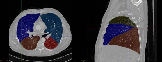
16 Perfusion SPECT- CT Ventilation SPECT-CT 16


17 Perfusion 46 year old male Known small cell lung cancer For preoperative assessment of lung resection MDCT 1 day prior Ventilation SPECT VQ fused with MDCT Mass effect of tumour on vessels and airways Possibility of doing lobar quantification Ventilation Perfusion 17
 A")
18 Lung resection surgery pneumonectomy /lobectomy Lung volume reduction surgery Radical radiotherapy field planning. Lung transplant and lung function after transplant After surgery for complex congenital heart disease Predicted post op FEV1 = FEV1 X Predicted % of remaining lung (after surgery/ radiotherapy) A postoperative or post RT FEV1= 700ml/min is required to avoid respirator dependence 51 patients with NSCLC Potential impact of VQ SPECT over QSPECT alone was assessed to plan high dose RT vs. RT avoidance. Abnormal VQ SPECT CT in all patients with tumour being most common and COPD as next cause of defect. Most defects were matched but 31% patients had reverse mismatch(v<q) Low V regions contribute to low O2 saturation and therefore need to be incorporated in RT plan. Shuanghu Yuan et al, Ann Arbor, University of Michigan J Thorac Oncol
 Quantitative lung scan %ppofev1>40% %ppotlco>40% Surgery %ppo FEV1<40% %ppo TLCO<40% Exercise testing VO 2 max >15ml/kg/min VO 2 max<15ml/kg/min Consider other options BTS")
19 Routine lung function tests FEV1>1.5 litre suitable for lobectomy FEV1>2.0 litre suitable for pneumonectomy FEV1<1.5 litre (Lobectomy) < 2.0 litre (Pneumonectomy) Quantitative lung scan %ppofev1>40% %ppotlco>40% Surgery %ppo FEV1<40% %ppo TLCO<40% Exercise testing VO 2 max >15ml/kg/min VO 2 max<15ml/kg/min Consider other options BTS Guidelines 2001 Different techniques have been used to predict post operative lung function. These have included various pulmonary function tests and quantitative ventilation/perfusion scintigraphy. In practice, scintigraphy is not widely employed in assessing patients for lobectomy, because of the difficulty in interpreting the contribution of individual lobes to the overall ventilation or perfusion. This may explain why several investigators have reported that the simple calculation using lung segment counting can predict post operative FEV 1 as accurately as ventilation/perfusion scintigraphy. Perfusion scintigraphy is the most widely used method to predict postoperative lung function in lung cancer patients undergoing pneumonectomy. 19


20 Most lung cancer patients have MDCT PACS, Data import and fusion software Ability to see finer anatomical details Future applications Identification of interlobar fissures. Lobar definition and possibility of improving quantification information in preoperative patients. Improved understanding of disease processes. More benefit for non PE applications, e.g. lung resection, LVRS, radiotherapy planning. 20


21 35% Contribution of RLL towards total perfusion Possible to quantify counts/vol of lung Acquire Q SPECT+LDCT Co register V SPECT to LDCT Fused dataset Identify fissures and define lobes on MDCT Fuse LDCT to MDCT Transfer fissures on SPECT volumes of V and Q Calculate lobar quantification in 3D 21


22 56 Y M Known Emhysema New lung mass in RUL Preoperative assessment Perfusion SPECT Ventilation SPECT 22


23 23


24 Ventilation Right lung % Left lung % RUL 6.4 LUL 48.3 RML 17.8 RLL 18.2 LLL 9.3 Total Right lung % Left lung % RUL 5.4 LUL 34.5 RML 14.9 RLL 27.5 LLL 17.7 Total Perfusion 24


25 Comparison of post-lobectomy FEV1 with predicted FEV1 by planar and SPECT quantification Spirometry Surgery Spirometry Actual ppo FEV1 Predicted ppo FEV1 25
Ventilation / Perfusion Imaging for Pulmonary Embolic Disease
 Ventilation / Perfusion Imaging for Pulmonary Embolic Disease 1. Purpose This guideline must be read in conjunction with the BNMS Generic Guidelines. The purpose of this guideline is to assist specialists
Chronic lung disease (CLD) is a major cause or mortality and morbidity world-wide.
 is a major cause or mortality and morbidity world-wide.") Reproducibility of a semi-quantitative lobar pulmonary ventilation and perfusion technique using SPET and CT Esber N El-Barhoun MBBS, FRANZCR, Ghee Chew MBBS, FRACP, FAANMS, Benjamin Crouch BSc (Hons),
Reproducibility of a semi-quantitative lobar pulmonary ventilation and perfusion technique using SPET and CT Esber N El-Barhoun MBBS, FRANZCR, Ghee Chew MBBS, FRACP, FAANMS, Benjamin Crouch BSc (Hons),
Potential of hybrid V/P SPECT low-dose CT in lung diagnostics
 Marika Bajc Department of Clinical Physiology, Lund University Hospital, 22 185 Lund, Sweden Department of Clinical Physiology, Lund University Hospital, 22 185 Lund, Sweden marika.bajc@med.lu.se Potential
Lung Imaging with SPECT-CT. Michael M. Graham University of Iowa
 Lung Imaging with SPECT-CT Michael M. Graham University of Iowa 1968 2016 Timeline of Lung Scintigraphy Perfusion MAA Microspheres Ventilation Xe-133 Aerosol Technegas Imaging Planar SPECT SPECT/CT 1960
Understanding the mode of action of a drug using Functional Respiratory Imaging (FRI) Roflumilast Study. Jan De Backer, MSc, PhD, MBA CEO
 Roflumilast Study. Jan De Backer, MSc, PhD, MBA CEO") Understanding the mode of action of a drug using Functional Respiratory Imaging (FRI) Roflumilast Study Jan De Backer, MSc, PhD, MBA CEO IPAC RS/UF Orlando Inhalation Conference March 20, 2014 OVERVIEW
Understanding the mode of action of a drug using Functional Respiratory Imaging (FRI) Roflumilast Study Jan De Backer, MSc, PhD, MBA CEO IPAC RS/UF Orlando Inhalation Conference March 20, 2014 OVERVIEW
Acute Management of Pulmonary Embolism
 Acute Management of Pulmonary Embolism Dr Alex West Respiratory Consultant Guy s and St Thomas Hospital London Declarations - none Order of Play Up date in Diagnostic Imaging - CTPA and V:Q SPECT Sub-massive
Reducing lung volume in emphysema Surgical Aspects
 Reducing lung volume in emphysema Surgical Aspects Simon Jordan Consultant Thoracic Surgeon Royal Brompton Hospital Thirteenth Cambridge Chest Meeting April 2015 Surgical aspects of LVR Why we should NOT
Review Article Ventilation/Perfusion SPECT for Diagnosis of Pulmonary Embolism and Other Diseases
 International Molecular Imaging Volume 2011, Article ID 682949, 7 pages doi:10.1155/2011/682949 Review Article / SPECT for Diagnosis of Pulmonary Embolism and Other Diseases Marika Bajc and Björn Jonson
Methods of nuclear medicine
 Methods of nuclear medicine Per Wollmer Dept. of Translational Medicine Lund University Gamma camera Positron camera Both frequently combined with CT Ventilation/perfusion scanning Perfusion: Albumin macroaggregates
Case 1. Technegas Case Studies. Prostate cancer. Finished treatment recently. Smoker. Angina. Presents sudden dyspnea and poorly defined chest pains.
 Case 1 Michel Leblanc MD; RCPSC; ABNM Head of the Nuclear Medicine Department, Centre Hospitalier Régional de Trois-Rivières. Clinical Professor, Centre Hospitalier Universitaire de Sherbrooke, Cannada.
Radiation Exposure in Pregnancy. John R. Mayo UNIVERSITY OF BRITISH COLUMBIA
 Radiation Exposure in Pregnancy John R. Mayo UNIVERSITY OF BRITISH COLUMBIA Illustrative Clinical Scenario 32 year old female 34 weeks pregnant with recent onset shortness of breath and central chest pain
Patient Dose in the Diagnosis of PE
 Patient Dose in the Diagnosis of PE IZAAZ BADSHAH 2018 CANM-CAMRT Joint Annual Meeting March 22-24, 2018 Disclosure I do not have a financial interest, arrangement or affiliation including receipt of honoraria
Preoperative assessment for lung resection. RA Dyer
 Preoperative assessment for lung resection RA Dyer 2016 The ideal assessment of operative risk would identify every patient who could safely tolerate surgery. This ideal is probably unattainable... Mittman,
Typical PET Image. Elevated uptake of FDG (related to metabolism) Lung cancer example: But where exactly is it located?
 Lung cancer example: But where exactly is it located?") Typical PET Image Elevated uptake of FDG (related to metabolism) Lung cancer example: But where exactly is it located? PET/CT Oncology Imaging Anatometabolic fusion images are useful in the management
Typical PET Image Elevated uptake of FDG (related to metabolism) Lung cancer example: But where exactly is it located? PET/CT Oncology Imaging Anatometabolic fusion images are useful in the management
Imaging Emphysema 3-Helium MR Imaging
 Imaging Emphysema 3-Helium MR Imaging Edwin J.R. van Beek MD PhD MEd FRCR Professor of Radiology and Medicine Carver College of Medicine, University of Iowa, USA. Permanent Visiting Professor of Radiology,
Calculation of Effective Doses for Radiotherapy Cone-Beam CT and Nuclear Medicine Hawkeye CT Laura Sawyer
 Calculation of Effective Doses for Radiotherapy Cone-Beam CT and Nuclear Medicine Hawkeye CT Laura Sawyer Department of Medical Physics and Bioengineering, Royal United Hospital, Bath Overview Varian Acuity
COMPREHENSIVE RESPIROMETRY
 INTRODUCTION Respiratory System Structure Complex pathway for respiration 1. Specialized tissues for: a. Conduction b. Gas exchange 2. Position in respiratory pathway determines cell type Two parts Upper
Epidermiology Early pulmonary embolism
 Epidermiology Early pulmonary embolism Sitang Nirattisaikul Faculty of Medicine, Prince of Songkla University 3 rd most common cause of cardiovascular death in the United States, following ischemic heart
Chest X-ray Interpretation
 Chest X-ray Interpretation Introduction Routinely obtained Pulmonary specialist consultation Inherent physical exam limitations Chest x-ray limitations Physical exam and chest x-ray provide compliment
Cardiac Imaging Tests
 Cardiac Imaging Tests http://www.medpagetoday.com/upload/2010/11/15/23347.jpg Standard imaging tests include echocardiography, chest x-ray, CT, MRI, and various radionuclide techniques. Standard CT and
Preoperative Workup for Pulmonary Resection. Kristen Bridges, M.D. Richmond University Medical Center January 21, 2016
 Preoperative Workup for Pulmonary Resection Kristen Bridges, M.D. Richmond University Medical Center January 21, 2016 Patient Presentation 50 yo male with 70 pack year smoking history Large R hilar lung
Journal of Nuclear Medicine, published on August 1, 2013 as doi: /jnumed
 Journal of Nuclear Medicine, published on August 1, 2013 as doi:10.2967/jnumed.113.124602 CONTINUING EDUCATION V/Q Scanning Using SPECT and SPECT/CT Paul J. Roach, Geoffrey P. Schembri, and Dale L. Bailey
Pulmonary Embolism. Thoracic radiologist Helena Lauri
 Pulmonary Embolism Thoracic radiologist Helena Lauri 8.5.2017 Statistics 1-2 out of 1000 adults annually are diagnosed with deep vein thrombosis (DVT) and/or pulmonary embolism (PE) About half of patients
Introduction to Chest Radiography
 Introduction to Chest Radiography RSTH 366: DIAGNOSTIC TECHNIQUES Alan Alipoon BS, RCP, RRT Instructor Department of Cardiopulmonary Sciences 1 Introduction Discovered in 1895 by Wilhelm Roentgen Terminology
A Snapshot on Nuclear Cardiac Imaging
 Editorial A Snapshot on Nuclear Cardiac Imaging Khalil, M. Department of Physics, Faculty of Science, Helwan University. There is no doubt that nuclear medicine scanning devices are essential tool in the
Tests Your Pulmonologist Might Order. Center For Cardiac Fitness Pulmonary Rehab Program The Miriam Hospital
 Tests Your Pulmonologist Might Order Center For Cardiac Fitness Pulmonary Rehab Program The Miriam Hospital BASIC ANATOMY OF THE LUNGS Lobes of Lung 3 lobes on the Right lung 2 lobes on the Left Blood
PULMONARY EMBOLISM ANGIOCT (CTA) ASSESSMENT OF VASCULAR OCCLUSION EXTENT AND LOCALIZATION OF EMBOLI 1. BACKGROUND
 ASSESSMENT OF VASCULAR OCCLUSION EXTENT AND LOCALIZATION OF EMBOLI 1. BACKGROUND") JOURNAL OF MEDICAL INFORMATICS & TECHNOLOGIES Vol. 11/2007, ISSN 1642-6037 Damian PTAK * pulmonary embolism, AngioCT, postprocessing techniques, Mastora score PULMONARY EMBOLISM ANGIOCT (CTA) ASSESSMENT
JOURNAL OF MEDICAL INFORMATICS & TECHNOLOGIES Vol. 11/2007, ISSN 1642-6037 Damian PTAK * pulmonary embolism, AngioCT, postprocessing techniques, Mastora score PULMONARY EMBOLISM ANGIOCT (CTA) ASSESSMENT
Lung Perfusion Analysis New Pathways in Lung Imaging. Case Study Brochure PLA 309 Hospital
 Lung Perfusion Analysis New Pathways in Lung Imaging Case Study Brochure PLA 309 Hospital http://www.toshibamedicalsystems.com Toshiba Medical Systems Corporation 2012 all rights reserved. Design and specifications
Calculation methods in Hermes Medical Solutions dosimetry software
 Calculation methods in Hermes Medical Solutions dosimetry software Helena McMeekin MSc. Clinical Applications Scientist, Hermes Medical Solutions MRTDosimetry Scientific Workshop The Principals and Clinical
Prapaporn Pornsuriyasak, M.D. Pulmonary and Critical Care Medicine Ramathibodi Hospital
 Prapaporn Pornsuriyasak, M.D. Pulmonary and Critical Care Medicine Ramathibodi Hospital Only 20-30% of patients with lung cancer are potential candidates for lung resection Poor lung function alone ruled
Respiratory Disease. Dr Amal Damrah consultant Neonatologist and Paediatrician
 Respiratory Disease Dr Amal Damrah consultant Neonatologist and Paediatrician Signs and Symptoms of Respiratory Diseases Cardinal Symptoms Cough Sputum Hemoptysis Dyspnea Wheezes Chest pain Signs and Symptoms
Ultralow Dose Chest CT with MBIR
 Ultralow Dose Chest CT with MBIR Ella A. Kazerooni, M.D. Professor & Director Cardiothoracic Radiology Associate Chair for Clinical Affairs University of Michigan Disclosures Consultant: GE Healthcare
Photon Attenuation Correction in Misregistered Cardiac PET/CT
 Photon Attenuation Correction in Misregistered Cardiac PET/CT A. Martinez-Möller 1,2, N. Navab 2, M. Schwaiger 1, S. G. Nekolla 1 1 Nuklearmedizinische Klinik der TU München 2 Computer Assisted Medical
SCINTIGRAPHY OF THE LUNGS THE VQ SCAN
 SCINTIGRAPHY OF THE LUNGS THE VQ SCAN By George N. Sfakianakis, M.D. Professor of Radiology and Pediatrics October 2009 PULMONARY EMBOLISM 94,000 cases annually in US. Not a disease by itself. A potentially
Pitfalls and Remedies in PET/CT imaging for RT planning
 Pitfalls and Remedies in PET/CT imaging for RT planning Tinsu Pan, Ph.D. M.D. Anderson Cancer Center The University of Texas Outlines Background Average CT (< 1 msv) to reduce mis-alignment of PET and
Cardiac Computed Tomography
 Cardiac Computed Tomography Authored and approved by Koen Nieman Stephan Achenbach Francesca Pugliese Bernard Cosyns Patrizio Lancellotti Anastasia Kitsiou Contents CARDIAC COMPUTED TOMOGRAPHY Page 1.
Audit of CT Pulmonary Angiogram in suspected pulmonary embolism patients
 Audit of CT Pulmonary Angiogram in suspected pulmonary embolism patients Poster No.: C-2511 Congress: ECR 2012 Type: Scientific Exhibit Authors: N. D. Gupta, M. K. Heir, P. Bradding; Leicester/UK Keywords:
Lung cancer is the leading cause of cancer death in the
 ORIGINAL ARTICLE Semiquantification and Classification of Local Pulmonary Function by V/Q Single Photon Emission Computed Tomography in Patients with Non-small Cell Lung Cancer Potential Indication for
CT Perfusion. U. Joseph Schoepf, MD, FAHA, FSCBT MR, FSCCT Professor of Radiology, Medicine, and Pediatrics Director of Cardiovascular Imaging
 CT Perfusion U. Joseph Schoepf, MD, FAHA, FSCBT MR, FSCCT Professor of Radiology, Medicine, and Pediatrics Director of Cardiovascular Imaging Disclosures Consultant for / research support from Bayer Bracco
Enhancing Lung Scintigraphy With Single-Photon Emission Computed Tomography
 Enhancing Lung Scintigraphy With Single-Photon Emission Computed Tomography Paul J. Roach, MBBS, FRACP,* Dale L. Bailey, PhD,* and Benjamin E. Harris, MBBS, PhD, FRACP, Although widely used for many years
SPECT-CT: Τι πρέπει να γνωρίζει ο Καρδιολόγος
 SPECT-CT: Τι πρέπει να γνωρίζει ο Καρδιολόγος Δρ Αναστασία Κίτσιου Διευθύντρια, Καρδιολογική Κλινική, Σισμανόγλειο ΓΝΑ Chair, Education Committee, Section on Nuclear Cardiology & Cardiac CT, EACVI, ESC
b) Tc-99m MAA may be used in adults as an imaging agent to aid in the evaluation of peritoneovenous (LeVeen) shunt patency.
 Tc-99m MAA may be used in adults as an imaging agent to aid in the evaluation of peritoneovenous (LeVeen) shunt patency.") 1. OVERVIEW a) Tc 99m Macroaggregated Albumin (MAA) is a lung imaging agent which may be used as an adjunct in the evaluation of pulmonary perfusion in adults and pediatric patients. b) Tc-99m MAA may
1. OVERVIEW a) Tc 99m Macroaggregated Albumin (MAA) is a lung imaging agent which may be used as an adjunct in the evaluation of pulmonary perfusion in adults and pediatric patients. b) Tc-99m MAA may
General Imaging. Imaging modalities. Incremental CT. Multislice CT Multislice CT [ MDCT ]
![General Imaging. Imaging modalities. Incremental CT. Multislice CT Multislice CT [ MDCT ]](/thumbs/76/74079340.jpg "General Imaging. Imaging modalities. Incremental CT. Multislice CT Multislice CT [ MDCT ]") General Imaging Imaging modalities Conventional X-rays Ultrasonography [ US ] Computed tomography [ CT ] Radionuclide imaging Magnetic resonance imaging [ MRI ] Angiography conventional, CT,MRI Interventional
General Imaging Imaging modalities Conventional X-rays Ultrasonography [ US ] Computed tomography [ CT ] Radionuclide imaging Magnetic resonance imaging [ MRI ] Angiography conventional, CT,MRI Interventional
Audit of CT Pulmonary Angiogram in suspected pulmonary embolism patients
 Audit of CT Pulmonary Angiogram in suspected pulmonary embolism patients Poster No.: C-2511 Congress: ECR 2012 Type: Scientific Exhibit Authors: N. D. Gupta, M. K. Heir, P. Bradding; Leicester/UK Keywords:
Nuclear Medicine in the Diabetic Foot
 26.11.2015, Uniklinik Balgrist Nuclear Medicine in the Diabetic Foot Martin Hüllner Nuklearmedizin und Neuroradiologie, USZ / UZH Outline A. Imaging modalities brief technical overview B. Nuclear medicine
Lung scintigraphy in the diagnosis of pulmonary embolism: current methods and interpretation criteria in clinical practice
 research article 113 Lung scintigraphy in the diagnosis of pulmonary embolism: current methods and interpretation criteria in clinical practice Ajda Skarlovnik 1, Damjana Hrastnik 2, Jure Fettich 3, Marko
Referring for specialist respiratory input. Dr Melissa Heightman Consultant respiratory physician, UCLH,WH, CNWL
 Referring for specialist respiratory input Dr Melissa Heightman Consultant respiratory physician, UCLH,WH, CNWL Respiratory Specialist- who? GPSI Community Team Secondary Care Respiratory physician and
PET-CT for radiotherapy planning in lung cancer: current recommendations and future directions
 PET-CT for radiotherapy planning in lung cancer: current recommendations and future directions Gerry Hanna Centre for Cancer Research and Cell Biology Queen s University of Belfast @gerryhanna Talk Outline
Measurement of Respiratory and Cardiac Motion Using a Multi Antenna Continuous Wave Radar Operating in the Near Field
 Measurement of Respiratory and Cardiac Motion Using a Multi Antenna Continuous Wave Radar Operating in the Near Field Florian Pfanner 1,2, Thomas Allmendinger 2, Thomas Flohr 2, and Marc Kachelrieß 1,3
Acute pulmonary embolism (PE) is a severe and
 is a severe and") Detection of Pulmonary Embolism with Combined Ventilation Perfusion SPECT and Low-Dose CT: Head-to-Head Comparison with Multidetector CT Angiography Henrik Gutte 1,2, Jann Mortensen 1, Claus Verner Jensen
Detection of Pulmonary Embolism with Combined Ventilation Perfusion SPECT and Low-Dose CT: Head-to-Head Comparison with Multidetector CT Angiography Henrik Gutte 1,2, Jann Mortensen 1, Claus Verner Jensen
Division of Nuclear Medicine Procedure / Protocol
 Division of Nuclear Medicine Procedure / Protocol MYOCARDIAL SPECT PERFUSION REST and REDISTRIBUTION CPT CODE: 78460, 78464 FOR USE WITH THALLIUM 201 ONLY UPDATED: MARCH 2012 Indications: Patient Prep:
4D PET: promises and limitations
 4D PET: promises and limitations Tinsu Pan, Ph.D. M.D. Anderson Cancer Center The University of Texas Background Outlines Gating techniques: Deep inspiration breath hold 4D PET/CT Non-gating techniques
Exercise Testing Interpretation in the Congenital Heart.
 Interpretation in the Congenital Heart. Stephen M. Paridon, MD Medical Director, Exercise Physiology Laboratory The Children s Hospital of Philadelphia Professor of Pediatrics The University of Pennsylvania
TW Hamilton, EP Ficaro, TA Mitchell, JN Kritzman, JR Corbett University of Michigan Health System, Ann Arbor, MI
 Accuracy and Variability of of 3D-MSPECT for Estimating the Left Ventricular Ejection Fraction as as a Function of of Gating Frames and Reconstruction Filters TW Hamilton, EP Ficaro, TA Mitchell, JN Kritzman,
Gastrointestinal tract
 Gastrointestinal tract Colloidal liver-spleen imaging Presented by: Jehad Felemban Introduction: To obtain better anatomic display of liver and spleen architecture, we use (CT Ultrasound). (Radionuclide
CYCLOPHARM (CYC) Investor Presentation. 4 April 2019 James McBrayer
 Investor Presentation. 4 April 2019 James McBrayer") CYCLOPHARM (CYC) Investor Presentation 4 April 2019 James McBrayer Disclaimer Certain views expressed here contain information derived from publicly available sources that have not been independently verified.
CYCLOPHARM (CYC) Investor Presentation 4 April 2019 James McBrayer Disclaimer Certain views expressed here contain information derived from publicly available sources that have not been independently verified.
Disclosures. GETTING TO THE HEART OF THE MATTER WITH MULTIMODALITY CARDIAC IMAGING Organ Review Meeting 25 September. Overview
 GETTING TO THE HEART OF THE MATTER WITH MULTIMODALITY CARDIAC IMAGING Organ Review Meeting 25 September Disclosures None relevant to this presentation Mini Pakkal Assistant Professor of Radiology University
HRCT V/S MDCT: IN DETECTION OF BRONCHIECTASIS Sowmya M 1, Shilpa Patel 2, Pravan Kumar Reddy 3
 HRCT V/S MDCT: IN DETECTION OF BRONCHIECTASIS Sowmya M 1, Shilpa Patel 2, Pravan Kumar Reddy 3 HOW TO CITE THIS ARTICLE: Sowmya M, Shilpa Patel, Pravan Kumar Reddy. HRCT v/s MDCT: In Detection of Bronchiectasis.
Critical review of SPECT imaging in pulmonary embolism
 Clin Transl Imaging (2014) 2:379 390 DOI 10.1007/s40336-014-0079-6 REVIEW ARTICLE Critical review of SPECT imaging in pulmonary embolism Paul D. Stein H. Dirk Sostman Fadi Matta Received: 22 March 2014
Planar ventilation perfusion (V/Q) lung scintigraphy was the
 lung scintigraphy was the") V/Q SPECT Interpretation for Pulmonary Embolism Diagnosis: Which Criteria to Use? Pierre-Yves Le Roux 1 3, Philippe Robin 1 3, Aurélien Delluc 1,2,4, Ronan Abgral 1 3, Alexandra Le Duc-Pennec 1 3, Emmanuel
V/Q SPECT Interpretation for Pulmonary Embolism Diagnosis: Which Criteria to Use? Pierre-Yves Le Roux 1 3, Philippe Robin 1 3, Aurélien Delluc 1,2,4, Ronan Abgral 1 3, Alexandra Le Duc-Pennec 1 3, Emmanuel
Dual Energy CT of the Heart: Perfusion and Beyond
 Dual Energy CT of the Heart: Perfusion and Beyond U. Joseph Schoepf, MD, FAHA, FSCBT MR, FSCCT Professor of Radiology, Medicine, and Pediatrics Director of Cardiovascular Imaging Disclosures Consultant
4D Radiotherapy in early ca Lung. Prof. Manoj Gupta Dept of Radiotherapy & oncology I.G.Medical College Shimla
 4D Radiotherapy in early ca Lung Prof. Manoj Gupta Dept of Radiotherapy & oncology I.G.Medical College Shimla Presentation focus on ---- Limitation of Conventional RT Why Interest in early lung cancer
GOSTT General concept
 GOSTT General concept Francesco GIAMMARILE «Aut tace aut loquere meliora silentio» Presentation Outline Introduction: GOSTT and radioguided surgery The Sentinel Node Concept Latest technological knowledge
Detectability of subsegmental pulmonary vessels in 64 MDCT-pulmonary angiography.
 ISPUB.COM The Internet Journal of Radiology Volume 11 Number 2 Detectability of subsegmental pulmonary vessels in 64 MDCT-pulmonary angiography. T Niemann, G Bongartz Citation T Niemann, G Bongartz. Detectability
Cardiac PET. John Buscombe
 Cardiac PET John Buscombe Why PET? Improved resolution-not really required in cardiology Improved sensitivity this may be important-financially as reduced acquisition time Improved attenuation correction-good
Fariba Rezaeetalab Associate Professor,Pulmonologist
 Fariba Rezaeetalab Associate Professor,Pulmonologist rezaitalabf@mums.ac.ir Patient related risk factors Procedure related risk factors Preoperative risk assessment Risk reduction strategies Age Obesity
Tc-99m Sestamibi/Tetrofosmin Stress-Rest Myocardial Perfusion Scintigraphy
 APPROVED BY: Director of Radiology Page 1 of 6 Tc-99m Sestamibi/Tetrofosmin Stress-Rest Myocardial Primary Indications: Evaluation of myocardial perfusion and viability in patients with known or suspected
Provider Led Entity. CDI Quality Institute PLE Chest / Pulmonary Embolus AUC 07/31/2018
 Provider Led Entity CDI Quality Institute PLE Chest / Pulmonary Embolus AUC 07/31/2018 Appropriateness of advanced imaging procedures* in patients with suspected or known pulmonary embolus and the following
Lung Cancer Clinical Guidelines: Surgery
 Lung Cancer Clinical Guidelines: Surgery 1 Scope of guidelines All Trusts within Manchester Cancer are expected to follow this guideline. This guideline is relevant to: Adults (18 years and older) with
Pulmonary Pathophysiology
 Pulmonary Pathophysiology 1 Reduction of Pulmonary Function 1. Inadequate blood flow to the lungs hypoperfusion 2. Inadequate air flow to the alveoli - hypoventilation 2 Signs and Symptoms of Pulmonary
Image Guided Stereotactic Radiotherapy of the Lung
 Image Guided Stereotactic Radiotherapy of the Lung Jamie Marie Harris, MS DABR Avera McKennan Radiation Oncology September 25, 2015 Stereotactic Body Radiotherapy - Clinical Dose/Fractionation - Normal
Austin Radiological Association Nuclear Medicine Procedure BONE MINERAL STUDY (Tc-99m-MDP, Tc-99m-HMDP)
") Austin Radiological Association Nuclear Medicine Procedure BONE MINERAL STUDY (Tc-99m-MDP, Tc-99m-HMDP) Overview The Bone Mineral Study, with either Tc-99m-MDP or Tc-99m-HMDP, depicts the distribution
Austin Radiological Association Nuclear Medicine Procedure BONE MINERAL STUDY (Tc-99m-MDP, Tc-99m-HMDP) Overview The Bone Mineral Study, with either Tc-99m-MDP or Tc-99m-HMDP, depicts the distribution
Pulmonary Embolism. Pulmonary Embolism. Pulmonary Embolism. PE - Clinical
 Pulmonary embolus - a practical approach to investigation and treatment Sam Janes Wellcome Senior Fellow and Respiratory Physician, University College London Background Diagnosis Treatment Common: 50 cases
Regional Comparisons of CT Air Trapping and MRI Ventilation Defect Percent in Asthma
 Regional Comparisons of CT Air Trapping and MRI Ventilation Defect Percent in Asthma V. A. Zavaletta 1, D.G. Mummy 4, T. Lampkins 4, W. Zha 4, M. L. Schiebler 1, N. Jarjour 3, L. Denlinger 3, and S.B.
Low-dose CT Lung Cancer Screening Guidelines for Pulmonary Nodules Management Version 2
 Low-dose CT Lung Cancer Screening Guidelines for Pulmonary Nodules Management Version 2 The Committee for Management of CT-screening-detected Pulmonary Nodules 2009-2011 The Japanese Society of CT Screening
Nuclear pulmonology. Katalin Zámbó Department of Nuclear Medicine
 Nuclear pulmonology Katalin Zámbó Department of Nuclear Medicine Imaging techniques Morphology Physiology Metabolism Molecules X-ray / CT MRI NM - SPECT/ PET MR spectroscopy fmri Ultrasound Hybrid imaging:
Precision of pre-sirt predictive dosimetry
 International Course on THERANOSTICS AND MOLECULAR RADIOTHERAPY Precision of pre-sirt predictive dosimetry Hugo Levillain Department of Nuclear Medicine Medical Physics Jules Bordet Institute, Université
Preview of Presentation
 Preview of Presentation Discuss Healthcare Environment Clinical Implementation of Technical Innovations SNMMI/ASNC Joint Statement on Rb-82 Cardiac PET Imaging Protocol Principles of Rb-82 Cardiac Imaging
Ventilation perfusion (V/Q) scintigraphy allows assessment
 scintigraphy allows assessment") Journal of Nuclear Medicine, published on September 9, 2011 as doi:10.2967/jnumed.111.093344 Ga PET/CT Ventilation Perfusion Imaging for Pulmonary Embolism: A Pilot Study with Comparison to Conventional
Journal of Nuclear Medicine, published on September 9, 2011 as doi:10.2967/jnumed.111.093344 Ga PET/CT Ventilation Perfusion Imaging for Pulmonary Embolism: A Pilot Study with Comparison to Conventional
New Criteria for Ventilation-Perfusion Lung Scan Interpretation: A Basis for Optimal Interaction with Helical CT Angiography 1
 1206 July-August 2000 RG Volume 20 Number 4 New Criteria for Ventilation-Perfusion Lung Scan Interpretation: A Basis for Optimal Interaction with Helical CT Angiography 1 Alexander Gottschalk, MD Introduction
SURGERY FOR GIANT BULLOUS EMPHYSEMA
 SURGERY FOR GIANT BULLOUS EMPHYSEMA Dr. Carmine Simone Head, Division of Critical Care & Thoracic Surgeon Department of Surgery December 15, 2006 OVERVIEW Introduction Classification Patient selection
8/3/2016. Background. CT-Ventilation. Validation, Clinical Endpoints and Opportunities for CT Ventilation. 4DCT-Ventilation Imaging.
 Validation, Clinical Endpoints and Opportunities for CT Ventilation Yevgeniy Vinogradskiy PhD University of Colorado School of Medicine Department Of Radiation Oncology 4DCT-Ventilation Imaging Background
ORIGINAL ARTICLE INTRODUCTION
 ORIGINAL ARTICLE Annals of Nuclear Medicine Vol. 16, No. 8, 549 555, 2002 Present diagnostic strategies for acute pulmonary thromboembolism; results of a questionnaire in a retrospective trial conducted
Combined Anatomical and Functional Imaging with Revolution * CT
 GE Healthcare Case studies Combined Anatomical and Functional Imaging with Revolution * CT Jean-Louis Sablayrolles, M.D. Centre Cardiologique du Nord, Saint-Denis, France Case 1 Whole Brain Perfusion and
State-of-the-Art SPECT/CT: Cardiac Imaging
 State-of-the-Art SPECT/CT: Cardiac Imaging Ernest V Garcia*, PhD Endowed Professor in Cardiac Imaging Director, Nuclear Cardiology R&D Laboratory Disclosure: Dr. Garcia receives royalties from the sale
Functional Chest MRI in Children Hyun Woo Goo
 Functional Chest MRI in Children Hyun Woo Goo Department of Radiology and Research Institute of Radiology Asan Medical Center, University of Ulsan College of Medicine, Seoul, Korea No ionizing radiation
Radiation Detection and Measurement
 Radiation Detection and Measurement Range of charged particles (e.g.,!: µm; ": mm) Range of high energy photons (cm) Two main types of interactions of high energy photons Compton scatter Photoelectric
Feasibility of a New C-arm angiography (DYNA-CT) based three-dimensional algorithm in combination with myocardial perfusion assessment
 based three-dimensional algorithm in combination with myocardial perfusion assessment") Feasibility of a New C-arm angiography (DYNA-CT) based three-dimensional algorithm in combination with myocardial perfusion assessment H. Rittger*, A.M.Sinha, J. Rieber±, G. Lauritsch+, J. Brachmann *Universitätsklinik
Feasibility of a New C-arm angiography (DYNA-CT) based three-dimensional algorithm in combination with myocardial perfusion assessment H. Rittger*, A.M.Sinha, J. Rieber±, G. Lauritsch+, J. Brachmann *Universitätsklinik
Chest Radiology Interpretation: Findings of Tuberculosis
 Chest Radiology Interpretation: Findings of Tuberculosis Get out your laptops, smart phones or other devices pollev.com/chestradiology Case #1 1 Plombage Pneumonia Cancer 2 Reading the TB CXR Be systematic!
CT Chest. Verification of an opacity seen on the straight chest X ray
 CT Chest Indications: To assess equivocal plain x-ray findings Staging of lung neoplasm Merastatic workup of extra thoraces malignancies Diagnosis of diffuse lung diseases with HRCT Assessment of bronchietasis
OTHER NON-CARDIAC USES OF Tc-99m CARDIAC AGENTS Tc-99m Sestamibi for parathyroid imaging, breast tumor imaging, and imaging of other malignant tumors.
 DEFINITION OF CARDIAC RADIOPHARMACEUTICAL: A radioactive drug which, when administered for purpose of diagnosis of heart disease, typically elicits no physiological response from the patient. Even though
Ventilation/perfusion tomography V/P-SPECT vs planar technique
 CASE REPORT Ventilation/perfusion tomography V/P-SPECT vs planar technique Amela Begic 1, Emina Opanković 1, Sadžida Begović-Hadžimuratović 1, Miran Konjić 2, Senahid Krekić 2 1. Clinic for Nuclear Medicine,
The radiation dose in retrospective
 The radiation dose in retrospective gated tdcoronary computed td tomography (CCT) Saeed AL Ahmari, Ghormallah AL Zahrani, Sumiah AL Helali, Samir AL Dulikan, Abdullah Bafagih, HibaKhashojji Prince Sultan
Reporting SPECT-VQ. Alp Notghi
 Reporting SPECT-VQ Alp Notghi 20 year old female 24 weeks pregnant Clinical History : SOB and chest pain for past 3 days.?pe Doppler USS excluded DVT Case 4413041 Normal Case 4413041 CXR report: The heart
CRUCIBLE. Feature Other Uses for Technegas (Part 2)
") CRUCIBLE THE CYCLOMEDICA NUCLEAR MEDICINE VOL 9 / NO. 1 - FEBRUARY 2016 Feature Other Uses for Technegas (Part 2) I n the last issue of the Crucible a brief overview of the usefulness of ventilation scans
CRUCIBLE THE CYCLOMEDICA NUCLEAR MEDICINE VOL 9 / NO. 1 - FEBRUARY 2016 Feature Other Uses for Technegas (Part 2) I n the last issue of the Crucible a brief overview of the usefulness of ventilation scans
SCLERODERMA LUNG DISEASE: WHAT THE PATIENT SHOULD KNOW
 SCLERODERMA LUNG DISEASE: WHAT THE PATIENT SHOULD KNOW Lung disease can be a serious complication of scleroderma. The two most common types of lung disease in patients with scleroderma are interstitial
Imaging of acute pulmonary thromboembolism*
 Silva, Isabela et al. Imaging of acute pulmonary thromboembolism Imaging of acute pulmonary thromboembolism* C. ISABELA S. SILVA, NESTOR L. MÜLLER The diagnosis of acute pulmonary thromboembolism is based
Fundamentals of Nuclear Cardiology. Terrence Ruddy, MD, FRCPC, FACC
 Fundamentals of Nuclear Cardiology Terrence Ruddy, MD, FRCPC, FACC Objectives To understand the Principles of Nuclear Cardiac Imaging Radiotracers Image acquisition and processing Stress protocols To appreciate
The GOSTT concept. (radio)guided intraoperative Scintigraphic Tumor Targeting. Emmanuel Deshayes. GOSTT = Radioguided Surgery
guided intraoperative Scintigraphic Tumor Targeting. Emmanuel Deshayes. GOSTT = Radioguided Surgery") IAEA WorkShop, November 2017 Emmanuel Deshayes With the kind help of Pr Francesco Giammarile The GOSTT concept GOSTT = Radioguided Surgery (radio)guided intraoperative Scintigraphic Tumor Targeting 1 Radioguided
IAEA WorkShop, November 2017 Emmanuel Deshayes With the kind help of Pr Francesco Giammarile The GOSTT concept GOSTT = Radioguided Surgery (radio)guided intraoperative Scintigraphic Tumor Targeting 1 Radioguided
Scintigraphic Lung Scans and Clinical Assessment in Critically Ill Patients With Suspected Acute Pulmonary Embolism*
 Scintigraphic Lung Scans and Clinical Assessment in Critically Ill Patients With Suspected Acute Pulmonary Embolism* Jerald W. Henry, MS; Paul D. Stein, MD, FCCP; Alexander Gottschalk, MD, FCCP; Bruce