Nuclear pulmonology. Katalin Zámbó Department of Nuclear Medicine
|
|
|
- Jonas Boyd
- 5 years ago
- Views:
Transcription
1 Nuclear pulmonology Katalin Zámbó Department of Nuclear Medicine

2 Imaging techniques Morphology Physiology Metabolism Molecules X-ray / CT MRI NM - SPECT/ PET MR spectroscopy fmri Ultrasound Hybrid imaging: SPECT/CT, PET/CT, (PET/MRI)
3 Radioactivity It is the spontaneous disintegration (decay) of the nucleus of a radioactive atom - in which the number of protons and neutrons are not stable - and different type of radiation (, -, +, ) comes out from the nucleus.
4 Number of protons = elemental identity number Number of protons and neutrons = mass number - Atoms with the same number of protons but differing number of neutrons are called isotopes of that element. - The behaviour of the different radioactive isotopes of an element is the same as the stable form in every conditions.
 Electromagnetic ray ( ) Lead Paper")
5 Rays of radioactive decay Corpuscular rays (, -, + ) Electromagnetic ray ( ) Lead Paper Penetration
6 Gamma radiation - really electromagnetic radiation - physically similar to X-rays, but it comes out from the nucleus of the atom - very penetrated and easily pass trough tissue - SO: it can be detected externally well, it can be used for diagnostics - 99meta-technetium (arteficial) - using the radioactive material as a tracer (Hevesy György 1923)

7 The equipments I. Gamma-camera - it sees the whole entire area below the detector
8 Structure of the gamma-camera DIGITAL PICTURE ARE PRINTED

9 The equipments II. - SPECT (Single Photon Emission Computer Tomograph) - SPECT/CT: multimodality!
10 The principle of the SPECT patient The detector whirls around the patient and makes pictures from different steps. The computer program reconstruates the transversal, sagittal and coronal slices of the organ.
11 Radionuclide studies - are based on the function of an organ or an organ system - are easily performed - need no any premedication - are not associated with any morbidity and complication, have only minimal risk - are very sensitive, but aspecific methods
12 Method - gamma emitting isotopes, which are detected outside - carrier molecules, which paricipate the function of the examined organs - together: radiopharmacon - they are usually administered intravenously in steril physiological NaCl solution - various delayed times before the examinations - imaging by scitillation detector
13 Nuclear medicine methods - Static examinations (scintigraphy): - an optimal time-period after the subject administration is delayed and several photos are made of the organ from different directions - Dynamic studies: - a frame-serie is stored in the computer from the time of the isotope injection during an optimal time-period of the examined organ function
14 Static studies - Negative scintigraphy: pathological decreased activity or lack of the activity (focal defect) - Positive scintigraphy: pathological increased activity (hot spot)

15 The lungs in the chest

16 The lung and the heart

17 The pulmonary vasculature and the bronchial tree
18 The pulmonary vasculature
19 Zones of the lung

20 Lung segmental anatomy
21 Pulmonary studies - Perfusion lung scintigraphy: shows the accurate assesment of the regional perfusion of the lung - Inhalation or ventillation examination: shows the distribution of the inhaled radioactive gases or aerosol
22 Perfusion lung scintigraphy - Injected subject: 99mTc-macro-albumon - with mean particles size of about 30 µm - blocks the precapillary arterioles, but only less than 0.1 % of and this is not permanent -Administration: intravenously in lying position (because of the perfusion zones of the lung) - Study can be started immediately
23 Indications of the perfusion scintigraphy - Verify the pulmonary embolism (couple of the chest X-ray!) - Evaluation of regional lung function in patients with lung tumor before the operation - Evaluation of regional lung function in asthmatic and obstructive lung diseases - Assessment of regional lung function after the therapy

24 Normal perfusion lung scan
25 Perfusion defects on the lung scan
26 Therapy: thrombolysis Indication: great perfusion defect on the scintigram, at least the half of the lungs + negative chest x-ray Contraindication: tumor, metastasis or infiltration on the chest x-ray previous operation or punctio hemorrhagical diseases Streptokinase, Urokinase, Actylise infusion it can be repeated in every day
27 Therapy of pulmonary embolism Before streptolysis One day after streptolysis Two days after streptolysis
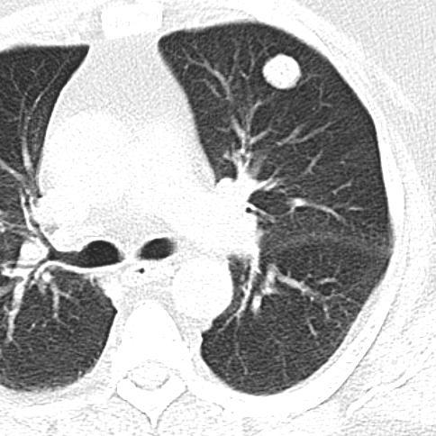
28 Inhalation study - Inhaled subject: 99mTc-DTPA aerosol - The examination can be started immediately - Indications: - pulmonary embolism (with perfusion study: mismatch ) - lung tumor (with perfusion study: match ) - obstructive lung diseses accumulation hot spots in the central bronchii
29 Ventilation study 81m-Krypton: from 81-Rubidium generator, T 1/2: 13 s, energy: 193 kev 133-Xenon: T1/2: 5.2 days, energy: 80 kev, during 3-5 minutes equilibrium 127-Xenon: T 1/2: 36 days, energy: 172, 203, 375 kev Technegas: 99mTc-DTPA is vapourised in argon atmosphere on grad Celsius behaves as a gas with µm particle size
30 Radioaerosol delivery system

31 Pulmonary embolism (mismatch)

32 Pulmonary embolism Perfusion scintigraphy Inhalation scintigraphy


33 Obstructive bronchitis Perfusion scintigraphy Inhalation scintigraphy
34 Central tumor in the right side (the lung is in the chest, but no any perfusion in the lung)

35 Bronchial carcinoma


36 Periferial tumor in the right lung The activity of the lungs: Both lung: cps Left lung: 48.3 % Right lung: 51.7 % Left upper lobe: 2597 cps Lingula: 2307 cps Left lower lobe: 3766 cps Right upper lobe: 3594 cps Right middle lobe: 2423 cps Right lower lobe: 3211 cps

37 Adenocarcinoma in the upper lobe of the right lung Pulmonary embolism in other localization?

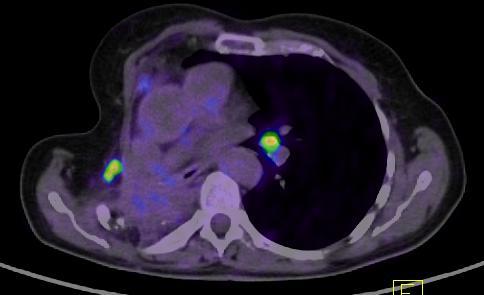
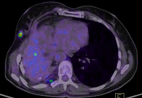
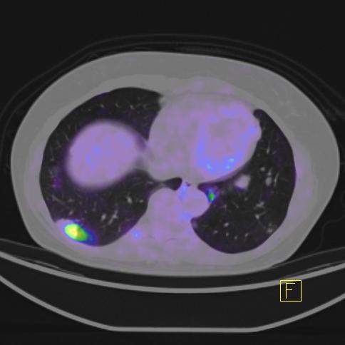
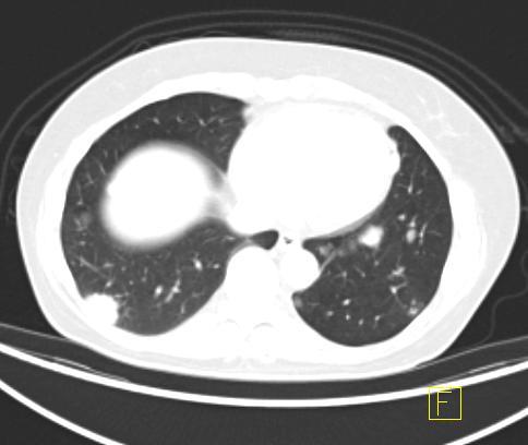
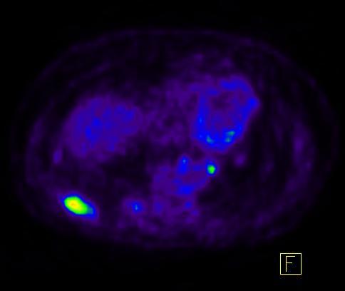
38 No embolism in the right side! CT SPECT SPECT/CT
39 No embolism, BUT: metastasis on the left side! CT SPECT SPECT/CT

40 Specific examination of pulmonary cancers 111In-octreotide (SSTR 2,3,5) and 99mTc-depreotide (SSTR 2) binding to the somatostatin receptors which are overexpressed on the surface of the tumor cells are suitable for the diagnosis SSTR Neospect

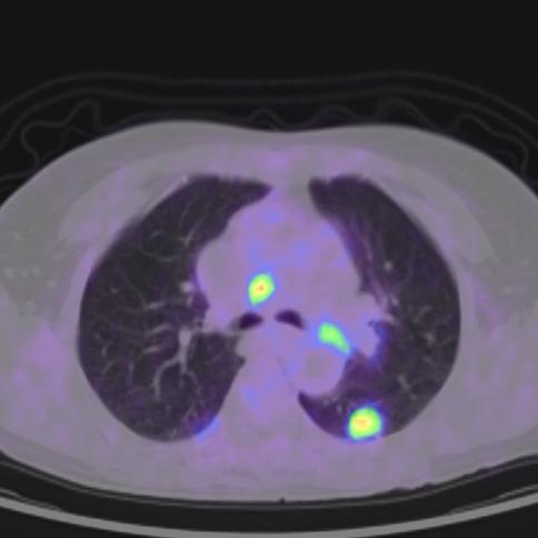
41 Adenocarcinoma in the right lung by 99mTc-Neospect SPECT imaging

42 Adenocarcinoma in the right lung by 18F-FDG PET imaging
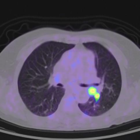
43 Adenocarcinoma in the left upper lobe by 99mTc-Neospect SPECT imaging

44 Adenocarcinoma in the left upper lobe by 18F-FDG PET imaging


45 Whole body scan Operation of lung carcinoid on the right side, metastasis on the left side CT SPECT SPECT/CT


46 Whole body scan Carcinoid in the left lung CT SPECT SPECT/CT
47 The gallium67 scintigraphy I. There is an isotope, which are binding aspecifically to the transferrine receptors of the tumor cells are found on their surface. Half-life: 78 hours Energy: 93 kev kev
48 The gallium67 scintigraphy II. Indications: - the localization, evaluation and follow up of patients with neoplastic disorders such as Hodgkin s and non- Hodgkin s disease, soft tissue sarcoma, bronchogenic carcinoma, melanoma and hepatoma - sarcoidosis of the lungs - the detection of focal inflammatory processes, abdominal abscesses, osteomyelitis

49 Anterior view of the chest Dg: Hodgkin disease Mediastinal mass

50 Anterior view and SPECT imaging of the chest Dg: Non-Hodgkin disease before radiation Mediastinal mass in the right side

51 Anterior view and SPECT imaging of the chest Dg: Non-Hodgkin disease after radiadion + recidiva in the opposite side New lung manifestation in the left side
52 +Beta (positron) radiation too many protons are in the nucleus its life is very short, when it slows down, it combines with a normal electron in a process known annihilation, which destroyes both the electron and positron and produces two energetic photons each with 511 kev they are used for PET examinations
 -")
53 The equipments III. - PET (Positron Emission Tomograph) - PET/CT: multimodality!

54 The principle of the PET imaging
")
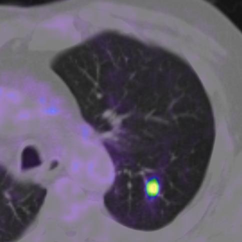
55 Peripherial lung tumor and mediastinal lymph node metastasis by 18F-FDG (fluorodesoxyglucose) PET/CT

56 Bronchogenic carcinoma by 18F-FDG PET/CT

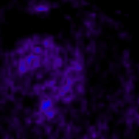
57 FDG avid lung metastasis and FDG negatíve benign tumor

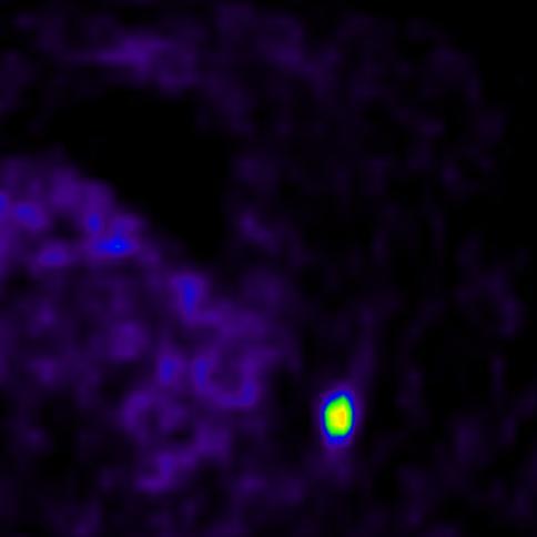
58 FDG avid adenocc in the right lung and 2 benign hamartomas in both side by 18F-FDG PET/CT


59 Recidiva of small cell lung cancer after pulmonectomy

60 Staging, restaging Metastases of adenocarcinoma
61 Localization of the biopsy
62 First passage study - The radioactive subject: 99mTc- DTPA (rapid movement from the body through the kidneys) - Fast dynamic through the heart and the lung - Bolus of the injection is important - Cardio-pulmonary circulation times - Cardiac output, stroke volume

63 The way of the bolus sup. v. cava right ventricle pulm. artery+lungs left ventricle curves ROIs
64 Time-activity curves and Right ventricle circulation times Bolus Lung Left ventricle
65 The report of the FP examination
66 Bone scintigraphy I. - Bone tissue has high activity for the intravenously injected phosphate agents (99mTc-MDP). The effectivity of the incorporation depends on the blood supply and on the calcium and phosphorus metabolism of the bone. - The method is very sensitive, which becomes positive in the earliest stage of the bone disorders. The increased bone metabolism already can be shown 6 months earlier than the changing of bone structure in the X-ray.
67 Bone scintigraphy II. - Injected subject: 99mTc-phosphate (MDP, EDP, Pyrophosphate) intravenously -The study can be started after 2-3 hours (slow metabolism) - Important: rich fluid input!
68 Indications of the bone scintigraphy - Metastases of the bone (mamma cc., prostatic cc, lung cc., malignant melonoma, and so on) - Primary bone tumors - Osteomyelitis, other inflammatory diseases - Fractures (pathologic and stress fracture) - Metabolic diseases (e.g. Paget disease) - Osteonecrosis (e.g. M. Perthes)

69 Normal whole body bone scintigraphy

70 Multifocal hot-spots in bronchial carcinoma


71 WBB Hot spots in the lumbal spine: degenerative or metastatic lesions? CT SPECT SPECT/CT Metastases

72 Thank you for your attention!
Itroduction to the Nuclear Medicine: biophysics and basic principles. Zámbó Katalin Department of Nuclear Medicine
 Itroduction to the Nuclear Medicine: biophysics and basic principles Zámbó Katalin Department of Nuclear Medicine NUCLEAR MEDICINE Application of the radioactive isotopes in the diagnostics and in the
Nuclear medicine studies of the digestiv system. Zámbó Katalin Department of Nuclear Medicine
 Nuclear medicine studies of the digestiv system Zámbó Katalin Department of Nuclear Medicine Imaging tehniques Anatomy Physiology Metabolism Molecular Rtg. / CT PET / SPECT MRI MR spectroscopy fmri Ultrasound
Nuclear cardiology. Zámbó Katalin Department of Nuclear Medicine
 Nuclear cardiology Zámbó Katalin Department of Nuclear Medicine Imaging techniques Morphology Physiology Metabolism Molecules X-ray / CT MRI NM - SPECT/ PET MR spectroscopy fmri Ultrasound Hybrid imaging:
Nuclear medicine in oncology. 1. Diagnosis 2. Therapy
 Nuclear medicine in oncology 1. Diagnosis 2. Therapy Diagnosis - Conventional methods - Nonspecific radiopharmaceuticals cumulating in tumours - Specific radiopharmaceuticals (receptor- and immunoscintigraphy)
PHYSICS 2: HSC COURSE 2 nd edition (Andriessen et al) CHAPTER 20 Radioactivity as a diagnostic tool (pages 394-5)
 CHAPTER 20 Radioactivity as a diagnostic tool (pages 394-5)") PHYSICS 2: HSC COURSE 2 nd edition (Andriessen et al) CHAPTER 20 Radioactivity as a diagnostic tool (pages 394-5) 1. (a) A radioisotope is an isotope that is unstable and will emit particles from the nucleus
PHYSICS 2: HSC COURSE 2 nd edition (Andriessen et al) CHAPTER 20 Radioactivity as a diagnostic tool (pages 394-5) 1. (a) A radioisotope is an isotope that is unstable and will emit particles from the nucleus
Nuclear Medicine: Manuals. Nuclear Medicine. Nuclear imaging. Emission imaging: study types. Bone scintigraphy - technique
 Nuclear Medicine - Unsealed radioactive preparations the tracer mixes with the patients body fluids on a molecular level (e.g. after intravenous injection) - 3 main fields: - In vitro : measuring concentrations
Radionuclides in Medical Imaging. Danielle Wilson
 Radionuclides in Medical Imaging Danielle Wilson Outline Definitions History and development Radionuclide applications & techniques in imaging Conclusion Definition #1 : Radionuclide An unstable nucleus
Nuclear medicine studies of the digestiv system. Zámbó Katalin Department of Nuclear Medicine
 Nuclear medicine studies of the digestiv system Zámbó Katalin Department of Nuclear Medicine Anatomy of the liver Liver scintigraphy The labelled colloid (200 MBq 99mTc-Fyton) is phagocyted by the Kuppfer-cells
Bone PET/MRI : Diagnostic yield in bone metastases and malignant primitive bone tumors
 Bone PET/MRI : Diagnostic yield in bone metastases and malignant primitive bone tumors Lars Stegger, Benjamin Noto Department of Nuclear Medicine University Hospital Münster, Germany Content From PET to
Option D: Medicinal Chemistry
 Option D: Medicinal Chemistry Basics - unstable radioactive nuclei emit radiation in the form of smaller particles alpha, beta, positron, proton, neutron, & gamma are all used in nuclear medicine unstable
Nuclear medicine. Zámbó Katalin Department of Nuclear Medicine
 Nuclear medicine Zámbó Katalin Department of Nuclear Medicine Imaging tehniques Anatomy Physiology Metabolism Molecular X-ray / CT Nuclear medicine / SPECT / PET MRI MR spectroscopy fmri Ultrasound Hybrid
Nuclear neurology. Zámbó Katalin Department of Nuclear Medicine
 Nuclear neurology Zámbó Katalin Department of Nuclear Medicine To refresh your memory Brain has a high rate of oxidative metabolism. It has no reserves either of oxygen or of glucose and has a very limited
Nuclear Medicine in Oncology
 Radiopharmaceuticals Nuclear Medicine in Oncology Practice Pharmaceutical Radionuc lide Function Tumor type Diphosphonates Tc-99m Osteoblast Bone tumor & metast. Ga-citrate Ga-67 Fe-analogue Bronchogenous
Medical imaging X-ray, CT, MRI, scintigraphy, SPECT, PET Györgyi Műzes
 Medical imaging X-ray, CT, MRI, scintigraphy, SPECT, PET Györgyi Műzes Semmelweis University, 2nd Dept. of Medicine Medical imaging: definition technical process of creating visual representations about
PET-MRI in malignant bone tumours. Lars Stegger Department of Nuclear Medicine University Hospital Münster, Germany
 PET-MRI in malignant bone tumours Lars Stegger Department of Nuclear Medicine University Hospital Münster, Germany Content From PET to PET/MRI General considerations Bone metastases Primary bone tumours
Chapter 1. Introduction
 Chapter 1 Introduction 1.1 PRINCIPLES OF NUCLEAR MEDICINE Nuclear medicine techniques use radioactive tracers and imaging devices, mainly to provide diagnostic information, but in some cases also for therapeutic
COMENIUS-Project: SM&CLIL Radiation & Medicine
 Medical imaging refers to the techniques and processes used to create images of the human body (or parts thereof) for clinical purposes. Thanks to modern mathematics and computer technology, medical imaging
Molecular Imaging and Cancer
 Molecular Imaging and Cancer Cancer causes one in every four deaths in the United States, second only to heart disease. According to the U.S. Department of Health and Human Services, more than 512,000
Gastrointestinal tract
 Gastrointestinal tract Colloidal liver-spleen imaging Presented by: Jehad Felemban Introduction: To obtain better anatomic display of liver and spleen architecture, we use (CT Ultrasound). (Radionuclide
POSITRON EMISSION TOMOGRAPHY (PET)
") Status Active Medical and Behavioral Health Policy Section: Radiology Policy Number: V-27 Effective Date: 08/27/2014 Blue Cross and Blue Shield of Minnesota medical policies do not imply that members should
Status Active Medical and Behavioral Health Policy Section: Radiology Policy Number: V-27 Effective Date: 08/27/2014 Blue Cross and Blue Shield of Minnesota medical policies do not imply that members should
HSC Physics. Module 9.6. Medical Physics
 HSC Physics Module 9.6 Medical Physics Contextual Outline 9.6 Medical Physics (28 indicative hours) The use of other advances in technology, developed from our understanding of the electromagnetic spectrum,
Austin Radiological Association Nuclear Medicine Procedure BONE MINERAL STUDY (Tc-99m-MDP, Tc-99m-HMDP)
") Austin Radiological Association Nuclear Medicine Procedure BONE MINERAL STUDY (Tc-99m-MDP, Tc-99m-HMDP) Overview The Bone Mineral Study, with either Tc-99m-MDP or Tc-99m-HMDP, depicts the distribution
Austin Radiological Association Nuclear Medicine Procedure BONE MINERAL STUDY (Tc-99m-MDP, Tc-99m-HMDP) Overview The Bone Mineral Study, with either Tc-99m-MDP or Tc-99m-HMDP, depicts the distribution
Special Imaging MUSCULOSKELETAL INFECTION. Special Imaging. Special Imaging. 18yr old male pt What is it? Additional Imaging
 MUSCULOSKELETAL INFECTION Additional Imaging May assist in diagnosis and, possibly, treatment Help create the picture May help differentiate from neoplasia 18yr old male pt What is it? Lymphoma Ewings
Molecular Imaging and Breast Cancer
 Molecular Imaging and Breast Cancer Breast cancer forms in tissues of the breast usually in the ducts, tubes that carry milk to the nipple, and lobules, the glands that make milk. It occurs in both men
A 64 y.o. man presents to the hospital with persistent cough and hemoptysis. Fernando Mut Montevideo - Uruguay
 A 64 y.o. man presents to the hospital with persistent cough and hemoptysis Fernando Mut Montevideo - Uruguay Teaching case Bone # 1 A 64 y.o. man presents to the hospital with persistent cough and hemoptysis.
General Nuclear Medicine
 General Nuclear Medicine What is General Nuclear Medicine? What are some common uses of the procedure? How should I prepare? What does the equipment look like? How does the procedure work? How is the procedure
Prof. Dr. NAGUI M. ABDELWAHAB,M.D.; MARYSE Y. AWADALLAH, M.D. AYA M. BASSAM, Ms.C.
 Role of Whole-body Diffusion MR in Detection of Metastatic lesions Prof. Dr. NAGUI M. ABDELWAHAB,M.D.; MARYSE Y. AWADALLAH, M.D. AYA M. BASSAM, Ms.C. Cancer is a potentially life-threatening disease,
Medical Use of Radioisotopes
 Medical Use of Radioisotopes Therapy Radioisotopes prove to be useful in the application of brachytherapy, the procedure for using temporary irradiation close to the area of disease (i.e. cancer) 10% Medical
Molecular Imaging and the Brain
 Molecular imaging technologies are playing an important role in neuroimaging, a branch of medical imaging, by providing a window into the living brain. Where CT and conventional MR imaging provide important
Positron Emission Tomography in Lung Cancer
 May 19, 2003 Positron Emission Tomography in Lung Cancer Andrew Wang, HMS III Patient DD 53 y/o gentleman presented with worsening dyspnea on exertion for the past two months 30 pack-year smoking Hx and
Laura Tormoehlen, M.D. Neurology and EM-Toxicology Indiana University
 Laura Tormoehlen, M.D. Neurology and EM-Toxicology Indiana University Disclosures! No conflicts of interest to disclose Neuroimaging 101! Plain films! Computed tomography " Angiography " Perfusion! Magnetic
An Introduction to PET Imaging in Oncology
 January 2002 An Introduction to PET Imaging in Oncology Janet McLaren, Harvard Medical School Year III Basics of PET Principle of Physiologic Imaging: Allows in vivo visualization of structures by their
Radiologic Imaging Magnetic Resonance Imaging (MRI)
") Radiologic Imaging X-ray has always been the golden rule in diagnosing and treating podiatric patients. Unfortunately, for some patients the diagnosis is not as evident. That is when we need to utilize
Radiologic Imaging X-ray has always been the golden rule in diagnosing and treating podiatric patients. Unfortunately, for some patients the diagnosis is not as evident. That is when we need to utilize
Nuclear medicine methods in the urogenital system
 Nuclear medicine methods in the urogenital system Anatomy of the kidneys I. Anatomy of the kidneys II. The types of examinations Static examinations (scintigraphy): 1) the radiopharmaceutical is administered
Ventilation / Perfusion Imaging for Pulmonary Embolic Disease
 Ventilation / Perfusion Imaging for Pulmonary Embolic Disease 1. Purpose This guideline must be read in conjunction with the BNMS Generic Guidelines. The purpose of this guideline is to assist specialists
Nuclear Medicine and PET. D. J. McMahon rev cewood
 Nuclear Medicine and PET D. J. McMahon 150504 rev cewood 2018-02-15 Key Points Nuclear Medicine and PET: Imaging: Understand how Nuc Med & PET differ from Radiography & CT by the source of radiation. Be
Basics of nuclear medicine
 Basics of nuclear medicine Prof. dr. Davor Eterović Prof. dr. Vinko Marković Radioisotopes are used both in diagnostics and in therapy Diagnostics gamma emitters are used since gamma rays can penetrate
Typical PET Image. Elevated uptake of FDG (related to metabolism) Lung cancer example: But where exactly is it located?
 Lung cancer example: But where exactly is it located?") Typical PET Image Elevated uptake of FDG (related to metabolism) Lung cancer example: But where exactly is it located? PET/CT Oncology Imaging Anatometabolic fusion images are useful in the management
Typical PET Image Elevated uptake of FDG (related to metabolism) Lung cancer example: But where exactly is it located? PET/CT Oncology Imaging Anatometabolic fusion images are useful in the management
Ga68 Imaging. Roland HUSTINX Division of Nuclear Medicine and Oncologic Imaging Centre Hospitalier Universitaire de Liège Belgium
 Ga68 Imaging Roland HUSTINX Division of Nuclear Medicine and Oncologic Imaging Centre Hospitalier Universitaire de Liège Belgium 68 Ga Produced by a 68 Ge/ 68 Ga generator Decays by positron emission
TOPICS FOR PRACTICAL LESSONS, DISCIPLINE RADIOLOGY For the IIIrd year students Faculty of Medicine, university year
 TOPICS FOR PRACTICAL LESSONS, DISCIPLINE RADIOLOGY For the IIIrd year students Faculty of Medicine, university year 2018-2019 I. Evolution of radiology. Notion of Radiophysics. 1. Medical imaging definition.
NUCLEAR MEDICINE Molecular Imaging + Endo-Radiotherapy
 NUCLEAR MEDICINE Molecular Imaging + Endo-Radiotherapy Istvan Szilvási Dept. of Nuclear Medicine Semmelweis University and HDF Medical Centre 2016 DEFINITION OF NUCLEAR MEDICINE Medical applications of
Radiopharmacy. Prof. Dr. Çetin ÖNSEL. CTF Nükleer Tıp Anabilim Dalı
 Prof. Dr. Çetin ÖNSEL CTF Nükleer Tıp Anabilim Dalı What is Nuclear Medicine? Nuclear Medicine is the branch of medicine concerned with the use of radionuclides in the study and the diagnosis of diseases.
Physical Bases : Which Isotopes?
 Physical Bases : Which Isotopes? S. Gnesin Institute of Radiation Physics, Lausanne University Hospital, Lausanne, Switzerland 1/53 Theranostic Bruxelles, 2 Octobrer 2017 Theranostic : use of diagnostic
Department of Nuclear Medicine with Positron Emission Tomography
 (PET) Unit [1] Contact information: Registration: +48 41 367 4850 Main office: +48 41 367 4860 Fax: +48 41 367 4887 e-mail: zmnsco@onkol.kielce.pl [2] Head of the Department: Professor Janusz Braziewicz
Index. Surg Oncol Clin N Am 16 (2007) Note: Page numbers of article titles are in boldface type.
 Note: Page numbers of article titles are in boldface type.") Surg Oncol Clin N Am 16 (2007) 465 469 Index Note: Page numbers of article titles are in boldface type. A Adjuvant therapy, preoperative for gastric cancer, staging and, 339 B Breast cancer, metabolic
Surg Oncol Clin N Am 16 (2007) 465 469 Index Note: Page numbers of article titles are in boldface type. A Adjuvant therapy, preoperative for gastric cancer, staging and, 339 B Breast cancer, metabolic
PET IMAGING (POSITRON EMISSION TOMOGRAPY) FACT SHEET
 FACT SHEET") Positron Emission Tomography (PET) When calling Anthem (1-800-533-1120) or using the Point of Care authorization system for a Health Service Review, the following clinical information may be needed to
Positron Emission Tomography (PET) When calling Anthem (1-800-533-1120) or using the Point of Care authorization system for a Health Service Review, the following clinical information may be needed to
Principles of nuclear metabolic imaging. Prof. Dr. Alex Maes AZ Groeninge Kortrijk and KULeuven Belgium
 Principles of nuclear metabolic imaging Prof. Dr. Alex Maes AZ Groeninge Kortrijk and KULeuven Belgium I. Molecular imaging probes A. Introduction - Chemical disturbances will precede anatomical abnormalities
OTHER NON-CARDIAC USES OF Tc-99m CARDIAC AGENTS Tc-99m Sestamibi for parathyroid imaging, breast tumor imaging, and imaging of other malignant tumors.
 DEFINITION OF CARDIAC RADIOPHARMACEUTICAL: A radioactive drug which, when administered for purpose of diagnosis of heart disease, typically elicits no physiological response from the patient. Even though
Nuclear Medicine Head and Neck Region. Bán Zsuzsanna, MD University of Pécs, Department of Nuclear Medicine
 Nuclear Medicine Head and Neck Region Bán Zsuzsanna, MD University of Pécs, Department of Nuclear Medicine Thyroid scintigraphy Parathyroid scintigraphy F18-FDG PET examinations in head and neck cancer
The Role of PET / CT in Lung Cancer Staging
 July 2004 The Role of PET / CT in Lung Cancer Staging Vlad Vinarsky, Harvard Medical School Year IV Patient AM HPI: 81 yo F p/w hemoptysis x 1 month LLL lesion on CXR, not responsive to Abx 35 pack-year
Applicable Neuroradiology
 For the Clinical Neurology Clerkship LSU Medical School New Orleans Amy W Voigt, MD Clerkship Director Introduction The field of Radiology first developed following the discovery of X-Rays by Wilhelm Roentgen
Using PET/CT in Prostate Cancer
 Using PET/CT in Prostate Cancer Legal Disclaimer These materials were prepared in good faith by MITA as a service to the profession and are believed to be reliable based on current scientific literature.
45 Hr PET Registry Review Course
 45 HR PET/CT REGISTRY REVIEW COURSE Course Control Document Timothy K. Marshel, MBA, R.T. (R), (N)(CT)(MR)(NCT)(PET)(CNMT) The PET/CT Training Institute, Inc. SNMMI-TS 028600-028632 45hr CEH s Voice Credits
Nuclear Medicine Diagnosis
 Anatomy : MRI Fonction : Nucl Med Nuclear Medicine Diagnosis Functional imaging = distribution of a (radio)- tracer in organs Each tracer is a SPY of a function, usually through a metabolic pathway flow
From 2015/2016 Batch
 Department of & Nuclear February 7, 2018 Medical Imaging Module 04 th Year 1 st and 2 nd Semesters From 2015/2016 Batch Topic Objectives Time Dept. T / L Activity Comments Understand the principles of
Radionuclide detection of sentinel lymph node
 Radionuclide detection of sentinel lymph node Sophia I. Koukouraki Assoc. Professor Department of Nuclear Medicine Medicine School, University of Crete 1 BACKGROUND The prognosis of malignant disease is
Isotopes in Functional Cancer Imaging
 Seeing and Believing: i Medical Isotopes in Functional Cancer Imaging François Bénard, MD, FRCPC BCCancer Cancer Agency and University of British Columbia Nuclear Medicine 101 A radioactive atom is produced
Whole body F-18 sodium fluoride PET/CT in the detection of bone metastases in patients with known malignancies: A pictorial review
 Whole body F-18 sodium fluoride PET/CT in the detection of bone metastases in patients with known malignancies: A pictorial review Poster No.: C-1196 Congress: ECR 2014 Type: Educational Exhibit Authors:
Nuclear Medicine in the Diabetic Foot
 26.11.2015, Uniklinik Balgrist Nuclear Medicine in the Diabetic Foot Martin Hüllner Nuklearmedizin und Neuroradiologie, USZ / UZH Outline A. Imaging modalities brief technical overview B. Nuclear medicine
F NaF PET/CT in the Evaluation of Skeletal Malignancy
 F NaF PET/CT in the Evaluation of Skeletal Malignancy Andrei Iagaru, MD September 26, 2013 School of of Medicine Ø Introduction Ø F NaF PET/CT in Primary Bone Cancers Ø F NaF PET/CT in Bone Metastases
SPECT-CT: Τι πρέπει να γνωρίζει ο Καρδιολόγος
 SPECT-CT: Τι πρέπει να γνωρίζει ο Καρδιολόγος Δρ Αναστασία Κίτσιου Διευθύντρια, Καρδιολογική Κλινική, Σισμανόγλειο ΓΝΑ Chair, Education Committee, Section on Nuclear Cardiology & Cardiac CT, EACVI, ESC
ADVANCES IN RADIATION TECHNOLOGIES IN THE TREATMENT OF CANCER
 ADVANCES IN RADIATION TECHNOLOGIES IN THE TREATMENT OF CANCER Bro. Dr. Collie Miller IARC/WHO Based on trends in the incidence of cancer, the International Agency for Research on Cancer (IARC) and WHO
Tests Your Pulmonologist Might Order. Center For Cardiac Fitness Pulmonary Rehab Program The Miriam Hospital
 Tests Your Pulmonologist Might Order Center For Cardiac Fitness Pulmonary Rehab Program The Miriam Hospital BASIC ANATOMY OF THE LUNGS Lobes of Lung 3 lobes on the Right lung 2 lobes on the Left Blood
PET/CT in lung cancer
 PET/CT in lung cancer Andrei Šamarin North Estonia Medical Centre 3 rd Baltic Congress of Radiology 08.10.2010 Imaging in lung cancer Why do we need PET/CT? CT is routine imaging modality for staging of
Subject: PET Scan With or Without CT Attenuation. Original Effective Date: 11/7/2017. Policy Number: MCR: 610. Revision Date(s): Review Date:
: Review Date:") Subject: PET Scan With or Without CT Attenuation Policy Number: MCR: 610 Revision Date(s): MHW Original Effective Date: 11/7/2017 Review Date: DISCLAIMER This Molina Clinical Review (MCR) is intended to
Subject: PET Scan With or Without CT Attenuation Policy Number: MCR: 610 Revision Date(s): MHW Original Effective Date: 11/7/2017 Review Date: DISCLAIMER This Molina Clinical Review (MCR) is intended to
Dr Alfred O Ankrah FCNP
 Dr Alfred O Ankrah FCNP Outline Introduction Brief history of Nuclear Medicine in Ghana Current situation of Nuclear Medicine in Ghana Use of Nuclear medicine in various disciplines Future of Nuclear Medicine
Radioactivity. Alpha particles (α) :
 :") Radioactivity It is the property of an element that causes it to emit radiation Discovered by Becquerel (1896) Radiation comes from the nucleus of the atom There are three types of radiation : alpha particles
Radioactivity It is the property of an element that causes it to emit radiation Discovered by Becquerel (1896) Radiation comes from the nucleus of the atom There are three types of radiation : alpha particles
Clinical indications for positron emission tomography
 Clinical indications for positron emission tomography Oncology applications Brain and spinal cord Parotid Suspected tumour recurrence when anatomical imaging is difficult or equivocal and management will
Methods of nuclear medicine
 Methods of nuclear medicine Per Wollmer Dept. of Translational Medicine Lund University Gamma camera Positron camera Both frequently combined with CT Ventilation/perfusion scanning Perfusion: Albumin macroaggregates
The Physics of Medical Imaging
 VEA Bringing Learning to Life Program Support Notes Senior Secondary The Physics of Medical Imaging 27mins Program Support Notes by Ian Walter, Dip.App.Chem.; G.Dip.Ed.Admin.; TTTC Produced by VEA Pty
MRI and CT of the CNS
 MRI and CT of the CNS Dr.Maha ELBeltagy Assistant Professor of Anatomy Faculty of Medicine The University of Jordan 2018 Computed Tomography CT is used for the detection of intracranial lesions. CT relies
Austin Radiological Association Nuclear Medicine Procedure PROSTATE CANCER STUDY (In-111-Capromab Pendetide [ProstaScint ])
![Austin Radiological Association Nuclear Medicine Procedure PROSTATE CANCER STUDY (In-111-Capromab Pendetide [ProstaScint ])](/thumbs/81/82771892.jpg "Austin Radiological Association Nuclear Medicine Procedure PROSTATE CANCER STUDY (In-111-Capromab Pendetide [ProstaScint ])") Austin Radiological Association Nuclear Medicine Procedure PROSTATE CANCER STUDY (In-111-Capromab Pendetide [ProstaScint ]) Overview Indications The Prostate Cancer Study with an indium-111 labeled murine
Austin Radiological Association Nuclear Medicine Procedure PROSTATE CANCER STUDY (In-111-Capromab Pendetide [ProstaScint ]) Overview Indications The Prostate Cancer Study with an indium-111 labeled murine
HEALTHFIRST 2011 RADIOLOGY PROGRAM CODE LIST
 HEALTHFIRST 2011 RADIOLOGY PROGRAM CODE LIST Outpatient Radiology utilization call Carecore at 1-877-773-6964 Modality CPT CODE Description CT SCANS 70450 CT HEAD/BRAIN W/O CONTRAST CT SCANS 70460 CT HEAD/BRAIN
weighing risks against benefits ALARA principle appropriate activities (radiopharmaceutical doses)
") weighing risks against benefits ALARA principle appropriate activities (radiopharmaceutical doses) based on EANM references adequate appointment method (patient booking system) Appropriate activities (doses)
weighing risks against benefits ALARA principle appropriate activities (radiopharmaceutical doses) based on EANM references adequate appointment method (patient booking system) Appropriate activities (doses)
FIRST COAST SERVICE OPTIONS FLORIDA MEDICARE PART B LOCAL COVERAGE DETERMINATION
 FIRST COAST SERVICE OPTIONS FLORIDA MEDICARE PART B LOCAL COVERAGE DETERMINATION CPT/HCPCS Codes 78300 Bone and/or joint imaging; limited area 78305 multiple areas 78306 whole body 78315 three phase study
PET/CT in oncology. Positron emission tomography
 Clinical Medicine 2012, Vol 12, No 4: 368 72 PET/CT in oncology Fahim-Ul-Hassan, SpR Nuclear Medicine, Guy s Hospital, London; Gary J Cook, professor of Clinical PET, KCL Division of Imaging Sciences &
Radiology Pathology Conference
 Radiology Pathology Conference Sharlin Johnykutty,, MD, Cytopathology Fellow Sara Majewski, MD, Radiology Resident Friday, August 28, 2009 Presentation material is for education purposes only. All rights
performed to help sway the clinician in what the appropriate diagnosis is, which can substantially alter the treatment of management.
 Hello, I am Maura Polansky at the University of Texas MD Anderson Cancer Center. I am a Physician Assistant in the Department of Gastrointestinal Medical Oncology and the Program Director for Physician
Brain Tumors. What is a brain tumor?
 Scan for mobile link. Brain Tumors A brain tumor is a collection of abnormal cells that grows in or around the brain. It poses a risk to the healthy brain by either invading or destroying normal brain
Positron Emission Tomography Computed Tomography (PET/CT)
") Positron Emission Tomography Computed Tomography (PET/CT) What is Positron Emission Tomography Computed Tomography (PET/CT) Scanning? What are some common uses of the procedure? How should I prepare for
Positron Emission Tomography Computed Tomography (PET/CT) What is Positron Emission Tomography Computed Tomography (PET/CT) Scanning? What are some common uses of the procedure? How should I prepare for
PSMA PET SCANNING AND THERANOSTICS IN PROSTATE CANCER KEVIN TRACEY, MD, FRCPC PRECISION DIAGNSOTIC IMAGING REGIONAL PET/CT CENTRE
 PSMA PET SCANNING AND THERANOSTICS IN PROSTATE CANCER KEVIN TRACEY, MD, FRCPC PRECISION DIAGNSOTIC IMAGING REGIONAL PET/CT CENTRE DISCLOSURES/CONFLICTS NONE OBJECTIVES Understand current diagnostic role
Radiation Detection and Measurement
 Radiation Detection and Measurement Range of charged particles (e.g.,!: µm; ": mm) Range of high energy photons (cm) Two main types of interactions of high energy photons Compton scatter Photoelectric
Lung. 10/24/13 Chest X-ray: 2.9 cm mass like density in the inferior lingular segment worrisome for neoplasm. Malignancy cannot be excluded.
 Lung Case Scenario 1 A 54 year white male presents with a recent abnormal CT of the chest. The patient has a history of melanoma, kidney, and prostate cancers. 10/24/13 Chest X-ray: 2.9 cm mass like density
Nuclear Sciences and Medicine
 Nuclear Sciences and Medicine Rethy Chhem, MD, PhD (Edu), PhD (His), FRCPC Division of Human Health Guest Professor, Medical University of Vienna International Atomic Energy Agency Medical Imaging X-rays
UWE has obtained warranties from all depositors as to their title in the material deposited and as to their right to deposit such material.
 King, S. (2010) SPECT/CT: A clinical testament. In: Hybrid Imaging Knowledge Exchange Event, UWE, Bristol, UK, 7 December 2010. Available from: http://eprints.uwe.ac.uk/25183 We recommend you cite the
PET Guidance of Therapy for BNCT and in vivo B-10 imaging
 INFN LNL Legnaro 17-19 Novembre 2009 Principles of Positron Emission Tomography and Radiopharmaceuticals PET Guidance of Therapy for BNCT and in vivo B-10 imaging Luca Menichetti, Ph.D C.N.R. Institute
Section IX Nuclear Radiology
 Figure 1A Figure 1B 366. A 34-year-old male presented with symptoms of irregular heartbeat and tremor. Thyroid function tests revealed an elevated serum T3, normal T4 and reduced TSH. The 24 hour I-123
CEREBRAL BLOOD FLOW AND METABOLISM
 Supported by: HURO/0901/069/2.3.1 HU-RO-DOCS CEREBRAL BLOOD FLOW AND METABOLISM Part 3 Modern imaging methods SPECT, PET, nmri History of Nuclear Medicine Starts with the invention of the X-ray 1946: radioactive
PET in Rectal Carcinoma
 Case Report PET in Rectal Carcinoma Josefina Jofré M 1, Paulina Sierralta C 1, José Canessa G 1,2, Pamela Humeres A 3, Gabriel Castro M 4, Teresa Massardo V 1,4 1 Centro PET de Imágenes Moleculares, Hospital
New Imaging Techniques in Diagnosing Cancer. Chris Kidd Evan McNabb
 New Imaging Techniques in Diagnosing Cancer Chris Kidd Evan McNabb Presentation Overview Cancer -Brief overview of cancer and related issues History of Diagnostic Imaging New Imaging Techniques -MRSI (Magnetic
Research Article Prevalence of Clinically Significant Extraosseous Findings on Unenhanced CT Portions of 18 F-Fluoride PET/CT Bone Scans
 The Scientific World Journal Volume 2012, Article ID 979867, 5 pages doi:10.1100/2012/979867 The cientificworldjournal Research Article Prevalence of Clinically Significant Extraosseous Findings on Unenhanced
Thoracic Diagnostic Assessment Program. Patient information for. Last revised: November
 Thoracic Diagnostic Assessment Program Patient information for Last revised: November 2016 1 A list of your tests and appointments Diagnostic tests 2 3 4 Specialist appointments Doctor: Specialty: Notes:
SCINTIGRAPHY OF THE CENTRAL NERVOUS SYSTEM Part 1: Introduction and BBB studies
 SCINTIGRAPHY OF THE CENTRAL NERVOUS SYSTEM Part 1: Introduction and BBB studies George N. Sfakianakis MD Professor of Radiology and Pediatrics Director, Division of Nuclear Medicine October 2009 FIRST
Medical Policy An independent licensee of the Blue Cross Blue Shield Association
 PET Scanning: Oncologic Applications Page 1 of 88 Medical Policy An independent licensee of the Blue Cross Blue Shield Association Title: Positron Emission Tomography (PET) Scanning: Oncologic Applications
Nuclear Medicine: Basics to therapy
 Nuclear Medicine: Basics to therapy RCP Medical careers day Dr Sabina Dizdarevic MD MSc PhD FRCP Dr Deena Neriman MBBS FRCR Ms Charlotte Weston CEO BNMS On behalf of the British Nuclear Medicine Society
MRI-PET: Oncologic Applications
 MRI-PET: Oncologic Applications Pablo R. Ros, MD University Hospitals Case Medical Center Case Western Reserve University SCBT-MR Boston, MA October, 2012 Pablo.Ros@UHhospitals.org Acknowledgement Osman
Research Article Real-Time Scintigraphic Assessment of Intravenous Radium-223 Administration for Quality Control
 BioMed Research International Volume 2015, Article ID 324708, 6 pages http://dx.doi.org/10.1155/2015/324708 Research Article Real-Time Scintigraphic Assessment of Intravenous Radium-223 Administration
Medical imaging. Medical imaging uses electromagnetic radiation, sound or ingestion of radioactive substances. 10/6/2011 Medical imaging 1
 Medical imaging Medical imaging uses electromagnetic radiation, sound or ingestion of radioactive substances 10/6/2011 Medical imaging 1 0 Ultrasound Imaging Transducer Reflector Use high-frequency sound
María del Pilar Garrido Ruiz Teresa Mendoza Dobaño Cristian Jesús Lucena Morales
 María del Pilar Garrido Ruiz Teresa Mendoza Dobaño Cristian Jesús Lucena Morales Index 1. Introduction. 2. Physical principles: annihilation reaction. 3. PET image creation. 4. Advantages of PET use. 5.
Non-Invasive Techniques
 Non-Invasive Techniques Key: Does not hurt the organism Psychology 372 Physiological Psychology Steven E. Meier, Ph.D. Listen to the audio lecture while viewing these slides or view the video presentation