Molecular Cytology. Anna M Bofin. Associate Professor of Pathology Department of Laboratory Medicine, Children s and Women s Health
|
|
|
- Maud Anthony
- 5 years ago
- Views:
Transcription
1 1 Molecular Cytology Anna M Bofin Associate Professor of Pathology Department of Laboratory Medicine, Children s and Women s Health
2 2 «Fine needle aspiration is a mechanicophysical tumour cell enrichment procedure» Professsor Bjørn Hagmar
3 3 What is molecular cytology? The in situ localization of selected molecules Utilization of advanced technologies that allow for the detection, analysis and quantification of molecules in their natural environment, such as cells, tissues, organs, embryos and tumors. in diagnostics and research. AMB May 2012
4 4 Why molecular cytology? Primary diagnostics Prognostic information Predictive information Determine treatment strategies Monitoring disease Diagnose recurrence Research AMB May 2012

5 5 Cytology Whole interphase cells A complete cell membrane A full set of chromosomes All the cytoplasm Air-dried Cell suspension Snap-frozen Fixed Henrietta Lacks Immortal Cells The Smithsonian Magazine 2010 AMB May 2012
6 6 Whole cells Exfoliated cells Urine, serous fluids, expectorat, vaginal cytology Brushed, scraped or washed from a surface Bronchial cytology, cervical cytology, skin scrape cytology, urinary bladder Fine needle cytology Fine needle aspiration (FNA), fine needle sampling (FNS)
7 7 Techniques In Situ Hybridization Immunocytochemistry PCR / RT-PCR Flow cytometry cdna microarray

8 8 The molecular biological pathway FISH/CISH/PCR PCR ICC/IHC
9 9 any cell with a nucleus can be examined using (F)ISH techniques JK Blancato, BR Haddad Medical Cytogenetics (Ed HFL Mark) 2000 pp 147
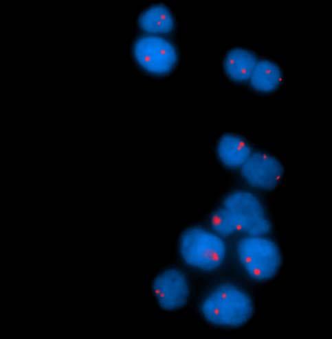
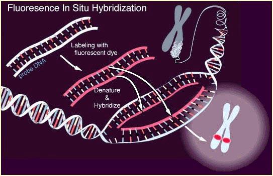
10 10 Fluorescence in situ hybridisation
11 11 Applications Aneuploidy Gene copy number Structural breakpoint analysis Translocation Microdeletion Gene mapping


12 12 ISH probes Metaphase Whole chromosome probes Alpha satellite/centromere probes Unique sequence probes Lodish et al Molecular Cell Biology 2005 Interphase

13 13 Alpha satellite/centromere probe Repetitive sequences Near chromosome centromeres Chromosome specific Detect aneuploidy

14 14 Unique sequence probes (locus specific probes) Target regions NOT repeated in the genome Chromosome deletions Oncogenes c-myc; HER2; EGFR Telomeric and subtelomeric probes
15 15

16 16 Amplification patterns Double minutes = extrachromosomal amplification MYCN FISH in neuroblastoma Interphase Metaphase

17 17 Amplification patterns Homogeneous staining regions = intrachromosomal amplification HER2 FISH in breast cancer Interphase

18 18 HER2/chromosome 17 FISH Looking for gene amplification - breast cancer FNA cytology Ratio 2:2 = 1.0 No gene amplification

19 19 HER2/chromosome 17 FISH Looking for gene amplification - breast cancer FNA cytology Ratio 2:>15=7.5 High grade gene amplification

20 20 Morphology in the fluorescence microscope

21 21 Morphology in the fluorescence microscope

22 22 Morphology in the fluorescence microscope

23 23 Morphology in the fluorescence microscope


24 24 HER2 TOP2A Chromosome17 Brystkreft BRCA1 HER2 TOP2A TP53 Bofin, et al Cytopathology 2003;14: Ingen HER2/TOP2A genamp amp HER2 amp/top2a nonamp
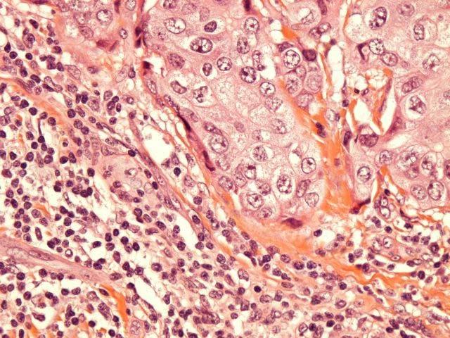
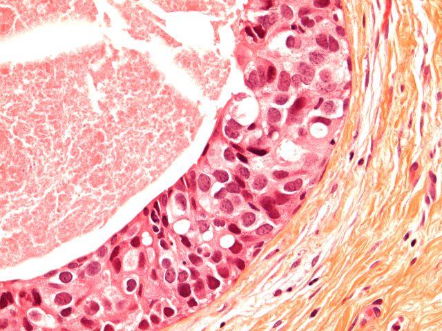
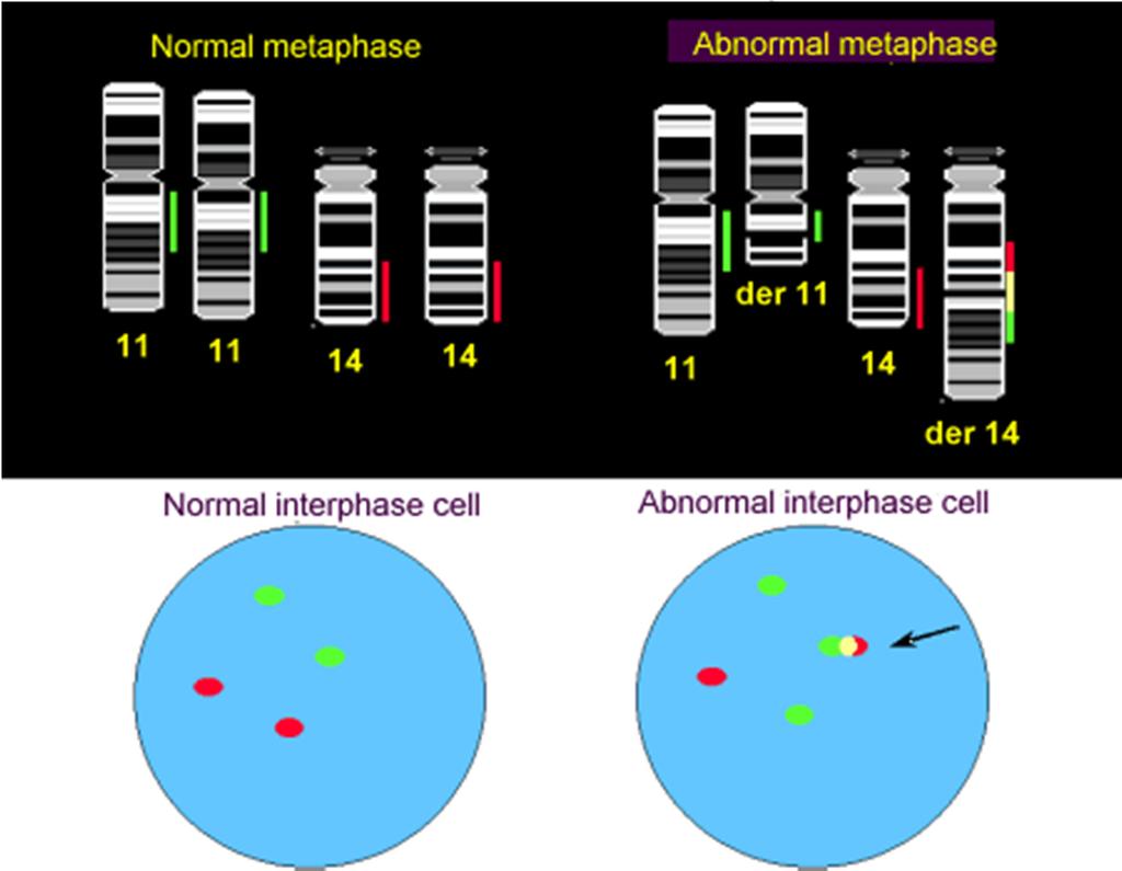
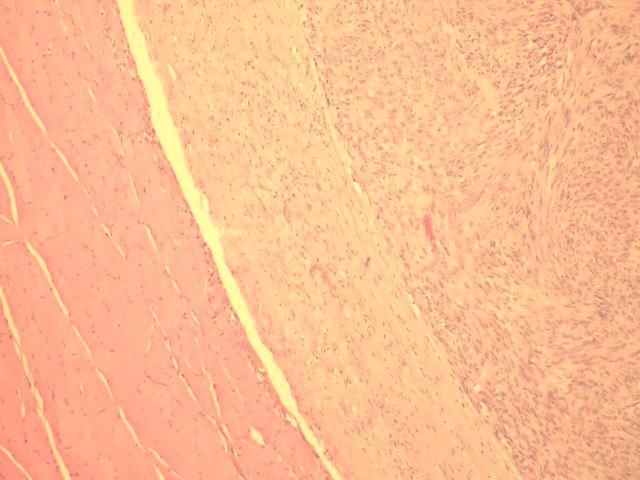
25 25 Translocations Locus specific probes Diagnostics Prognostication Follow-up Leukemia CML brc/abl translokasjon Lymphoma Mantle cell lymphoma t(11;14)(q13;q32) Sarcoma synovial sarcoma - t(x;18)

26 26 Fusion and split translocations Red and green co-localise to detect a known translocation Red and green separate to detect a break without the need to know the translocation partner
27 27 Translocations - fusions

28 28 Translocations - Splits Bishop R Bioscience Horizons 2010;3:85-95
29 29 Screening for disease or relapse Multicolour FISH Cancer of the urinary bladder urine cytology Copy number changes in ch. 3, 7, or 17 in at least 4 cells OR Loss of 9p21 in at least 12 cells Ferras S et al Cancer Cytopathol 2009 Feb 25;117(1):7-14.
30 30 Chromosome copy number Urinary cytology FISH Chromosomes 3,7,17 Locus 9p21 E Berggren/A Bofin


31 31 Pap Urothelial carcinoma WHO grade 2-3 FISH Pap Urothelial carcinoma, WHO grade 3 FISH E Berggren/A Bofin
 Pap")

32 32 Pap Urothelial carcinoma, WHO grade 3 (FISH) Pap Cis,/Urothelial carcinoma, WHO grade 2 FISH E Berggren/A Bofin
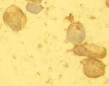
:1679-80 Croes R et al Adult sclerosing rhabdomyosarcoma: cytogenetic link with embryonal rhabdomyosarcomavirchows Arch.")
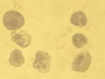
33 33 Cytogenetic analyses Karyotyping FISH Lipoma Synovial sarcoma t(x;18) (SYT;SSX) Rhabdomyosarcoma Cohen IJ et al Synovial sarcoma of bone delineated by spectral karyotyping.lancet ;350(9092): Croes R et al Adult sclerosing rhabdomyosarcoma: cytogenetic link with embryonal rhabdomyosarcomavirchows Arch Jan;446(1):64-7 Sandberg AA Updates on the cytogenetics and molecular genetics of bone and soft tissue tumors: lipoma. Cancer Genet Cytogenet Apr 15;150(2):93-115
34 34 FISH in the path lab Contributes to diagnosis Surveillance in patient follow-up Prognostic and predictive information Determining treatment strategies
35 35 FISH - pros and cons Simpler protocol minimal pretreatment No formalin-fixation artefacts Whole nuclei - no truncation artefacts Little background fluorescence Recognisable morphology compared to routine stained smears Cytology not always available
36 36 Artefacts/Problems Smearing artefacts Non-specific background fluorescence Low stringency washing Cross-hybridisation Poorly prepared probe Probe too small Amplification techniques (TSA)
37 37 Immunocytochemistry Cell smears Fine needle aspirates Liquid-based cytology Smears Cell suspension Snap frozen Cell blocks «pseudohistology»

38 38 Immunocytochemistry Cervix Liquid-based cytology HSIL MIB1 p16 INK4a Sahebali et al Immunocytochemistry in liquid based cervical cytology: Analysis of clinical use following a cross sectional study International Journal of Cancer 2005;118:


39 39 Immunocytochemistry Breast carcinoma Diff Quick atypical ductal cells Oestrogen receptor
40 40 Immunocytochemistry Breast carcinoma Reliability of a negative result Snap freezing Fixative/air-drying The epitope may become slightly deformed resultiong in a false neg. Result Cuthbert et al Cytopathology 1990;1: Sauer T, Beraki E, Jebsen P. Anal Quant Cytol Histol 1998;20:122-6 A negative ER on FNA should prompt ER on tissue biopsy Lea D, Bofin A. Er hormonundersøkelse av cytologisk materiale i brystkreftsvulster til å stole på? Bioingeniøren 2011:3;6-12


41 41 Immunocytochemistry Malignant melanoma Atypical melanocytes Immunocytochemistry:HMB45
42 42 Painful subcutaneous tumour S100 protein
43 43 Painful subcutaneous tumour Neurofibroma


44 44 Large tumour in the shoulder FNA smear LCA B-cell lymphoma CD20 CD99


45 45 Soft tissue tumour in a child FNA smear MGG Desmin Alveolar rhabdomyosarcoma Lisa Walaas, Rikshospitalet, Oslo
46 46 ICC - pros and cons Simple procedure air-dried smears LBC Cell blocks Provides additional information in inoperable patients Enables pre-operative chemotherapy Not always enough material for multiple ICC s
47 47 Polymerase Chain Reaction(PCR) Finding small sequences of DNA and making them visible DNA mrna converted to cdna by means of reverse transcriptase DNA is amplified: 5 3 DNA DNA
48 48 RT-PCR (reverse transcriptase) Fluorochrome Probe Quencher DNA DNA

49 49 Real time RT-PCR Relative quantification
50 50 B-RAF mutation in thyroid FNA Diagnostics Determining treatment strategy B-REF mutation detection in papillary carcinoma (45 %) BRAF-mutation in a follicular-like neoplasm indicates a follicular variant of a papillary carcinoma AND total thyroidectomy rather than a diagnostic hemithyroidectomy Canadas-Garre et al Ann Surg 2012 May;255(5):986-92
51 51 PCR- pros and cons Costly, perhaps complicated procedure Requires a reasonable amount of material Quantitation or relative quantitation of small amounts of RNA/DNA
52 52 Flow cytometry Flow cytometry is a method allowing the analysis of of cells or particles suspended and separated in a fluid. The fluid flows past a focused laser beam. The cells are usually labelled using fluorescent probes which bind to specific cell associated molecules Phenotypic, biochemical and molecular characteristics of the individual cells Visualised as a scatter chart or graph

53 53 Flow cytometry Rudbeck laboratory, University of Uppsala
54 54 Flow cytometry FNS/FNA from lymph nodes/lymphoid tissue Particularly in non-hodgkins lymphoma Requires a certain number of cells No morphology keep some material back for microscopy Zeppa et al Cytopathology 2010;21:
55 55 Quality assurance/control Follow procedure particularly posthybridisation washes Qualified readers National and international QA programmes


56 56 Take home message Think special tests - Always
From Morphology to Molecular Pathology: A Practical Approach for Cytopathologists Part 1-Cytomorphology. Songlin Zhang, MD, PhD LSUHSC-Shreveport
 From Morphology to Molecular Pathology: A Practical Approach for Cytopathologists Part 1-Cytomorphology Songlin Zhang, MD, PhD LSUHSC-Shreveport I have no Conflict of Interest. FNA on Lymphoproliferative
Neoplasia 2018 lecture 11. Dr H Awad FRCPath
 Neoplasia 2018 lecture 11 Dr H Awad FRCPath Clinical aspects of neoplasia Tumors affect patients by: 1. their location 2. hormonal secretions 3. paraneoplastic syndromes 4. cachexia Tumor location Even
Application. Application of molecular biology methods in hematology and oncology. Oncogenes are involved in:
 Application Application of molecular biology methods in hematology and oncology Research on the etiology of cancer Diagnostics / Differential diagnostics Stratifying for treatment Prognosing the outcome
Characterisation of structural variation in breast. cancer genomes using paired-end sequencing on. the Illumina Genome Analyser
 Characterisation of structural variation in breast cancer genomes using paired-end sequencing on the Illumina Genome Analyser Phil Stephens Cancer Genome Project Why is it important to study cancer? Why
Role of FISH in Hematological Cancers
 Role of FISH in Hematological Cancers Thomas S.K. Wan PhD,FRCPath,FFSc(RCPA) Honorary Professor, Department of Pathology & Clinical Biochemistry, Queen Mary Hospital, University of Hong Kong. e-mail: wantsk@hku.hk
ACMG/CAP Cytogenetics CY
 www.cap.org Cytogenetics Analytes/procedures in bold type are regulated for proficiency testing by the Centers for Medicare & Medicaid Services ACMG/CAP Cytogenetics CY Analyte CY Challenges per Shipment
Fellowship in Cytopathology Department of Pathology. All India Institute of Medical Sciences (AIIMS) Jodhpur, Rajasthan, India
 Jodhpur, Rajasthan, India") Fellowship in Cytopathology Department of Pathology All India Institute of Medical Sciences (AIIMS) Jodhpur, Rajasthan, India Syllabus for Fellowship in Cytopathology: FNAC Direct, Guided, EUS Exfoliative
Fellowship in Cytopathology Department of Pathology All India Institute of Medical Sciences (AIIMS) Jodhpur, Rajasthan, India Syllabus for Fellowship in Cytopathology: FNAC Direct, Guided, EUS Exfoliative
Significance of Chromosome Changes in Hematological Disorders and Solid Tumors
 Significance of Chromosome Changes in Hematological Disorders and Solid Tumors Size of Components of Human Genome Size of haploid genome 3.3 X 10 9 DNA basepairs Estimated genetic constitution 30,000
Significance of Chromosome Changes in Hematological Disorders and Solid Tumors
 Significance of Chromosome Changes in Hematological Disorders and Solid Tumors Size of Components of Human Genome Size of haploid genome! Estimated genetic constitution! Size of average chromosome
INTRODUCTION TO PATHOLOGICAL TECHNIQUES. 1. Types of routine biopsy procedures 2. Special exams (IHC, FISH)
") INTRODUCTION TO PATHOLOGICAL TECHNIQUES 1. Types of routine biopsy procedures 2. Special exams (IHC, FISH) Biopsy-Indications Diffuse/multifocal lesions (neoplastic, inflammatory, etc) Etiology of the
INTRODUCTION TO PATHOLOGICAL TECHNIQUES 1. Types of routine biopsy procedures 2. Special exams (IHC, FISH) Biopsy-Indications Diffuse/multifocal lesions (neoplastic, inflammatory, etc) Etiology of the
Chapter 4 Cellular Oncogenes ~ 4.6 -
 Chapter 4 Cellular Oncogenes - 4.2 ~ 4.6 - Many retroviruses carrying oncogenes have been found in chickens and mice However, attempts undertaken during the 1970s to isolate viruses from most types of
What kind of material should we use for ICC in our daily routine. Torill Sauer Department of Pathology, Akershus University Hospital
 What kind of material should we use for ICC in our daily routine Torill Sauer Department of Pathology, Akershus University Hospital Diversity of preparing cytological material Cell block Direct smears
Applications of IHC. Determination of the primary site in metastatic tumors of unknown origin
 Applications of IHC Determination of the primary site in metastatic tumors of unknown origin Classification of tumors that appear 'undifferentiated' by standard light microscopy Precise classification
Molecular Diagnosis. Nucleic acid based testing in Oncology
 Molecular Diagnosis Nucleic acid based testing in Oncology Objectives Describe uses of NAT in Oncology Diagnosis, Prediction, monitoring. Genetics Screening, presymptomatic testing, diagnostic testing,
Nucleic Acid Testing - Oncology. Molecular Diagnosis. Gain/Loss of Nucleic Acid. Objectives. MYCN and Neuroblastoma. Molecular Diagnosis
 Nucleic Acid Testing - Oncology Molecular Diagnosis Nucleic acid based testing in Oncology Gross alterations in DNA content of tumors (ploidy) Gain/Loss of nucleic acids Markers of Clonality Oncogene/Tumor
Determination of Genomic Imbalances by Genome-wide Screening Approaches
 Overview Determination of Genomic Imbalances by Genome-wide Screening Approaches Károly Szuhai Introduction/Methodologies Applications/Results Conclusion Approaches Introduction/Methodologies Chromosome
Anti-hTERT Antibody (SCD-A7)
") Quality in Control Anti-hTERT Antibody (SCD-A7) htert_ab_pi_v1 Product Code: HCL025 Contents htert and Telomerase 2 Negative htert 3 Positive htert 4 Guidance and additional data 5 Case Study: RMH12-001
Quality in Control Anti-hTERT Antibody (SCD-A7) htert_ab_pi_v1 Product Code: HCL025 Contents htert and Telomerase 2 Negative htert 3 Positive htert 4 Guidance and additional data 5 Case Study: RMH12-001
Case 3. Ann T. Moriarty,MD
 Case 3 Ann T. Moriarty,MD Case 3 59 year old male with asymptomatic cervical lymphadenopathy. These images are from a fine needle biopsy of a left cervical lymph node. Image 1 Papanicolaou Stained smear,100x.
Klinisch belang van chromosomale translocatie detectie in sarcomen
 Translocations in sarcomas Klinisch belang van chromosomale translocatie detectie in sarcomen Judith V.M.G. Bovée, M.D., Ph.D. Department of Pathology Leiden University Medical Center RNA binding DNA binding
ADx Bone Marrow Report. Patient Information Referring Physician Specimen Information
 ADx Bone Marrow Report Patient Information Referring Physician Specimen Information Patient Name: Specimen: Bone Marrow Site: Left iliac Physician: Accession #: ID#: Reported: 08/19/2014 - CHRONIC MYELOGENOUS
Providence Medford Medical Center Pathology Department
 Providence Medford Medical Center Pathology Department Anatomic pathology services including histology, cytology and autopsies are offered through Providence Medford Medical Center Pathology Department.
USCAP 2012: Companion Meeting of the AAOOP. Update on lacrimal gland neoplasms: Molecular pathology of interest
 USCAP 2012: Companion Meeting of the AAOOP Vancouver BC, Canada, March 17, 2012 Update on lacrimal gland neoplasms: Molecular pathology of interest Valerie A. White MD, MHSc, FRCPC Department of Pathology
MECHANISMS OF HUMAN DISEASE: LABORATORY SESSION CYTOPATHOLOGY Monday, April 26, 2013 FACULTY COPY
 GOAL: MECHANISMS OF HUMAN DISEASE: LABORATORY SESSION CYTOPATHOLOGY Monday, April 26, 2013 FACULTY COPY 1. Understated the role of cytopathology in the clinical management of the patient and recognize
American Society of Cytopathology Core Curriculum in Molecular Biology
 American Society of Cytopathology Core Curriculum in Molecular Biology American Society of Cytopathology Core Curriculum in Molecular Biology Chapter 1 Molecular Basis of Cancer Molecular Oncology Keisha
I have no relevant conflicts of interest to disclose. John T. Seykora MD PhD Departments of Dermatology & Pathology and Laboratory Medicine
 Molecular Characterization of Stage 1-3 Melanoma: Are we close to accurate prognostication and prediction? I have no relevant conflicts of interest to disclose. John T. Seykora MD PhD Departments of Dermatology
Disclosures. An update on ancillary techniques in the diagnosis of soft tissue tumors. Ancillary techniques. Introduction
 Disclosures An update on ancillary techniques in the diagnosis of soft tissue tumors. I have nothing to disclose. Andrew Horvai, MD, PhD Clinical Professor, Pathology Introduction Ancillary techniques
American Society of Cytopathology Core Curriculum in Molecular Biology
 American Society of Cytopathology Core Curriculum in Molecular Biology American Society of Cytopathology Core Curriculum in Molecular Biology Chapter 6 Fluorescence in situ Hybridization (FISH) Principles
The Pathology of Neoplasia Part II
 The Pathology of Neoplasia Part II February 2018 PAUL BOGNER, MD A S S O C I A T E P R O F E S S O R O F O N C O L O G Y P A T H O L O G Y A N D D E R M A T O L O G Y Clinical goals of cancer pathology
CODING TUMOUR MORPHOLOGY. Otto Visser
 CODING TUMOUR MORPHOLOGY Otto Visser INTRODUCTION The morphology describes the tissue of the tumour closest to normal tissue Well differentiated tumours are closest to normal Undifferentiated tumours show
SPECIMEN PREPARATION AND ADEQUACY OF THE MATERIAL
 SPECIMEN PREPARATION AND ADEQUACY OF THE MATERIAL Guido FADDA, MD, MIAC Head, Cytopathology Section Department of Anatomic Pathology and Laboratory Medicine Agostino Gemelli School of Medicine and Hospital
A. Incorrect! All the cells have the same set of genes. (D)Because different types of cells have different types of transcriptional factors.
Because different types of cells have different types of transcriptional factors.") Genetics - Problem Drill 21: Cytogenetics and Chromosomal Mutation No. 1 of 10 1. Why do some cells express one set of genes while other cells express a different set of genes during development? (A) Because
Genetics - Problem Drill 21: Cytogenetics and Chromosomal Mutation No. 1 of 10 1. Why do some cells express one set of genes while other cells express a different set of genes during development? (A) Because
PD-L1 Analyte Control DR
 Quality in Control PD-L1 Analyte Control DR PD-L1_PI_v2 Product Codes: HCL019, HCL020 and HCL021 Contents PD-L1 Analyte Control DR 2 What is PD-L1? 3 The Role of PD-L1 in Cancer 3 PD-L1 Assessment 4 PD-L1
Oncology Genetics: Cytogenetics and FISH 17/09/2014
 Oncology Genetics: Cytogenetics and FISH 17/09/2014 Chris Wragg Head of Oncology Genomics, BGL BGL Bristol Genetics Laboratory (BGL) CPA accredited Genetics laboratory serving a core population of 4-5million
Dr. Issraa Ali Hussein
 CLINICAL 09888888;rCYTOLOGY Dr. Issraa Ali Hussein objectives Define diagnostic cytology (clinical cytology). Explain the differences between histopathology and cytopathology. Recognize the methods for
Current Status of Biomarkers (including DNA Tumor Markers and Immunohistochemistry in the Laboratory Diagnosis of Tumors)
") Current Status of Biomarkers (including DNA Tumor Markers and Immunohistochemistry in the Laboratory Diagnosis of Tumors) Kael Mikesell, DO McKay-Dee Hospital May 14, 2015 Outline Update to DNA Testing
Current Status of Biomarkers (including DNA Tumor Markers and Immunohistochemistry in the Laboratory Diagnosis of Tumors) Kael Mikesell, DO McKay-Dee Hospital May 14, 2015 Outline Update to DNA Testing
Presentation material is for education purposes only. All rights reserved URMC Radiology Page 1 of 98
 Presentation material is for education purposes only. All rights reserved. 2011 URMC Radiology Page 1 of 98 Radiology / Pathology Conference February 2011 Brooke Koltz, Cytopathology Resident Presentation
Hematopathology Case Study
 www.medfusionservices.com Hematopathology Case Study CV3515-14 JUNE Clinical Presentation: Clinical Information: A 42 year old male with history of chronic myelogenous leukemia (CML) presents with an elevated
Molecular Genetics of Paediatric Tumours. Gino Somers MBBS, BMedSci, PhD, FRCPA Pathologist-in-Chief Hospital for Sick Children, Toronto, ON, CANADA
 Molecular Genetics of Paediatric Tumours Gino Somers MBBS, BMedSci, PhD, FRCPA Pathologist-in-Chief Hospital for Sick Children, Toronto, ON, CANADA Financial Disclosure NanoString - conference costs for
Cytogenetics Technologies, Companies & Markets
 Cytogenetics Technologies, Companies & Markets By Prof. K. K. Jain MD, FRACS, FFPM Jain PharmaBiotech Basel, Switzerland November 2018 A Jain PharmaBiotech Report A U T H O R ' S B I O G R A P H Y Professor
Select analysis on the next pages. Sample request and sending address see last page. Institut für Pathologie und Molekularpathologie
 Diagnostic Tumor Genome Analysis Schmelzbergstrasse 12 8091 Zürich Tel.: (+41) 044 255 3929 Fax.: (+41) 044 255 4416 Client (address, telephone number): ngs.pathologie@usz.ch www.pathologie.usz.ch Sample-Nr:
INTRODUCTION TO PATHOLOGY
 INTRODUCTION TO PATHOLOGY The literal translation of the word pathology is the study (logos) of suffering (pathos). It is a discipline that bridges clinical practice and basic sciences. Pathology is concerned
Objectives. Morphology and IHC. Flow and Cyto FISH. Testing for Heme Malignancies 3/20/2013
 Molecular Markers in Hematologic Malignancy: Ways to locate the needle in the haystack. Objectives Review the types of testing for hematologic malignancies Understand rationale for molecular testing Marcie
An Overview of Cytogenetics. Bridget Herschap, M.D. 9/23/2013
 An Overview of Cytogenetics Bridget Herschap, M.D. 9/23/2013 Objectives } History and Introduction of Cytogenetics } Overview of Current Techniques } Common cytogenetic tests and their clinical application
CYTOGENETICS INTRODUCTION SPECIAL INSTRUCTIONS ON SAMPLE COLLECTION AND HANDLING
 INTRODUCTION The Cytogenetics Laboratory offers a comprehensive array of chromosome investigations for cancers, constitutional abnormalities, and prenatal and postnatal diagnosis. Analyses are performed
Molecular Methods in the Diagnosis and Prognostication of Melanoma: Pros & Cons
 Molecular Methods in the Diagnosis and Prognostication of Melanoma: Pros & Cons Ben J. Friedman, MD Senior Staff Physician Department of Dermatology Department of Pathology and Laboratory Medicine Henry
Cytology meets Molecular: Don t throw away your microscope! British Association for Cytopathology November 4, 2017
 Cytology meets Molecular: Don t throw away your microscope! British Association for Cytopathology November 4, 2017 Andrew Fischer, M.D. Director of Cytopathology University of Massachusetts Theme: How
Ascitic Fluid and Use of Immunocytochemistry. Mercè Jordà, University of Miami
 Ascitic Fluid and Use of Immunocytochemistry Mercè Jordà, University of Miami Is It Malignant? Yes? No Ascitic Fluid Cytomorphologic Useful Findings Tight clusters with smooth borders Cellular and nuclear
TEST MENU BY SPECIALTY
 1 TEST MENU BY SPECIALTY Breast Pathology Surgical excisions, needle core biopsies, and plastic surgery accepted from all sites Assessment of margins Axillary lymph node dissections Sentinel lymph nodes
SELECTED DILEMMAS IN RESPIRATORY CYTOPATHOLOGY (2 CASES)
") SELECTED DILEMMAS IN RESPIRATORY CYTOPATHOLOGY (2 CASES) Dr. Mariamma Joseph Professor of Pathology Division Head Cytopathology Department of Pathology and Laboratory Medicine LHSC and Western University
SELECTED DILEMMAS IN RESPIRATORY CYTOPATHOLOGY (2 CASES) Dr. Mariamma Joseph Professor of Pathology Division Head Cytopathology Department of Pathology and Laboratory Medicine LHSC and Western University
Classification of Hematologic Malignancies. Patricia Aoun MD MPH
 Classification of Hematologic Malignancies Patricia Aoun MD MPH Objectives Know the basic principles of the current classification system for hematopoietic and lymphoid malignancies Understand the differences
Molecular Probes Introducing 14 new probes
 Molecular Probes Introducing 14 new probes Gene and Chromosome Probes Dual Colour ISH INFORM HER2 Dual ISH DNA Probe Cocktail Assay Product Part Number INFORM HER2 Dual ISH DNA Probe Cocktail 800-4422
Cytology for the Endocrinologist. Nicole Massoll M.D
 Cytology for the Endocrinologist Nicole Massoll M.D Objectives Discuss slide preperation Definitions of adequacy ROSE (Rapid On-Site Evaluation) Thyroid Cytology Adequacy Nicole Massoll M.D. University
Reporting cytogenetics Can it make sense? Daniel Weisdorf MD University of Minnesota
 Reporting cytogenetics Can it make sense? Daniel Weisdorf MD University of Minnesota Reporting cytogenetics What is it? Terminology Clinical value What details are important Diagnostic Tools for Leukemia
Disclosures. Molecular Cytopathology. Update on Molecular Testing of Cytology Specimens: Beyond the Cell Block. Molecular Cytopathology Solid Tumor
 Update on Molecular Testing of Cytology Specimens: Beyond the Cell Block Disclosures None relevant to this talks Dara L. Aisner, M.D., Ph.D. Assistant Professor Co-Director, Colorado Molecular Correlates
Anatomic Molecular Pathology: An Emerging Field
 Anatomic Molecular Pathology: An Emerging Field Antonia R. Sepulveda M.D., Ph.D. University of Pennsylvania asepu@mail.med.upenn.edu 2008 ASIP Annual Meeting Anatomic pathology (U.S.) is a medical specialty
Cancer. The fundamental defect is. unregulated cell division. Properties of Cancerous Cells. Causes of Cancer. Altered growth and proliferation
 Cancer The fundamental defect is unregulated cell division. Properties of Cancerous Cells Altered growth and proliferation Loss of growth factor dependence Loss of contact inhibition Immortalization Alterated
The role of the cytologist in breast cancer screening
 The role of the cytologist in breast cancer screening I.Seili-Bekafigo, MD, PhD Clinical cytologist KBC Rijeka Croatian Society for Clinical Cytology Fine needle aspiration (FNA, FNAB, FNAC) Fine needle
Evolution of Pathology
 1 Traditional pathology Molecular pathology 2 Evolution of Pathology Gross Pathology Cellular Pathology Morphologic Pathology Molecular/Predictive Pathology Antonio Benivieni (1443-1502): First autopsy
BHS training course. Laboratory Hematology Cytogenetics. Lucienne Michaux. Centrum voor Menselijke Erfelijkheid, UZLeuven
 BHS training course Laboratory Hematology Cytogenetics Lucienne Michaux Centrum voor Menselijke Erfelijkheid, UZLeuven 18/11/2017 Organization of the Lecture Definition and principles Tools Applications
Disclosures. An update on ancillary techniques in the diagnosis of soft tissue tumors. Ancillary techniques. Introduction
 Disclosures An update on ancillary techniques in the diagnosis of soft tissue tumors. I have nothing to disclose. Andrew Horvai, MD, PhD Clinical Professor, Pathology Introduction Ancillary techniques
Kimberly Rohan ANP-BC, AOCN Nurse Practitioner Edward Cancer Center
 Kimberly Rohan ANP-BC, AOCN Nurse Practitioner Edward Cancer Center Objective The nurse will be able to explain biomarkers and their implications to patient s prescribed treatment plan Biomarkers Use in
Update on Thyroid FNA The Bethesda System. Shikha Bose M.D. Associate Professor Cedars Sinai Medical Center
 Update on Thyroid FNA The Bethesda System Shikha Bose M.D. Associate Professor Cedars Sinai Medical Center Thyroid Nodules Frequent occurrence Palpable: 4-7% of adults Ultrasound: 10-31% Majority benign
Case #1: 75 y/o Male (treated and followed by prostate cancer oncology specialist ).
.") SOLID TUMORS WORKSHOP Cases for review Prostate Cancer Case #1: 75 y/o Male (treated and followed by prostate cancer oncology specialist ). January 2009 PSA 4.4, 20% free; August 2009 PSA 5.2; Sept 2009
SOLID TUMORS WORKSHOP Cases for review Prostate Cancer Case #1: 75 y/o Male (treated and followed by prostate cancer oncology specialist ). January 2009 PSA 4.4, 20% free; August 2009 PSA 5.2; Sept 2009
Cytopathology. Robert M Genta Pathologie Clinique Université de Genève
 Cytopathology Robert M Genta Pathologie Clinique Université de Genève Learning objectives At the end of this hour you will know: 1. What cytopathology is 2. How specimens are collected, processed, and
HEMATOPATHOLOGY SUMMARY REPORT RL;MMR;
 HEMATOPATHOLOGY SUMMARY REPORT RL;MMR; Page 1 of 1 05/15/20XX HP000000-20XX 05/21/20XX (212) 123-457 (51) 32-3455 (51) 123-457 Age: 78 DOB: 0/05/19XX SS#: 45-45-45 Clinical Information: 78 y/o female with
Cutaneous Mesenchymal Neoplasms with EWSR1 Rearrangement
 Cutaneous Mesenchymal Neoplasms with EWSR1 Rearrangement By Konstantinos Linos MD, FCAP, FASDP Bone, Soft Tissue and Dermatopathology Assistant Professor of Pathology Dartmouth-Hitchcock Medical Center
Dr. dr. Primariadewi R, SpPA(K)
") Curriculum Vitae Dr. dr. Primariadewi R, SpPA(K) Education : Medical Doctor from UKRIDA Doctoral Degree from Faculty of Medicine University of Indonesia Pathologist Specialist and Consultant from Faculty
Curriculum Vitae Dr. dr. Primariadewi R, SpPA(K) Education : Medical Doctor from UKRIDA Doctoral Degree from Faculty of Medicine University of Indonesia Pathologist Specialist and Consultant from Faculty
ROSE in EUS guided FNA of Pancreatic Lesions
 ROSE in EUS guided FNA of Pancreatic Lesions Guy s Hospital, London, 16 April 2018 Laxmi Batav Imperial College NHS Trust Imperial College NHS Trust Cytology Workload Cervical Cytology 57,500 (decreases
HER-2/neu amplification detected by fluorescence in situ hybridization in fine needle aspirates from primary breast cancer
 Original article Annals of Oncology 13: 1398 1403, 2002 DOI: 10.1093/annonc/mdf217 HER-2/neu amplification detected by fluorescence in situ hybridization in fine needle aspirates from primary breast cancer
Use of MYC, BCL2 and BCL6 FISH for investigations of high grade B cell lymphoma
 Use of MYC, BCL2 and BCL6 FISH for investigations of high grade B cell lymphoma Dr Anthony Bench Haematopathology and Oncology Diagnostic Service Cambrıdge Unıversıty Hospitals NHS Foundatıon Trust Cambridge
Diplomate of the American Board of Pathology in Anatomic and Clinical Pathology
 A 33-year-old male with a left lower leg mass. Contributed by Shaoxiong Chen, MD, PhD Assistant Professor Indiana University School of Medicine/ IU Health Partners Department of Pathology and Laboratory
Management of Neck Metastasis from Unknown Primary
 Management of Neck Metastasis from Unknown Primary.. Definition Histologic evidence of malignancy in the cervical lymph node (s) with no apparent primary site of original tumour Diagnosis after a thorough
Activation of cellular proto-oncogenes to oncogenes. How was active Ras identified?
 Dominant Acting Oncogenes Eugene E. Marcantonio, M.D. Ph.D. Oncogenes are altered forms of normal cellular genes called proto-oncogenes that are involved in pathways regulating cell growth, differentiation,
CYTOLOGY: What every general practitioner should know ( and every future specialist)
") CYTOLOGY: What every general practitioner should know ( and every future specialist) 16 April 2014 Dr C Crause Pathologist and Senior Lecturer Department of Anatomical Pathology University of Pretoria/NHLS
CYTOLOGY: What every general practitioner should know ( and every future specialist) 16 April 2014 Dr C Crause Pathologist and Senior Lecturer Department of Anatomical Pathology University of Pretoria/NHLS
FISH VALIDATION: HOW I DO IT!
 FISH VALIDATION: HOW I DO IT! Theresa C. Brown, PhD, FACMG, CG(ASCP) CM Director, Cytogenetics laboratory Hayward Genetics Center Instructor Tulane University School of Medicine WHERE DO I GET THIS INFORMATION
Case 2. Dr. Sathima Natarajan M.D. Kaiser Permanente Medical Center Sunset
 Case 2 Dr. Sathima Natarajan M.D. Kaiser Permanente Medical Center Sunset History 24 year old male presented with a 3 day history of right flank pain, sharp in nature Denies fever, chills, hematuria or
Understanding the Human Karyotype Colleen Jackson Cook, Ph.D.
 Understanding the Human Karyotype Colleen Jackson Cook, Ph.D. SUPPLEMENTAL READING Nussbaum, RL, McInnes, RR, and Willard HF (2007) Thompson and Thompson Genetics in Medicine, 7th edition. Saunders: Philadelphia.
Fluorescence in-situ Hybridization (FISH) ETO(RUNX1T1)/AML1(RUNX1) or t(8;21)(q21.3;q22)
 ETO(RUNX1T1)/AML1(RUNX1) or t(8;21)(q21.3;q22)") PML/RARA t(15;17) Translocation Assay Result : nuc ish(pml 2)(RARA 2)[200] : 200/200(100%) interphase nuclei show normal 2O 2G signals for PML/RARA : is Negative for t(15;17)(q22;q21.1) 2 Orange 2 Green
PML/RARA t(15;17) Translocation Assay Result : nuc ish(pml 2)(RARA 2)[200] : 200/200(100%) interphase nuclei show normal 2O 2G signals for PML/RARA : is Negative for t(15;17)(q22;q21.1) 2 Orange 2 Green
Differentiation of Tumors with Specific Red Cell Adherence (SRCA) test
 test") 753 Differentiation of Tumors with Specific Red Cell Adherence (SRCA) test Dr. Abhishek A Mangaonkar *, Dr. A G Valand 1 Intern, Grant Medical College and Sir J.J. Group of Hospitals, Mumbai, India 2 Professor,
753 Differentiation of Tumors with Specific Red Cell Adherence (SRCA) test Dr. Abhishek A Mangaonkar *, Dr. A G Valand 1 Intern, Grant Medical College and Sir J.J. Group of Hospitals, Mumbai, India 2 Professor,
Personalized Medicine: Lung Biopsy and Tumor
 Personalized Medicine: Lung Biopsy and Tumor Mutation Testing Elizabeth H. Moore, MD Personalized Medicine: Lung Biopsy and Tumor Mutation Testing Genomic testing has resulted in a paradigm shift in the
at least 5 probes standard 8 probes (13, 15, 16, 18, 21, 22, 15, X, Y) at least 5 probes standard 8 probes (13, 15, 16, 18, 21, 22, X, Y)
 at least 5 probes standard 8 probes (13, 15, 16, 18, 21, 22, X, Y)") Management of FISH probe testing Petra Musilová et al. Repromeda, Brno, Czech Rep. Veterinary Research Institute, Brno Genprogress, Brno, Czech Rep. Aneuploidy screening at least 5 probes standard 8 probes
Management of FISH probe testing Petra Musilová et al. Repromeda, Brno, Czech Rep. Veterinary Research Institute, Brno Genprogress, Brno, Czech Rep. Aneuploidy screening at least 5 probes standard 8 probes
Cancer. The fundamental defect is. unregulated cell division. Properties of Cancerous Cells. Causes of Cancer. Altered growth and proliferation
 Cancer The fundamental defect is unregulated cell division. Properties of Cancerous Cells Altered growth and proliferation Loss of growth factor dependence Loss of contact inhibition Immortalization Alterated
CYTOGENETICS Dr. Mary Ann Perle
 CYTOGENETICS Dr. Mary Ann Perle I) Mitosis and metaphase chromosomes A) Chromosomes are most fully condensed and clearly distinguishable during mitosis. B) Mitosis (M phase) takes 1 to 2 hrs and is divided
Copy number and somatic mutations drive tumors
 Detection of copy number alterations, ploidy and loss of heterozygosity across the genome in FFPE specimens Utility for diagnosis and treatment with comparison to FISH-based and as a complement to sequencing
The role of Electron Microscopy in the study of cytologic specimens. Elba A. Turbat-Herrera, MD
 The role of Electron Microscopy in the study of cytologic specimens. Elba A. Turbat-Herrera, MD Louisiana State University Health Sciences Center Shreveport, LA, USA Introduction The field of Cytology
40 TH EUROPEAN CONGRESS 0F CYTOLOGY LIVERPOOL, UK October 2-5, 2016
 Outcomes from the diagnostic approach of thyroid lesions using US-FNA and LBC in clinical practice Emmanouel Mastorakis MD PhD Cytopathologist Director in Cytopathology Laboratory Regional General Hospital
Evidence for a Tetraploid Intermediate in the Development of Cervical Cancer
 Evidence for a Tetraploid Intermediate in the Development of Cervical Cancer David A. Eastmond, Andrew Olaharski, Maria Gonsebatt University of California, Riverside and Universidad Nacional Autonoma de
Differential diagnosis of hematolymphoid tumors composed of medium-sized cells. Brian Skinnider B.C. Cancer Agency, Vancouver General Hospital
 Differential diagnosis of hematolymphoid tumors composed of medium-sized cells Brian Skinnider B.C. Cancer Agency, Vancouver General Hospital Lymphoma classification Lymphoma diagnosis starts with morphologic
Molecular Markers. Marcie Riches, MD, MS Associate Professor University of North Carolina Scientific Director, Infection and Immune Reconstitution WC
 Molecular Markers Marcie Riches, MD, MS Associate Professor University of North Carolina Scientific Director, Infection and Immune Reconstitution WC Overview Testing methods Rationale for molecular testing
Barriers to Understanding
 Behind the Scenes: The Critical Importance of Cancer Cell Pathology and the Pathologist Sherry T. Emery, M.D., Chief of Pathology Northeast Health System Barriers to Understanding Questions for 2010 What
GOALS AND OBJECTIVES CYTOPATHOLOGY
 GOALS AND OBJECTIVES CYTOPATHOLOGY LEVEL: PGY2, PGY4, PGY5 The 1st block in PGY2 is an introductory in nature and is followed by two more blocks in PGY-4 (please, see core rotation for PGY4 below) and
Surgical Pathology Evening Specialty Conference USCAP 2015
 Surgical Pathology Evening Specialty Conference USCAP 2015 John R. Goldblum, M.D. Chairman, Department of Pathology, Cleveland Clinic Professor of Pathology, Cleveland Clinic Lerner College of Medicine
Goals and Objectives for Cytopathology Rotation
 Goals and Objectives for Cytopathology Rotation Level: PGY3, PGY4, PGY5 The 1st block in PGY3 is an introductory in nature and is followed by three more blocks in PGY-4 (please, see core rotation for PGY4
Integrated Diagnostic Approach to the Classification of Myeloid Neoplasms. Daniel A. Arber, MD Stanford University
 Integrated Diagnostic Approach to the Classification of Myeloid Neoplasms Daniel A. Arber, MD Stanford University What is an integrated approach? What is an integrated approach? Incorporating all diagnostic
Neoplasia 2018 lecture 4. Dr Heyam Awad MD, FRCPath
 Neoplasia 2018 lecture 4 Dr Heyam Awad MD, FRCPath ILOS To understand the concept of the hallmarks of cancer and that they are phenotypic changes needed in all cancer cells. To list the tumor enablers
Contributions to Anatomic Pathology, over the years
 Contributions to Anatomic Pathology, over the years Anatomic Pathology, part 1 G.B. Morgagni Xavier Bichat Rudolf Wirchow Anatomic Pathology, part 1 Anatomic pathology materials: morphological samples
Introduction to Genetics
 Introduction to Genetics Table of contents Chromosome DNA Protein synthesis Mutation Genetic disorder Relationship between genes and cancer Genetic testing Technical concern 2 All living organisms consist
Molecular Detection of BCR/ABL1 for the Diagnosis and Monitoring of CML
 Molecular Detection of BCR/ABL1 for the Diagnosis and Monitoring of CML Imran Mirza, MD, MS, FRCPC Pathology & Laboratory Medicine Institute Sheikh Khalifa Medical City, Abu Dhabi, UAE. imirza@skmc.ae
Chromosome Abnormalities
 Chromosome Abnormalities Chromosomal abnormalities vs. molecular mutations Simply a matter of size Chromosomal abnormalities are big errors Two types of abnormalities 1. Constitutional problem present
Follow up of the Guidelines for Cytopathologic Diagnosis of Malignant Mesothelioma
 Follow up of the Guidelines for Cytopathologic Diagnosis of Malignant Mesothelioma Assoc. Prof. Katalin Dobra, Senior Lecturer in Molecular Pathology Karolinska University Hospital Stockholm, Sweden Disclosure