Imaging Work-Up of a Neck Mass - Adults & Children
|
|
|
- Oswin Copeland
- 6 years ago
- Views:
Transcription
1 Disclosures Imaging Work-Up of a Neck Mass - Adults & Children I have nothing to disclose Christine M Glastonbury MBBS Professor of Radiology & Biomedical Imaging Otolaryngology-Head & Neck Surgery and Radiation Oncology University of California, San Francisco November 7, 2014 Plan & Summary Modalities: P & C, I & CI Masses When in doubt start with CECT? Thyroid mass U/S, NECT, MR Pediatric case U/S, MR, unless urgent clinical problem Almost no utility of PET-CT for initial mass evaluation CT (NECT/CECT) MRI PET-CT ULTRASOUND CT NECT CECT MRI PET-CT Ultrasound Modalities 1
2 CT (NECT/CECT) MRI PET-CT ULTRASOUND CT CT (NECT/CECT) MRI MRI PET-CT ULTRASOUND P: Fast, readily available, multiplanar, bone detail, reproducible studies, relatively cheap, C: Limited ST characterization, radiation I: Emergent situations, staging, bone... CI:?? Pediatric patients Thyroid carcinoma P: Excellent soft tissue contrast, no radiation, few contrast allergies C: 45min study, motion etc artifacts, $$ I: Characterize mass, determine deep extent, perineural tumor, avoid radiation CI: Pacemaker, shrapnel Claustrophobia, No gad if renal failure CT (NECT/CECT) MRI PET-CT PET-CT ULTRASOUND CT (NECT/CECT) MRI PET-CT ULTRASOUND U/S P: Physiological information, localize sites of active disease C: Inflammation FDG-avid, tumor may not be!, radiation, $$ Many different PET-CT flavors. Many different readers of PET-CT I: Staging, Tx response, surveillance? CI: Tumor not FDG-avid P: Real time, see flow, may characterize masses, guide FNA, rapidly obtained, no ionizing radiation C: Limited FOV, user dependant I: Thyroid mass, superficial lesions, nodal evaluation, pediatric patients CI:? Large masses, deep extension, RPN 2


3 Indications thyroid parotid adenopathy unknown primary pediatric Thyroid mass?thyroid mass Parotid mass Adenopathy Unknown primary Pediatric mass Multinodular goiter Benign thyroid lesion Thyroid malignancy Invasion of adjacent tissues Nodal disease 31yF Enlarging neck masses 55yM 3


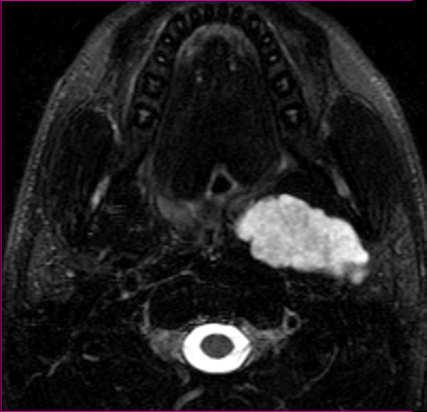
4 thyroid parotid adenopathy unknown primary pediatric thyroid parotid adenopathy unknown primary pediatric Thyroid mass Parotid mass U/S Excellent first line, FNA guidance MR & CT Good anatomic, limited morphologic info Extent of MNG, invasion of tissues Avoid iodine contrast if suspect malignancy FDG-PET Negative ¹³¹I scan + TG, NHL, Hürthle Inflammatory disease Calculi, symmetrical changes Neoplasms May be invisible on CT Need MRI for PNT Benign and malignant may be FDG-avid TMJ masses CT or MRI CECT Acinic cell ca *Parotid masses may be occult on CT thyroid parotid adenopathy unknown primary pediatric Deep Parotid lobe Oropharyngeal pleomorphic deep lobe adenoma mass *MR allows better characterization of parotid masses 55yM. Palpable left neck mass T1 MR T2 MR 4


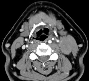
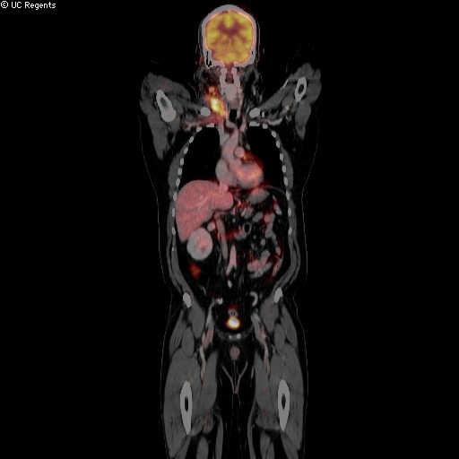
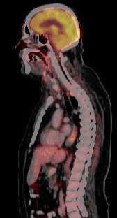
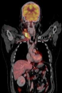
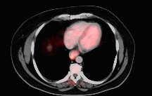
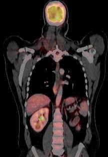
5 thyroid parotid adenopathy unknown primary pediatric thyroid parotid adenopathy unknown primary pediatric Parotid mass Adenopathy MRI preferred imaging method Localize and characterize Perineural & deep extension CT for inflammatory disease U/S may help to localize, guide FNA No role for FDG-PET in assessing mass CT and MR probably equivalent for detection of neoplastic nodes Easier with MR to see RPN U/S difficult for evaluating entire neck for nodes (or RPN) Excellent for suspicious node, guiding FNA FDG-PET not taken up by cystic nodes thyroid parotid adenopathy unknown primary Unknown Primary pediatric 59yM. Left neck mass CECT often first line study PET/CT used if primary not seen on CECT We prefer MRI Better soft tissue characterization Unless adenopathy is supraclavicular No real role for U/S 5


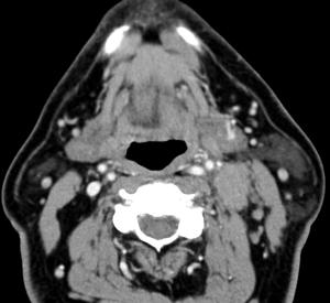
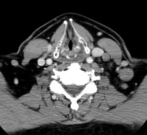

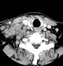
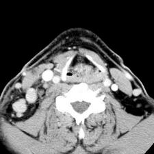

6 63yM. Left neck masses FNA = SCCa 55yM. Right neck masses 6

 Branchial")
 85% Second")
")
 *MR")


7 Congenital Pediatric Neck Mass Thyroglossal duct cyst (53%) Branchial cleft fistula /cyst (22%) 85% Second Dermoid cyst (11%) Hemangioma (7%) Venous /lymphatic malformation (6%) *MR allows better evaluation T2 FS MR of extent T1+C FS and MR characterization of mass Al-Khateeb TH. J Oral Maxillofacial Surg 2007;65: T1 MR T2 FS MR T1+C FS MR Pediatric Neck Mass Image gently Second branchial cleft fistula Avoid ionizing radiation when possible U/S, MRI as first line whenever possible Minimize radiation dose when CT is necessary 11 wk old with right neck dimple draining mucoid material 7
8 Which test? Summary Indications Thyroid mass Parotid mass Adenopathy Unknown primary Pediatric Modalities Ultrasound NECT or MRI if suspect malignancy MRI CECT MRI, PET/CT Acute infections CECT Congenital malf MRI Superficial -?U/S When in doubt start with CECT? Thyroid mass U/S, NECT, MR Pediatric case U/S, MR, unless urgent clinical problem U/S may be excellent additional tool FNA guidance PET/CT largely reserved for evaluation of known malignant (metastatic) disease 8
Evaluation of Neck Mass. Disclosure. Learning Objectives 3/24/2014. Karen T. Pitman MD, FACS Banner MDACC, Gilbert AZ. Nothing to disclose
 Evaluation of Neck Mass Karen T. Pitman MD, FACS Banner MDACC, Gilbert AZ Nothing to disclose Disclosure Learning Objectives 1. Describe a systematic method to evaluate a patient with a neck mass 2. Select
Thyroid Nodule. Disclosure. Learning Objectives P A P A P A 3/18/2014. Nothing to disclose.
 Thyroid Nodule Evaluating the patient with a thyroid nodule and some management options. Miguel V. Valdez PA C Disclosure Nothing to disclose. Learning Objectives Examination of thyroid gland Options for
Neckmasses in infancy and childhood: Clinical and radiological classification and imaging approaches M. Mearadji
 Neckmasses in infancy and childhood: Clinical and radiological classification and imaging approaches M. Mearadji International Foundation for Pediatric Imaging Aid Introduction Neck masses are a frequent
Index. Surg Oncol Clin N Am 16 (2007) Note: Page numbers of article titles are in boldface type.
 Note: Page numbers of article titles are in boldface type.") Surg Oncol Clin N Am 16 (2007) 465 469 Index Note: Page numbers of article titles are in boldface type. A Adjuvant therapy, preoperative for gastric cancer, staging and, 339 B Breast cancer, metabolic
Surg Oncol Clin N Am 16 (2007) 465 469 Index Note: Page numbers of article titles are in boldface type. A Adjuvant therapy, preoperative for gastric cancer, staging and, 339 B Breast cancer, metabolic
Evaluation and Management of Thyroid Nodules. Nick Vernetti, MD, FACE Palm Medical Group Las Vegas, Nevada
 Evaluation and Management of Thyroid Nodules Nick Vernetti, MD, FACE Palm Medical Group Las Vegas, Nevada Disclosure Consulting Amgen Speaking Amgen Objectives Understand the significance of incidental
Radiology- Pathology Conference 4/29/2012. Lymph Nodes. John McGrath
 Radiology- Pathology Conference 4/29/2012 Lymph Nodes John McGrath 1 Presentation material is for education purposes only. All rights reserved. 2012 URMC Radiology Page 1 of 24 Case 1: 51 year-old male
PEDIATRICS WK 3 HEAD AND NECK ALISON WALLACE MD, PHD
 PEDIATRICS WK 3 HEAD AND NECK ALISON WALLACE MD, PHD Topics 1. Cervical lymphadenopathy 2. Lymphatic malformation 3. Thyroglossal duct cysts 4. Branchial cleft cysts 5. Thyroid masses CASE 1 Case 1 A 2
Neck Imaging Reporting and Data System: An Atlas of NI-RADS Categories for Head and Neck Cancer
 Neck Imaging Reporting and Data System: An Atlas of NI-RADS Categories for Head and Neck Cancer Bethany Cavazuti Patricia Hudgins Tanya Rath Char Branstetter Kristen Baugnon Amanda Corey Ashley Aiken Disclosures
Introduction to ENT Imaging. Disclosure. Objectives 3/18/2014. Barton F. Branstetter IV. No commercial relationships to disclose
 Introduction to ENT Imaging Barton F. Branstetter IV Chief of Neuroradiology University of Pittsburgh Medical Center University of Pittsburgh School of Medicine Disclosure No commercial relationships to
Thyroid nodules. Most thyroid nodules are benign
 Thyroid nodules Postgraduate Course in General Surgery Jessica E. Gosnell MD Assistant Professor March 22, 2011 Most thyroid nodules are benign thyroid nodules occur in 77% of the world s population palpable
Sonographic Features of Thyroid Nodules & Guidelines for Management
 Sonographic Features of Thyroid Nodules & Guidelines for Management Mark A. Lupo, MD, FACE, ECNU Thyroid & Endocrine Center of Florida Assistant Clinical Professor of Medicine Florida State University,
Contents. Basic Ultrasound Principles and Terminology. Ultrasound Nodule Characteristics
 Contents Basic Ultrasound Principles and Terminology Basic Ultrasound Principles... 1 Ultrasound System... 2 Linear Transducer for Superficial Images and Ultrasound-Guided FNA... 3 Scanning Planes... 4
Imaging in Pediatric Thyroid disorders: US and Radionuclide imaging. Deepa R Biyyam, MD Attending Pediatric Radiologist
 Imaging in Pediatric Thyroid disorders: US and Radionuclide imaging Deepa R Biyyam, MD Attending Pediatric Radiologist Imaging in Pediatric Thyroid disorders: Imaging modalities Outline ACR-SNM-SPR guidelines
Thyroid Ultrasound for the Endocrine Surgeon: A Valuable Clinical Tool that Enhances Diagnostic and Therapeutic Outcomes
 Thyroid Ultrasound for the Endocrine Surgeon: A Valuable Clinical Tool that Enhances Diagnostic and Therapeutic Outcomes Allan Siperstein MD The Cleveland Clinic Audience Quiz Taken ultrasound course Perform
PAPILLARY THYROID CARCINOMA PRESENTING AS A LATERAL NECK MASS MASS. Dr. Pamela Hanson DO PGY3
 PAPILLARY THYROID CARCINOMA PRESENTING AS A LATERAL NECK MASS MASS Dr. Pamela Hanson DO PGY3 MK CASE PRESENTATION 28 yo Female presented to the ENT Clinic in October 2016, with the complaint of chronic
LYMPHATIC DRAINAGE IN THE HEAD & NECK
 LYMPHATIC DRAINAGE IN THE HEAD & NECK Like other parts of the body, the head and neck contains lymph nodes (commonly called glands). Which form part of the overall Lymphatic Drainage system of the body.
objectives Pitfalls and Pearls in PET/CT imaging Kevin Robinson, DO Assistant Professor Department of Radiology Michigan State University
 objectives Pitfalls and Pearls in PET/CT imaging Kevin Robinson, DO Assistant Professor Department of Radiology Michigan State University To determine the regions of physiologic activity To understand
Osman Ilkay Ozdamar, 1 Gul Ozbilen Acar, 1 Cigdem Kafkasli, 1 M. Tayyar Kalcioglu, 1 Tulay Zenginkinet, 2 and H. Gonca Tamer 3. 1.
 Case Reports in Otolaryngology Volume 2015, Article ID 79658, 4 pages http://dx.doi.org/10.1155/2015/79658 Case Report Papillary Thyroid Microcarcinoma with a Large Cystic Dilated Lymph Node Metastasis
PARATHYROID NUCLEAR MEDICINE IMAGING REVIEW DISCLOSURES
 PARATHYROID NUCLEAR MEDICINE IMAGING REVIEW Miguel Hernandez Pampaloni, M.D., Ph.D. Chief, Nuclear Medicine Assistant Professor of Radiology UCSF Department of Radiology and Biomedical Imaging DISCLOSURES
Women s Health in General Practice Symposium 2015 Thyroid & Parathyroid Cases
 Women s Health in General Practice Symposium 2015 Thyroid & Parathyroid Cases Bill Fleming Epworth Freemasons Hospital 1 Common Endocrine Presentations anatomical problems thyroid nodule / goitre embryological
Ultrasound Interpretation of Non-Thyroid Neck Pathology
 Ultrasound Interpretation of Non-Thyroid Neck Pathology Kevin T. Brumund, M.D., F.A.C.S. Associate Professor of Surgery Head and Neck Surgery University of California, San Diego Health Sciences VA Medical
Cystic Head and Neck Lesions
 Cystic Head and Neck Lesions Disclosures None Brad Wright, MD 19 March 2018 Key points Huge variety of cystic lesions in H&N May be cystic, necrotic, or solid but cystic-appearing Patient age, clinical
Thyroid Ultrasonography: clinical and radiological correlations
 Thyroid Ultrasonography: clinical and radiological correlations Dr.M.Thijs Radiology Anatomy Inflammatory Thyroid Disease Benign lesions Thyroid tumors Thyroglossal duct cyst Anatomy Transverse Longitudinal
DISORDERS OF THE SALIVARY GLANDS Neoplasms Dr.M.Baskaran Selvapathy S IV
 DISORDERS OF THE SALIVARY GLANDS Neoplasms Dr.M.Baskaran Selvapathy S IV NEOPLASMS A) Epithelial I. Benign Pleomorphic adenoma( Mixed tumour) Adenolymphoma (Warthin s tumour) Oxyphil adenoma (Oncocytoma)
performed to help sway the clinician in what the appropriate diagnosis is, which can substantially alter the treatment of management.
 Hello, I am Maura Polansky at the University of Texas MD Anderson Cancer Center. I am a Physician Assistant in the Department of Gastrointestinal Medical Oncology and the Program Director for Physician
Thyroid Nodules. Family Medicine Refresher Course Geeta Lal MD, FACS April 2, No financial disclosures
 Thyroid Nodules Family Medicine Refresher Course Geeta Lal MD, FACS April 2, 2014 No financial disclosures Objectives Review epidemiology Work up of Thyroid nodules Indications for FNAB Evolving role of
Thyroid nodules 3/22/2011. Most thyroid nodules are benign. Thyroid nodules: differential diagnosis
 Most thyroid nodules are benign Thyroid nodules Postgraduate Course in General Surgery thyroid nodules occur in 77% of the world s population palpable thyroid nodules occur in about 5% of women and 1%
5/18/2013. Most thyroid nodules are benign. Thyroid nodules: new techniques in evaluation
 Most thyroid nodules are benign Thyroid nodules: new techniques in evaluation Incidence Etiology Risk factors Diagnosis Gene classification system Treatment Postgraduate Course in General Surgery Jessica
How good are we at finding nodules? Thyroid Nodules Thyroid Cancer Epidemiology Initial management Long-term follow up Disease-free status
 New Perspectives in Thyroid Cancer Jennifer Sipos, MD Assistant Professor of Medicine Division of Endocrinology The Ohio State University Outline Thyroid Nodules Thyroid Cancer Epidemiology Initial management
Neck mass Evaluation & Management OTOLARYNGOLOGY, HEAD & NECK SURGICAL ONCOLOGY
 Neck mass Evaluation & Management MOHAMMED ALESSA MBBS,FRCSC ASSISTANT PROFESSOR CONSULTANT OTOLARYNGOLOGY, HEAD & NECK SURGICAL ONCOLOGY KSU, MEDICAL CITY & KKUH Objectives Obtain map overview in neck
USGFNA of thyroid nodules
 US Guided FNA (USGFNA) of neck masses INTERVENTIONAL HEAD & NECK ULTRASOUND Brendan C. Stack, Jr., MD., FACS, FACE Professor Otolaryngology-Head and Neck Surgery Indications Technique Interpretation Results
Clinical indications for positron emission tomography
 Clinical indications for positron emission tomography Oncology applications Brain and spinal cord Parotid Suspected tumour recurrence when anatomical imaging is difficult or equivocal and management will
Unknown Cases from the Participants
 Unknown Cases from the Participants Case 1: 1 Case 1: Case 1: DDX? Answer on next slide Case 1: MS V5 Neuropathy Case 2: Case 2: 76 year old woman Ultrasound for multinodular goiter finds suspicious nodule
Thyroid Nodules. Dr. HAKIMI, SpAK Dr. MELDA DELIANA, SpAK Dr. SISKA MAYASARI LUBIS, SpA
 Thyroid Nodules ENDOCRINOLOGY DIVISION ENDOCRINOLOGY DIVISION Dr. HAKIMI, SpAK Dr. MELDA DELIANA, SpAK Dr. SISKA MAYASARI LUBIS, SpA Anatomical Considerations The Thyroid Nodule Congenital anomalies Thyroglossal
HEAD AND NECK ENDOCRINE SURGERY
 HEAD AND NECK ENDOCRINE SURGERY OCTOBER 22-23, 2010 THE MARK HOPKINS SAN FRANCISCO, CA THYROID FNA AND CYTOPATHOLOGY THEODORE R. MILLER, MD THE THYROID NODULE Prevalence of palpable nodule: Female ~ 6%
Salivary Gland Imaging. Mary Scanlon MD FACR October 2016
 Salivary Gland Imaging Mary Scanlon MD FACR October 2016 Objectives Recognize normal and abnormal anatomy Discuss work up, management and differential diagnosis of commonly referred clinical scenarios
Fusion Ultrasound: Characterization of Abdominal Masses with MR, CT, PET, and Contrast Ultrasound
 Fusion Ultrasound: Characterization of Abdominal Masses with MR, CT, PET, and Contrast Ultrasound Mollie Rashid, MD Corinne Deurdulian, MD Hisham Tchelepi, MD Keck School of Medicine, University of Southern
Evaluation of Head and Neck Masses in Adults
 Evaluation of Head and Neck Masses in Adults Kristi Chang, MD Associate Professor Department of Otolaryngology-Head and Neck Surgery University of Iowa Hospitals and Clinics Annual Refresher Course for
NI-RADS: Head & Neck Cancer Imaging Surveillance. Goals of the RADS templates: Goals: Disclosures ACR NI-RADS
 NI-RADS: Head & Neck Cancer Imaging Surveillance ASHNR 2017 none Disclosures Ashley H. Aiken, M.D. Associate Professor of Radiology Emory University School of Medicine ACR RADS (Reporting and Data Systems)
Dr Nick McIvor. Dr John Chaplin. Head & Neck Surgeon Auckland City Hospital Auckland. Auckland Head & Neck Surgeon Gillies Hospital Auckland
 Dr Nick McIvor Head & Neck Surgeon Auckland City Hospital Auckland Dr John Chaplin Auckland Head & Neck Surgeon Gillies Hospital Auckland 14:00-14:55 WS #148: Case Studies of Lumps in the Neck 15:05-16:00
PARATHYROID IMAGING. James Lee, MD Chief, Endocrine Surgery Co-Director NY Thyroid-Parathyroid Center Columbia University Medical Center
 PARATHYROID IMAGING James Lee, MD Chief, Endocrine Surgery Co-Director NY Thyroid-Parathyroid Center Columbia University Medical Center NO DISCLOSURES Overview The hallmarks of the ideal test Benefits
FDG PET/CT STAGING OF LUNG CANCER. Dr Shakher Ramdave
 FDG PET/CT STAGING OF LUNG CANCER Dr Shakher Ramdave FDG PET/CT STAGING OF LUNG CANCER FDG PET/CT is used in all patients with lung cancer who are considered for curative treatment to exclude occult disease.
42 yr old male with h/o Graves disease and prior I 131 treatment presents with hyperthyroidism and undetectable TSH. 2 hr uptake 20%, 24 hr uptake 50%
 Pinhole images of the neck are acquired in multiple projections, 24hrs after the oral administration of approximately 200 µci of I123. Usually, 24hr uptake value if also calculated (normal 24 hr uptake
MEASUREMENT OF EFFECT SOLID TUMOR EXAMPLES
 MEASUREMENT OF EFFECT SOLID TUMOR EXAMPLES Although response is not the primary endpoint of this trial, subjects with measurable disease will be assessed by standard criteria. For the purposes of this
Disclosures. Diffusion and Perfusion Imaging in the Head and Neck. Learning objectives ???
 Disclosures No relevant financial disclosures Diffusion and Perfusion Imaging in the Head and Neck Ashok Srinivasan, MD Associate Professor Director of Neuroradiology University of Michigan Health System
ACOS Inquiry and Response Selected Inquires CS Tumor Size/Extension Evaluation, CS Lymph Nodes Evaluation, CS Metastasis at Diagnosis Evaluation *
 ACOS Inquiry and Response Selected Inquires CS Tumor Size/Extension Evaluation, CS Lymph Nodes Evaluation, CS Metastasis at Diagnosis Evaluation * CS Tumor Size/Extension Evaluation 24842 12/11/2007: Q:
Enterprise Interest None
 Enterprise Interest None Risk stratification of salivary gland lesions on cytology based on the proposed Milan System for reporting salivary gland cytopathology: A pilot study Kartik Viswanathan, M.D.,
Preoperative Evaluation
 Preoperative Evaluation Lateral compartment lymph nodes are easier to detect and are amenable to FNA Central compartment lymph nodes are much more difficult to detect and FNA (Tg washout testing is compromised)
Tania Gallant MD, FRCPC Internal Medicine Update April
 Tania Gallant MD, FRCPC Internal Medicine Update April 28 2017 Disclosures Honoraria/Ad board: Sanofi-Aventis, Janssen, Merck Frosst, Eli-Lilly, Astra Zeneca, Boehringer-Ingelheim Objectives By the end
Effective Utilization of Imaging. John V. Roberts, M.D. Premier Radiology Abdominal Imaging
 Effective Utilization of Imaging John V. Roberts, M.D. Premier Radiology Abdominal Imaging Safety Contrast and Radiation What to order Abdomen/Pelvis Brain/Spine Chest Musculoskeletal Ob/Gyn Head and Neck
Esophageal Cancer: A Multimodality Approach to Detection and Staging
 Esophageal Cancer: A Multimodality Approach to Detection and Staging, MBA Topic: Esophageal Cancer Imaging: A multimodality approach Conference: Society of Thoracic Imaging Location: Date/Time: March 14,
The International Federation of Head and Neck Oncologic Societies. Current Concepts in Head and Neck Surgery and Oncology
 The International Federation of Head and Neck Oncologic Societies Current Concepts in Head and Neck Surgery and Oncology www.ifhnos.net The International Federation of Head and Neck Oncologic Societies
Disclosures. Learning objectives. Case 1A. Autoimmune Thyroid Disease: Medical and Surgical Issues. I have nothing to disclose.
 Disclosures Autoimmune Thyroid Disease: Medical and Surgical Issues I have nothing to disclose. Chrysoula Dosiou, MD, MS Clinical Assistant Professor Division of Endocrinology Stanford University School
Dr Sneha Shah Tata Memorial Hospital, Mumbai.
 Dr Sneha Shah Tata Memorial Hospital, Mumbai. Topics covered Lymphomas including Burkitts Pediatric solid tumors (non CNS) Musculoskeletal Ewings & osteosarcoma. Neuroblastomas Nasopharyngeal carcinomas
Disclosure. Acknowledgement. What is the Best Workup for Rectal Cancer Staging: US/MRI/PET? Rectal cancer imaging. None
 What is the Best Workup for Rectal Cancer Staging: US/MRI/PET? Zhen Jane Wang, MD Assistant Professor in Residence UC SF Department of Radiology Disclosure None Acknowledgement Hueylan Chern, MD, Department
5/3/2017. Ahn et al N Engl J Med 2014; 371
 Alan Failor, M.D. Clinical Professor of Medicine Division of Metabolism, Endocrinology and Nutrition University of Washington April 20, 2017 No disclosures to report 1. Appropriately evaluate s in adult
Dr Claire Smith, Consultant Radiologist St James University Hospital Leeds
 Dr Claire Smith, Consultant Radiologist St James University Hospital Leeds Imaging in jaundice and 2ww pathway Image protocol Staging Limitations Pancreatic cancer 1.2.4 Refer people using a suspected
STAGING AND FOLLOW-UP STRATEGIES
 ATHENS 4-6 October 2018 European Society of Urogenital Radiology STAGING AND FOLLOW-UP STRATEGIES Ahmet Tuncay Turgut, MD Professor of Radiology Hacettepe University, Faculty of Medicine Ankara 2nd ESUR
THYROID CANCER IN CHILDREN. Humberto Lugo-Vicente MD FACS FAAP Professor Pediatric Surgery UPR School of Medicine
 THYROID CANCER IN CHILDREN Humberto Lugo-Vicente MD FACS FAAP Professor Pediatric Surgery UPR School of Medicine Thyroid nodules Rare Female predominance 4-fold as likely to be malignant Hx Radiation exposure?
WTC 2013 Panel Discussion: Minimal disease
 WTC 2013 Panel Discussion: Minimal disease Susan J. Mandel MD MPH Panelists Ken Ain Yasuhiro Ito Stephanie Lee Erich Sturgis Mark Urken Faculty/Presenter Disclosure Relationships with commercial interests
Shadow because the air
 Thyroid Ultrasound Thyroid US examination needs: 1. high frequency transducer 2. extended patient's neck 3. check all the neck area because the swelling could be in areas other than the thyroid such as
Case Scenario 1 Worksheet. Primary Site C44.4 Morphology 8743/3 Laterality 0 Stage/ Prognostic Factors
 CASE SCENARIO 1 9/10/13 HISTORY: Patient is a 67-year-old white male and presents with lesion located 4-5cm above his right ear. The lesion has been present for years. No lymphadenopathy. 9/10/13 anterior
PET/CT Frequently Asked Questions
 PET/CT Frequently Asked Questions General Q: Is FDG PET specific for cancer? A: No, it is a marker of metabolism. In general, any disease that causes increased metabolism can result in increased FDG uptake
Value of true whole-body FDG- PET/CT scanning protocol in oncology and optimization of its use based on primary malignancy
 Value of true whole-body FDG- PET/CT scanning protocol in oncology and optimization of its use based on primary malignancy Ronnie Sebro MD, Ph.D Carina Mari Aparici MD, Miguel Hernandez Pampaloni MD, PhD
Zubair W. Baloch, MD, PhD: Consultant for Veracyyte, INC Tarik M. Elsheikh, MD: Nothing to disclose
 Cytology Works shop #8 Zubair W. Baloch, MD, PhD: Consultantt for Veracyte, INC Tarik M. Elsheik kh, MD: Nothing to disclose Controversies and Diagnostic Challenges in Head and Neck Cytopathology Zubair
Clinical summary. Male 30 year-old with past history of non-seminomous germ cell tumour. Presents with retroperitoneal lymphadenopathy on CT.
 Clinical summary Male 30 year-old with past history of non-seminomous germ cell tumour. Presents with retroperitoneal lymphadenopathy on CT. For restaging PET/CT. PET/CT findings No significant FDG uptake
NI-RADS: Structured Reporting for Head and Neck Cancer
 NI-RADS: Structured Reporting for Head and Neck Cancer ASHNR 2018 Disclosures Part of group that initiated NI-RADS Colleagues A. Aiken & P. Hudgins: ACR NIRADS committee members Kristen Lloyd Baugnon,
Thyroid nodules - medical and surgical management. Endocrinology and Endocrine Surgery Manchester Royal Infirmary
 Thyroid nodules - medical and surgical management JRE Davis NR Parrott Endocrinology and Endocrine Surgery Manchester Royal Infirmary Thyroid nodules - prevalence Thyroid nodules common, increase with
Imaging: When to get MRI, CT or PET-CT?
 Imaging: When to get MRI, CT or PET-CT? Alina Uzelac, D.O. Assistant Clinical Professor Neuroradiology UCSF Department of Radiology and Biomedical Imaging San Francisco General Hospital Overview CT MRI
"Mummy what's this on my neck? - A pictorial review of paediatric neck masses"
 "Mummy what's this on my neck? - A pictorial review of paediatric neck masses" Poster No.: C-0405 Congress: ECR 2014 Type: Educational Exhibit Authors: A. Farrugia, A. S. Gatt; Msida/MT Keywords: Education,
AACE/ACE Principles of Endocrine Neck Sonography Course
 AACE/ACE Principles of Endocrine Neck Sonography Course Primary objective of thyroid ultrasound: assess for malignant disease Nodular Disease Benign Malignant Goiter Iodine deficient Thyroiditis Organification
Ultrasound of soft-tissue vascular anomalies
 Ultrasound of soft-tissue vascular anomalies Oscar M. Navarro Associate Professor, University of Toronto Dept. of Diagnostic Imaging, The Hospital for Sick Children Toronto, Canada Declaration of Disclosure
Endobronchial Ultrasound in the Diagnosis & Staging of Lung Cancer
 Endobronchial Ultrasound in the Diagnosis & Staging of Lung Cancer Dr Richard Booton PhD FRCP Lead Lung Cancer Clinician, Consultant Respiratory Physician & Speciality Director Manchester University NHS
Thyroid US. Background: Thyroid/Neck US. Use of Office Ultrasound in the Thyroid Surgery Practice
 2010 UCSF Head and Neck Endocrine Surgery Course Use of Office Ultrasound in the Thyroid Surgery Practice Lisa A. Orloff, MD FACS Dept of Otolaryngology-Head and Neck Surgery University of California,
Study of validity of ultrasonographic diagnosis in relation to Fine Needle Aspiration Cytology (FNAC) diagnosis
 diagnosis") Original article: Study of validity of ultrasonographic diagnosis in relation to Fine Needle Aspiration Cytology (FNAC) diagnosis *Dr Rajvi Matalia, ** Dr Y.P.Sachdev, ***Dr D.S.Kulkarni *Junior Resident,
Original article: Study of validity of ultrasonographic diagnosis in relation to Fine Needle Aspiration Cytology (FNAC) diagnosis *Dr Rajvi Matalia, ** Dr Y.P.Sachdev, ***Dr D.S.Kulkarni *Junior Resident,
THE ROLE OF CONTEMPORARY IMAGING AND HYBRID METHODS IN THE DIAGNOSIS OF CUTANEOUS MALIGNANT MELANOMA(CMM) AND MERKEL CELL CARCINOMA (MCC)
 AND MERKEL CELL CARCINOMA (MCC)") THE ROLE OF CONTEMPORARY IMAGING AND HYBRID METHODS IN THE DIAGNOSIS OF CUTANEOUS MALIGNANT MELANOMA(CMM) AND MERKEL CELL CARCINOMA (MCC) I.Kostadinova, Sofia, Bulgaria CMM some clinical facts The incidence
THE ROLE OF CONTEMPORARY IMAGING AND HYBRID METHODS IN THE DIAGNOSIS OF CUTANEOUS MALIGNANT MELANOMA(CMM) AND MERKEL CELL CARCINOMA (MCC) I.Kostadinova, Sofia, Bulgaria CMM some clinical facts The incidence
Sonography of soft-tissue vascular lesions
 Sonography of soft-tissue vascular lesions Oscar M. Navarro Associate Professor, University of Toronto Dept. of Diagnostic Imaging, The Hospital for Sick Children Toronto, Canada Declaration of Disclosure
Imaging of liver and pancreas
 Imaging of liver and pancreas.. Disease of the liver Focal liver disease Diffusion liver disease Focal liver disease Benign Cyst Abscess Hemangioma FNH Hepatic adenoma HCC Malignant Fibrolamellar carcinoma
AACE Thyroid Cancer Tumor board 25 years of the Endocrine and Surgery collaboration
 AACE Thyroid Cancer Tumor board 25 years of the Endocrine and Surgery collaboration Dr. Peter Singer, Endocrinology Dr. Peter Sadow, Pathology Moderator Dr. Greg Randolph, Otolaryngology Relevant Financial
Parotid Disease Case Discussions. Valerie Jefford November 28, 2002
 Parotid Disease Case Discussions Valerie Jefford November 28, 2002 Case 1 44 y.o. man referred with lump anterior to R ear. Q1 What do you want to know? no pain 2 years but bigger now Smoker Q2 What to
Objectives. Salivary Gland FNA: The Milan System. Role of Salivary Gland FNA 04/26/2018
 Salivary Gland FNA: The Milan System Dr. Jennifer Brainard Section Head Cytopathology Cleveland Clinic Objectives Introduce the Milan System for reporting salivary gland cytopathology Define cytologic
Management of Thyroid Nodules
 Management of Thyroid Nodules 38 y/o female with solid 1.5 cm right Thyroid nodule. TSH=0.68 Vincent J. Reid, MD., FACS Thyroid Cancer Incidence & Mortality 1974 to 2004 Overall Women Men Mortality 1 Cancer
Management of Salivary Gland Malignancies. No Disclosures or Conflicts of Interest. Anatomy 10/4/2013
 Management of Salivary Gland Malignancies Daniel G. Deschler, MD Director: Division of Head and Neck Surgery Massachusetts Eye & Ear Infirmary Massachusetts General Hospital Professor Harvard Medical School
Pre-operative Ultrasound of Lymph Nodes in Thyroid Cancer
 Pre-operative Ultrasound of Lymph Nodes in Thyroid Cancer AACE - Advances in Medical and Surgical Management of Thyroid Cancer - 2018 Robert A. Levine, MD, FACE, ECNU Thyroid Center of New Hampshire Geisel
Update on RECIST and Staging of Common Pediatric Tumors Ethan A. Smith, MD
 Update on RECIST and Staging of Common Pediatric Tumors Ethan A. Smith, MD Section of Pediatric Radiology C.S. Mott Children s Hospital University of Michigan ethans@med.umich.edu Disclosures No relevant
PEDIATRIC Ariel Katz MD
 PEDIATRIC Ariel Katz MD Dept. Otolaryngology Head &Neck Surgery Wolfson Medical Center Holon, Israel OBJECTIVES Overview/Background Epidemiology/Etiology Intro to Guidelines Workup Treatment Follow-Up
Workshop 2. Controversies and Diagnostic Challenges in Head and Neck Cytopathology. Zubair Baloch, MD,PhD. Veracyte, Inc: Consultant
 Workshop 2 Controversies and Diagnostic Challenges in Head and Neck Cytopathology Zubair Baloch, MD,PhD Veracyte, Inc: Consultant Tarik Elsheikh, MD There are no disclosures necessary. Controversies and
Hybrid Imaging SPECT/CT PET/CT PET/MRI. SNMMI Southwest Chapter Aaron C. Jessop, MD
 Hybrid Imaging SPECT/CT PET/CT PET/MRI SNMMI Southwest Chapter 2014 Aaron C. Jessop, MD Assistant Professor, Department of Nuclear Medicine UT MD Anderson Cancer Center, Houston, Texas Complimentary role
Michigan AACE: Case Presentation
 Michigan AACE: Case Presentation Marco De Santis D.O. Endocrinology Fellow McLaren Medical Center Macomb Background O 83 year old female with fatigue and TSH of 0.13 O Medical history T2DM, Osteoporosis,
Salivary Glands tumors
 Salivary Glands tumors Sal.Gl. 1 Salivary Glands tumors Work-up procedure TNM staging Primary treatment Follow-up Treatment of recurrent and/or metastatic disease References Sal.Gl. 2 Standard clinical
AACE/ACE Advanced Endocrine Neck Ultrasound Training Course 2016
 AACE/ACE Advanced Endocrine Neck Ultrasound Training Course 2016 This 9mm left inferior nodule should remind us all why we re here! There is no absolute number of images required for documentation
Neuroradiology Case of the Day
 Neuroradiology Case of the Day 76 th CAR Annual Meeting, Montreal, Quebec April 27, 2013 Eugene Yu, MD Assistant Professor of Radiology and Otolaryngology-Head and Neck Surgery Head and Neck Imaging Princess
Ultrasound imaging of vascular anomalies: pearls and pitfalls
 Ultrasound imaging of vascular anomalies: pearls and pitfalls Oscar Navarro, MD Dept. of Medical Imaging, University of Toronto Dept. of Diagnostic Imaging, The Hospital for Sick Children Declaration of
Liver Cancer (Hepatocellular Carcinoma or HCC) Overview
 Overview") Liver Cancer (Hepatocellular Carcinoma or HCC) Overview Recent advances in liver cancer care seek to address the rising incidence of liver cancer, which has steadily increased over the past three decades.
Liver Cancer (Hepatocellular Carcinoma or HCC) Overview Recent advances in liver cancer care seek to address the rising incidence of liver cancer, which has steadily increased over the past three decades.
J of Evolution of Med and Dent Sci/ eissn , pissn / Vol. 3/ Issue 09/ Mar 3, 2014 Page 2087
 ACCURACY OF FINE NEEDLE ASPIRATION CYTOLOGY IN THE DIAGNOSIS OF NECK MASSES Mudassar Ahmed Shariff 1, Srinivasa V 2, Elangovan S 3, Manikandan D 4, Jarvis Raju 5 HOW TO CITE THIS ARTICLE: Mudassar Ahmed
4/22/2010. Hakan Korkmaz, MD Assoc. Prof. of Otolaryngology Ankara Dıșkapı Training Hospital-Turkey.
 Management of Differentiated Thyroid Cancer: Head Neck Surgeon Perspective Hakan Korkmaz, MD Assoc. Prof. of Otolaryngology Ankara Dıșkapı Training Hospital-Turkey Thyroid gland Small endocrine gland:
Ultrasonography of the Neck as an Adjunct to FNA. Nicole Massoll M.D.
 Ultrasonography of the Neck as an Adjunct to FNA Nicole Massoll M.D. Basic Features of Head and Neck Ultrasound and Anatomy Nicole Massoll M.D. University of Arkansas for Medical Sciences, Little Rock
8/3/2016. Consultant for / research support from: Astellas Bayer Bracco GE Healthcare Guerbet Medrad Siemens Healthcare. Single Energy.
 U. Joseph Schoepf, MD Prof. (h.c.), FAHA, FSCBT-MR, FNASCI, FSCCT Professor of Radiology, Medicine, and Pediatrics Director, Division of Cardiovascular Imaging Consultant for / research support from: Astellas
Disclosure of Relevant Financial Relationships
 Squamous entities of the thyroid: Reactive to Neoplastic Michelle D. Williams Associate Professor Dept of Pathology, Head & Neck Section University of Texas MD Anderson Cancer Center Disclosure of Relevant
Radiology Pathology Conference
 Radiology Pathology Conference Sharlin Johnykutty,, MD, Cytopathology Fellow Sara Majewski, MD, Radiology Resident Friday, August 28, 2009 Presentation material is for education purposes only. All rights