Optical biopsy of early gastroesophageal cancer by catheter-based reflectance-type laser-scanning confocal microscopy
|
|
|
- Ada Booker
- 5 years ago
- Views:
Transcription
1 Optical biopsy of early gastroesophageal cancer by catheter-based reflectance-type laser-scanning confocal microscopy Madoka Nakao, M.D. 1, Shigeto Yoshida, M.D. 2, Shinji Tanaka, M.D. 2, Yoshito Takemura, M.D. 1, Shiro Oka, M.D. 2, Masaharu Yoshihara, M.D. 3, Kazuaki Chayama, M.D. 1 1 Department of Medicine and Molecular Science, Division of Frontier Medical Science, Programs for Biomedical Research, Graduate School of Biomedical Sciences, Hiroshima University, Hiroshima, Japan 2 Department of Endoscopy, Hiroshima University Hospital, Hiroshima, Japan 3 Department of Health Service Center, Hiroshima University, Hiroshima, Japan Address for correspondence: Shigeto Yoshida, M.D., PhD Department of Endoscopy, Hiroshima University Hospital, Kasumi, Minami-ku, Hiroshima, , Japan Telephone: Facsimile: yoshida7@hiroshima-u.ac.jp 1
2 Abstract Magnified endoscopic observation of the gastrointestinal tract has become possible. However, such observation at the cellular level remains difficult. Laser-scanning confocal microscopy (LCM) is a novel, noninvasive optical imaging method that provides instant microscopic images of untreated tissue under endoscopy. We compared prototype catheter-based reflectance-type LCM images in vivo and histologic images of early gastroesophageal cancer to assess the usefulness of LCM in diagnosing such cancer. Twenty sites in the esophagus and 40 sites in the stomach were examined by LCM under endoscopy prior to endoscopic or surgical resection. A prototype catheter LCM system, equipped with a semiconductor laser that oscillates at 685 nm and analyzes reflected light (Mauna Kea Technologies, Paris, France; Fujinon, Saitama, Japan), was used in vivo without fluorescent agent. In all normal esophageal mucosa and esophageal cancers, the nuclei were visualized. In 9 of the 10 normal esophageal mucosa, cell membranes were visualized, and in 5 of the 10 esophageal cancers, cell membranes were visualized. In all normal gastric mucosa, nuclei and cell membranes were not visualized, but in 10 of the 20 gastric cancers, nuclei were visualized. This novel method will aid in immediate diagnosis under endoscopy without the need for biopsy. 2
3 Introduction Detailed endoscopic observation of the esophagus, stomach, and colon has become possible due to advances in magnifying endoscopy and conventional endoscopy. 1,2 However, magnified observation at the cellular level remains difficult under endoscopic examination, thus often making histopathologic examination via biopsy a necessity. Laser-scanning confocal microscopy (LCM) provides in vivo images that are close in quality to histopathologic images. This technology is currently being applied clinically in the field of gastroenterology. Most reports are of fluorescence-type LCM, which require a fluorescent agent Reflectance-type LCM, which does not need fluorescence, is in the investigational stage, and most reports are of in vitro studies. 12 We compared in vivo LCM images and histologic images of early gastroesophageal cancer and normal mucosa to assess the usefulness of a newly developed prototype catheter-based reflectance-type LCM system (Mauna Kea Technologies, Paris, France; Fujinon, Saitama, Japan) for diagnosing gastroesophageal cancer. Materials and Methods Instrument specifications We used a prototype LCM system equipped with a pulsed semiconductor laser centered at 685 nm. The combination of pulsed illumination (15 ns pulse width, 80 ns repetition 3
4 period) with a time-gated detection of the reflected light (15 ns detection window, 40 ns delay) permits to overcome the back-reflections onto the proximal optics by means of light travel-time differentiation. 13 The flexible catheter probe was 2.6 mm in outer diameter and 3876 mm long (Fig. 1). The scanning field was 30,000 pixels-the number of fibers in the bundle and the frame rate was 12 images per second (Fig. 2). An objective with a numerical aperture of 1.2 was placed in contact with the tissue, with a focus of 30 μm from the objective lens, a lateral resolution of less than 1μm, and an observation area 160 μm in diameter. The catheter probes were connected to the laser scanning unit and introduced under direct endoscopic visualization, LCM was performed after the flexible confocal catheter probe was introduced through the instrument channel of the endoscope (Fig. 3). All images of the LCM examinations were inspected, recorded, and stored digitally as real-time video sequences with the use of software on a PowerMac G5 Dual 1.8GHz personal computer (Apple Computer, Cupertino, CA, USA). In addition, still LCM images were saved for future review. Patients and comparison of LCM images Ten patients with esophageal cancer and 20 patients with gastric cancer underwent reflectance-type LCM examination after white-light endoscopic examination at Hiroshima University Hospital during the period April 2007 through July Clinical 4
5 characteristics of the lesions are presented in Table 1. The distal tip of the LCM catheter was placed gently against the mucosa, and an endoscopist captured LCM images of normal mucosa near the cancer and images of the cancer in the esophagus (normal mucosa, n = 10; cancer, n = 10) or stomach (normal mucosa, n = 20; cancer, n = 20). After endoscopic examination, a gastroenterologist (S.Y., who had analyzed LCM images previously) judged whether the cell nuclei and membranes were visible on the LCM images. 12 After these procedures, patients underwent endoscopic mucosal resection (EMR), endoscopic submucosal dissection (ESD), endoscopic aspiration mucosectomy (EAM), or surgery. The resected specimens were fixed in formalin, embedded in paraffin, sliced with a microtome, deparaffinized, and stained with hematoxylin-eosin for light microscopic examination. After these procedures, the histologic diagnosis was confirmed. The endoscopic system used in this study was a VP-4400 endoscope processor and an EG-590WR or EG-450D upper gastrointestinal endoscope (Fujinon). The study protocol conformed to the tenets of the Declaration of Helsinki and was approved by our institutional ethics committee. Results With the distal tip of the catheter placed gently against the mucosa and the cancer, real-time LCM images of the normal mucosa and of the cancer of the esophagus and 5
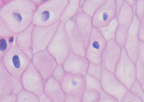
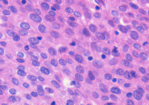
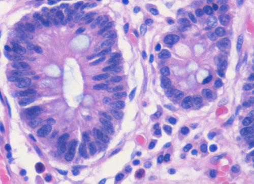
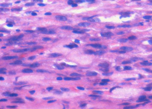
6 stomach were obtained safely and easily, and the influence of slight motion was ignore. Even few water or blood was existed in the normal mucosa and cancer, the influence of water or blood was also ignore. The time required for scanning each normal and cancer site ranged between 16 and 390 seconds. Normal esophageal mucosa In LCM images of normal esophageal mucosa, high-reflectivity spots were observed near the center of honeycomb-like structures of high reflectivity. These high reflectivity spots and structures in the LCM images appeared to correspond to nuclei and cell membranes, respectively, in the histologic images of hematoxylin-eosin-stained sections (Fig. 4). Esophageal cancer In LCM images of esophageal cancer, high-reflectivity spots that were considered nuclei were observed. The nucleus-to-cytoplasm (N/C) ratio was much increased, and honeycomb-like structures of high reflectivity, considered cell membranes, were not observed (Fig. 5). Normal gastric mucosa In LCM images of normal gastric mucosa, cell membranes and nuclei were not visualized. However, the crypt cells were arranged like flower petals surrounding the 6
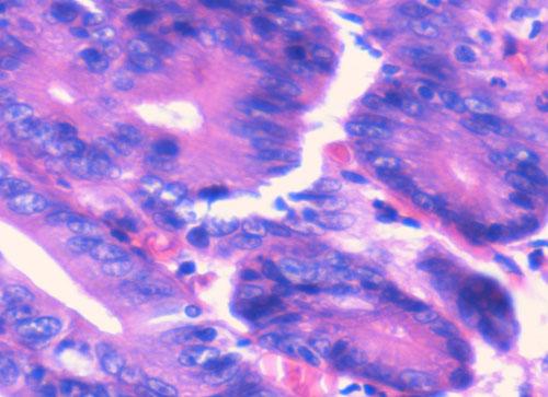
7 gastric pit (Fig. 6). Gastric cancer In LCM images of differentiated adenocarcinoma of the stomach, cell membranes were not visualized, and a disorganized configuration of glands with high-reflectivity spots that were considered nuclei was observed (Fig. 7). In LCM images of undifferentiated adenocarcinoma, no ductal structure was recognized; only a amorphous structure was seen. Cell membranes and nuclei were not visualized (Fig. 8). Visualization of nuclei and cell membranes in LCM images in relation to histologic diagnoses is shown in Table 2. In all normal esophageal mucosa and esophageal cancers, the nuclei were visualized. In 9 of the 10 (90%) normal esophageal mucosa, cell membranes were visualized, and in 5 of the 10 (50%) esophageal cancers, cell membranes were visualized. In all normal gastric mucosa, nuclei and cell membranes were not visualized, but in 10 of the 20 (50%) gastric cancers, nuclei were visualized. The storoma was visulized as high reflectivity. In some case, low-reflectivity spots were also observed in the LCM images which was considered mucin in goblet cells in the hematoxylin-eosin-stained specimen. Discussion Recent advances in endoscopic technology have afforded high-quality, detailed 7
8 diagnosis of gastrointestinal diseases. To confirm the presence of malignancy, however, snip biopsy is often performed under endoscopy when endoscopic examination reveals an abnormality. Thus, biopsy is performed for many lesions that are subsequently determined not be malignant. Histologic analysis of biopsy material remains the gold standard for the final diagnosis of a gastrointestinal lesion. Histologic diagnosis via biopsy involves the following process: formalin fixation of the specimen, cutting the specimen into small columns, paraffin embedding, ultra-thin slicing, deparaffinization, staining, glass slide, mounting, and finally light microscopic observation. Moreover, it takes several days to obtain a diagnosis. Also, snip biopsy is associated with bleeding, apparent endoscopic disappearance of cancer cells after biopsy, and artificial ulceration, which make endoscopic treatment, e.g. EMR, ESD, and EAM, difficult. In addition, because of the bleeding, biopsy cannot be easily performed in patients taking anticoagulants. Being able to accurately image a lesion in vivo at the time of endoscopic examination without biopsy allows for prompt diagnosis and treatment. Fluorescence-type LCM is reported to be a promising tool for in vivo histopathologic examination during endoscopy and might overcome the disadvantages associated with conventional biopsy In recent years, there have been several reports describing the ability to 8
9 obtain an LCM image that corresponds precisely to the histopathologic tissue diagnosis in cases of gastrointestinal tract disease However, many of the reports were based on observations made on excised specimens or with fluorescence-type LCM. We too have previously used probe-based reflectance-type LCM to obtain images that are close to histopathologic specimens in vitro. 12 In the present study, however, we conducted examinations in vivo using catheter-based reflectance-type LCM, which enabled us to insert the microscope through the instrument channel of endoscope and to capture images at a single depth of 30 microns below the tissue surface. In our LCM observations of the esophagus, nuclei were detected at both sites of normal mucosa and cancer. In our LCM observations of the stomach, nuclei were not recognized in normal mucosa but were recognized in 50% of cancer sites. Because the slice of LCM was very thin, we assumed that the nuclei in the normal esophageal mucosa were easily visualized because the cells were composed of stratified squamous epithelial cells. Likewise, because the N/C ratio increased, we assumed that the nuclei of the esophageal cancer and gastric cancer were visualized, whereas the nuclei of the normal gastric mucosa were not visualized. Although further prospective and large number study, e.g., immediate diagnosis of neoplasia versus inflammation, is needed, we have shown that catheter-based reflectance-type LCM can provide images at the cellular level in vivo, 9
10 suggesting the possibility of immediate cancer diagnosis under endoscopic observation without the need for biopsy. A fluorescence-type LCM system that uses a catheter was recently developed by Mauna Kea Technologies. 3-8 This LCM system has the capability to provide dynamic (12 frames/second) ultrahigh resolution images at the cellular level on a field of view as wide as μm with 1.5 lateral and 10-μm axial resolutions, at 60 µm working depth. To overcome the limits of the field of view, an image reconstruction algorithm that uses video mosaicing has been developed. 3 One advantage of catheter-based LCM is that it allows the capture of an image during conventional endoscopic examination without changing to a specialized scope. The catheter-based reflectance-type LCM used in this study is of a size and flexibility to pass through the endoscopic instrument channel and to be placed accurately on the mucosa with guidance from the white-light endoscopic image. Furthermore, there is a report of in vivo acquisition of real-time and dynamic histologic images of the peritoneum, liver, and spleen during a novel, minimally invasive transgastric approach to surgery termed natural orifice transluminal endoscopic surgery (NOTES). 4 Compared to reflectance-type LCM, fluorescence-type LCM can provide images with higher signal-to-noise ratios (although a fluorescence agent is needed). The fluorescence-type 10
11 LCM device used for diagnosing cancer, 11 visualizing lymphoepithelial lesions in gastric mucosa-associated lymphoid tissue-type lymphoma, 5 detecting angiodysplasia, 6 visualizing Helicobacter pylori, 9 diagnosing lymphocytic colitis, 7 diagnosing a dysplasia-associated lesional mass or adenoma-like mass in patients with ulcerative colitis, 10 and for functional examinations that provide moving images with visualization of blood flow through microvessels. 8 Unlike fluorescence-type LCM systems, reflectance-type LCM collects and counts the reflective laser beam and therefore requires no staining process. The fluorescence-type LCM requires some staining to obtain clear images, but the acquired image is of high quality, and signal-to-noise ratio is better than with the reflectance-type LCM. Further comparison of the two systems is needed but we believe that these two instruments complement each other. In summary, this feasibility study showed that catheter-based reflectance-type LCM can be used in clinical practice to provide instant images that correspond well with hematoxylin-eosin-stained microscopic images. Therefore, we expect that this novel method will aid in immediate diagnosis under endoscopy without the need for biopsy. Disclosure System control software and prototype confocal catheter probes were provided on loan by Mauna Kea Technologies, Paris, France, and Fujinon, Saitama, Japan at no charge. 11
12 References 1. S. Nagata, S.Tanaka, K. Haruma, M. Yoshihara, K. Sumii, G. Kajiyama, and F. Shimamoto, Pit pattern diagnosis of early colorectal carcinoma by magnifying colonoscopy: clinical and histological implications, Int J Oncol. 16(5), (2000). 2. M. Hirata, S. Tanaka, S. Oka, I. Kaneko, S. Yoshida, M. Yoshihara, and K. Chayama, Magnifying endoscopy with narrow band imaging for diagnosis of colorectal tumors, Gassrointest Endosc. 65(7), (2007). 3. V. Becker, T. Vercauteren, C. H. von Weyhern, C. Prinz, R. M. Schmid, and A. Meining, High-resolution miniprobe-based confocal microscopy in combination with video mosaicing (with video), Gastrointest Endosc. 66(5), (2007). 4. S. von Delius, H. Feussner, D. Wilhelm, A. Karagianni, J. Henke, R. M. Schmid, and A. Meining, Transgastric in vivo histology in the peritoneal cavity using miniprobe-based confocal fluorescence microscopy in an acute porcine model, Endoscopy. 39(5), (2007). 12
13 5. A. Morgner, M. Stolte, and S. Miehlke, Visualization of lymphoepithelial lesions in gastric mucosa-associated lymphoid tissue-type lymphoma by miniprobe confocal laser microscopy, Clin Gastroenterol Hepatol. 5(9), e37 (2007). 6. A. Meining, M. Bajbouj, and R. M. Schmid, Confocal fluorescence microscopy for detection of gastric angiodysplasia, Endoscopy. Epub ahead of print (2007). 7. A. Meining, S. Schwendy, V. Becker, R. M. Schmid, and C. Prinz, In vivo histopathology of lymphocytic colitis, Gastrointest Endosc. 66(2), (2007). 8. T. D. Wang, S. Friedland, P. Sahbaie, R. Soetikno, P. L. Hsiung, J. T. Liu, J. M. Crawford, and C. H. Contag, Functional imaging of colonic mucosa with a fibered confocal microscope for real-time in vivo pathology, Clin Gastroenterol Hepatol. 5(11), (2007). 9. R. Kiesslich, M. Goetz, J. Burg, M. Stolte, E. Siegel, M. J. Maeurer, S. Thomas, D. Strand, P. R. Galle, and M. F. Neurath, Diagnosing Helicobacter pylori in vivo by confocal laser endoscopy, Gastroenterology. 128(7), (2007). 10. D. P. Hurlstone, M. Thomson, S. Brown, N. Tiffin, S. S. Cross, and M. D. Hunter, Confocal Endomicroscopy in Ulcerative Colitis: Differentiating 13
14 Dysplasia-Associated Lesional Mass and Adenoma-Like Mass, Clin Gastroenterol Hepatol. 5(10), (2007). 11. S. Kitabatake, Y. Niwa, R. Miyahara, A. Ohashi, T. Matsuura, Y. Iguchi, Y. Shimoyama, T. Nagasaka, O. Maeda, T. Ando, N. Ohmiya, A. Itoh, Y. Hirooka, and H. Goto, Confocal endomicroscopy for the diagnosis of gastric cancer in vivo, Endoscopy. 38(11), (2006). 12. S. Yoshida, S. Tanaka, M. Hirata, R. Mouri, I. Kaneko, S. Oka, M. Yoshihara, and K. Chayama, Optical biopsy of GI lesions by reflectance-type laser-scanning confocal microscopy, Gastrointest Endosc. 66(1), (2007). 13. A. Osdoit, M. Genet M, A. Perchant, S. Loiseau, B. Abrat, and F. Lacombe, In vivo fibered confocal reflectance imaging: totally non-invasive morphological cellular imaging brought to the endoscopist (Proceedings Paper), Endoscopic Microscopy (Proceedings Volume). 6082, (2006). 14
15 Figure legends Fig. 1. Catheter-based reflectance-type laser-scanning confocal microscope (Mauna Kea Technologies, Paris, France; Fujinon, Saitama, Japan). Fig. 2. Schema of the catheter-based reflectance-type laser-scanning confocal microscopy. Fig. 3. LCM examination for early gastric cancer under endoscopy. Fig.4. Images of normal esophageal mucosal. a, Laser-scanning confocal microscopy image. b, Hematoxylin-eosin-stained tissue from the same specimen. Fig. 5. Images of esophageal cancer. a, Laser-scanning confocal microscopy image. b, Hematoxylin-eosin-stained tissue from the same specimen. Fig. 6. Images of normal gastric mucosa. a, Laser-scanning confocal microscopy image. b, Hematoxylin-eosin-stained tissue from the same specimen. Fig. 7. Images of differentiated adenocarcinoma of the stomach. a, Laser-scanning confocal microscopy image. b, Hematoxylin-eosin-stained tissue from the same specimen. Fig. 8. Images of undifferentiated adenocarcinoma of the stomach. a, Laser-scanning confocal microscopy image. b, Hematoxylin-eosin-stained tissue from the same specimen. 15
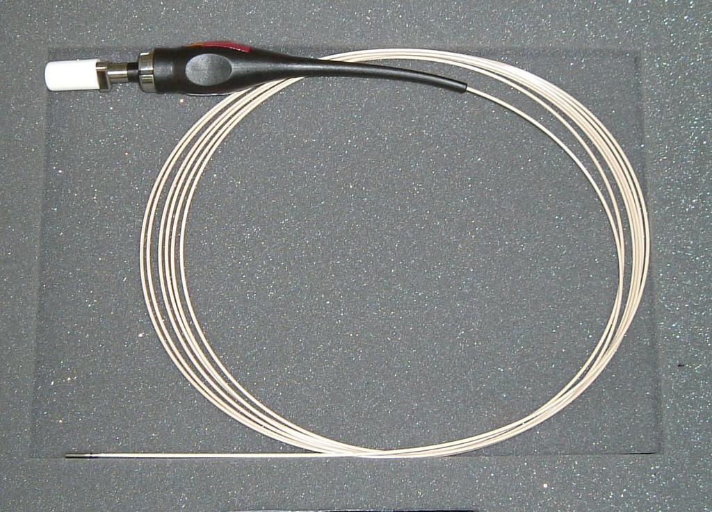
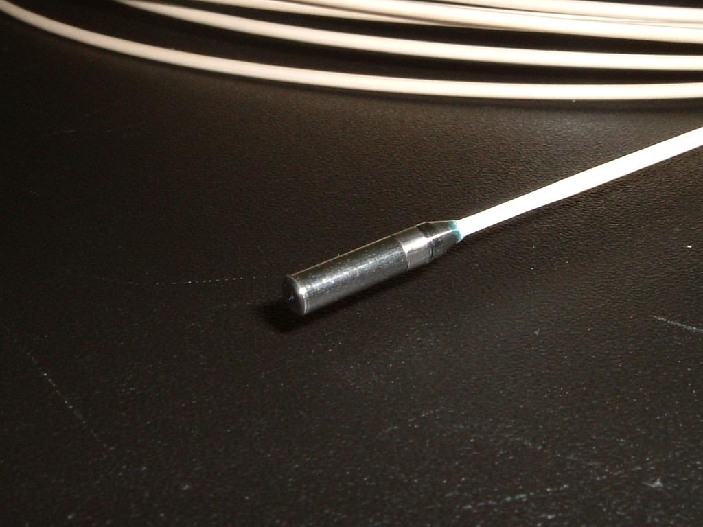
16 Fig. 1
17 Fig. 2
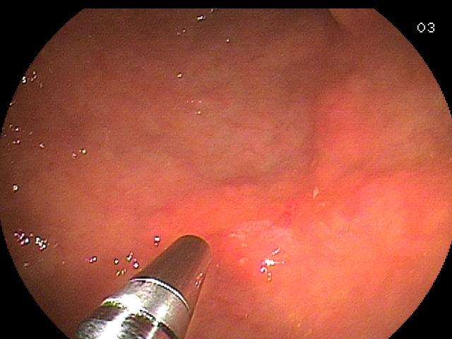
18 Fig. 3

19 a b Fig. 4a Fig. 4b

20 a b Fig. 5a Fig. 5b

21 a A Fig. 6a b Fig. 6b

22 a b Fig. 7a Fig. 7b

23 a µm b Fig. 8a Fig. 8b
24 Table 1. Clinical Characteristics of the lesions Esophagus Tumor size (mm) Mean Range Localization of tumor Esophagus Depth of invasion Mucosa Submucosa Histologic type Squamous cell carcinoma 22.8± Stomach Tumor size (mm) Mean Range Localization of tumor Antrum Angle Corpus Cardia Depth of invasion Mucosa Submucosa Histologic type Well Moderately Papillary Poorly Signet ring cell 16.3±
25 Table 2. Visualization of nuclei and cell membrane in LCM images in relation to histologic diagnoses Histologic diagnosis Nucleus Cell membrane Normal esophageal mucosa (n=10) Esophageal cancer (n=10) Normal gastric mucosa (n=20) Gastric cancer: well (n=10) moderately (n=6) papillary (n=1) poorly (n=1) signet ring cell (n=2) 10 (100) 10 (100) 0 (0) 6 (60) 4 (66.7) 0 (0) 0 (0) 0 (0) 9 (90) 5 (50) 0 (0) 0 (0) 0 (0) 0 (0) 0 (0) 0 (0) Number (and percentage) of samples are shown
ORIGINAL ARTICLES ALIMENTARY TRACT
 CLINICAL GASTROENTEROLOGY AND HEPATOLOGY 2007;5:1261 1267 ORIGINAL ARTICLES ALIMENTARY TRACT In Vivo Histopathology for Detection of Gastrointestinal Neoplasia With a Portable, Confocal Miniprobe: An Examiner
Advances in Endoscopic Imaging
 Advances in Endoscopic Imaging SGNA meeting February 20, 2010 Amar R. Deshpande, MD Asst Professor of Medicine Division of Gastroenterology University of Miami Miller School of Medicine Objectives To recognize
Histopathology of Endoscopic Resection Specimens from Barrett's Esophagus
 Histopathology of Endoscopic Resection Specimens from Barrett's Esophagus Br J Surg 38 oct. 1950 Definition of Barrett's esophagus A change in the esophageal epithelium of any length that can be recognized
University Mainz. Early Gastric Cancer. Ralf Kiesslich. Johannes Gutenberg University Mainz, Germany. Early Gastric Cancer 15.6.
 Ralf Kiesslich Johannes Gutenberg University Mainz, Germany DIAGNOSIS Unmask lesions - Chromoendoscopy -NBI Red flag technology - Autofluorescence Surface and detail analysis - Magnifying endoscopy - High
Image Analysis of Magnifying Endoscopy for Differentiation between Early Gastric Cancers and Gastric Erosions
 Showa Univ J Med Sci 29 3, 297 306, September 2017 Original Image Analysis of Magnifying Endoscopy for Differentiation between Early Gastric Cancers and Gastric Erosions Shotaro HANAMURA, Kuniyo GOMI,
Magnifying Endoscopy and Chromoendoscopy of the Upper Gastrointestinal Tract
 Magnifying Endoscopy and Chromoendoscopy of the Upper Gastrointestinal Tract Alina M.Boeriu 1, Daniela E.Dobru 1, Simona Mocan 2 1) Department of Gastroenterology, University of Medicine and Pharmacy;
Confocal Laser Endomicroscopy of the Colon
 clinical imaging Confocal Laser Endomicroscopy of the Colon Dan Ionut Gheonea, Adrian Saftoiu, Tudorel Ciurea, Carmen Popescu, Claudia Valentina Georgescu, Anca Malos Research Center of Gastroenterology
Chromoendoscopy and Endomicroscopy for detecting colonic dysplasia
 Chromoendoscopy and Endomicroscopy for detecting colonic dysplasia Ralf Kiesslich I. Medical Department Johannes Gutenberg University Mainz, Germany Cumulative cancer risk in ulcerative colitis 0.5-1.0%
Philip Chiu Associate Professor Department of Surgery, Prince of Wales Hospital The Chinese University of Hong Kong
 Application of Chromoendoscopy, NBI and AFI in Esophagus why, who, and how? Philip Chiu Associate Professor Department of Surgery, Prince of Wales Hospital The Chinese University of Hong Kong Cancer of
Helicobacter pylori Improved Detection of Helicobacter pylori
 DOI:http://dx.doi.org/10.7314/APJCP.2016.17.4.2099 RESEARCH ARTICLE Improved Detection of Helicobacter pylori Infection and Premalignant Gastric Mucosa Using Conventional White Light Source Gastroscopy
Paris classification (2003) 삼성의료원내과이준행
 삼성의료원내과이준행") Paris classification (2003) 삼성의료원내과이준행 JGCA classification - Japanese Gastric Cancer Association - Type 0 superficial polypoid, flat/depressed, or excavated tumors Type 1 polypoid carcinomas, usually attached
Paris classification (2003) 삼성의료원내과이준행 JGCA classification - Japanese Gastric Cancer Association - Type 0 superficial polypoid, flat/depressed, or excavated tumors Type 1 polypoid carcinomas, usually attached
RESEARCH ARTICLE. Abstract. Introduction
 DOI:http://dx.doi.org/10.7314/APJCP.2013.14.5.2765 RESEARCH ARTICLE Correlation between Magnifying Narrow-band Imaging Endoscopy Results and Organoid Differentiation Indicated by Cancer Cell Differentiation
Esophageal seeding after endoscopic ultrasound-guided fine-needle aspiration of a mediastinal tumor
 Esophageal seeding after endoscopic ultrasound-guided fine-needle aspiration of a mediastinal tumor Authors Kensuke Yokoyama 1,JunUshio 1,NorikatsuNumao 1, Kiichi Tamada 1, Noriyoshi Fukushima 2, Alan
Oesophagus and Stomach update dysplasia and early cancer
 Oesophagus and Stomach update dysplasia and early cancer Dr Tim Bracey STR teaching 13/4/16 Please check pathkids.com for previous talks One of the biggest units in the country (100 major resections per
ARTICLE IN PRESS. tumor grade or stage information. Procedures
 Optical Biopsy of Human Bladder Neoplasia With In Vivo Confocal Laser Endomicroscopy Geoffrey A. Sonn, Sha-Nita E. Jones, Tatum V. Tarin, Christine B. Du, Kathleen E. Mach, Kristin C. Jensen and Joseph
American Journal of Gastroenterology. Volumetric Laser Endomicroscopy Detects Subsquamous Barrett s Adenocarcinoma
 Volumetric Laser Endomicroscopy Detects Subsquamous Barrett s Adenocarcinoma Journal: Manuscript ID: AJG-13-1412.R1 Manuscript Type: Letter to the Editor Keywords: Barrett-s esophagus, Esophagus, Endoscopy
Correlation between Gastric Mucosal Morphologic Patterns and Histopathological Severity of
 Hindawi Publishing Corporation BioMed Research International Volume 2015, Article ID 808505, 7 pages http://dx.doi.org/10.1155/2015/808505 Research Article Correlation between Gastric Mucosal Morphologic
Comparison of the Diagnostic Usefulness of Conventional Magnification and Near-focus Methods with Narrow-band Imaging for Gastric Epithelial Tumors
 ORIGINAL ARTICLE ISSN 1738-3331, http://dx.doi.org/10.7704/kjhugr.2015.15.1.39 The Korean Journal of Helicobacter and Upper Gastrointestinal Research, 2015;15(1):39-43 Comparison of the Diagnostic Usefulness
Emerging Interventions in Endoscopy. Margaret Vance Nurse Consultant in Gastroenterology St Mark s Hospital
 Emerging Interventions in Endoscopy Margaret Vance Nurse Consultant in Gastroenterology St Mark s Hospital Colon Cancer Colon cancer is common. 1 in 20 people in the UK will develop the disease 19 000
Corporate Medical Policy
 Corporate Medical Policy Chromoendoscopy as an Adjunct to Colonoscopy File Name: Origination: Last CAP Review: Next CAP Review: Last Review: chromoendoscopy_as_an_adjunct_to_colonoscopy 7/2012 11/2017
Endoscopic Submucosal Dissection ESD
 Endoscopic Submucosal Dissection ESD Peter Draganov MD Professor of Medicine Division of Gastroenterology, Hepatology and Nutrition University of Florida Gastrointestinal Cancer Lesion that Can be Treated
Role of Helicobacter pylori Infection and Chronic Inflammation in Gastric Cancer in the Cardia
 Original Article Japanese Journal of Clinical Oncology Advance Access published June 19, 2007 Jpn J Clin Oncol doi:10.1093/jjco/hym029 Role of Helicobacter pylori Infection and Chronic Inflammation in
Review Article Confocal Endomicroscopy of Colorectal Polyps
 Gastroenterology Research and Practice Volume 2012, Article ID 545679, 6 pages doi:10.1155/2012/545679 Review Article Confocal Endomicroscopy of Colorectal Polyps Vivian M. Ussui and Michael B. Wallace
Histopathology: gastritis and peptic ulceration
 Histopathology: gastritis and peptic ulceration These presentations are to help you identify, and to test yourself on identifying, basic histopathological features. They do not contain the additional factual
B Barrett neoplasia, early, endoscopic mucosal resection of, in Europe, 297
 Note: Page numbers of article titles are in boldface type. A Achalasia, treatment of, history of, 258 259 Achalasia myotomy, 270 Adenocarcinomas, endoscopic management of, in Asia, 289 290 B Barrett neoplasia,
Magnifying image-enhanced endoscopy for collagenous colitis
 Magnifying image-enhanced endoscopy for collagenous colitis Authors Masaaki Kobayashi 1, Takahiro Hoshi 1, Shin-ich Morita 1,TsutomuKanefuji 1, Takeshi Suda 1,GoHasegawa 2, Shuji Terai 3 Institutions 1
Information Technology Solutions
 2016 2014 CPT Esophagoscopy Changes - Gastroenterology CPT Changes Information Technology Solutions ASGE LOGO AND INFO Esophagogastroduodenoscopy CPT Codes 43235-43270 The American Society for Gastrointestinal
malignant polyp Daily Challenges in Digestive Endoscopy for Endoscopists and Endoscopy Nurses BSGIE Annual Meeting 18/09/2014 Mechelen
 Plan Incidental finding of a malignant polyp 1. What is a polyp malignant? 2. Role of the pathologist and the endoscopist 3. Quantitative and qualitative risk assessment 4. How to decide what to do? Hubert
Delayed Perforation Occurring after Endoscopic Submucosal Dissection for Early Gastric Cancer
 CASE REPORT Clin Endosc 2015;48:251-255 Print ISSN 2234-2400 / On-line ISSN 2234-2443 http://dx.doi.org/10.5946/ce.2015.48.3.251 Open Access Delayed Perforation Occurring after Endoscopic Submucosal Dissection
Endoscopic Corner CASE 1. Kimtrakool S Aniwan S Linlawan S Muangpaisarn P Sallapant S Rerknimitr R
 170 Endoscopic Corner Kimtrakool S Aniwan S Linlawan S Muangpaisarn P Sallapant S Rerknimitr R CASE 1 A 54-year-old woman underwent a colorectal cancer screening. Her fecal immunochemical test was positive.
The white globe appearance (WGA): a novel marker for a correct diagnosis of early gastric cancer by magnifying. endoscopy with narrow-band imaging (M-
: a novel marker for a correct diagnosis of early gastric cancer by magnifying. endoscopy with narrow-band imaging (M-") E120 The white globe appearance (WGA): a novel marker for a correct diagnosis of early gastric cancer by magnifying endoscopy with narrow-band imaging (M-NBI) Authors Hisashi Doyama 1, Naohiro Yoshida
E120 The white globe appearance (WGA): a novel marker for a correct diagnosis of early gastric cancer by magnifying endoscopy with narrow-band imaging (M-NBI) Authors Hisashi Doyama 1, Naohiro Yoshida
Supplementary Information. Detection and delineation of oral cancer with a PARP1 targeted optical imaging agent
 Supplementary Information Detection and delineation of oral cancer with a PARP1 targeted optical imaging agent Authors: Susanne Kossatz a, Christian Brand a, Stanley Gutiontov b, Jonathan T.C. Liu c, Nancy
Page 1. Is the Risk This High? Dysplasia in the IBD Patient. Dysplasia in the Non IBD Patient. Increased Risk of CRC in Ulcerative Colitis
 Screening for Colorectal Neoplasia in Inflammatory Bowel Disease Francis A. Farraye MD, MSc Clinical Director, Section of Gastroenterology Co-Director, Center for Digestive Disorders Boston Medical Center
위암내시경진단 (2019) - 융기형위암을중심으로 성균관대학교의과대학내과이준행
 - 융기형위암을중심으로 성균관대학교의과대학내과이준행") 위암내시경진단 (2019) - 융기형위암을중심으로 성균관대학교의과대학내과이준행 위암내시경진단 (2019) 위암검진에대한짧지않은 comment 융기형암은융기되어있는가? 함몰형암은함몰되어있는가? Semi-pedunculated polyp Sentinel polyp or EGJ cancer? IIa + IIc 위암검진에대한짧지않은 comment 성균관대학교의과대학내과이준행
위암내시경진단 (2019) - 융기형위암을중심으로 성균관대학교의과대학내과이준행 위암내시경진단 (2019) 위암검진에대한짧지않은 comment 융기형암은융기되어있는가? 함몰형암은함몰되어있는가? Semi-pedunculated polyp Sentinel polyp or EGJ cancer? IIa + IIc 위암검진에대한짧지않은 comment 성균관대학교의과대학내과이준행
COLON: Innovations 3 steps, 3 parts..
 COLON: Innovations 3 steps, 3 parts.. Detection: I see an abnormality (usually a polyp) Characterization: Is this abnormality neoplastic? (for example: an adenoma) Treatment: it is neoplastic. Can I treat
Synchronous and Subsequent Lesions of Serrated Adenomas and Tubular Adenomas of the Colorectum
 Tsumura T, et al 1 Synchronous and Subsequent Lesions of Serrated Adenomas and Tubular Adenomas of the Colorectum T. Tsumura a T. Hiyama d S. Tanaka b M. Yoshihara d K. Arihiro c K. Chayama a Departments
The Use of Pancreatoscopy in the Diagnosis of Intraductal Papillary Mucinous Tumor Lesions of the Pancreas
 CLINICAL GASTROENTEROLOGY AND HEPATOLOGY 2005;3:S53 S57 The Use of Pancreatoscopy in the Diagnosis of Intraductal Papillary Mucinous Tumor Lesions of the Pancreas KENJIRO YASUDA, MUNEHIRO SAKATA, MOOSE
What Every Pathologist Wants the GI Nurse to Know (and how you can help us help you)
") What Every Pathologist Wants the GI Nurse to Know (and how you can help us help you) Jonathan N. Glickman MD PhD Director, GI Pathology, Caris Diagnostics, Newton, MA Associate Professor of Pathology,
What Every Pathologist Wants the GI Nurse to Know (and how you can help us help you) Jonathan N. Glickman MD PhD Director, GI Pathology, Caris Diagnostics, Newton, MA Associate Professor of Pathology,
Feasibility of endoscopic mucosa-submucosa clip closure method (with video)
") Feasibility of endoscopic mucosa-submucosa clip closure method (with video) Authors Toshihiro Nishizawa 1, Shigeo Banno 2, Satoshi Kinoshita 1,HidekiMori 2, Yoshihiro Nakazato 3,YuichiroHirai 2,Yoko Kubosawa
Feasibility of endoscopic mucosa-submucosa clip closure method (with video) Authors Toshihiro Nishizawa 1, Shigeo Banno 2, Satoshi Kinoshita 1,HidekiMori 2, Yoshihiro Nakazato 3,YuichiroHirai 2,Yoko Kubosawa
How to treat early gastric cancer? Endoscopy
 How to treat early gastric cancer? Endoscopy Presented by Pierre H. Deprez Institution Cliniques universitaires Saint-Luc, Brussels Université catholique de Louvain 2 3 4 5 6 Background Diagnostic or therapeutic
EMR, ESD and Beyond. Peter Draganov MD. Professor of Medicine Division of Gastroenterology, Hepatology and Nutrition University of Florida
 EMR, ESD and Beyond Peter Draganov MD Professor of Medicine Division of Gastroenterology, Hepatology and Nutrition University of Florida Gastrointestinal Cancer Lesion that Can be Treated by Endoscopy
Barrett s Esophagus: Old Dog, New Tricks
 Barrett s Esophagus: Old Dog, New Tricks Stuart Jon Spechler, M.D. Chief, Division of Gastroenterology, VA North Texas Healthcare System; Co-Director, Esophageal Diseases Center, Professor of Medicine,
Colon Polyps: Detection, Inspection and Characteristics
 Colon Polyps: Detection, Inspection and Characteristics Stephen Kim, M.D. Assistant Professor of Medicine Interventional Endoscopy Services UCLA Division of Digestive Diseases September 29, 2018 1 Disclosures
Factors for Endoscopic Submucosal Dissection in Early Colorectal Neoplasms: A Single Center Clinical Experience in China
 ORIGINAL ARTICLE Clin Endosc 2015;48:405-410 http://dx.doi.org/10.5946/ce.2015.48.5.405 Print ISSN 2234-2400 On-line ISSN 2234-2443 Open Access Factors for Endoscopic Submucosal Dissection in Early Colorectal
Chromoendoscopy as an Adjunct to Colonoscopy
 Chromoendoscopy as an Adjunct to Colonoscopy Policy Number: 2.01.84 Last Review: 1/2018 Origination: 7/2017 Next Review: 7/2018 Policy Blue Cross and Blue Shield of Kansas City (Blue KC) will not provide
INTRODUCTION. Key Words: Gastroesophageal reflux; Agreement; Experience. ORiginal Article
 Gut and Liver, Vol. 8, No. 2, March 2014, pp. 154-159 ORiginal Article Endoscopic Experience Improves Interobserver Agreement in the Grading of Esophagitis by Los Angeles Classification: Conventional Endoscopy
153 esophagus. We also aim to evaluate this technology for the early detection of precancerous changes in the specialized columnar epithelium of Barrr
 152 Department of Endoscopy Hisao Tajiri, Professor Hiroshi Arakawa, Assistant Professor Hiroo Imazu, Assistant Professor Keiichi Ikeda, Assistant Professor Kazuki Sumiyama, Assistant Professor Tomohiro
BENEFIT APPLICATION BLUE CARD/NATIONAL ACCOUNT ISSUES
 Medical Policy BCBSA Ref. Policy: 2.01.84 Last Review: 11/15/2018 Effective Date: 11/15/2018 Section: Medicine Related Policies 2.01.87 Confocal Laser Endomicroscopy 6.01.32 Virtual Colonoscopy/Computed
ESD for EGC with undifferentiated histology
 ESD for EGC with undifferentiated histology Jun Haeng Lee, Department of Medicine, Samsung Medical Center, Sungkyunkwan University School of Medicine, Seoul, Korea Biopsy: M/D adenocarcinoma ESD: SRC >>
Volumetric laser endomicroscopy can target neoplasia not detected by conventional endoscopic measures in long segment Barrett s esophagus
 E318 Volumetric laser endomicroscopy can target neoplasia not detected by conventional endoscopic measures in long segment esophagus Authors Institution Arvind J. Trindade, Benley J. George, Joshua Berkowitz,
Application of magnifying narrow-band imaging endoscopy for diagnosis of early gastric cancer and precancerous lesion
 RESEARCH ARTICLE Open Access Application of magnifying narrow-band imaging endoscopy for diagnosis of early gastric cancer and precancerous lesion Jing Zhang 1,2, Shi-Bin Guo 1 and Zhi-Jun Duan 1* Abstract
The Usefulness Of Narrow Band Imaging Endoscopy For The Real Time Characterization Of Colonic Lesions
 Acta Medica Marisiensis 2016;62(2):182-186 DOI: 10.1515/amma-2016-0004 RESEARCH ARTICLE The Usefulness Of Narrow Band Imaging Endoscopy For The Real Time Characterization Of Colonic Lesions Boeriu Alina
Endoscopic Mucosal Resection (EMR) & Endoscopic Submucosal Dissection (ESD)
 & Endoscopic Submucosal Dissection (ESD)") Endoscopic Mucosal Resection (EMR) & Endoscopic Submucosal Dissection (ESD) Minimally Invasive Polyp Removal IE-02700-Understanding EMR and ESD Brochure_R3.indd 1 Occasionally, a polyp that infiltrates
Endoscopic Mucosal Resection (EMR) & Endoscopic Submucosal Dissection (ESD) Minimally Invasive Polyp Removal IE-02700-Understanding EMR and ESD Brochure_R3.indd 1 Occasionally, a polyp that infiltrates
ASGE and AGA Issue Consensus Statement on Surveillance and Management of Dysplasia in Patients With Inflammatory Bowel Disease
 ASGE and AGA Issue Consensus Statement on Surveillance and Management of Dysplasia in Patients With Inflammatory Bowel Disease DOWNERS GROVE, Ill., (March 5, 2015) The American Society for Gastrointestinal
Gastroenterology Tutorial
 Gastroenterology Tutorial Gastritis Poorly defined term that refers to inflammation of the stomach. Infection with H. pylori is the most common cause of gastritis. Most patients remain asymptomatic Some
Devices To Improve Colon Polyp Detection
 Devices To Improve Colon Polyp Detection ACG/VGS Regional Postgraduate Course Sep 10-11, 2016 Williamsburg, VA VIVEK KAUL, MD, FACG Segal-Watson Professor of Medicine Chief, Division of Gastroenterology
Risk factors for lymph node metastasis in histologically poorly differentiated type early gastric cancer
 498 Original article Risk factors for lymph node metastasis in histologically poorly differentiated type early gastric cancer Authors C. Kunisaki 1, M. Takahashi 2, Y. Nagahori 3, T. Fukushima 3, H. Makino
Probe-based confocal endomicroscopy is accurate for differentiating gastric lesions in patients in a Western center
 Original Article Probe-based confocal endomicroscopy is accurate for differentiating gastric lesions in patients in a Western center Adriana Vaz Safatle-Ribeiro, Elisa Ryoka Baba, Rodrigo Corsato Scomparin,
Quiz Adenocarcinoma of the distal stomach has been increasing in the last 20 years. a. True b. False
 Quiz 1 1. Which of the following are risk factors for esophagus cancer. a. Obesity b. Gastroesophageal reflux c. Smoking and Alcohol d. All of the above 2. Adenocarcinoma of the distal stomach has been
Management of Barrett s: From Imaging to Resection
 Management of Barrett s: From Imaging to Resection Michael Wallace, MD, MPH, FACG Professor of Medicine Mayo Clinic Florida Goals of Endoscopic Evaluation in Barrett s Detect Barrett s and dysplasia Reduce/eliminate
Do any benign polyps require an operation?
 Do any benign polyps require an operation? Kevin Waschke MD.,CM., FRCPC, FASGE McGill University Health Center President Elect Canadian Association of Gastroenterology Colonoscopy Education Day - Tuesday
Prognostic analysis of gastric mucosal dysplasia after endoscopic resection: A single-center retrospective study
 JBUON 2019; 24(2): 679-685 ISSN: 1107-0625, online ISSN: 2241-6293 www.jbuon.com E-mail: editorial_office@jbuon.com ORIGINAL ARTICLE Prognostic analysis of gastric mucosal dysplasia after endoscopic resection:
Quantitative analysis of high-resolution microendoscopic images for diagnosis of neoplasia in patients with Barrett s esophagus
 Washington University School of Medicine Digital Commons@Becker Open Access Publications 2016 Quantitative analysis of high-resolution microendoscopic images for diagnosis of neoplasia in patients with
Risk factors for non-curative resection of early gastric neoplasms with endoscopic submucosal dissection: Analysis of 1,123 lesions
 EXPERIMENTAL AND THERAPEUTIC MEDICINE 9: 1209-1214, 2015 Risk factors for non-curative resection of early gastric neoplasms with endoscopic submucosal dissection: Analysis of 1,123 lesions TATSUYA TOYOKAWA
Diagnostic accuracy of pit pattern and vascular pattern in colorectal lesions
 Diagnostic accuracy of pit pattern and vascular pattern in colorectal lesions Digestive Disease Center, Showa University Northern Yokohama Hospital Department of Pathology Yoshiki Wada, Shin-ei Kudo, Hiroshi
Analysis of microvascular density in early gastric carcinoma using magnifying endoscopy with narrow-band imaging
 E832 THIEME Analysis of microvascular density in early gastric using magnifying endoscopy with narrow-band imaging Authors Masashi Kawamura 1, Hiroshi Naganuma 2, Rie Shibuya 2, Tatsuya Kikuchi 1, Yoshitaka
Chromoendoscopy or Narrow Band Imaging with Targeted biopsies Should be the Cancer Surveillance Endoscopy Procedure of Choice in Ulcerative Colitis
 Chromoendoscopy or Narrow Band Imaging with Targeted biopsies Should be the Cancer Surveillance Endoscopy Procedure of Choice in Ulcerative Colitis Bret A. Lashner, M.D. Professor of Medicine Director,
Endoscopy in IBD. F.Hartmann K.Kasper-Kliniken (St.Marienkrankenhaus) Frankfurt/M.
 Frankfurt/M.") F.Hartmann K.Kasper-Kliniken (St.Marienkrankenhaus) Frankfurt/M. F.Hartmann@em.uni-frankfurt.de Indications for endoscopy Diagnosis Management Surveillance Diagnosis Single most valuable tool: ileocolonoscopy
F.Hartmann K.Kasper-Kliniken (St.Marienkrankenhaus) Frankfurt/M. F.Hartmann@em.uni-frankfurt.de Indications for endoscopy Diagnosis Management Surveillance Diagnosis Single most valuable tool: ileocolonoscopy
Treatment Strategy for Non-curative Resection of Early Gastric Cancer. Jun Haneg Lee. Sungkyunkwan University, Samsung Medical Center, Seoul Korea
 Treatment Strategy for Non-curative Resection of Early Gastric Cancer Jun Haneg Lee. Sungkyunkwan University, Samsung Medical Center, Seoul Korea Classic EMR/ESD data analysis style Endoscopic resection
Case Scenario 1. The patient has now completed his neoadjuvant chemoradiation and has been cleared for surgery.
 Case Scenario 1 July 10, 2010 A 67-year-old male with squamous cell carcinoma of the mid thoracic esophagus presents for surgical resection. The patient has completed preoperative chemoradiation. This
SAM PROVIDER TOOLKIT
 THE AMERICAN BOARD OF PATHOLOGY Maintenance of Certification (MOC) Program SAM PROVIDER TOOLKIT Developing Self-Assessment Modules (SAMs) www.abpath.org The American Board of Pathology (ABP) approves educational
Narrow Band Imaging for the Detection of Gastric Intestinal Metaplasia and Dysplasia During Surveillance Endoscopy
 Dig Dis Sci (2010) 55:3442 3448 DOI 10.1007/s10620-010-1189-2 ORIGINAL ARTICLE Narrow Band Imaging for the Detection of Gastric Intestinal Metaplasia and Dysplasia During Surveillance Endoscopy Lisette
How to characterize dysplastic lesions in IBD?
 How to characterize dysplastic lesions in IBD? Name: Institution: Helmut Neumann, MD, PhD, FASGE University Medical Center Mainz What do we know? Patients with IBD carry an increased risk of developing
Citation Acta Medica Nagasakiensia. 1992, 37
 NAOSITE: Nagasaki University's Ac Title Author(s) Clinicopathological Study on Gastri Murata, Ikuo; Oda, Hidetoshi; Muta, Nakamuta, Kohji; Tsuruta, Hideo; Oh Makiyama, Kazuya; Hara, Kohei Citation Acta
Confocal laser endomicroscopy is a new field of endoluminal
 Imaging and Advanced Technology Michael B. Wallace, Section Editor Probe-Based Confocal Laser Endomicroscopy MICHAEL B. WALLACE* and PAUL FOCKENS *Mayo Clinic, Jacksonville, Florida; and Academic Medical
Evaluation of the visibility of early gastric cancer using linked color imaging and blue laser imaging
 Yoshifuku et al. BMC Gastroenterology (2017) 17:150 DOI 10.1186/s12876-017-0707-5 RESEARCH ARTICLE Open Access Evaluation of the visibility of early gastric cancer using linked color imaging and blue laser
REPORT. McMahon Publishing Group. PENTAX Medical i-scan Technology for Improved Endoscopic Evaluations
 Brought to You by MAY 2014 PENTAX Medical i-scan Technology for Improved Endoscopic Evaluations Endoscopy plays a vital role in the diagnosis and clinical management of diseases of the gastrointestinal
Confocal Endomicroscopy: In Vivo Diagnosis of Neoplastic Lesions of the Gastrointestinal Tract
 Review Confocal Endomicroscopy: In Vivo Diagnosis of Neoplastic Lesions of the Gastrointestinal Tract MARTIN GOETZ and RALF KIESSLICH Johannes Gutenberg-University Mainz, 55131 Mainz, Germany Abstract.
Tools of the Gastroenterologist: Introduction to GI Endoscopy
 Tools of the Gastroenterologist: Introduction to GI Endoscopy Objectives Endoscopy Upper endoscopy Colonoscopy Endoscopic retrograde cholangiopancreatography (ERCP) Endoscopic ultrasound (EUS) Endoscopic
Abstracting Upper GI Cancer Incidence and Treatment Data Quiz 1 Multiple Primary and Histologies Case 1 Final Pathology:
 Abstracting Upper GI Cancer Incidence and Treatment Data Quiz 1 Multiple Primary and Histologies Case 1 A 74 year old male with a history of GERD presents complaining of dysphagia. An esophagogastroduodenoscopy
Pathology in Slovenian CRC screening programme:
 Pathology in Slovenian CRC screening programme: Findings, organisation and quality assurance Snježana Frković Grazio University Medical Center Ljubljana, Slovenia Slovenia s population: 2 million Incidence
Diagnosis of colorectal lesions with a novel endocytoscopic classification a pilot study
 Original article 869 Diagnosis of colorectal lesions with a novel endocytoscopic classification a pilot study Authors S-E Kudo 1, K. Wakamura 1, N. Ikehara 1,Y.Mori 1, H. Inoue 1, S. Hamatani 2 Institutions
Vital staining and Barrett s esophagus
 Marcia Irene Canto, MD, MHS Baltimore, Maryland Vital staining or chromoendoscopy refers to staining of endoscopic tissue or topical application of chemical stains or pigments to alter tissue appearances
Confocal Laser Endomicroscopy
 Confocal Laser Endomicroscopy Policy Number: 2.01.87 Last Review: 3/2018 Origination: 3/2013 Next Review: 9/2018 Policy Blue Cross and Blue Shield of Kansas City (Blue KC) will not provide coverage for
Research Article Characteristics of Metachronous Gastric Tumors after Endoscopic Submucosal Dissection for Gastric Intraepithelial Neoplasms
 Gastroenterology Research and Practice, Article ID 863595, 6 pages http://dx.doi.org/1.1155/214/863595 Research Article Characteristics of Metachronous Gastric Tumors after Endoscopic Submucosal Dissection
Shou Jiang Tang, MD, FASGE. Director of Endoscopic Research Professor in Medicine
 Shou Jiang Tang, MD, FASGE Director of Endoscopic Research Professor in Medicine Through-the-scope clipping devices Over-the-scope clipping devices First reported clipping device Hayshi T, Yonezawa M,
Barrett Esophagus - RadioFrequency Ablation (BE-RFA) - Project manual + FAQ
 - Project manual + FAQ") Barrett Esophagus - RadioFrequency Ablation (BE-RFA) - Project manual + FAQ Table of contents 1 General project information...3 1.1 Inclusion criteria...3 1.2 Registration time points...3 1.3 Project variable
Barrett Esophagus - RadioFrequency Ablation (BE-RFA) - Project manual + FAQ Table of contents 1 General project information...3 1.1 Inclusion criteria...3 1.2 Registration time points...3 1.3 Project variable
Gastric Polyps. Bible class
 Gastric Polyps Bible class 29.08.2018 Starting my training in gastroenterology, some decades ago, my first chief always told me that colonoscopy may seem technically more challenging but gastroscopy has
Barrett s Esophagus. Abdul Sami Khan, M.D. Gastroenterologist Aurora Healthcare Burlington, Elkhorn, Lake Geneva, WI
 Barrett s Esophagus Abdul Sami Khan, M.D. Gastroenterologist Aurora Healthcare Burlington, Elkhorn, Lake Geneva, WI A 58 year-old, obese white man has had heartburn for more than 20 years. He read a magazine
Advanced techniques for resection of large polyps. John G. Lee, MD February 2, 2018
 Advanced techniques for resection of large polyps John G. Lee, MD February 2, 2018 Background 1cm - large polyp on screening 2cm - large for polypectomy 3cm giant polyp 10-15% of polyps can t be removed
The utility of a novel colonoscope with retroflexion for colorectal endoscopic submucosal dissection
 Original article The utility of a novel colonoscope with retroflexion for colorectal endoscopic submucosal dissection Authors Hidenori Tanaka 1,ShiroOka 2, Shinji Tanaka 1, Katsuaki Inagaki 2,YukiOkamoto
Definition of GERD American College of Gastroenterology
 Definition of GERD American College of Gastroenterology GERD is defined as chronic symptoms or mucosal damage produced by the abnormal reflux of gastric contents into the esophagus DeVault et al. Am J
Is intestinal metaplasia a necessary precursor lesion for adenocarcinomas of the distal esophagus, gastroesophageal junction and gastric cardia?
 Diseases of the Esophagus (2007) 20, 36 41 DOI: 10.1111/j.1442-2050.2007.00638.x Blackwell Publishing Asia Original article Is intestinal metaplasia a necessary precursor lesion for adenocarcinomas of
A Proposed Strategy for Treatment of Superficial Carcinoma. in the Thoracic Esophagus Based on an Analysis. of Lymph Node Metastasis
 Kitakanto Med J 2002 ; 52 : 189-193 189 A Proposed Strategy for Treatment of Superficial Carcinoma in the Thoracic Esophagus Based on an Analysis of Lymph Node Metastasis Susumu Kawate,' Susumu Ohwada,'
Dysplasia 4/19/2017. How do I practice Chromoendoscopy for Surveillance of Colitis? SCENIC: Polypoid Dysplasia in UC. Background
 SCENIC: Polypoid in UC Definition How do I practice for Surveillance of Colitis? Themos Dassopoulos, M.D. Director, BSW Center for IBD Themistocles.Dassopoulos@BSWHealth.org Tel: 469-800-7189 Cell: 314-686-2623
Esophageal Adenocarcinoma Developing after Eradication of Helicobacter pylori
 355 This is an Open Access article licensed under the terms of the Creative Commons Attribution- NonCommercial-NoDerivs 3.0 License (www.karger.com/oa-license), applicable to the online version of the
Identification of gastric atrophic changes: from histopathology to endoscopy
 Evidence in perspective 533 Identification of gastric atrophic changes: from histopathology to endoscopy Authors Mário Dinis-Ribeiro 1,2, Ernst J. Kuipers 3 Institutions Bibliography DOI http://dx.doi.org/
Utility of Probe-based (Cellvizio) Confocal Laser Endomicroscopy in Gastroenterology
 Confocal Laser Endomicroscopy in Gastroenterology") Review articles Utility of Probe-based (Cellvizio) Confocal Laser Endomicroscopy in Gastroenterology Elías Alfonso Forero Piñeros, MD, 1 Héctor José Cardona, MD, 2 Kunal Karia, MD, 3 Amrita Sethi, MD,
Review articles Utility of Probe-based (Cellvizio) Confocal Laser Endomicroscopy in Gastroenterology Elías Alfonso Forero Piñeros, MD, 1 Héctor José Cardona, MD, 2 Kunal Karia, MD, 3 Amrita Sethi, MD,
Gastric atrophy: use of OLGA staging system in practice
 Gastroenterology and Hepatology From Bed to Bench. 2016 RIGLD, Research Institute for Gastroenterology and Liver Diseases ORIGINAL ARTICLE Gastric atrophy: use of OLGA staging system in practice Mahsa
Morphologic Criteria of Invasive Colonic Adenocarcinoma on Biopsy Specimens
 ISPUB.COM The Internet Journal of Pathology Volume 12 Number 1 Morphologic Criteria of Invasive Colonic Adenocarcinoma on Biopsy Specimens C Rose, H Wu Citation C Rose, H Wu.. The Internet Journal of Pathology.