Novel Functions of LBP-5 in Caenorhabditis elegans Fat Metabolism
|
|
|
- Toby Stephens
- 5 years ago
- Views:
Transcription
1 SUPPLEMENTAL DATA (Xu et al., JBC/2011/ R2) Clean Version Novel Functions of LBP-5 in Caenorhabditis elegans Fat Metabolism Mo Xu, Hyoe-Jin Joo and Young-Ki Paik * Supplemental Table S1. A List of Oligonucleotides That Have Been Used for RNAi Primers Gene name Primers lbp-1 Sense: 5 -TACTCTAGAATGTGCGCTAAAATCGCT-3 Antisense: 5 -TACAAGCTTTTATGGGAGTCGTTTGTA-3 lbp-2 Sense: 5 -TACTCTAGAATGTCTTCGAAATTCCTC-3 Antisense: 5 -TACCTCGAGTTATGGGAGTCTCTTGTA-3 lbp-3 Sense: 5 -TACTCTAGAATGAATCTGTACTTAACT-3 Antisense: 5 -TACAAGCTTCTACTTCTTTCCGGTCGA-3 lbp-4 Sense: 5 -TACTCTAGAATGTCAGTGCCAGACAAG-3 Antisense: 5 -TACAAGCTTTCACTTCTGCTCGACTCT-3 lbp-5 Sense: 5 -TACTCTAGAATGTCTGCTGAACAATTT-3 Antisense: 5 -TACCTCGAGCTATTCACGAATATAGGC-3 lbp-6 Sense: 5 -TACTCTAGAGTTGGACGCTGGAAGCTC-3 Antisense: 5 -TACCTCGAGCGGATCCAGATTCAAGAG-3 lbp-7 Sense: 5 -TACTCTAGAATGGCATCTATGAATGAC-3 Antisense: 5 -TACAAGCTTTTATTCTCTCTCCCACTC-3 lbp-8 Sense: 5 -TACTCTAGAGAGTTTATTGGACGATGG-3 Antisense: 5 -TACAAGCTTTTTTGAAAGCGAGCTTGTTG-3 lbp-9 Sense: 5 -TACTCTAGAATGCCAATTCAAACCGATC-3 Antisense: 5 -TACCTCGAGTTAGGCAGCCTTCTCGTAG-3 Supplemental Table S2. Differential Efficiency of Ligand Displacement from LBP-5 by DAUDA Fatty Acid Ligand displacement a Oleic Acid (Δ9, 18:1) 68.9±5.1 Lauric Acid (C12:0) 31.6±3.4 Pentadecanoic Acid (C15:0) 32.3±5.0 Palmitic Acid (C16:0) 33.1±2.7 Stearic Acid (C18:0) 30.8±6.1 Arachidic Acid (C20:0) 26.1±4.8 Arachidonic Acid (C20:4, ω-6) 62.1±7.2 Alpha-Linolenic Acid (C18:3, ω-3) 4.6±0.7 a Values represent the percent drop in peak fluorescence emission (470 nm) of each ligand (oleic acid, lauric acid, pentadecanoic acid, palmitic acid, stearic acid, arachidic acid, arachidonic acid and alphalinolenic acid) in LBP-5-DAUDA complex when each fatty acid was added. Concentration of both DAUDA and LBP-5 was 1 μm; that of ligand (fatty acid) was 1 μm. The intensity of fluorescence emission was recorded at the peak emission wavelength (See also supplemental Fig. S5). 1
 are shown in red; acidic residues (DE) are shown in blue; basic residues (RK) are shown in magenta; Hydroxyl + Amine + Basic residues (STYHCNGQ) are shown in")
2 Figure S1 Figure S1. Multiple Sequence Alignment of lbp Genes. Small and hydrophobic residues (AVFPMILW) are shown in red; acidic residues (DE) are shown in blue; basic residues (RK) are shown in magenta; Hydroxyl + Amine + Basic residues (STYHCNGQ) are shown in green; and all other residues are shown in gray. Asterisks indicate residues that are identical in all sequences in the alignment. Colons indicate conserved substitutions. Periods indicate semi-conserved substitutions. Square frames indicate conserved fatty acid binding domain. 2
3 Figure S2 A B LBP-2 LBP-3 LBP-4 LBP-9 LBP-5 LBP-6 LBP-7 LBP-8 LBP LBP LBP LBP LBP LBP LBP LBP-7 70 Figure S2. Relationships of LBP Family Proteins. (A) Phylogenetic relationships of the LBP protein sequences of C. elegans. Relative distances are indicated. The phylogenetic tree was constructed as detailed in the Experimental Procedures and was visualized using Treeview. (B) Sequence identity of LBP family members. 3

4 Figure S3 A B 4
 and nhr-49(nr2041). (C) Rescue of the lbp-5(tm1618) mutant exhibiting high-fat phenotype.")
, and lbp- 5(tm1618);Ex[lbp-5::gfp] worms using fixative dyes Oil Red O and Sudan Black. Scale bars, 50 μm.")
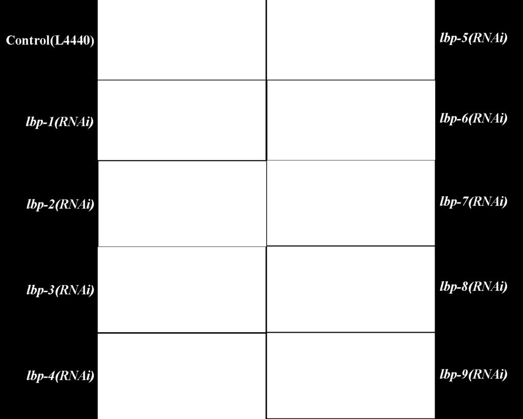
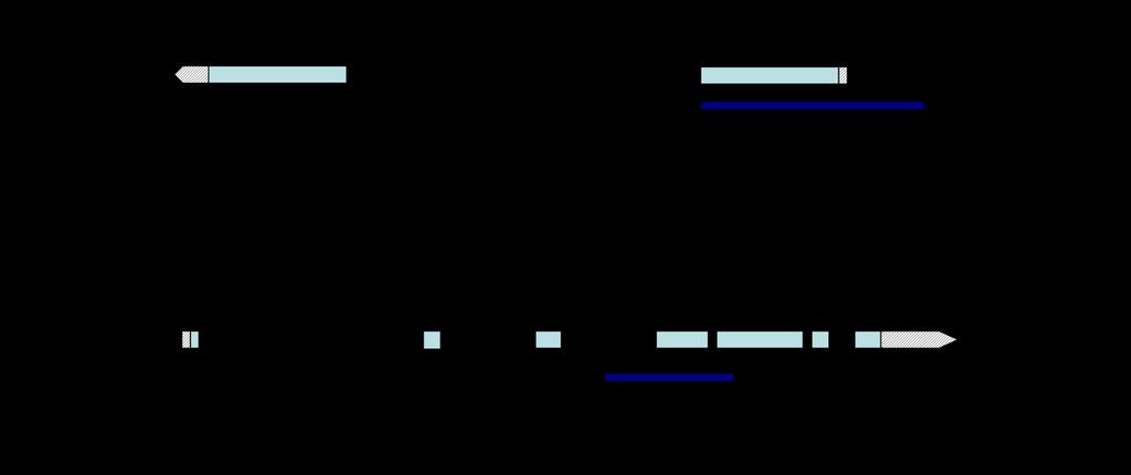
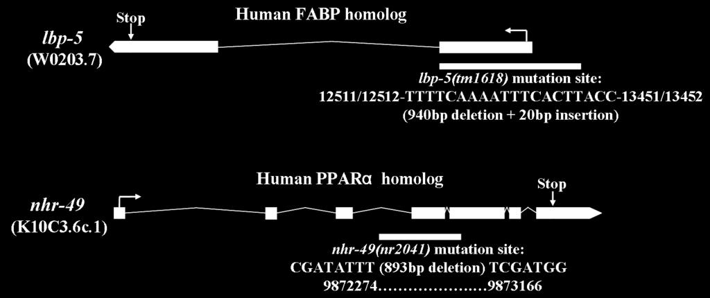
5 C Figure S3. (A) Sudan black staining of lbp(rnai) animals (adult). Scale bars, 50 μm. (B) Structural organization of the C. elegans lbp-5 and nhr-49 genes. Mutation sites are underlined for lbp-5(tm1618) and nhr-49(nr2041). (C) Rescue of the lbp-5(tm1618) mutant exhibiting high-fat phenotype. Lipid staining with either Oil Red O staining or Sudan Black staining (left panel) and quantitative analysis (right panel) indicate that high fat lbp-5 mutant is suppressed in the rescued worms. Shown here is wisualization of fat stores in fixed N2, lbp-5(tm1618), and lbp- 5(tm1618);Ex[lbp-5::gfp] worms using fixative dyes Oil Red O and Sudan Black. Scale bars, 50 μm. Triglyceride contents for wild type N2, lbp-5(tm1618), and lbp-5(tm1618);ex[lbp-5::gfp] worms. At least two independent experiments were performed. Error bars indicate the standard deviation. An asterisk (*) indicates a significant difference from the control sample (P < 0.05 as calculated by t- test). 5


 1D gel image")


6 Figure S4 A B C D Figure S4. Production of Recombinant GST-LBP-5. (A) 1D gel image of purified recombinant LBP-5 fusion protein. Separation and confirmation of the 6
7 size of purified recombinant LBP-5-GST fusion protein (42.5 kda including GST) by SDS-gel electrophoresis. (B) Map of LBP-5-GST fusion plasmid. (C) MALDI-TOF-MS/MS analysis of recombinant LBP-5. Purified LBP-5 protein was digested with trypsin (1:50, w/w) followed by analysis by MALDI-TOF-MS/MS (Applied Biosystems 4800) and then validated by peptide mass fingerprinting of the selected peaks of two representative peptides (top: EVGVAVLLR, bottom: EVGVAVLLR NTTLEFTLGVEFDETTPDGR) by NCBInr Database ( sequences: residues). For LBP-5, a total of 7 peaks were detected with 64% coverage. (D) MALDI-MS peptide mass spectrum of purified LBP-5 protein following tryptic digestion and trypsin autolytic fragments. indicates matched peptides. Matched peptides in sequence: MSAEQFVGRW KLVESENFED YLKEVGVGLL LRKAACAAKP TLEIKVNGNK WHVNQLSTFK NTTLEFTLGV EFDETTPDGR QFKSTITIED GKVVHVQKRI KDSDHDSVIT RWFEGEKLIT TLQSGSVISR RAYIRE. Experimental data were then compared to the peptide mass database Peptide Mass Fingerprint (PMF). After accurate determination of the peptide masses, databases were searched to identify the LBP-5 protein. 7

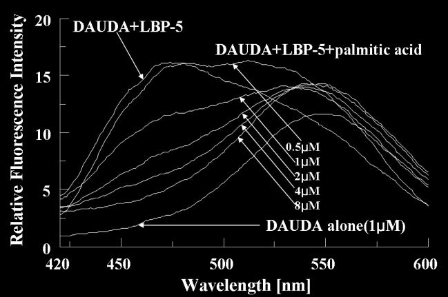
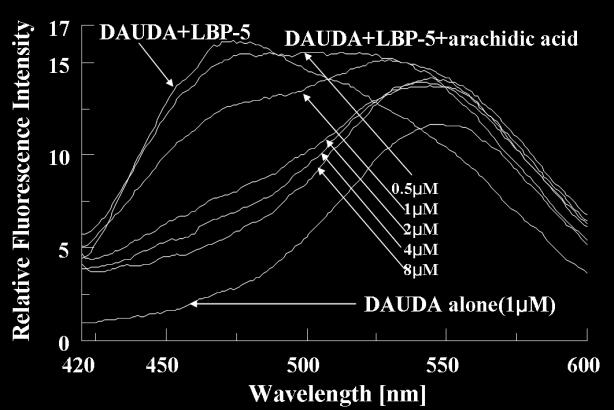
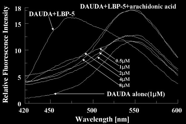
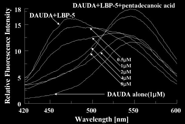
8 Figure S5 A B C D E F G 8
, pentadecanoic acid (B), palmitic acid (C),")
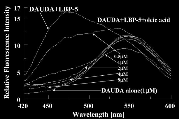
 of 1 µm DAUDA alone or with 1 µm LBP-5 monomer.")
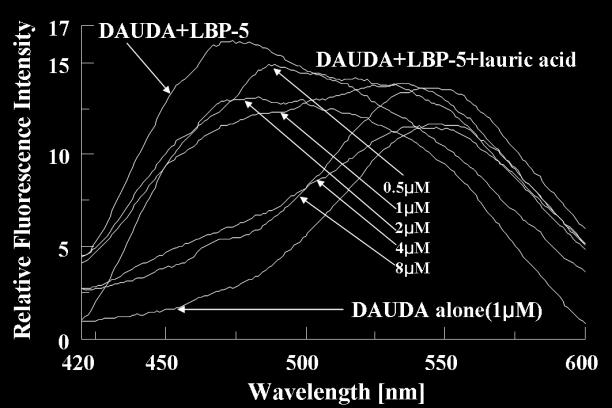
9 Figure S5. LBP-5 Binds Fatty Acids with Different Affinities. Binding of DAUDA to recombinant LBP-5 and competition with a fatty acid (lauric acid (A), pentadecanoic acid (B), palmitic acid (C), oleic acid (D), arachidic acid (E), arachidonic acid (F) and alpha-linolenic acid (G)). Fluorescence emission spectra (excitation at 345 nm) of 1 µm DAUDA alone or with 1 µm LBP-5 monomer. Also shown is the reversal of changes in DAUDA emission by competition with fatty acid (0.5, 1, 2, 4, and 8 µm) added to the LBP-5-DAUDA complex. 9
10 Figure S6 A B C D Figure S6. Relationship between lbp-5 and nhr-49 Gene Expression. (A) Gene expression levels of NHR-49 targets involved in mitochondrial β-oxidation in N2, lbp- 5(tm1618), nhr-49(nr2041), and lbp-5(tm1618); nhr-49(nr2041) animals by qrt-pcr. (B) Gene expression levels of NHR-49 targets involved in peroxisomal β-oxidation in N2, lbp-5(tm1618), nhr- 49(nr2041), and lbp-5(tm1618); nhr-49(nr2041) animals. (C) Gene expression levels of NHR-49 targets involved in fatty acid desaturation/elongation in N2, lbp-5(tm1618), nhr-49(nr2041), and lbp- 5(tm1618); nhr-49(nr2041) animals. (D) Gene expression levels of NHR-49 targets involved in gluconeogenesis in N2, lbp-5(tm1618), nhr-49(nr2041), and lbp-5(tm1618); nhr-49(nr2041) animals. Actin served as an internal control. At least three independent experiments were performed. Error bars indicate the standard deviation. 10
11 Figure S7 Figure S7. Relative Gene Expression of Non-nhr-49 Target Genes. Gene expression levels of non-nhr-49 targets involved in mitochondrial β-oxidation (ech-1 and T08B2.7), peroxisomal β-oxidation (acs-1 and F53A2.7) and gluconeogenesis (sdha-1) in N2, lbp- 5(tm1618), nhr-49(nr2041) and lbp-5(tm1618); nhr-49(nr2041) animals by qrt-pcr. Actin served as an internal control. At least three independent experiments were performed. Error bars indicate S.D. 11
, nhr-49(nr2041) and lbp- 5(tm1618); nhr-49(nr2041) mutant worms which were fed with fatty acid (oleic acid, palmitic acid,")
, nhr- 49(nr2041) and lbp-5(tm1618); nhr-49(nr2041) mutant worms indicates any fatty acid was actually taken up by the worms.")
12 Figure S8 Figure S8. Relative Fatty Acids Contents Measured by GC-MS. Total fatty acids were extracted from wild type N2 worms, lbp-5(tm1618), nhr-49(nr2041) and lbp- 5(tm1618); nhr-49(nr2041) mutant worms which were fed with fatty acid (oleic acid, palmitic acid, stearic acid, arachidonic acid and alpha-linolenic acid), then were analyzed by GC-MS. Worm of control group were fed with NP40. Relative content of each fatty acid in N2, lbp-5(tm1618), nhr- 49(nr2041) and lbp-5(tm1618); nhr-49(nr2041) mutant worms indicates any fatty acid was actually taken up by the worms. In detail, each determined contents of fatty acid (oleic acid, palmitic acid, stearic acid, arachidonic acid and alpha-linolenic acid) in each control group (N2, lbp-5(tm1618), nhr- 49(nr2041) and lbp-5(tm1618); nhr-49(nr2041) worms fed with NP40) was as a baseline, while the content of each specific fatty acid of each group worms fed with this specific fatty acid was just compared with its baseline. Pentadecanoic acid (C15:0) served as an internal standard. Three replicates of fatty acid methyl esters were prepared and three independent experiments were performed. Error bars indicate S.D. 12

13 Figure S9 A B C D 13


14 E F G H I J 14

15 K L M N O P 15
16 Q R S Figure S9. nhr-49 Target Gene Expression in Different Conditions. Gene expression levels of nhr-49 targets involved in mitochondrial β-oxidation, peroxisomal β- oxidation, fatty acid desaturation/elongation and gluconeogenesis in N2, lbp-5(tm1618), nhr- 49(nr2041) and lbp-5(tm1618); nhr-49(nr2041) worms fed with oleic acid, palmitic acid, arachidonic acid and alpha-linolenic acid by qrt-pcr. Actin served as an internal control. At least three independent experiments were performed. Error bars indicate S.D. 16
17 Figure S10 Figure S10. Relative expression levels of lbp-5 at different stages of N2 and dauer animals. Actin served as an internal control. Graph shows the mean values (± S.D.) from three independent experiments. 17
18 Supplementary Experimental Procedures Identification of Fatty Acid Binding Domain in C. elegans LBP Family Members-C. elegans lipid binding protein (LBP) family members (lbp-1 through -9) were previously reported (1). In this study, the sequences of nine lbp were obtained from WormBase ( and the conserved fatty acid binding domain of all these nine LBP proteins as well as their protein sequences were aligned by Vector NTI software package. Information on the conserved sequences among LBPs was obtained using ScanProsite ( RNAi by Feeding-The primers used to construct lbp RNAi vectors are shown in supplemental Table S1. These clones were digested with the appropriate restriction enzymes and inserted into the ppd (L4440) vector containing two convergent T7 polymerase promoters in opposite orientations and separated by a multiple cloning site. Plasmid DNA was transformed into E. coli HT115 (DE3) cells. HT115, which carries the ppd plasmid with no insert, was used as a control. Cells carrying plasmid DNA were directly applied onto agar plates composed of standard NGM/agar medium supplemented with 50 µg/ml ampicillin and 0.5 mm isopropyl beta-d-1- thiogalactopyranoside (IPTG), and then cultured overnight at room temperature. Beginning the next day, N2 worms were grown on the plates containing transformed E. coli HT115 (DE3) cells producing lbp dsrna. Expression of the corresponding endogenous C. elegans gene was knocked down by RNAi (2). Nile Red Lipid Staining-As a preliminary examination procedure, Nile red was used to stain and assess the amount of lipid present in worms (3). Nile red powder (Molecular Probes, Eugene, OR) was dissolved in acetone as a stock solution of 500 µg/ml, and diluted to 1 µg/ml in M9 buffer before 200 µl was added to E. coli (OP50)-seeded 50-mm NGM plates, resulting in a final concentration of 0.05 µg/ml. Synchronized cohorts of C. elegans were transferred to these plates, left overnight, and the fluorescence intensity was measured on the following day using a rhodamine filter. Nile red was visualized using a Zeiss AX10 microscope (Jena, Germany). All Nile red images were acquired using identical settings and exposure times. Source of lbp-5(tm1618) and nhr-49(nr2041)-the lbp-5(tm1618) mutant was created with 940-bp deletion and 20-bp insertion at the 3 region, which was prepared by NBRP, Japan. The nhr- 49(nr2041) mutant has an 893-bp deletion and was a gift from Carl Johnson at Nemapharm Pharmaceuticals. Oil Red O Staining-Worms were washed three times with 1 x PBS and then suspended in 200 µl of PBS to which an equal volume of 2X MRWB buffer (160 mm KCl, 40 mm NaCl, 14 mm Na 2 EGTA, PIPES ph 7.4, 1 mm spermidine, 0.4 mm spermine, 30 mm, 2% paraformaldehyde, 0.2% 18
19 beta-mercaptoethanol) was added. The worms were taken through 1 freeze-thaw cycle between liquid nitrogen and warm running tap water, followed by spinning at 14000g and washing once in PBS to remove paraformaldehyde. After fixation, worms were resuspended and dehydrated in 60% isopropanol. Approximately 450 ml of 60% Oil Red O stain (Cat. No. O9755, Sigma-Aldrich, St. Louis, MO, USA) was added to each sample, and samples were incubated overnight at room temperature (4). Animals were mounted and imaged with a Zeiss AX10 microscope (Jena, Germany). Construction of LBP-5 Expression Plasmid and Recombinant LBP-5-Primers were designed to PCR amplify the LBP-5 coding region from the reverse-transcribed C. elegans RNA. The PCR fragment was sub-cloned into the pgex4t3 expression plasmid, and transformed first into DH5α cells and then into BL21 (DE3) E. coli for recombinant protein expression. Transformed E. coli BL21 (DE3) were cultured overnight at 37 C in Luria Bertani medium containing 50 µg/ml ampicillin. The stock culture was diluted 1:100 in the same medium and the bacteria were grown to A 600 =0.5 before induction with 1 mm isopropyl beta-d-1-thiogalactopyranoside (IPTG). After 4 hr at 37 C, the cells were collected by centrifugation at 4,000 g and resuspended in pre-cold PBS plus lysozyme at a final concentration of 0.2 mg/ml, then sonicated on ice for five 1-min bursts. Following centrifugation at 15,000 g, the supernatant was applied to glutathione Sepharose 4B (GE Healthcare) packed in a 2-ml column. The washing procedure and elution of the fusion protein were carried out according to the manufacturer s instructions ( The concentration of the protein solution was measured using a Bradford assay. Aliquots of protein solution were subjected to SDS-PAGE analysis on 8% gels followed by staining with Coomassie Brilliant Blue R- 250 (Amersham Pharmacia). The recombinant LBP-5 was confirmed by MS/MS analysis and further characterized by various chemical methods. Identification of Recombinant LBP-5 Protein by MALDI-TOF-Protein bands that were not present in control samples on Coomassie blue-stained SDS-PAGE gels were excised, destained, and digested by 10 μg/ml trypsin (Promega, Southampton, UK) in 50 mm ammonium bicarbonate. Mixtures were processed for mass spectrometry analysis as previously described (5). Recovered peptides were prepared for MALDI-TOF MS by mixing with CHCA, 1% formic acid in 50% ACN, and droplets were allowed to dry on the MALDI sample plate. The peptide mass fingerprinting (PMF) was analyzed with the 4800 MALDI-TOF mass spectrometer (Applied Biosystems, Foster City, CA, USA). The mass spectra were obtained in reflect/delayed extraction mode with an accelerating voltage of 20 kv and sum data from 1,000 laser pulses. Proteins were identified from the PMF using MASCOT ( in search of protein database and NCBInr database with the Matrix Science search engine. Circular Dichroism Analysis-CD spectra were collected using a Jasco J810 19
20 spectropolarimeter with a thermostatically controlled cell holder. A fused quartz cell with a path length of 0.1 cm was used for CD measurements, using 0.67 mg/ml of recombinant LBP-5. All measurements were made in 10 mm potassium phosphate buffer at ph 7.4 and ambient temperature. The spectra measured in the far UV-region nm were averages of four scans and were normalized by subtracting the baseline of the buffer (6,7). Fluorescent Probes and Competitors-The fluorescent fatty acid analogue 11-((5- dimethylaminonaphthalene-1-sulfonyl) amino) undecanoic acid (DAUDA) was obtained from Molecular Probes. Oleic acid (Δ9, 18:1), lauric acid (12:0), pentadecanoic acid (15:0), palmitic acid (16:0), stearic acid (18:0), arachidic acid (C20:0), arachidonic acid (C20:4, ω-6) and alpha-linolenic acid (C18:3, ω-3) were obtained from Sigma. The fluorescent compound DAUDA was prepared as a 10 mm stock solution in ethanol, stored in the dark at 20 C, and freshly diluted in PBS for use in the fluorescence experiments. Competitors of fluorescent fatty acid binding were prepared as stock solutions in ethanol at approximately 10 mm and diluted in PBS for use. Spectrofluorimetry Analysis-Fluorescence intensities were measured at 20 C in a Jasco FP Spectrofluorimeter using 200 μl samples in a quartz cuvette. Raman and background scattering by the solvent was corrected for using appropriate blank solutions, if necessary. Binding Affinity Assay-The ligand binding capacity of the recombinant LBP-5 was investigated with DAUDA. This fluorescent lipid shows a blue shift of emission wavelength maximum from 535 nm to 470 nm when it binds to protein. Fixed amounts (1 μm) of recombinant LBP-5 were incubated with 0-10 μm DAUDA in PBS to a total volume of 200 μl. Fluorescence intensity was measured (Ex345nm, Em nm) after equilibration at 25 C for 20 min. The fluorescence enhancement that occurred with the probe binding to LBP-5 was compared to the amount of fluorescence of the same probe in buffer alone. The data were subjected to Scatchard analysis. Apparent dissociation constants [K d ] and Bmax were calculated from the Scatchard Curves. If the fluorescent probe bound to the fatty acid-binding site of LBP-5 caused displacement of the probe, this was detectable as a concomitant decrease in the fluorescence intensity with increasing fatty acid concentration. We assumed that the maximal fluorescence achieved in the presence of excess DAUDA represented 100% binding and that the amount of DAUDA bound was proportional to the relative intensity (1,8-10). Fatty Acid Analysis by GC-MS-For quantitative analysis of the fatty acid composition, worms were washed with S-basal medium for three times and the buffer should be completely removed. Fatty acid methyl esters were prepared as described (11,12). As an internal standard, 50 µg of pentadecanoic acid (C15:0) was added to each worm sample. About 1 ml of 2.5% methanolic H 2 SO 4 was then added, and the worm samples were boiled at 90 C for 1 hr. After cooling the samples, 1 ml of hexane and 1.5 ml H 2 O were added into the sample and mixed thoroughly. Methyl 20
21 esters in the hexane layer were analyzed using an Agilent HP6890 gas chromatograph (GC)-HP5973N mass selective detector (MSD) system (Agilent, Palo Alto, CA, USA) equipped with an HP-5 column (30 m 0.25 mm, 0.25 µm). Three replicates of fatty acid methyl esters were prepared and analyzed for each worm sample. Life Span Assay-Life spans were measured in synchronized populations from the onset of the L4 stage. For each experiment, five animals per strain were placed on each 50-mm seeded NGM plate and checked daily until expiration. During the egg-laying period, animals were transferred daily to fresh plates. At all other times, they were transferred approximately once per week to prevent starvation. Worms were classified as dead when no movement was detected following a gentle prod to the anterior end with a worm pick. Worms that died because they crawled off the plates, exploded (i.e., had a gonad extruding through their vulva), or bagged (i.e., experienced internal hatching) were excluded from this study, because they did not die of old age. Brood Size Assay-The brood sizes of mutant and wild-type strains were determined by placing synchronized late-l4 animals on seeded NGM plates at 20 C and recording when egg laying began. From this point onward, animals were transferred daily to fresh plates, and the number of eggs laid per day was scored. Plates containing the eggs were checked a few days later to determine the proportion that had hatched and developed (13). Supplemental References 1. Plenefisch, J., Xiao, H., Mei, B., Geng, J., Komuniecki, P. R., and Komuniecki, R. (2000) Mol Biochem Parasitol 105(2), Kim, S., and Paik, Y. K. (2008) Biochem Biophys Res Commun 368(3), Ashrafi, K., Chang, F. Y., Watts, J. L., Fraser, A. G., Kamath, R. S., Ahringer, J., and Ruvkun, G. (2003) Nature 421(6920), Soukas, A. A., Kane, E. A., Carr, C. E., Melo, J. A., and Ruvkun, G. (2009) Genes Dev 23(4), Cho, S. Y., Park, K. S., Shim, J. E., Kwon, M. S., Joo, K. H., Lee, W. S., Chang, J., Kim, H., Chung, H. C., Kim, H. O., and Paik, Y. K. (2002) Proteomics 2(9), Provencher, S. W., and Glockner, J. (1981) Biochemistry 20(1), Sreerama, N., and Woody, R. W. (1993) Anal Biochem 209(1), Motola, D. L., Cummins, C. L., Rottiers, V., Sharma, K. K., Li, T., Li, Y., Suino-Powell, K., Xu, H. E., Auchus, R. J., Antebi, A., and Mangelsdorf, D. J. (2006) Cell 124(6), Prior, A., Jones, J. T., Blok, V. C., Beauchamp, J., McDermott, L., Cooper, A., and Kennedy, M. W. (2001) Biochem J 356(Pt 2), Kennedy, M. W., Britton, C., Price, N. C., Kelly, S. M., and Cooper, A. (1995) J Biol Chem 270(33), Watts, J. L., and Browse, J. (2002) Proc Natl Acad Sci U S A 99(9), Joo, H. J., Yim, Y. H., Jeong, P. Y., Jin, Y. X., Lee, J. E., Kim, H., Jeong, S. K., Chitwood, D. J., and Paik, Y. K. (2009) Biochem J 422(1), Janssen, D., and Barrett, J. (1995) Biochem J 311(Pt 1),
A ph-dependent Charge Reversal Peptide for Cancer Targeting
 Supporting Information A ph-dependent Charge Reversal Peptide for Cancer Targeting Naoko Wakabayashi 1, Yoshiaki Yano 1, Kenichi Kawano 1, and Katsumi Matsuzaki 1 1 Graduate School of Pharmaceutical Sciences,
Luminescent platforms for monitoring changes in the solubility of amylin and huntingtin in living cells
 Electronic Supplementary Material (ESI) for Molecular BioSystems. This journal is The Royal Society of Chemistry 2016 Contents Supporting Information Luminescent platforms for monitoring changes in the
In-Solution Digestion for proteomics
 In-Solution Digestion for proteomics Guidelines for sample preparation (How to protect your samples from contamination with keratin) 1. Try to avoid any contact of samples and solutions with dust, skin
Improve Protein Analysis with the New, Mass Spectrometry- Compatible ProteasMAX Surfactant
 Improve Protein Analysis with the New, Mass Spectrometry- Compatible Surfactant ABSTRACT Incomplete solubilization and digestion and poor peptide recovery are frequent limitations in protein sample preparation
Trypsin Mass Spectrometry Grade
 058PR-03 G-Biosciences 1-800-628-7730 1-314-991-6034 technical@gbiosciences.com A Geno Technology, Inc. (USA) brand name Trypsin Mass Spectrometry Grade A Chemically Modified, TPCK treated, Affinity Purified
SUPPLEMENTAL INFORMATION
 SUPPLEMENTAL INFORMATION EXPERIMENTAL PROCEDURES Tryptic digestion protection experiments - PCSK9 with Ab-3D5 (1:1 molar ratio) in 50 mm Tris, ph 8.0, 150 mm NaCl was incubated overnight at 4 o C. The
Double charge of 33kD peak A1 A2 B1 B2 M2+ M/z. ABRF Proteomics Research Group - Qualitative Proteomics Study Identifier Number 14146
 Abstract The 2008 ABRF Proteomics Research Group Study offers participants the chance to participate in an anonymous study to identify qualitative differences between two protein preparations. We used
Supporting Information
 Supporting Information The Effects of Spacer Length and Composition on Aptamer-Mediated Cell-Specific Targeting with Nanoscale PEGylated Liposomal Doxorubicin Hang Xing +, [a] Ji Li +, [a] Weidong Xu,
Supplementary Figure 1. Chemical structures of activity-based probes (ABPs) and of click reagents used in this study.
 and of click reagents used in this study.") Supplementary Figure 1. Chemical structures of activity-based probes (ABPs) and of click reagents used in this study. In this study, one fluorophosphonate (FP, 1), three nitrophenol phosphonate probes
Supplementary Figure 1. Chemical structures of activity-based probes (ABPs) and of click reagents used in this study. In this study, one fluorophosphonate (FP, 1), three nitrophenol phosphonate probes
O. Repeat the measurement in all relevant modes used in your experiments (e.g. settings for orbital averaging).
.") Before You Begin Read through this entire protocol sheet carefully before you start your experiment and prepare any materials you may need. This year, in order to improve reproducibility, we are requiring
Before You Begin Read through this entire protocol sheet carefully before you start your experiment and prepare any materials you may need. This year, in order to improve reproducibility, we are requiring
Work-flow: protein sample preparation Precipitation methods Removal of interfering substances Specific examples:
 Dr. Sanjeeva Srivastava IIT Bombay Work-flow: protein sample preparation Precipitation methods Removal of interfering substances Specific examples: Sample preparation for serum proteome analysis Sample
Protocol for purification of recombinant protein from 300 ml yeast culture
 Protocol for purification of recombinant protein from 300 ml yeast culture Equipment and reagents needed: Zirconia beads (0.5 mm diameter from BSP, Germany) Paint Shaker (at 4 C) Tube rotator for 15 ml
Supplementary Information
 Supplementary Information Supplementary Figure 1. CD4 + T cell activation and lack of apoptosis after crosslinking with anti-cd3 + anti-cd28 + anti-cd160. (a) Flow cytometry of anti-cd160 (5D.10A11) binding
TECHNICAL BULLETIN. R 2 GlcNAcβ1 4GlcNAcβ1 Asn
 GlycoProfile II Enzymatic In-Solution N-Deglycosylation Kit Product Code PP0201 Storage Temperature 2 8 C TECHNICAL BULLETIN Product Description Glycosylation is one of the most common posttranslational
Cholesterol determination using protein-templated fluorescent gold nanocluster probes
 Electronic Supplementary Information for Cholesterol determination using protein-templated fluorescent gold nanocluster probes Xi Chen and Gary A. Baker* Department of Chemistry, University of Missouri-Columbia,
<Supplemental information>
 The Structural Basis of Endosomal Anchoring of KIF16B Kinesin Nichole R. Blatner, Michael I. Wilson, Cai Lei, Wanjin Hong, Diana Murray, Roger L. Williams, and Wonhwa Cho Protein
Agilent Protein In-Gel Tryptic Digestion Kit
 Agilent 5188-2749 Protein In-Gel Tryptic Digestion Kit Agilent Protein In-Gel Tryptic Digestion Kit Instructions Kit Contents The Protein In-Gel Tryptic Digestion Kit includes sufficient reagents for approximately
MEK1 Assay Kit 1 Catalog # Lot # 16875
 MEK1 Assay Kit 1 Kit Components Assay Dilution Buffer (ADB), Catalog # 20-108. Three vials, each containing 1.0ml of assay dilution buffer (20mM MOPS, ph 7.2, 25mM ß-glycerol phosphate, 5mM EGTA, 1mM sodium
Triptycene-Based Small Molecules Modulate (CAG) (CTG) Repeat Junctions
 (CTG) Repeat Junctions") Electronic Supplementary Material (ESI) for Chemical Science. This journal is The Royal Society of Chemistry 2015 Triptycene-Based Small Molecules Modulate (CAG) (CTG) Repeat Junctions Stephanie A. Barros
Electronic Supplementary Material (ESI) for Chemical Science. This journal is The Royal Society of Chemistry 2015 Triptycene-Based Small Molecules Modulate (CAG) (CTG) Repeat Junctions Stephanie A. Barros
Supplementary Figure 1. Method development.
 Supplementary Figure 1 Method development. Titration experiments to determine standard antibody:lysate concentration. Lysates (~2 mg of total proteins) were prepared from cells expressing FLAG- tagged
Taiwan. University, Hualien 970, Taiwan
 Glutathione-Bound Gold Nanoclusters for Selective-Binding and Detection of Glutathione S-Transferase-Fusion Proteins from Cell Lysates Cheng-Tai Chen, 1 Wei-Jen Chen, 1 Chao-Zong Liu, 2,3 Ling-Ya Chang,
ARTESUNATE TABLETS: Final text for revision of The International Pharmacopoeia (December 2009) ARTESUNATI COMPRESSI ARTESUNATE TABLETS
 ARTESUNATI COMPRESSI ARTESUNATE TABLETS") December 2009 ARTESUNATE TABLETS: Final text for revision of The International Pharmacopoeia (December 2009) This monograph was adopted at the Forty-fourth WHO Expert Committee on Specifications for Pharmaceutical
December 2009 ARTESUNATE TABLETS: Final text for revision of The International Pharmacopoeia (December 2009) This monograph was adopted at the Forty-fourth WHO Expert Committee on Specifications for Pharmaceutical
Instructions. Fuse-It-Color. Overview. Specifications
 Membrane fusion is a novel and highly superior method for incorporating various molecules and particles into mammalian cells. Cargo-specific liposomal carriers are able to attach and rapidly fuse with
Fig.S1 ESI-MS spectrum of reaction of ApA and THPTb after 16 h.
 Electronic Supplementary Material (ESI) for RSC Advances. This journal is The Royal Society of Chemistry 2014 Experiment Cleavage of dinucleotides Dinucleotides (ApA, CpC, GpG, UpU) were purchased from
Characterization of the DNA-mediated Oxidation of Dps, a Bacterial Ferritin
 SUPPORTING INFORMATION Characterization of the DNA-mediated Oxidation of Dps, a Bacterial Ferritin Anna R. Arnold, Andy Zhou, and Jacqueline K. Barton Division of Chemistry and Chemical Engineering, California
SUPPLEMENTARY INFORMATION
 Supplementary Figures Supplementary Figure S1. Binding of full-length OGT and deletion mutants to PIP strips (Echelon Biosciences). Supplementary Figure S2. Binding of the OGT (919-1036) fragments with
Supporting Information for:
 Supporting Information for: Methylerythritol Cyclodiphosphate (MEcPP) in Deoxyxylulose Phosphate Pathway: Synthesis from an Epoxide and Mechanisms Youli Xiao, a Rodney L. Nyland II, b Caren L. Freel Meyers
SUPPORTING INFORMATION. For. ACS Applied Materials & Interfaces
 SUPPRTIG IFRMATI For ACS Applied Materials & Interfaces S-1 Specific Fluorescence Probes for Lipid Droplets Based on Simple AIEgens Zhiming Wang,,,, # Chen Gui,,, Engui Zhao,, Jing Wang, # Xiaodong Li,
Supporting Information. Ligand-mediated Coating of Liposomes with Human Serum Albumin
 Supporting Information Ligand-mediated Coating of Liposomes with Human Serum Albumin Hikari Sato, 1 Elnaz Nakhaei, 1 Takahito Kawano, 2,3 Masaharu Murata, 2,3 Akihiro Kishimura, 1,4,5,6 Takeshi Mori, 1,4,5,
Supporting Information
 Notes Bull. Korean Chem. Soc. 2013, Vol. 34, No. 1 1 http://dx.doi.org/10.5012/bkcs.2013.34.1.xxx Supporting Information Chemical Constituents of Ficus drupacea Leaves and their α-glucosidase Inhibitory
Supporting Information. A Two-In-One Fluorescent Sensor With Dual Channels to. Discriminate Zn 2+ and Cd 2+
 Electronic Supplementary Material (ESI) for RS Advances Supporting Information A Two-In-One Fluorescent Sensor With Dual hannels to Discriminate Zn 2 and d 2 Li-Kun Zhang, a Guang-Fu Wu, a Ying Zhang,
Supplementary material: Materials and suppliers
 Supplementary material: Materials and suppliers Electrophoresis consumables including tris-glycine, acrylamide, SDS buffer and Coomassie Brilliant Blue G-2 dye (CBB) were purchased from Ameresco (Solon,
Supplementary Fig. 1. Identification of acetylation of K68 of SOD2
 Supplementary Fig. 1. Identification of acetylation of K68 of SOD2 A B H. sapiens 54 KHHAAYVNNLNVTEEKYQEALAK 75 M. musculus 54 KHHAAYVNNLNATEEKYHEALAK 75 X. laevis 55 KHHATYVNNLNITEEKYAEALAK 77 D. rerio
Trypsin Digestion Mix
 G-Biosciences 1-800-628-7730 1-314-991-6034 technical@gbiosciences.com A Geno Technology, Inc. (USA) brand name 239PR Trypsin Digestion Mix Provides optimal buffered conditions for in gel trypsin digestion
Nature Methods: doi: /nmeth Supplementary Figure 1
 Supplementary Figure 1 Subtiligase-catalyzed ligations with ubiquitin thioesters and 10-mer biotinylated peptides. (a) General scheme for ligations between ubiquitin thioesters and 10-mer, biotinylated
VaTx1 VaTx2 VaTx3. VaTx min Retention Time (min) Retention Time (min)
 Retention Time (min)") a Absorbance (mau) 5 2 5 3 4 5 6 7 8 9 6 2 3 4 5 6 VaTx2 High Ca 2+ Low Ca 2+ b 38.2 min Absorbance (mau) 3 2 3 4 5 3 2 VaTx2 39.3 min 3 4 5 3 2 4. min 3 4 5 Supplementary Figure. Toxin Purification For
a Absorbance (mau) 5 2 5 3 4 5 6 7 8 9 6 2 3 4 5 6 VaTx2 High Ca 2+ Low Ca 2+ b 38.2 min Absorbance (mau) 3 2 3 4 5 3 2 VaTx2 39.3 min 3 4 5 3 2 4. min 3 4 5 Supplementary Figure. Toxin Purification For
Fatty Acid Mass Spectrometry Protocol Updated 10/11/2007 By Daren Stephens
 Fatty Acid Mass Spectrometry Protocol Updated 10/11/2007 By Daren Stephens Synopsis: This protocol describes the standard method for extracting and quantifying free fatty acids found in cells and media
DetergentOUT Tween. DetergentOUT GBS10. OrgoSol DetergentOUT
 252PR 01 G-Biosciences, St Louis, MO. USA 1-800-628-7730 1-314-991-6034 technical@gbiosciences.com A Geno Technology, Inc. (USA) brand name DetergentOUT Detergent Removal Systems For the Removal of Detergents
Sequence Identification And Spatial Distribution of Rat Brain Tryptic Peptides Using MALDI Mass Spectrometric Imaging
 Sequence Identification And Spatial Distribution of Rat Brain Tryptic Peptides Using MALDI Mass Spectrometric Imaging AB SCIEX MALDI TOF/TOF* Systems Patrick Pribil AB SCIEX, Canada MALDI mass spectrometric
Supplementary Material
 Supplementary Material Nuclear import of purified HIV-1 Integrase. Integrase remains associated to the RTC throughout the infection process until provirus integration occurs and is therefore one likely
ProteaseMAX Surfactant, Trypsin Enhancer
 Technical Bulletin ProteaseMAX Surfactant, Trypsin Enhancer INSTRUCTIONS FOR USE OF PRODUCTS V2071 AND V2072. PRINTED IN USA. Revised 1/10 ProteaseMAX Surfactant, Trypsin Enhancer All technical literature
Supplementary Materials for
 advances.sciencemag.org/cgi/content/full/2/4/e1500980/dc1 Supplementary Materials for The crystal structure of human dopamine -hydroxylase at 2.9 Å resolution Trine V. Vendelboe, Pernille Harris, Yuguang
Supplementary Data. Different volumes of ethanol or calcium solution were slowly added through one of four
 Supplementary Data METHODS Liposome preparation Different volumes of ethanol or calcium solution were slowly added through one of four methods: Method I, no ethanol or calcium solution; Method II, exactly
Data Sheet TIGIT / NFAT Reporter - Jurkat Cell Line Catalog #60538
 Data Sheet TIGIT / NFAT Reporter - Jurkat Cell Line Catalog #60538 Background: TIGIT is a co-inhibitory receptor that is highly expressed in Natural Killer (NK) cells, activated CD4+, CD8+ and regulatory
Heparin Sodium ヘパリンナトリウム
 Heparin Sodium ヘパリンナトリウム Add the following next to Description: Identification Dissolve 1 mg each of Heparin Sodium and Heparin Sodium Reference Standard for physicochemical test in 1 ml of water, and
An optical dosimeter for the selective detection of gaseous phosgene with ultra-low detection limit
 Supporting information for An optical dosimeter for the selective detection of gaseous phosgene with ultra-low detection limit Alejandro P. Vargas, Francisco Gámez*, Javier Roales, Tània Lopes-Costa and
TENOFOVIR TABLETS: Final text for addition to The International Pharmacopoeia (June 2010)
") June 2010 TENOFOVIR TABLETS: Final text for addition to The International Pharmacopoeia (June 2010) This monograph was adopted at the Forty-fourth WHO Expert Committee on Specifications for Pharmaceutical
June 2010 TENOFOVIR TABLETS: Final text for addition to The International Pharmacopoeia (June 2010) This monograph was adopted at the Forty-fourth WHO Expert Committee on Specifications for Pharmaceutical
Nature Protocols: doi: /nprot Supplementary Figure 1. Fluorescent titration of probe CPDSA.
 Supplementary Figure 1 Fluorescent titration of probe CPDSA. Fluorescent titration of probe CPDSA (10 um) upon addition of GSH in HEPES (10 mm, ph = 7.4) containing 10% DMSO. Each spectrum was recorded
Supporting information
 Electronic Supplementary Material (ESI) for ChemComm. This journal is The Royal Society of Chemistry 2014 Supporting information Glycan Reductive Isotope-coded Amino Acid Labeling (GRIAL) for Mass Spectrometry-based
Tenofovir disoproxil fumarate (Tenofoviri disoproxili fumaras)
") C 19 H 30 N 5 O 10 P. C 4 H 4 O 4 Relative molecular mass. 635.5. Chemical names. bis(1-methylethyl) 5-{[(1R)-2-(6-amino-9H-purin-9-yl)-1-methylethoxy]methyl}-5-oxo-2,4,6,8-tetraoxa-5-λ 5 - phosphanonanedioate
C 19 H 30 N 5 O 10 P. C 4 H 4 O 4 Relative molecular mass. 635.5. Chemical names. bis(1-methylethyl) 5-{[(1R)-2-(6-amino-9H-purin-9-yl)-1-methylethoxy]methyl}-5-oxo-2,4,6,8-tetraoxa-5-λ 5 - phosphanonanedioate
Total Phosphatidic Acid Assay Kit
 Product Manual Total Phosphatidic Acid Assay Kit Catalog Number MET- 5019 100 assays FOR RESEARCH USE ONLY Not for use in diagnostic procedures Introduction Phosphatidic Acid (PA) is a critical precursor
Nanoglassified, Optically-Active Monolayer Films of Gold Nanoparticles for in situ Orthogonal Detection by LSPR and SALDI-MS
 SUPPLEMENTARY INFORMATION Nanoglassified, Optically-Active Monolayer Films of Gold Nanoparticles for in situ Orthogonal Detection by LSPR and SALDI-MS Chih-Yuan Chen,, Samuel S. Hinman,, Jicheng Duan,
In-Gel Tryptic Digestion Kit
 INSTRUCTIONS In-Gel Tryptic Digestion Kit 3747 N. Meridian Road P.O. Box 117 Rockford, IL 61105 89871 1468.2 Number Description 89871 In-Gel Tryptic Digestion Kit, sufficient reagents for approximately
Characterization of Disulfide Linkages in Proteins by 193 nm Ultraviolet Photodissociation (UVPD) Mass Spectrometry. Supporting Information
 Mass Spectrometry. Supporting Information") Characterization of Disulfide Linkages in Proteins by 193 nm Ultraviolet Photodissociation (UVPD) Mass Spectrometry M. Montana Quick, Christopher M. Crittenden, Jake A. Rosenberg, and Jennifer S. Brodbelt
Characterization of Disulfide Linkages in Proteins by 193 nm Ultraviolet Photodissociation (UVPD) Mass Spectrometry M. Montana Quick, Christopher M. Crittenden, Jake A. Rosenberg, and Jennifer S. Brodbelt
Table S1. Sequence of human and mouse primers used for RT-qPCR measurements.
 Table S1. Sequence of human and mouse primers used for RT-qPCR measurements. Ca9, carbonic anhydrase IX; Ndrg1, N-myc downstream regulated gene 1; L28, ribosomal protein L28; Hif1a, hypoxia inducible factor
PHOSPHOPEPTIDE ANALYSIS USING IMAC SAMPLE PREPARATION FOLLOWED BY MALDI-MS and MALDI PSD MX
 PHOSPHOPEPTIDE ANALYSIS USING IMAC SAMPLE PREPARATION FOLLOWED BY MALDI-MS and MALDI PSD MX E. Claude 1, E. Emekwue 2, M. Snel 1, T. McKenna 1 and J. Langridge 1 1: Waters Corporation, Manchester, UK 2:
Recipes for Media and Solution Preparation SC-ura/Glucose Agar Dishes (20mL/dish, enough for 8 clones)
") Protocol: 300 ml Yeast culture preparation Equipment and Reagents needed: Autoclaved toothpicks Shaker Incubator set at 30 C Incubator set at 30 C 60 mm 2 sterile petri dishes Autoclaved glass test tubes
Protocol: 300 ml Yeast culture preparation Equipment and Reagents needed: Autoclaved toothpicks Shaker Incubator set at 30 C Incubator set at 30 C 60 mm 2 sterile petri dishes Autoclaved glass test tubes
DetergentOUT Detergent Removal Systems
 252PR-04 G-Biosciences 1-800-628-7730 1-314-991-6034 technical@gbiosciences.com A Geno Technology, Inc. (USA) brand name DetergentOUT Detergent Removal Systems For the Removal of Detergents from Peptide
Superior Fluorescent Labeling Dyes Spanning the Full Visible Spectrum...1. Trademarks: HiLyte Fluor (AnaSpec, Inc.)
") Table of Contents Fluor TM Labeling Dyes Superior Fluorescent Labeling Dyes Spanning the Full Visible Spectrum....1 Fluor TM 405 Dye, an Excellent Alternative to Alexa Fluor 405 & DyLight 405....2 Fluor
Table of Contents Fluor TM Labeling Dyes Superior Fluorescent Labeling Dyes Spanning the Full Visible Spectrum....1 Fluor TM 405 Dye, an Excellent Alternative to Alexa Fluor 405 & DyLight 405....2 Fluor
End of the Note Book
 End of the Note Book 16 September 2016 Colonies PCR Mix (25 µl total volume reaction): + clones - 12.5 µl DreamTaq PCR Mastermix 2X (ThermoScientific) - 2.5 µl 10X DreamTaq Green Buffer (ThermoScientific)
klp-18 (RNAi) Control. supplementary information. starting strain: AV335 [emb-27(g48); GFP::histone; GFP::tubulin] bleach
![klp-18 (RNAi) Control. supplementary information. starting strain: AV335 [emb-27(g48); GFP::histone; GFP::tubulin] bleach](/thumbs/91/104639484.jpg "klp-18 (RNAi) Control. supplementary information. starting strain: AV335 [emb-27(g48); GFP::histone; GFP::tubulin] bleach") DOI: 10.1038/ncb1891 A. starting strain: AV335 [emb-27(g48); GFP::histone; GFP::tubulin] bleach embryos let hatch overnight transfer to RNAi plates; incubate 5 days at 15 C RNAi food L1 worms adult worms
DOI: 10.1038/ncb1891 A. starting strain: AV335 [emb-27(g48); GFP::histone; GFP::tubulin] bleach embryos let hatch overnight transfer to RNAi plates; incubate 5 days at 15 C RNAi food L1 worms adult worms
PosterREPRINT A NOVEL APPROACH TO MALDI-TOF-MS SAMPLE PREPARATION. Presented at ABRF 2002, Austin, Texas, USA, 9th - 12th March 2002.
 Introduction A NOVEL APPROACH TO MALDI-TOF-MS SAMPLE PREPARATION Ed Bouvier 2, Jeff Brown 1, Emmanuelle Claude 1, John L. Gebler 2, Weibin Chen 2, *Dominic Gostick 1, Kevin Howes 1, James Langridge 1,
Midi Plant Genomic DNA Purification Kit
 Midi Plant Genomic DNA Purification Kit Cat #:DP022MD/ DP022MD-50 Size:10/50 reactions Store at RT For research use only 1 Description: The Midi Plant Genomic DNA Purification Kit provides a rapid, simple
Free Fatty Acid Uptake Assay Kit (Fluorometric)
") ab176768 Free Fatty Acid Uptake Assay Kit (Fluorometric) Instructions for Use For measurement of fatty acid uptake in cells containing fatty acid transporters. This product is for research use only and
ab176768 Free Fatty Acid Uptake Assay Kit (Fluorometric) Instructions for Use For measurement of fatty acid uptake in cells containing fatty acid transporters. This product is for research use only and
SUPPLEMENTARY MATERIAL
 SUPPLEMENTARY MATERIAL Purification and biochemical properties of SDS-stable low molecular weight alkaline serine protease from Citrullus Colocynthis Muhammad Bashir Khan, 1,3 Hidayatullah khan, 2 Muhammad
Supplementary Figure 1. Ent inhibits LPO activity in a dose- and time-dependent fashion.
 Supplementary Information Supplementary Figure 1. Ent inhibits LPO activity in a dose- and time-dependent fashion. Various concentrations of Ent, DHBA or ABAH were pre-incubated for 10 min with LPO (50
Analysis of Triglycerides in Cooking Oils Using MALDI-TOF Mass Spectrometry and Principal Component Analysis
 Analysis of Triglycerides in Cooking Oils Using MALDI-TOF Mass Spectrometry and Principal Component Analysis Kevin Cooley Chemistry Supervisor: Kingsley Donkor 1. Abstract Triglycerides are composed of
High-Resolution Analysis of Intact Triglycerides by Reversed Phase HPLC Using the Agilent 1290 Infinity LC UHPLC System
 High-Resolution Analysis of Intact Triglycerides by Reversed Phase HPLC Using the Agilent 1290 Infinity LC UHPLC System Application Note Food, Hydrocarbon Processing Authors Michael Woodman Agilent Technologies,
Supporting Information
 Electronic Supplementary Material (ESI) for ChemComm. This journal is The Royal Society of Chemistry 2015 Supporting Information Enzyme-activatable Probe with a Self-immolative Linker for Rapid and Sensitive
Instructions. Fuse-It-mRNA easy. Shipping and Storage. Overview. Kit Contents. Specifications. Note: Important Guidelines
 Membrane fusion is a highly efficient method for transfecting various molecules and particles into mammalian cells, even into sensitive and primary cells. The Fuse-It reagents are cargo-specific liposomal
LC/MS Method for Comprehensive Analysis of Plasma Lipids
 Application Note omics LC/MS Method for Comprehensive Analysis of Plasma s Authors Tomas Cajka and Oliver Fiehn West Coast Metabolomics Center, University of California Davis, 451 Health Sciences Drive,
Structural Characterization of Prion-like Conformational Changes of the Neuronal Isoform of Aplysia CPEB
 Structural Characterization of Prion-like Conformational Changes of the Neuronal Isoform of Aplysia CPEB Bindu L. Raveendra, 1,5 Ansgar B. Siemer, 2,6 Sathyanarayanan V. Puthanveettil, 1,3,7 Wayne A. Hendrickson,
Dental Research Institute, Faculty of Dentistry, University of Toronto, Toronto, Canada *For correspondence:
 Zymogram Assay for the Detection of Peptidoglycan Hydrolases in Streptococcus mutans Delphine Dufour and Céline M. Lévesque * Dental Research Institute, Faculty of Dentistry, University of Toronto, Toronto,
SUPPLEMENTARY INFORMATION
 In the format provided by the authors and unedited. SUPPLEMENTARY INFORMATION DOI: 10.1038/NCHEM.2919 Direct observation of the influence of cardiolipin and antibiotics on lipid II binding to MurJ Jani
N α -Acetylation of yeast ribosomal proteins and its effect on protein synthesis
 JOURNAL OF PROTEOMICS 74 (2011) 431 441 available at www.sciencedirect.com www.elsevier.com/locate/jprot N α -Acetylation of yeast ribosomal proteins and its effect on protein synthesis Masahiro Kamita
Lutein Esters from Tagetes Erecta
 Residue Monograph prepared by the meeting of the Joint FAO/WHO Expert Committee on Food Additives (JECFA), 82 nd meeting 2016 Lutein Esters from Tagetes Erecta This monograph was also published in: Compendium
Protocol for Gene Transfection & Western Blotting
 The schedule and the manual of basic techniques for cell culture Advanced Protocol for Gene Transfection & Western Blotting Schedule Day 1 26/07/2008 Transfection Day 3 28/07/2008 Cell lysis Immunoprecipitation
Tunable Hydrophobicity in DNA Micelles Anaya, Milena; Kwak, Minseok; Musser, Andrew J.; Muellen, Klaus; Herrmann, Andreas; Müllen, Klaus
 University of Groningen Tunable Hydrophobicity in DNA Micelles Anaya, Milena; Kwak, Minseok; Musser, Andrew J.; Muellen, Klaus; Herrmann, Andreas; Müllen, Klaus Published in: Chemistry DOI: 10.1002/chem.201001816
Data Sheet. CD28:B7-2[Biotinylated] Inhibitor Screening Assay Kit Catalog # Size: 96 reactions
![Data Sheet. CD28:B7-2[Biotinylated] Inhibitor Screening Assay Kit Catalog # Size: 96 reactions](/thumbs/88/115250702.jpg "Data Sheet. CD28:B7-2[Biotinylated] Inhibitor Screening Assay Kit Catalog # Size: 96 reactions") Data Sheet CD28:B7-2[Biotinylated] Inhibitor Screening Assay Kit Catalog # 72062 Size: 96 reactions BACKGROUND: The activation of naïve T cells requires two signals; the specific T cell receptor recognition
Data Sheet CD28:B7-2[Biotinylated] Inhibitor Screening Assay Kit Catalog # 72062 Size: 96 reactions BACKGROUND: The activation of naïve T cells requires two signals; the specific T cell receptor recognition
Optimization of the Fuse-It-mRNA Protocol for L929 Cells in the µ-plate 24 Well
 Optimization of the Fuse-It-mRNA Protocol for L929 Cells in the µ-plate 24 Well 1. General Information... 1 2. Background... 1 3. Material and Equipment Required... 2 4. Experimental Procedure and Results...
Data Sheet. PCSK9[Biotinylated]-LDLR Binding Assay Kit Catalog # 72002
![Data Sheet. PCSK9[Biotinylated]-LDLR Binding Assay Kit Catalog # 72002](/thumbs/79/80181511.jpg "Data Sheet. PCSK9[Biotinylated]-LDLR Binding Assay Kit Catalog # 72002") Data Sheet PCSK9[Biotinylated]-LDLR Binding Assay Kit Catalog # 72002 DESCRIPTION: The PCSK9[Biotinylated]-LDLR Binding Assay Kit is designed for screening and profiling purposes. PCSK9 is known to function
Data Sheet PCSK9[Biotinylated]-LDLR Binding Assay Kit Catalog # 72002 DESCRIPTION: The PCSK9[Biotinylated]-LDLR Binding Assay Kit is designed for screening and profiling purposes. PCSK9 is known to function
Williams Lab Recipes ANTIBIOTICS
 Williams Lab Recipes ANTIBIOTICS 1000x Ampicillin (sodium salt) 100mg/ml recipe 1. Measure out 1 g of Ampicillin tri hydrate 2. Add Milli-Q H2O to 10 ml 3. Add ~.1 g of NaOH pellets (half pellet or more
Kit for assay of thioredoxin
 FkTRX-02-V2 Kit for assay of thioredoxin The thioredoxin system is the major protein disulfide reductase in cells and comprises thioredoxin, thioredoxin reductase and NADPH (1). Thioredoxin systems are
Chromatin IP (Isw2) Fix soln: 11% formaldehyde, 0.1 M NaCl, 1 mm EDTA, 50 mm Hepes-KOH ph 7.6. Freshly prepared. Do not store in glass bottles.
 Fix soln: 11% formaldehyde, 0.1 M NaCl, 1 mm EDTA, 50 mm Hepes-KOH ph 7.6. Freshly prepared. Do not store in glass bottles.") Chromatin IP (Isw2) 7/01 Toshi last update: 06/15 Reagents Fix soln: 11% formaldehyde, 0.1 M NaCl, 1 mm EDTA, 50 mm Hepes-KOH ph 7.6. Freshly prepared. Do not store in glass bottles. 2.5 M glycine. TBS:
Chromatin IP (Isw2) 7/01 Toshi last update: 06/15 Reagents Fix soln: 11% formaldehyde, 0.1 M NaCl, 1 mm EDTA, 50 mm Hepes-KOH ph 7.6. Freshly prepared. Do not store in glass bottles. 2.5 M glycine. TBS:
doi: /j.bbrc
 doi: 10.1016/j.bbrc.2009.11.006 Fatty Acid Metabolism is Involved in Stress Resistance Mechanisms of Caenorhabditis elegans Makoto Horikawa a, b, Kazuichi Sakamoto a, 1 a Graduate School of Life and Environmental
Lipid Droplets Fluorescence Assay Kit
 Lipid Droplets Fluorescence Assay Kit Item No. 500001 www.caymanchem.com Customer Service 800.364.9897 Technical Support 888.526.5351 1180 E. Ellsworth Rd Ann Arbor, MI USA TABLE OF CONTENTS GENERAL INFORMATION
ARTENIMOLUM ARTENIMOL. Adopted revised text for addition to The International Pharmacopoeia
 February 2012 ARTENIMOLUM ARTENIMOL Adopted revised text for addition to The International Pharmacopoeia This monograph was adopted at the Forty-sixth WHO Expert Committee on Specifications for Pharmaceutical
ab65336 Triglyceride Quantification Assay Kit (Colorimetric/ Fluorometric)
") Version 10 Last updated 19 December 2017 ab65336 Triglyceride Quantification Assay Kit (Colorimetric/ Fluorometric) For the measurement of triglycerides in various samples. This product is for research
Version 10 Last updated 19 December 2017 ab65336 Triglyceride Quantification Assay Kit (Colorimetric/ Fluorometric) For the measurement of triglycerides in various samples. This product is for research
Residue Monograph prepared by the meeting of the Joint FAO/WHO Expert Committee on Food Additives (JECFA), 82 nd meeting 2016.
, 82 nd meeting 2016.") Residue Monograph prepared by the meeting of the Joint FAO/WHO Expert Committee on Food Additives (JECFA), 82 nd meeting 2016 Aspartame This monograph was also published in: Compendium of Food Additive
Residue Monograph prepared by the meeting of the Joint FAO/WHO Expert Committee on Food Additives (JECFA), 82 nd meeting 2016 Aspartame This monograph was also published in: Compendium of Food Additive
Manja Henze, Dorothee Merker and Lothar Elling. 1. Characteristics of the Recombinant β-glycosidase from Pyrococcus
 S1 of S17 Supplementary Materials: Microwave-Assisted Synthesis of Glycoconjugates by Transgalactosylation with Recombinant Thermostable β-glycosidase from Pyrococcus Manja Henze, Dorothee Merker and Lothar
Fatty acid profile analysis: Grape Seed Oil Sample Set Two ( )
") Fatty acid profile analysis: Grape Seed Oil Sample Set Two (1-21-2014) Overview: Samples were provided of oil pressed from grape seeds and analysis of fatty acid content requested. Fatty acid profiles
Fatty acid profile analysis: Grape Seed Oil Sample Set Two (1-21-2014) Overview: Samples were provided of oil pressed from grape seeds and analysis of fatty acid content requested. Fatty acid profiles
Annexin V-PE Apoptosis Detection Kit
 Annexin V-PE Apoptosis Detection Kit Catalog Number KA0716 100 assays Version: 02 Intended for research use only www.abnova.com Table of Contents Introduction... 3 Background... 3 General Information...
MALDI-TOF. Introduction. Schematic and Theory of MALDI
 MALDI-TOF Proteins and peptides have been characterized by high pressure liquid chromatography (HPLC) or SDS PAGE by generating peptide maps. These peptide maps have been used as fingerprints of protein
Transient Ribosomal Attenuation Coordinates Protein Synthesis and Co-translational Folding
 SUPPLEMENTARY INFORMATION: Transient Ribosomal Attenuation Coordinates Protein Synthesis and Co-translational Folding Gong Zhang 1,2, Magdalena Hubalewska 1 & Zoya Ignatova 1,2 1 Department of Cellular
SUPPLEMENTARY INFORMATION. Bacterial strains and growth conditions. Streptococcus pneumoniae strain R36A was
 SUPPLEMENTARY INFORMATION Bacterial strains and growth conditions. Streptococcus pneumoniae strain R36A was grown in a casein-based semisynthetic medium (C+Y) supplemented with yeast extract (1 mg/ml of
MicroRNA sponges: competitive inhibitors of small RNAs in mammalian cells
 MicroRNA sponges: competitive inhibitors of small RNAs in mammalian cells Margaret S Ebert, Joel R Neilson & Phillip A Sharp Supplementary figures and text: Supplementary Figure 1. Effect of sponges on
Multi-Parameter Apoptosis Assay Kit
 Multi-Parameter Apoptosis Assay Kit Catalog Number KA1335 5 x 96 assays Version: 05 Intended for research use only www.abnova.com Table of Contents Introduction... 3 Background... 3 Principle of the Assay...
Multiplex Protein Quantitation using itraq Reagents in a Gel-Based Workflow
 Multiplex Protein Quantitation using itraq Reagents in a Gel-Based Workflow Purpose Described herein is a workflow that combines the isobaric tagging reagents, itraq Reagents, with the separation power
Universal sample preparation method for proteome analysis
 nature methods Universal sample preparation method for proteome analysis Jacek R Wi niewski, Alexandre Zougman, Nagarjuna Nagaraj & Matthias Mann Supplementary figures and text: Supplementary Figure 1