University of Zurich. Ultrasonography of the gastrointestinal tract in cattle. Zurich Open Repository and Archive. Braun, U.
|
|
|
- Elfrieda Jefferson
- 6 years ago
- Views:
Transcription
. Ultrasonography of the gastrointestinal tract in cattle. Veterinary Clinics of North America: Food Animal Practice, 25(3):567-590.")
1 University of Zurich Zurich Open Repository and Archive Winterthurerstr. 190 CH-8057 Zurich Year: 2009 Ultrasonography of the gastrointestinal tract in cattle Braun, U Braun, U (2009). Ultrasonography of the gastrointestinal tract in cattle. Veterinary Clinics of North America: Food Animal Practice, 25(3): Postprint available at: Posted at the Zurich Open Repository and Archive, University of Zurich. Originally published at: Veterinary Clinics of North America: Food Animal Practice 2009, 25(3):
2 Ultrasonography of the gastrointestinal tract in cattle Abstract Ultrasonography is an ideal diagnostic tool for the investigation of bovine gastrointestinal disorders, the most common of which are traumatic reticuloperitonitis, left and right displacement of the abomasum, ileus of the small intestine, and dilatation and displacement of the cecum. An ultrasonographic examination is performed on nonsedated, standing cattle using a 3.5 MHz to 5.0 linear or convex transducer. When a tentative diagnosis has been made based on the clinical findings, often only the region in question is examined. For example, in cases with suspected traumatic reticuloperitonitis, the examination is performed in the sternal and parasternal regions, and in cattle suspected of having cholestasis, only the costal part of the abdominal wall on the right side is examined. Even experienced clinicians may not be able to pinpoint the organ affected and make a diagnosis in patients in which abdominal disease is suspected, however. In such cases, both sides of the abdomen are examined. Normally, the reticulum,1,2 spleen,1-4 rumen,2 and parts of the abomasum5,6 are seen on the left side, and the liver,7-9 omasum,10,11 parts of the abomasum,5,6 small intestine,12,13 large intestine,14,15 and right kidney16 are seen on the right. The uterus may be seen on either side depending on the stage of pregnancy. This article describes the ultrasonographic techniques used for examination of the reticulum, rumen, omasum, abomasum, small intestine, and large intestine. The normal findings are presented followed by a description of the most important diseases of these organs.
3 Ultrasonography of the gastrointestinal tract in cattle Ueli Braun, Prof Dr med vet, Dr med vet h c Dipl. ECBHM Ueli Braun, Prof Dr Med Vet, Professor for Internal Medicine of Cattle, Department of Farm Animals, University of Zurich, Winterthurerstrasse 260, CH-8057 Zürich, Switzerland address: ubraun@vetclinics.uzh.ch Telephone number: Fax number: Key words: Cattle, ultrasonography, reticulum, omasum, abomasum, intestine
4 Abstract Ultrasonography is an ideal diagnostic tool for investigating gastrointestinal disorders in cattle. It is performed on standing non-sedated cattle using a 3.5 to 5.0 MHz linear or convex transducer. In animals with traumatic reticuloperitonitis, inflammatory fibrinous changes and abscesses can be imaged. Ultrasonography can be used to assess the size, position and contents of the abomasum. In left displacement of the abomasum, the abomasum is seen between the left abdominal wall and the rumen. It contains fluid ingesta ventrally and a gas cap of varying size dorsally. Occasionally, the abomasal folds are seen in the ingesta. In cattle with right displacement of the abomasum or abomasal volvulus, the liver is displaced medially from the right abdominal wall by the abomasum, which has an ultrasonographic appearance similar to that described for left displacement. Motility and diameter of the intestine are the most important criteria for ultrasonographic assessment of ileus. However, the cause of the ileus is rarely determined via ultrasonography. In cases with ileus of the small intestine, there is at least one region of dilatation of the intestine and motility is reduced or absent. In cattle with caecal dilatation, the caecum can always be imaged from the right lateral abdominal wall. The wall of the caecum closest to the tranducer appears as a thick, echogenic, semi-circular line. Introduction Ultrasonography is an ideal diagnostic tool for the investigation of bovine gastrointestinal disorders, the most common of which include traumatic reticuloperitonitis, left and right displacement of the abomasum, ileus of the small intestine and dilatation and displacement of the caecum. An ultrasonographic examination is performed on non-sedated, standing cattle using a 3.5 MHz to 5.0. linear or convex transducer. When a tentative diagnosis has been made based on the clinical findings, often only the region in question is examined. For 2
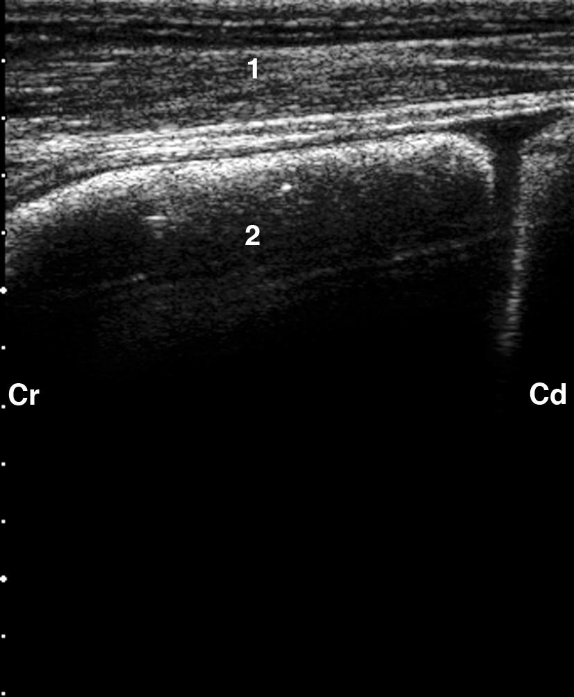
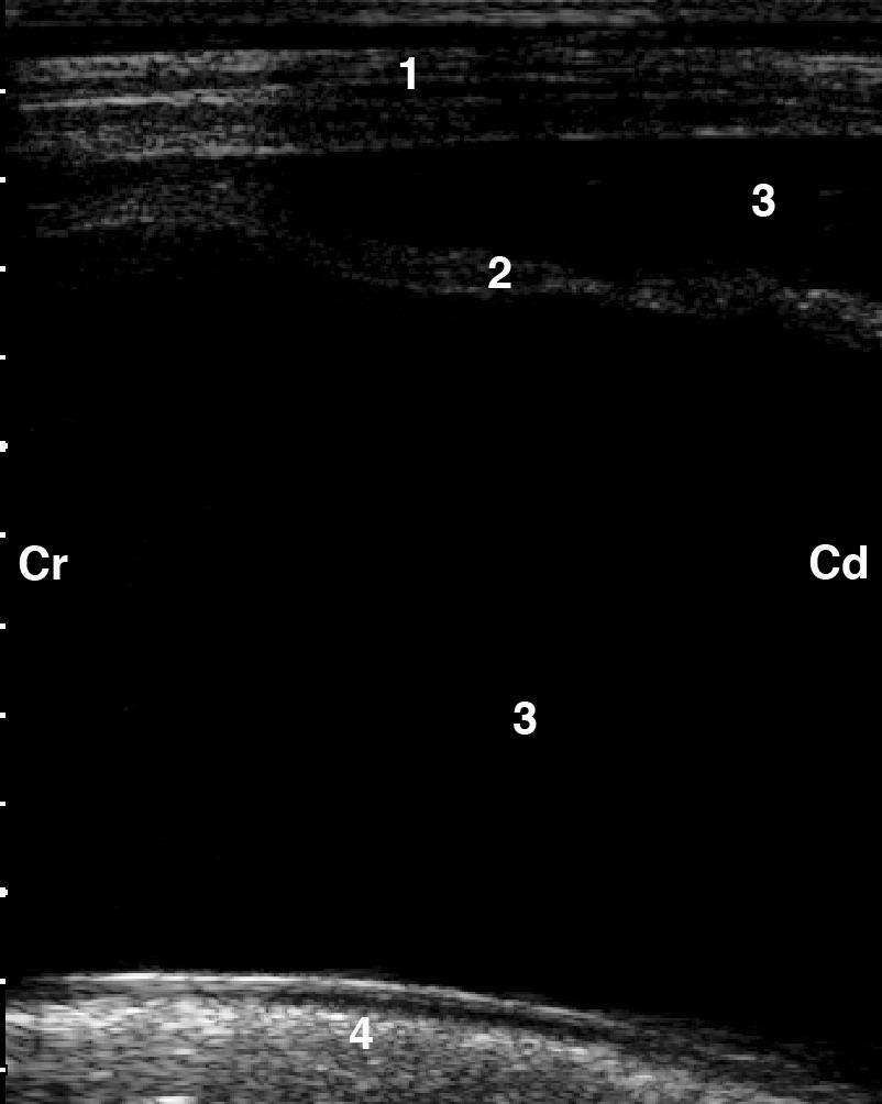
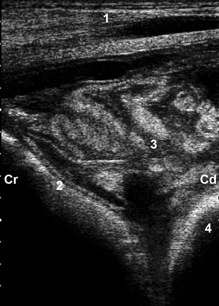
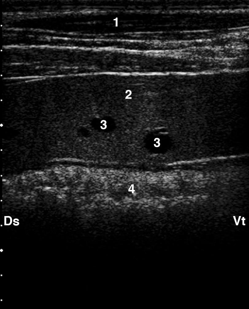
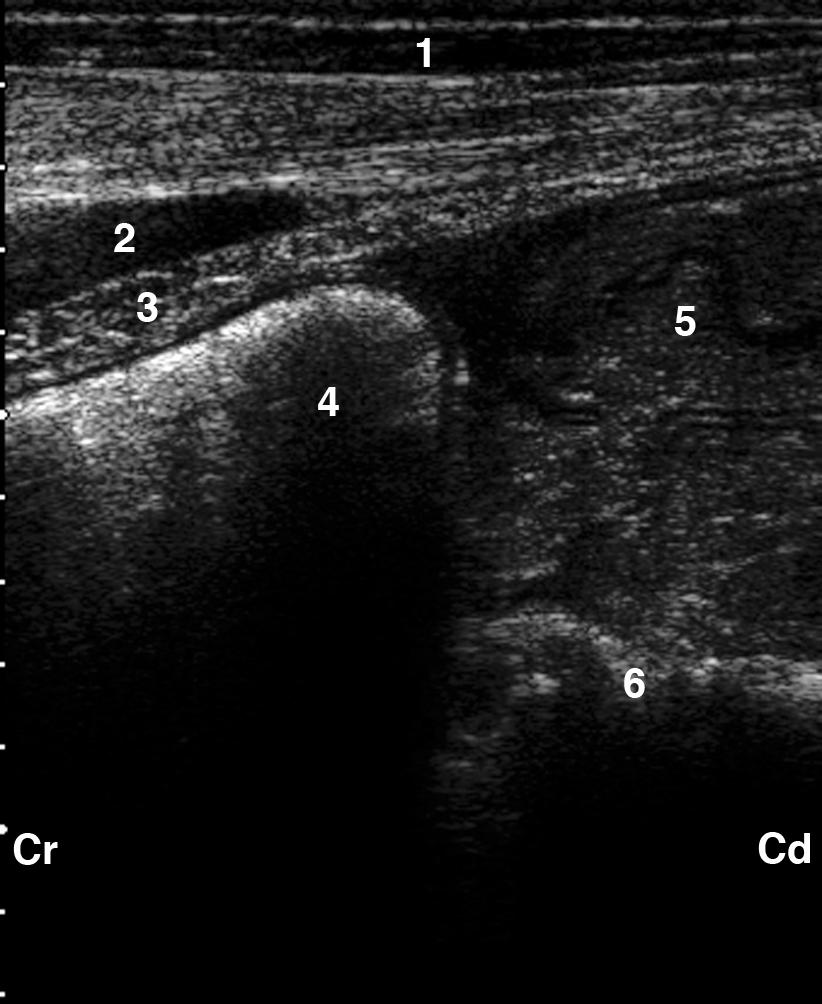
5 example, in cases with suspected traumatic reticuloperitonitis, the examination is carried out in the sternal and parasternal regions, and in cattle suspected of having cholestasis, only the costal part of the abdominal wall on the right side is examined. However, even experienced clinicians may not be able to pinpoint the organ affected and make a diagnosis in patients in which abdominal disease is suspected. In such cases, both sides of the abdomen are examined. Normally, the reticulum [1, 2], spleen [1-4], rumen [2] and parts of the abomasum [5, 6] are seen on the left side and the liver [7-9], omasum [10,11], parts of the abomasum [5, 6], small intestine [12, 13], large intestine [14, 15] and right kidney [16] on the right. The uterus may be seen on either side depending on the stage of pregnancy. This review describes the ultrasonographic techniques used for examination of the reticulum, rumen, omasum, abomasum, small intestine and large intestine. The normal findings are presented followed by a description of the most important diseases of these organs. Reticulum/Rumen Ultrasonographic examination of the reticulum and normal findings For ultrasonographic examination of the reticulum, the transducer is applied to the ventral aspect of the thorax on the left and right of the sternum as well as to the left and right lateral thorax up to the level of the elbow [1, 2, 17, 18]. The reticulum is first examined from the left side and then the right. The normal reticulum appears as a half-moon-shaped structure with an even contour (Fig. 1). It contracts at regular intervals and when relaxed, is situated immediately adjacent to the diaphragm and ventral portion of the abdominal wall. The different layers of the reticular wall usually cannot be imaged, and the honeycomb-like structure of the mucosa is not often seen clearly. In cattle with ascites, the tunica serosa of the reticulum appears as a narrow echogenic line, the tunica muscularis is seen as a hypoechogenic line and the tunica mucosa as a wider echogenic line (Fig. 2). Contents of the reticulum cannot be normally imaged because of their partly gaseous composition. Foreign bodies and magnets also cannot usually be seen in the reticulum because of the gas content of 3
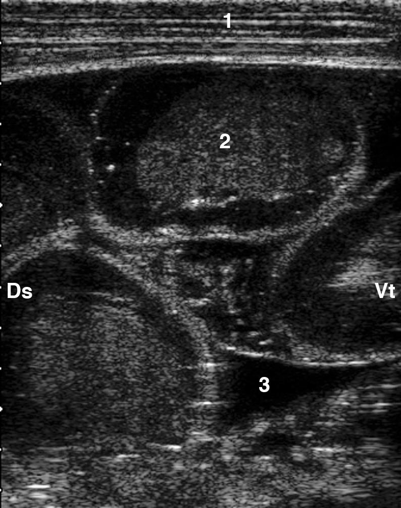
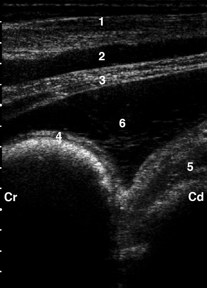
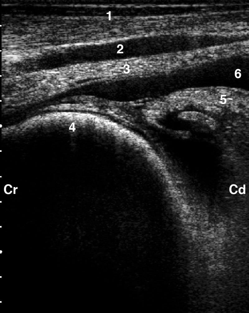
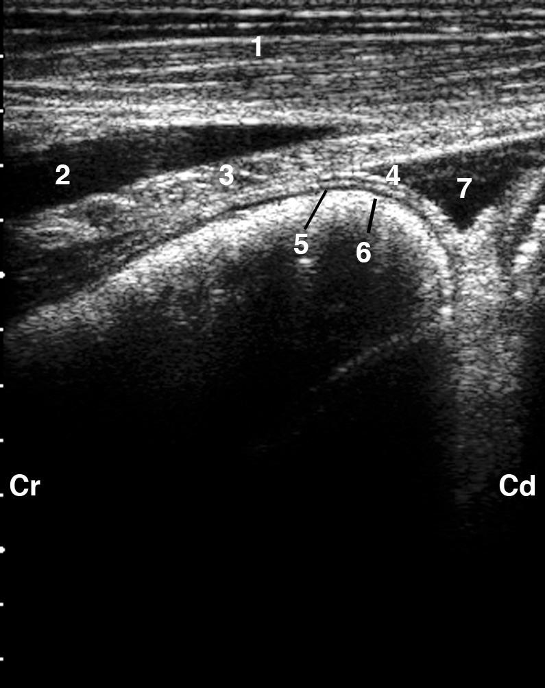
6 the reticulum. Radiography is the method of choice for identifying radiodense foreign bodies and magnets [19]. The craniodorsal blind sac of the rumen and the transition to the ventral sac of the rumen can be seen caudal to the reticulum (Fig. 3). The abomasum is frequently seen between the craniodorsal blind sac of the rumen or rumen and ventral abdominal wall, immediately caudal to the reticulum. For assessment of reticular motility, the transducer is placed on the left ventral thoracic region. The reticulum is located and observed for three minutes without moving the transducer. The number, amplitude and speed of reticular contractions and the duration of the interval of relaxation between two biphasic reticular contractions are assessed. The reticulum normally contracts once per minute in a biphasic manner, in which the first contraction is incomplete [1, 2]. Thus, in the three-minuteobservation period, the reticulum has three biphasic contractions. Contraction of the craniodorsal blind sac of the rumen is seen immediately after the second reticular contraction. Rumination is associated with an additional ruminal contraction, which occurs immediately prior to the biphasic contraction. Control of reticular motility Reticular motility is initiated and controlled by the vagus nerve, which in turn is under the influence of the gastric center in the medulla oblongata [20]. A number of factors, including eating, rumination and stress, affect reticular motility [21, 22]. The number of reticular contractions is highest during eating (approximately 1.5/min) and lowest when the cow is stressed (a little less than 1.0/min). In cattle with vagal indigestion, the number of reticular contractions may be reduced, normal or increased [21, 23]. In 144 cows with vagal indigestion, the number of contractions ranged from 0 to 12 per 3 minutes. Cows with reticulo-omasal stenosis had significantly more reticular contractions (4.6 contractions/3 min) than cows with functional stenosis of the pylorus (3.6 contraction/3 min). Reticular hypermotility also commonly occurs in cows with 4
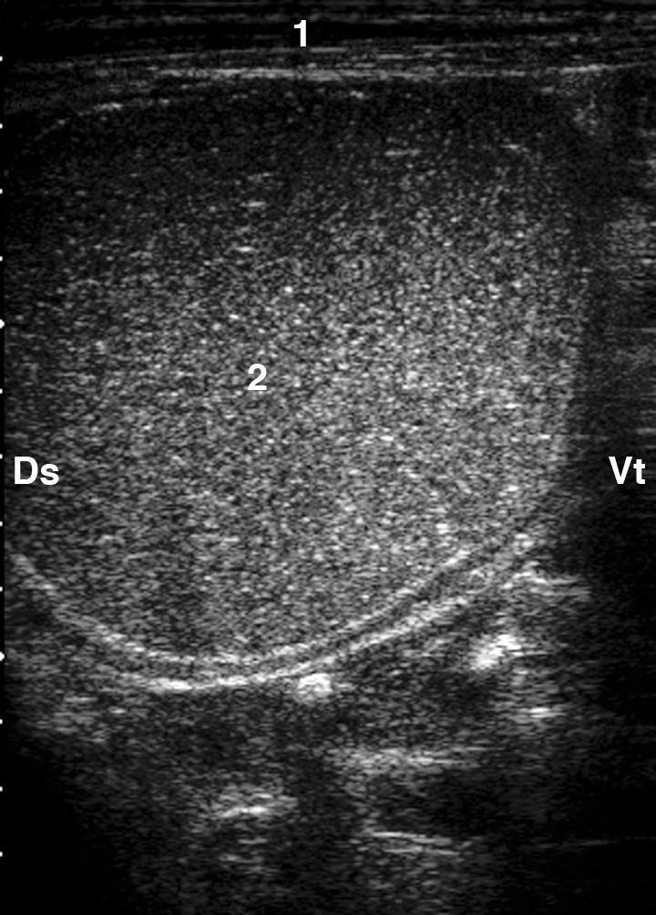
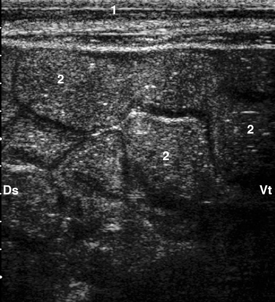
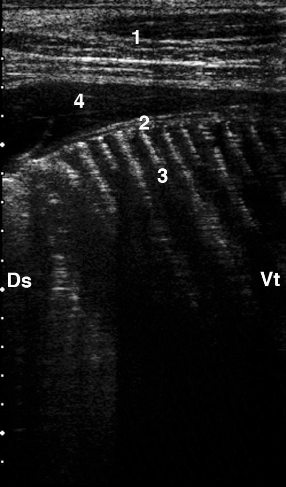
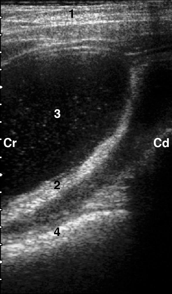
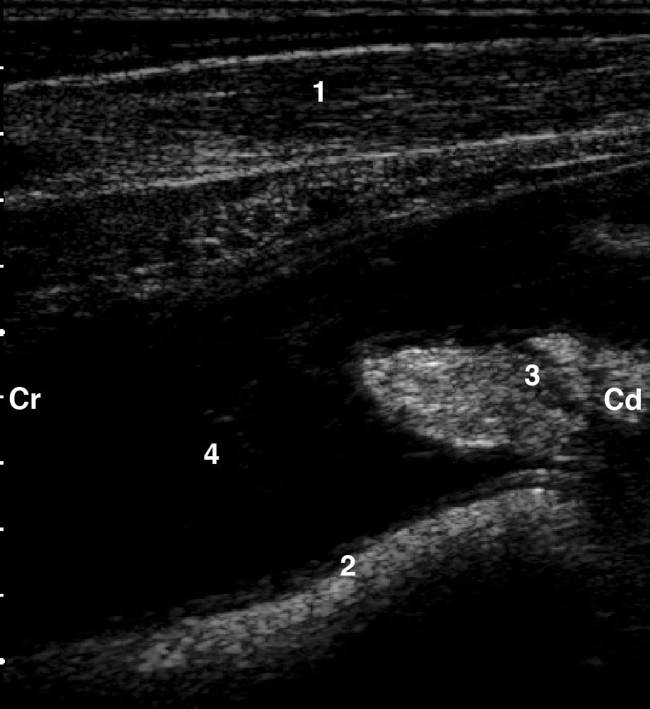
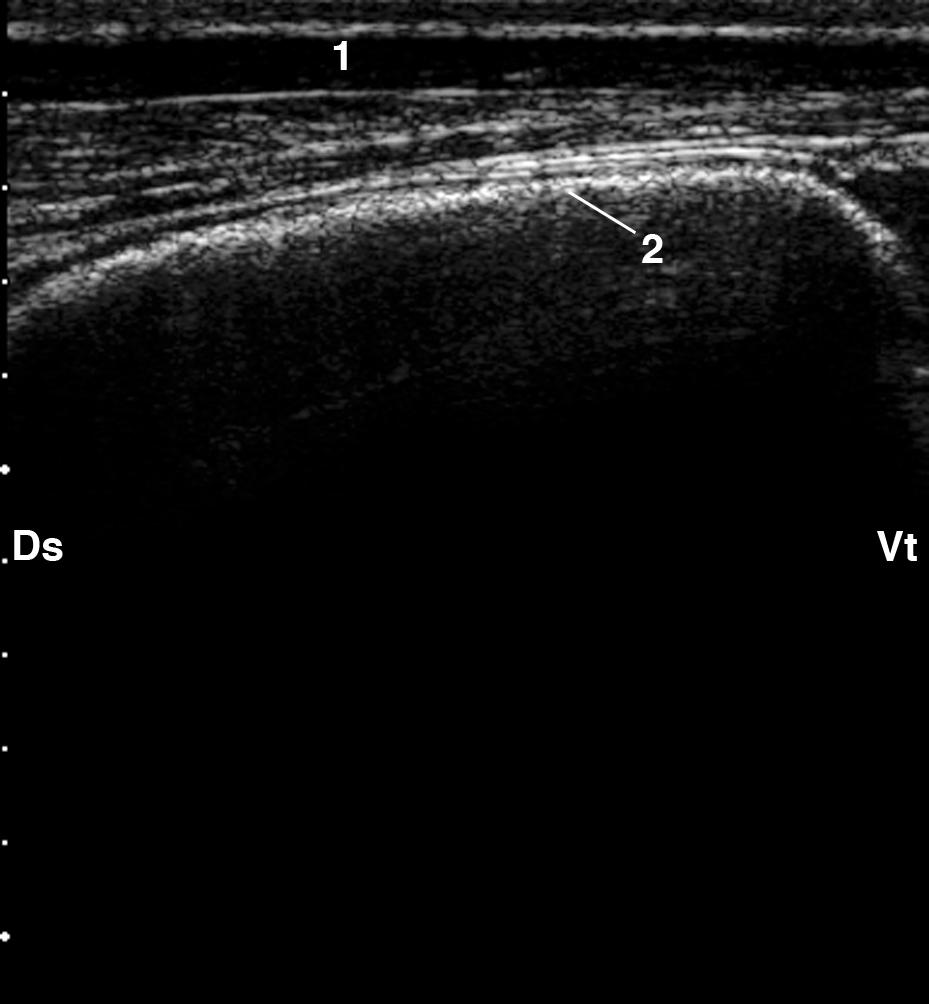
7 mechanical stenosis in the region of the reticulum [24]; two of three cows with reticuloomasal stenosis caused by a rope foreign body had 6 contractions per 3 minutes. (for reticular motility in traumatic reticuloperitonitis, see traumatic reticuloperitonitis). Drugs also affect reticular motility. Atropine, scopolamine and xylazine inhibit reticular motility [25, 26]. Reticular atony occurred within 0 to 3 minutes of intravenous administration of these drugs and lasted 3 to 111 minutes. The drug dose was directly proportional to the onset and duration of atony. Rumen The rumen can be imaged in the region of the left costal part of the abdominal wall. From dorsal to ventral are the dorsal sac of the rumen, the longitudinal groove of rumen and the ventral sac of the rumen. The ruminal wall appears echogenic. Reverberation artefacts running parallel to the ruminal wall are seen in the region of the dorsal gas cap [27]. The ingesta are located in the middle of the rumen and appear echogenic with gaseos inclusions. The fluid in the ventral aspect of the rumen is hypoechogenic [27]. Spleen The spleen is located craniodorsally on the rumen. The spleen can be seen in the intercostal spaces 7 to 12 [3, 4]. It is 2.0 to 5.0 cm thick, and tapered ventrally. The splenic capsule appears as an echogenic line. The splenic parenchyma consists of numerous small regularly spaced echoes, and vessels within the parenchyma appear as anechoic round to oval or elongated images (Fig. 4). The long axis is oblique, running from caudodorsal to cranioventral. Traumatic reticuloperitonitis 5
8 In cattle with traumatic reticuloperitonitis, ultrasonography can be used to identify morphological changes in the region of the cranial, ventral or caudal reticular wall [28]. The caudoventral reticular wall is the most frequently affected, often in association with the craniodorsal blind sac of the rumen. The changes in the contour of the reticulum depend on the severity of the inflammatory changes. Deposits of fibrinous tissue interspersed with fluid pockets are frequently seen on the reticular serosa and structures adjacent to the reticulum (Fig. 5-8). The extent of the inflammatory lesions varies greatly, but may occasionally extend to the region of the flank fold. Remarkably, these inflammatory lesions can resolve within six months of effective treatment [29]; ultrasonography revealed that nine of 16 cows had no remaining adhesions six months after treatment and the adhesions in the other seven cows had markedly regressed [29]. Reticular abscesses have an echogenic capsule of varying thickness, which surrounds a homogeneous hypoechogenic to moderately echogenic centre (Fig. 9, 10). The contents of an abscess are frequently partitioned by echogenic septa. Abscesses are usually caudoventral to the reticulum, but may be cranial or lateral to the reticulum. Abscesses are often seen between the reticulum and spleen, reticulum and liver or reticulum and omasum or abomasum. Reticular abscesses vary in diameter from a few centimetres to more than 15 cm. In certain cases, it is possible to drain abscesses through an ultrasound-guided transcutaneous incision [30]. However, the abscess must be immediately adjacent to and attached to the abdominal wall, and the intercostal space over the abscess must be large enough. Reticular activity is almost always affected in cattle with traumatic reticuloperitonitis. The frequency, amplitude or speed of contractions, singly or combined, may be abnormal. The frequency can be reduced from three to two, one or no contractions per three minutes. The reduction in the amplitude of contractions varies; when formation of adhesions is extensive, reticular contractions appear indistinct via ultrasonography. Although the pattern of 6
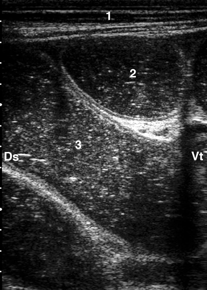
9 biphasic contraction often is maintained, the reticulum contracts by only 1 to 3 cm. The speed of reticular contractions may be normal but can be markedly reduced. An inflammatory effusion is often seen near the reticulum in cattle with traumatic reticuloperitonitis. Peritoneal effusion is visible ultrasonographically as an accumulation of fluid without an echogenic margin and restricted to the reticular area (Fig. 11). Depending on the fibrin and cell content, the fluid may be anechoic or hypoechogenic. Fibrinous deposits are easily identified in the fluid, and sometimes, bands of fibrin are seen within the effusion. Occasionally, the peritoneal effusion is considerable and extends to the caudal abdomen (Fig. 12). The lesions may be within (omental bursitis) or outside the omentum (peritonitis). In such cases, the greater omentum is commonly seen as an echogenic structure surrounded by fluid. The spleen, particularly its distal portion, is often affected in cattle with traumatic reticuloperitonitis. Fibrinous changes are frequently seen as echogenic deposits of varying thickness (Fig. 13), often surrounded by fluid, between the spleen and reticulum or rumen. The spleen may be covered with fibrinous deposits. Occasionally, one or more splenic abscesses are visible (Fig. 14), and the vasculature may be dilated indicating splenitis. Omasum The omasum is visible in the 6th to 11th intercostal spaces [10, 11]. It has a crescent shape with only the wall closest to the transducer visible as a thick echogenic line (Fig15). The size of the omasum varies with the intercostal spaces from 16.3 ± 1.5 cm to 56.9 ± 10.0 cm. The size of the omasum is greatest in the 9th intercostal space and decreases cranially and caudally. The omasum is located closest to the right abdominal wall in the 8th and 9th intercostal spaces and in many cows it is immediately adjacent to it in this region. Unlike the reticulum, the omasum of healthy cows does not have any apparent active motility of its own. The omasal laminae cannot be imaged in healthy cows because of intraluminal gas. 7
10 Occasionally, the attachments of the omasal laminae are seen as short echogenic cone-shaped structures that protrude from the inner wall of the omasum. Occasionally the omasal laminae can be seen in ill cattle (Fig.16) with an increased fluid content in the omasum, usually caused by abomasal reflux. The position and size of the omasum change in cattle with right and left displacement of the abomasum, abomasal volvulus, traumatic reticuloperitonitis, ileus and reticulo-omasal stenosis [10, 31]. Omasal motility can be observed in rare cases of reticuloomasal stenosis. Primary diseases of the omasum are rare; one publication described the ultrasonographics findings in a cow that had a leiomyoma in the omasum [32]. Abomasum Ultrasonography is a valuable technique for the assessment of the size, position and content of the abomasum. The abomasum can be visualised approximately 10 cm caudal to the xyphoid process from the left and right paramedian regions and from the ventral midline [5, 6]. The bulk of the abomasum is situated to the right of the ventral midline. The abomasum is frequently seen immediately caudal to the reticulum between the craniodorsal blind sac of the rumen, or the rumen, and the ventral abdominal wall (Fig. 3). The wall of the abomasum appears at the most as a thin echogenic line. However, the abomasum is easily differentiated from neighbouring organs by the ultrasonographic appearance of its contents, which are seen as a heterogeneous moderately echogenic mass with echogenic stippling. Parts of the abomasal folds can occasionally be seen as echogenic structures within the content of the abomasum. Passive and slow movement of the abomasal contents are frequently seen. The position of the abomasum changes in late pregnancy because of the expanding uterus [33-35]. The abomasum changes from a more longitudinal to a transverse position, but within 14 days of parturition resumes its normal longitudinal position. Abomasocentesis 8
11 Percutaneous ultrasound-guided abomasocentesis can be performed to evaluate the nature and chemical composition of abomasal contents [36]. Centesis is performed at a site where the abomasum appears large and where no other organs are in the way. Abomasal fluid is assessed mainly for colour, smell and the presence of blood, and the ph is determined. Blood does not normally occur in abomasal fluid. In the majority of cases, blood originates from an abomasal ulcer [36] and rarely from the rumen or small intestine. Left displacement of the abomasum An ultrasonographic examination is useful to confirm the diagnosis of left displacement of the abomasum in unclear cases [37]. The last three intercostal spaces on the left side are examined ventrally to dorsally with the transducer held parallel to the ribs. Normally, the rumen is immediately adjacent to the left abdominal wall. Thus, on ultrasonograms, the ruminal wall is medial to the abdominal wall and from ventral to dorsal appears as a smooth, thick, echogenic line, which is indented at the left longitudinal sulcus. With left displacement of the abomasum, the wall of the rumen often is still immediately adjacent to the abdominal wall in the ventral region. When the transducer is moved dorsally, it becomes apparent that the wall of the rumen is pushed medially and then can no longer be imaged ultrasonographically. Instead the abomasum is seen, located between the abdominal wall and rumen. Moving the transducer further dorsally, the abomasum disappears and the rumen reappears on the screen. The abomasal contents do not appear uniform because, ventrally, there are fluid ingesta, and dorsally, there is a gas cap that varies in extent. The ingesta visible ventrally in the abomasum appear hypoechogenic (Fig. 17). Occasionally, the abomasal folds are visible among the ingesta and appear as elongated, echogenic, sickle-shaped structures. The ruminal wall can often be seen medial to the ingesta. The abomasal gas cap, seen further dorsally, is characterised by reverberation artifacts (Fig. 18) similar to those observed during the ultrasonographic examination of lung. 9
12 Right displacement of the abomasum and abomasal volvulus Ultrasonography also is a useful diagnostic tool in doubtful cases of right displacement of the abomasum and abomasal volvulus. The area immediately caudal to the last rib and the caudal three to four intercostal spaces on the right side are examined from ventral to dorsal with the transducer held parallel to the ribs [18, 38, 39]. Normally, loops of small intestine are imaged in cross-section and, less commonly, longitudinally in the ventral abdomen, and further dorsally, the liver is seen immediately adjacent to the right abdominal wall. In animals with right displacement of the abomasum or abomasal volvulus, the liver is displaced from the abdominal wall. The abomasum is seen where the liver would normally be, immediately adjacent to the right abdominal wall. Its ultrasonographic appearance is the same as that described previously for left displacement. The ultrasonographic appearance of right displacement of the abomasum cannot be differentiated from abomasal volvulus [38, 39]. In both disorders, the liver, omasum and small and large intestine may be displaced depending on the size of the abomasum [38, 40]. In contrast to healthy cows in which the liver can always be imaged on the right side, there is a significant decrease in the frequency with which the liver is seen from the different intercostal spaces in cows with abomasal displacement or volvulus; in fact, sometimes the liver cannot be imaged on the right side at all. This is because the liver is displaced from the abdominal wall by the abomasum. The omasum and small and large intestine are also less frequently seen in cows with right displacement of the abomasum and abomasal voluvlus compared with healthy cows. Defects in abomasal emptying Defects in emptying of the abomasum occur with functional or mechanical pyloric stenosis. Abomasal emptying can also be impaired secondary to intestinal ileus. With all these disorders, the abomasum is dilated but not displaced dorsally or torsed. Depending on its 10
13 degree of fill, the dilated abomasum can be imaged on the right side from the ventral region of intercostal spaces 8 to 12 or even from the ventral abdomen caudal to the ribs. In contrast to left or right displacement, there is no accumulation of gas in the non-displaced, dilated abomasum. The abomasal contents appear predominantly hypoechogenic and homogeneous, and because of sequestration of hypochloric acid, are frequently fluid in nature. This often allows for good visualisation of the abomasal folds, which appear as thin, echogenic, wavy structures (Fig. 19). In animals with ileus, the loops of intestine are dilated and clearly visible. With pyloric stenosis, the small intestine is empty. Abomasitis/abomasal ulcers Discrete lesions of the abomasal mucosa such as chronic abomasitis, parasitic nodules, erosions and type 1 ulcers cannot be imaged ultrasonographically using a 3.5 MHz or 5.0 MHz transducer. Type 3 abomasal ulcers produce abdominal changes similar to those of fibrinous traumatic reticuloperitonitis with abscessation. Type 4 abomasal ulcers produce signs of generalized peritonitis with ascites, fibrinous adhesions and thickening of the intestinal wall. Abomasal ulcers have never been imaged with ultrasound. Intestine Normal small intestine For ultrasonography of the small intestine in cattle, the area from the tuber coxae to the eighth intercostal space and from the transverse processes of the vertebrae to the linea alba on the right side is examined [12, 13]. The appearance of loops of small intestine and their diameter, contents and motility are assessed. The wall of the normal small intestine is 2 to 3 mm thick and its luminal diameter is 2 to 4 cm [13]. Evaluation of the contents of the small intestine in cattle is usually straight forward because there is generally no gas. This is because, unlike small animals and humans, ruminants digest carbohydrates principally in the forestomachs, from which the gas is eructated [41]. The ultrasonographic appearance of the 11
14 contents of the small intestine varies. Most commonly, the intestine contains mucus or feed, which appears hyperechogenic. In these cases, not only the intestinal wall closest to the transducer, but also the intestinal contents and the wall furthest from the transducer can be visualised. This is also true for intestine filled with fluid, which is hypoechogenic. In rare cases with gaseous intestinal contents, the intestinal wall closest to the transducer appears as a hyperechogenic line adjacent to an acoustic shadow. Because of reflection of the ultrasound waves at the soft tissue-air interface, the intestinal contents and the intestinal wall furthest from the transducer cannot be visualised. In contrast with results in dogs [42], there is no difference between the diameter of most of the small intestine before and after feeding in cattle. This uniformity is probably because the forestomachs constitute a feed reservoir and ingesta pass along the intestine continuously, independent of feed intake, and without changes in the diameter of the intestinal lumen, which would be measurable by ultrasonography. The cranial part of the duodenum is relatively easy to identify because it originates from the abomasum and is in close proximity with the liver and gallbladder. It is seen in cross section or longitudinally medial or ventral to the gallbladder and almost always from the 10th or 11th intercostal space. The diameter of the cranial part of the duodenum ranges from 0.9 to 5.5 cm [13]. The descending duodenum also is quite easily imaged in most cattle. From the 10th, 11th or 12th intercostal space and from the dorsal region of the right flank, it is observed in cross section or longitudinally. The following characteristics allow accurate identification of the descending duodenum: it is situated immediately adjacent to the abdominal wall and runs horizontally and caudally between the serosal lamellae of the omentum, which appears as an echogenic envelope around the descending duodenum. It then courses caudally to form the caudal duodenal flexure at the level of the tuber coxae. After this, it runs medially and cranially to form the ascending duodenum. The diameter of the descending duodenum varies from 1.5 to 3.5 cm. The ascending duodenum is situated more than 20.0 cm from the right abdominal wall and as a result cannot be imaged via ultrasonography. 12
15 The jejunum and ileum form the longest part of the small intestine and cannot be differentiated from one another ultrasonographically. It is typical to see more than ten loops of jejunum and ileum immediately adjacent to one another from the flank and lateral abdominal wall and from intercostal spaces 9 to 12 [13]. The loops of small intestine are usually seen in cross-section and occasionally longitudinally. They can be differentiated from the descending duodenum because they are not surrounded by omentum and because they are constantly in motion (Fig. 20). Loops of jejunum and ileum are seen from the 10th intercostal space in approximately 70 % of cows and from the 9th intercostal space in approximately 10 % of cows. The diameter of the jejunum and ileum ranges from 2.0 to 4.0 cm. The number of loops of jejunum and ileum visible longitudinally and in cross-section are approximately the same when imaged from the flank and the 12th intercostal space, but it decreases when imaged from the successive intercostal spaces. Ileus of the small intestine When ileus of the small intestine is suspected, an ultrasonographic examination should evaluate the diameter, motility and anatomical arrangement of the small intestine, evidence of peritonitis and the possible cause of the ileus [18, 43]. The most important parameters are diameter and motility of the small intestine; identification of the cause of ileus via ultrasonography is rarely possible. In cattle with ileus, the small intestine is dilated in at least one area and has a diameter of more than 3.5 cm [43]. Moreover, the motility of the small intestine is usually reduced or absent. Sometimes, hypoechogenic fluid, attributable to transudation, is visible between the dilated loops of intestine. Independent of the localisation of ileus and its cause, the loops of small intestine are most commonly imaged in cross-section (Fig. 21), often in both cross-section and longitudinally (Fig. 22) but rarely only longitudinally. 13
16 The site of ileus markedly affects the number of dilated loops of intestine seen in crosssection and longitudinally from either the flank or each intercostal space. When only one or a few, usually markedly dilated, loops of small intestine are seen (Fig. 23), ileus of the duodenum is most likely [43, 44]. More than five loops of small intestine seen in one area usually indicates ileus of the jejunum or ileum. Rarely, when the ileus is localised in the proximal jejunum, only one or two dilated loops of small intestine are imaged. The number of dilated loops of small intestine increases if the localisation of the ileus is more distal. Conversely, in the 8th and 9th intercostal spaces, the number of dilated loops of small intestine generally decreases. The loops become more dilated with ileus of the proximal small intestine. The largest diameter of intestine measured from the 12th intercostal space varied from 6.5 to 9.9 cm (7.7 ± 1.9) in animals with ileus of the duodenum, from 3.5 to 9.8 cm (5.5 ± 1.7) in animals with ileus of the jejunum and from 4.4 to 5.5 cm (5.0 ± 0.4 cm) in animals with ileus of the ileum [43]. When interpreting the diameter of the intestine, it is important to remember that in healthy cows, in which the intestine is full of ingesta, all parts of the intestine will have a similar diameter. By contrast, in animals with ileus, in addition to the extremely dilated loops of intestine proximal to the ileus, there are usually empty loops of intestine distal to the ileus. Furthermore, the intestinal lumen of a healthy cow is constantly changing, whereas the increased intestinal diameter of a cow with ileus remains unchanged because the intestinal motility is markedly reduced or absent. The contents of the small intestine appear predominantly echogenic and rarely anechoic. Different parts of the intestine of the same animal may be echogenic or anechogenic. Intraluminal gas, which is associated with reverberation artifacts, is rarely observed. In the majority of cows with ileus of the small intestine, intestinal motility is markedly reduced or absent. However, movement of intestinal contents often is apparent although no intestinal contractions are visible. This flowing movement is presumably due to the passive 14
17 movement of the intestine by respiratory activity and possibly by the movement of adjacent organs such as the rumen or abomasum. The cause of ileus can seldom be determined ultrasonographically. Often, this is because the cause of ileus is further from the abdominal wall than the penetration capacity of the transducer. A common cause of ileus is intussusception, the ultrasonographic appearance of which in cross-section has been described as bowel within bowel, bull's eye lesion, target pattern or as multiple layered, onion ring-type mass with varing echogenicities. Typically, the invaginated intestinal wall is swollen. Depending on the severity of oedema and the imaging plane, the affected area of intestine may appear hyperechogenic or hypoechogenic. Viewed longitudinally, the typical lumen-within-a-lumen can be clearly identified and has been described as a "sandwich" configuration. In rare cases, compression of the small intestine by abscesses in the region of the liver or compression of the duodenum between the liver and gallbladder can be identified. Ileus can also be caused by generalised peritonitis with fibrinous adhesions involving the small intestine. In such cases, thickening of the intestinal wall, fibrinous deposits and accumulation of intraabdominal fluid are usually seen. In cattle with hemorrhagic bowel syndrome (HBS), the blood clots can occasionally be imaged as an echogenic masses in the lumen of the small intestine (Fig. 24) [45]. In a heifer with duodenal ileus, ultrasonography showed that the gallbladder was not in its normal position, and exploratory laparotomy revealed that the displaced gallbladder was causing obstruction of the duodenum [46]. Failure of ingesta to pass through the small intestine results in delayed passage of ingesta through the abomasum, omasum and rumen, which consequently become dilated (see Defects in abomasal emptying). Uncomplete perforation of the intestine usually leads to chronic peritonitis. Ultrasonographically, this is seen as intraabdominal fluid with bands of fibrin, which may have a spiderweb-type of appearance among the loops of intestine and organs. Free intra- 15
18 abdominal gas with its associated reverberation artifacts may be seen if intestinal gas has leaked through the perforation. Normal large intestine Carbohydrates remaining in the ingesta after their passage through the forestomachs are fermented in the large intestine, and the gas produced makes it more difficult to image this section of the bowel [12]. The large intestine is always visible from the flank and is situated medial to the descending duodenum, whereby the colon is more dorsal and the proximal loop of the colon and caecum are more ventral. The large intestine is usually easy to differentiate from the small intestine based on its marked gas content [12, 14, 15]. Because of the gas, only the wall of the large intestine closest to the transducer can usually be imaged and appears as a thick echogenic line. However, reverberation artifacts originating from the tissue-gas interphase may become superimposed on the image of the wall and obscure it. The wall of the large intestine furthest from the transducer cannot be imaged. Usually, the proximal loop of the large colon, the caecum and the colon can be visualised. The proximal loop of the large colon and the caecum appear as thick, echogenic, continuous and slightly curved lines. The spiral loop of the colon has the appearance of a garland with a number of echogenic arched lines next to each other. In contrast to the small intestine, which has vigorous peristaltic activity and segmental contractions, very few contractions are observed in the large intestine. Caecal dilatation Diagnosis of caecal dilatation usually is straightforward but may be difficult when the dilatation is complicated by retroflexion of the caecum. In that case, either no abnormal transrectal findings can be palpated, or a distended viscus can be palpated only with the tips of the fingers. The differential diagnoses must include ileus of the small intestine and right displacement of the abomasum, respectively. A diagnosis by clinical examination alone may 16
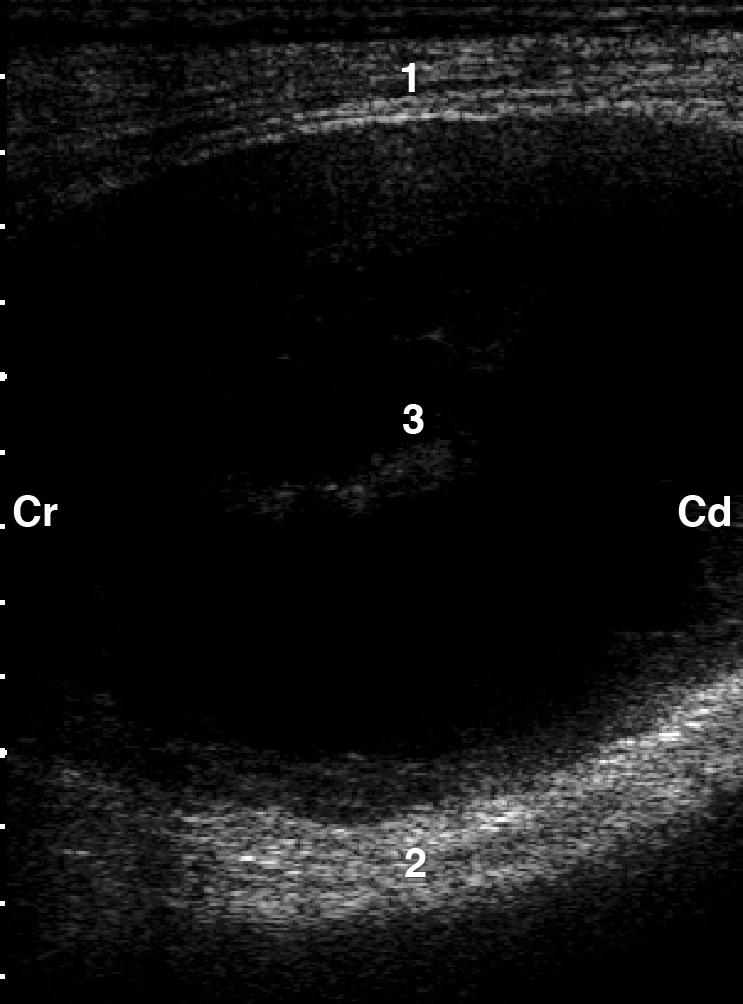
19 not be possible, but ultrasonography allows the differentiation between right displacement of the abomasum, ileus of the small intestine and caecal dilatation. A dilated caecum can always be imaged from the lateral abdominal wall [14, 47] and in some cases, it may be seen from the 12th, 11th and 10th intercostal spaces. The dilated caecum and the proximal loop of the colon are almost always immediately adjacent to the abdominal wall. Because of the gaseous contents, only the wall of the dilated caecum and proximal loop of the colon closest to the transducer are seen ultrasonographically and appear as thick, echogenic, semi-circular lines (Fig. 25). In cases with fluid ingesta instead of gaseous contents in the caecum and proximal loop of the colon, the lumen appears moderately echogenic. Differentiation of caecum and proximal loop of the colon may be difficult ultrasonographically unless the ileocaecal fold of the peritoneum between the two can be identified. References [1] Götz M. Sonographische Untersuchungen an der Haube des Rindes. Dissertation, Faculty of Veterinary Medicine, University of Zurich, [2] Braun U, Götz M. Ultrasonography of the reticulum in cows. Am J Vet Res 1994;55(3): [3] Sicher D. Sonographische Untersuchungen an Lunge, Mediastinum und Milz des Rindes. Dissertation, Vetsuisse Faculty, University of Zurich, [4] Braun U, Sicher D. Ultrasonography of the spleen in 50 healthy cows. Vet J 2006;171(3): [5] Wild K. Sonographische Untersuchungen am Labmagen des Rindes. Dissertation, Vetsuisse Faculty, University of Zurich, [6] Braun U, Wild K, Guscetti F. Ultrasonographic examination of the abomasum of 50 cows. Vet Rec 1997;140(4):93-8. [7] Braun U. Ultrasonographic examination of the liver in cows. Am J Vet Res 1990;51(10):
20 [8] Gerber D. Sonographische Befunde an der Leber des Rindes. Dissertation, Vetsuisse Faculty, University of Zurich, [9] Braun U, Gerber D. Influence of age, breed, and stage of pregnancy on hepatic ultrasonographic findings in cows. Am J Vet Res 1994;55(9): [10] Blessing S. Sonographische Untersuchungen am Psalter des Rindes. Dissertation, Vetsuisse Faculty, University of Zurich, [11] Braun U., Blessing S. Ultrasonographic examination of the omasum in 30 healthy cows. Vet Rec 2006;159(24): [12] Marmier O. Sonographische Untersuchungen am Darm des Rindes. Dissertation, Vetsuisse Faculty, University of Zurich, [13] Braun U, Marmier O. Ultrasonographic examination of the small intestine of cows. Vet Rec 1995;136(10): [14] Amrein EM. Ultraschalluntersuchungen bei Kühen mit Blinddarmdilatation. Dissertation, Faculty of Veterinary Medicine, University of Zurich, [15] Braun U, Amrein E. Ultrasonographic examination of the caecum and proximal and spiral ansa of the colon of cattle. Vet Rec 2001;149(2):45-8. [16] Braun U. Ultrasonographic examination of the right kidney in cows. Am J Vet Res 1991;52(12): [17] Kaske M, Midasch A, Rehage J. Sonographic investigation of reticular contractions in healthy sheep, cows and goats and in cows with traumatic reticulo-peritonitis. J Vet Med A 1994;41(10): [18] Braun U. Atlas und Lehrbuch der Ultraschalldiagnostik beim Rind. Berlin: Parey Buchverlag; [19] Braun U, Flückiger M, Nägeli F. Radiography as an aid in the diagnosis of traumatic reticuloperitonitis in cattle. Vet Rec 1993;132(5):
21 [20] Constable PD, Hoffsis GF, Rings DM. The reticulorumen: Normal and abnormal motor function. Part I. Primary contraction cycle. Comp Cont Educ Pract Vet 1990;12(7): [21] Rauch S. Haubenmotorik bei gesunden Kühen und bei Kühen mit Hoflund-Syndrom. Dissertation, Vetsuisse Faculty, University of Zurich, [22] Braun U, Rauch S. Ultrasonographic evaluation of reticular motility during rest, eating, rumination and stress in 30 healthy cows. Vet Rec 2008;163(19): [23] Braun U, Rauch S, Haessig M. Ultrasonographic evaluation of reticular motility in 144 cattle with vagal indigestion. Vet Rec 2009;164(1):11-3. [24] Braun U, Schweizer G, Flückiger M. Radiographic and ultrasonographic findings in three cows with reticulo-omasal obstruction due to a foreign body. Vet Rec 2002;150(18): [25] Gansohr B. Untersuchungen zur Eingabe von Fremdkörpernacktmagneten beim Rind. Dissertation, Vetsuisse Faculty, University of Zurich, [26] Braun U, Gansohr B, Haessig M. Ultrasonographic evaluation of reticular motility in cows after administration of atropine, scopolamine and xylazine. J Vet Med A 2002;49(6): [27] Tschuor A, Clauss M. Investigations on the stratification of forestomach contents in ruminants: an ultrasonographic approach. Eur J Wildl Res, in press. DOI /s [28] Braun U, Götz M, Marmier O. Ultrasonographic findings in cows with traumatic reticuloperitonitis. Vet Rec 1993;133(17): [29] Herzog K, Kaske M, Bischoff C, et al. Post surgical development of inflammatory adhesions and reticular function in cows suffering from traumatic reticuloperitonitis. Dtsch tierärztl Wschr 2004;111(2):
22 [30] Braun U, Iselin U, Lischer C, et al. Ultrasonographic findings in five cows before and after treatment of reticular abscesses. Vet Rec 1998;142(8): [31] Braun U, Blessing S, Lejeune B, et al. Ultrasonography of the omasum in cows with various gastrointestinal diseases. Vet Rec 2007;160(25): [32] Mohamed T, Oikawa S, Koiwa K, et al. Ultrasonographic diagnosis of omasal leiomyoma in a cow. Vet Rec 2004;155(17): [33] Van Winden SC, Brattinga CR, Müller KE, et al. Position of the abomasum in dairy cows during the first six weeks after calving. Vet Rec 2002;151(15): [34] Sendag S, Seeger T, Wehrend A. Sonographische Untersuchung über die Lageänderungen des Labmagens bei Kühen im peripartalen Zeitraum. Dtsch tierärztl Wschr 2005;112(9): [35] Wittek T, Constable PD, Morin DE. Ultrasonographic assessment of change in abomasal position during the last three months of gestation and first three months of lactation in Holstein-Friesian cows. J Am Vet Med Assoc 2005;227(9): [36] Braun U, Wild K, Merz M, et al. Percutaneous ultrasound-guided abomasocentesis in cows. Vet Rec 1997;140(23): [37] Braun U, Pusterla N, Schönmann M. Ultrasonographic findings in cows with left displacement of the abomasum. Vet Rec 1997;141(13): [38] Feller B. Sonographische Untersuchungen bei Kühen mit rechtsseitiger Labmagenverlagerung mit und ohne Torsion. Dissertation, Vetsuisse Faculty, University of Zurich, [39] Braun U, Feller B. Ultrasonographic findings in cows with right displacement of the abomasum and abomasal volvulus. Vet Rec 2008;162(10): [40] Braun U, Feller B, Haessig M, et al. Ultrasonographic examination of the omasum, liver and small and large intestines in cows with right displacement of the abomasum and abomasal volvulus. Am J Vet Res 2008;69(6):
23 [41] Gürtler H. Die Physiologie der Verdauung und Absorption. In: Kolb E, editor. Lehrbuch der Physiologie der Haustiere. Stuttgart: VEB Gustav Fischer; p [42] Penninck DG, Nyland TG, Fisher PE, et al. Ultrasonography of the normal canine gastrointestinal tract. Vet Radiol 1989;30(6): [43] Braun U, Marmier O, Pusterla N. Ultrasonographic examination of the small intestine of cows with ileus of the duodenum, jejunum or ileum. Vet Rec 1995;137(9): [44] Lejeune B, Lorenz I. Ultrasonographic findings in 2 cows with duodenal obstruction. Can Vet J 2008;49(4): [45] Dennison AC, VanMetre DC, Callan RJ, Dinsmore P, Mason GL, Ellis RP. Hemorrhagic bowel syndrome in dairy cattle: 22 cases ( ). J Am Vet Med Assoc 2002;221(5): [46] Boerboom D, Mulon PY, Desrochers A. Duodenal obstruction caused by malposition of the gallbladder in a heifer. J Am Vet Med Assoc. 2003;223(10): [47] Braun U, Amrein E, Koller U, et al. Ultrasonographic findings in cows with dilatation, torsion and retroflexion of the caecum. Vet Rec 2002;150(3):
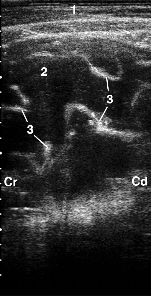
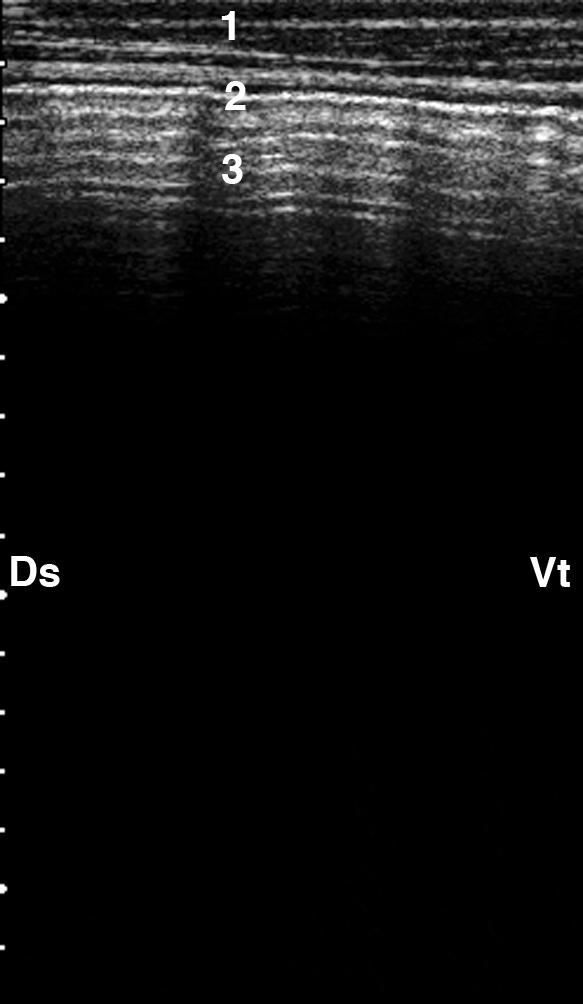
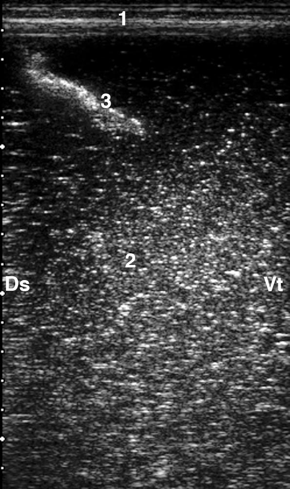
24 Legend to Figures Fig. 1: Ultrasonogram of the normal reticulum imaged from the left sternal region. 1 Ventral abdominal wall, 2 Reticulum, Cr Cranial, Cd Caudal. Fig. 2: Ultrasonogram of the normal reticulum in a cow with mild ascites imaged from the left sternal region. 1 Ventral abdominal wall, 2 Musculophrenic vein, 3 Diaphragm, 4 Tunica serosa of the reticulum, 5 Tunica muscularis of the reticulum, 6 Tunica mucosa of the reticulum, 7 Mild ascites, Cr Cranial, Cd Caudal Fig. 3 : Ultrasonogram of the reticulum, craniodorsal blind sac of the rumen and abomasum imaged from the left sternal region. 1 Ventral abdominal wall, 2 Musculophrenic vein, 3 Diaphragm, 4 Reticulum, 5 Abomasum, 6 Craniodorsal blind sac of the rumen, Cr Cranial, Cd Caudal. Fig. 4: Ultrasonogram of the normal spleen imaged from the distal portion of the 6th intercostal space. 1 Lateral thoracic wall, 2 Spleen, 3 Spleen vessels, 4 Rumen, Ds Dorsal, Vt Ventral. Fig. 5: Ultrasonogram of echogenic deposits on the reticulum and accumulation of fluid in a cow with traumatic reticuloperitonitis imaged from the left ventral thorax. 1 Ventral abdominal wall, 2 Musculophrenic vein, 3 Diaphragm, 4 Reticulum, 5 Echogenic deposits of fibrin, 6 Accumulation of fluid, Cr Cranial, Cd Caudal. Fig. 6: Ultrasonogram of fluid accumulation interspersed with fibrin in a cow with traumatic reticuloperitonitis imaged from the left ventral thorax. 1 Ventral abdominal wall, 2 Caudal 22
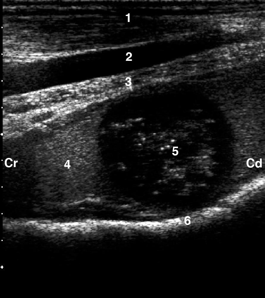
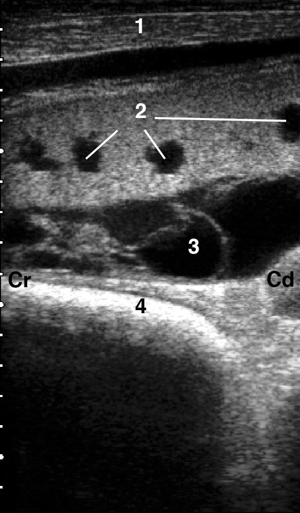
25 wall of the reticulum, 3 Craniodorsal blind sac of the rumen, 4 Strands of fibrin, 5 Fluid accumulation, Cr Cranial, Cd caudal. Fig. 7: Ultrasonogram of echogenic deposits on the reticulum and accumulation of fluid in a cow with traumatic reticuloperitonitis imaged from the left ventral thorax. 1 Ventral abdominal wall, 2 Reticulum, 3 Echogenic deposits of fibrin, 4 Accumulation of fluid, Cr Cranial, Cd Caudal. Fig. 8: Ultrasonogram of echogenic deposits between the reticulum and the craniodorsal blind sac of the rumen in a cow with traumatic reticuloperitonitis imaged from the left ventral thorax. 1 Ventral abdominal wall, 2 Reticulum, 3 Echogenic deposits of fibrin, 4 Craniodorsal blind sac of the rumen, Cr Cranial, Cd Caudal. Fig. 9: Ultrasonogram of an abscess ventral to the reticulum of a cow with traumatic reticuloperitonitis imaged from the left ventral thorax. 1 Ventral abdominal wall, 2 Reticulum, 3 Abscess, Cr Cranial, Cd Caudal. Fig. 10: Ultrasonogram of an abscess ventral to the reticulum in a cow with traumatic reticuloperitonitis imaged from the left ventral thorax. 1 Ventral abdominal wall, 2 Abscess capsule, 3 Abscess lumen, 4 Reticulum, Cr Cranial, Cd Caudal. Fig. 11: Ultrasonogram of a small fluid accumulation caudal to the reticulum in a cow with traumatic reticuloperitonitis imaged from the sternal part of the ventral abdomen. 1 Ventral abdominal wall, 2 Musculophrenic vein, 3 Diaphragm, 4 Reticulum, 5 Craniodorsal blind sac of the rumen with fibrin depositis, 6 Hypoechoic (due to inflammation) fluid accumulation, Cr Cranial, Cd Caudal. 23
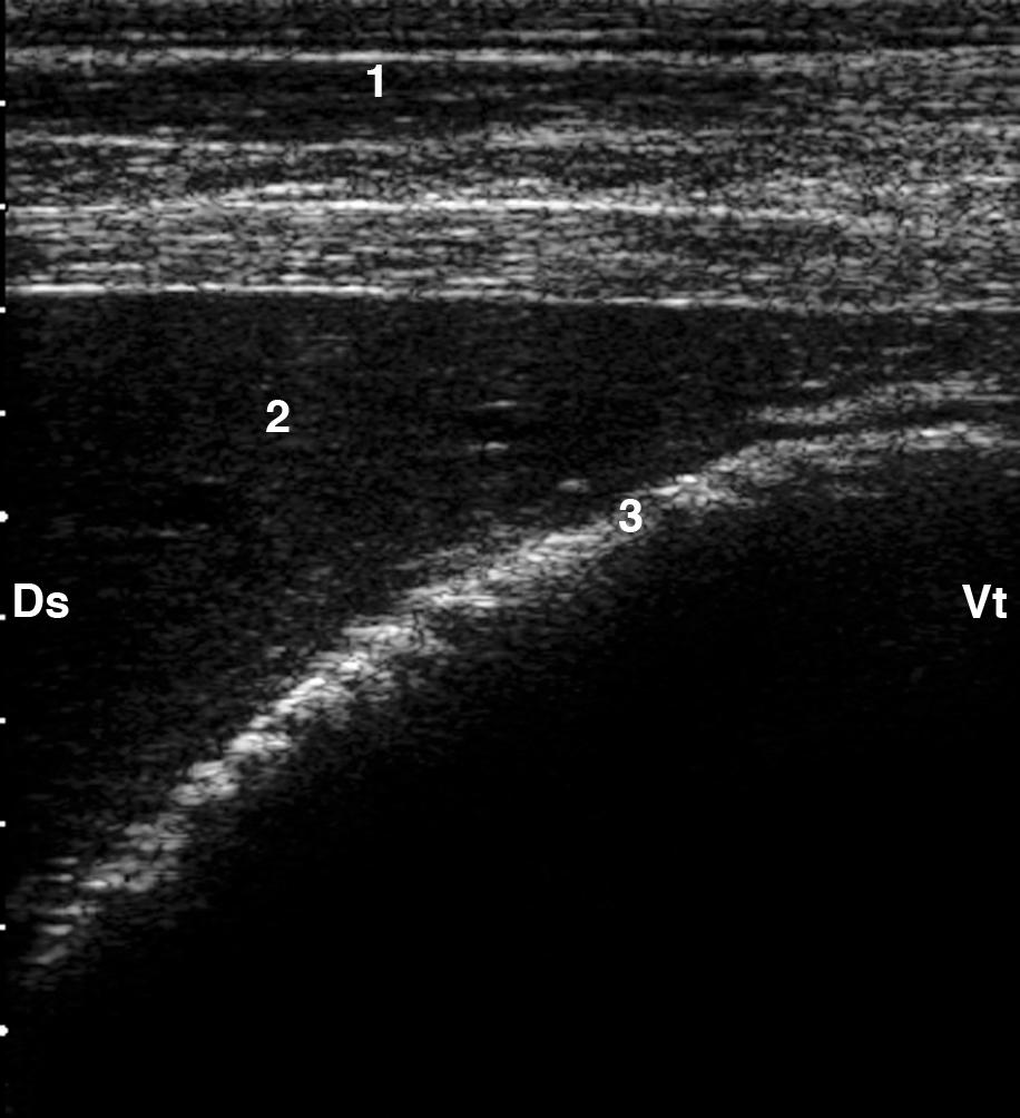
26 Fig. 12: Ultrasonogram of a large fluid accumulation due to peritonitis in a cow with traumatic reticuloperitonitis ventral the rumen imaged from the left side of the ventral abdomen. 1 Ventral abdominal wall, 2 Omentum, 3 Fluid accumulation, 4 Rumen, Cr Cranial, Cd Caudal. Fig. 13: Ultrasonogram of inflammatory changes between the reticulum and the spleen in a cow with traumatic reticuloperitonitis imaged from the left ventral thorax. 1 Ventral abdominal wall, 2 Spleen with dilated vessels, 3 Fluid containing fibrin between the reticulum and the spleen, 4 Reticulum, Cr Cranial, Cd Caudal. Fig. 14 : Ultrasonogram of a large abscess in the spleen imaged from the left ventral thorax. 1 Ventral thoracic wall, 2 Musculophrenic vein, 3 Diaphragm, 4 Spleen, 5 Abscess in the spleen, 6 Reticulum, indented by the spleen, Cr Cranial, Cd Caudal. Fig. 15 : Ultrasonogram of the normal omasum imaged from the 8th intercostal space of the right side. The wall of the omasum appears as a curved echogenic line. The liver is situated on the omasum dorsolaterally. 1 Costal part of the abdominal wall, 2 Liver, 3 Omasal wall, Ds Dorsal, Vt Ventral. Fig. 16: Ultrasonogram of the omasum of a cow with abomasal volvulus imaged from the 10th intercostals space. The omasal laminae appear as thin echogenic lines. 1 Costal part of the abdominal wall, 2 Omasal wall, 3 Omasal laminae, 4 Greater omentum, Ds Dorsal, Vt Ventral. 24
27 Fig. 17: Ultrasonogram of left displacement of the abomasum imaged from the ventral region of the 12th intercostal space. 1 Abdominal wall, 2 Abomasum with hypoechogenic ingesta, 3 Abomasal fold, Ds Dorsal, Vt Ventral. Fig. 18: Ultrasonogram of left displacement of the abomasum imaged from the dorsal region of the 12th intercostal space. The abomasal gas cap is not visible because of reverberation artifacts at the abomasal surface. 1 Abdominal wall, 2 Abomasal wall, 3 Reverberation artifacts, Ds Dorsal, Vt Ventral. Fig. 19: Ultrasonogram of the abomasum dilated secondary to ileus of the jejunum imaged from the right paramedian region. The abomasal contents appear hypochechogenic and the abomasal folds are clearly visible as thin, echogenic wavy structures. 1 Abdominal wall, 2 Abomasum, 3 Abomasal folds, Cr Cranial, Cd Caudal. Fig. 20: Ultrasonogram of cross-sections through loops of the jejunum imaged from the right flank. Several loops of jejunum, seen in cross-section, are situated immediately adjacent to one another. 1 Abdominal wall, 2 Loops of jejunum, Ds Dorsal, Vt Ventral. Fig. 21 : Ultrasonogram of cross sections through dilated loops of the jejunum in a cow with ileus imaged from the 12th intercostal space. The contents of the loops appear echogenic. There is anechoic fluid between the loops. 1 Abdominal wall, 2 Dilated loops of the jejunum, 3 Anechoic fluid between the loops of the jejunum, Ds Dorsal, Vt Ventral. Fig. 22 : Ultrasonogram of cross and longitudinal sections through dilated loops of the jejunum in a cow with ileus imaged from the 12th intercostal space. The contents of the loops 25
28 appear echogenic. 1 Abdominal wall, 2 Loop of jejunum in cross section, 3 Loop of jejunum in longitudinal section, Ds Dorsal, Vt Ventral. Fig. 23: Ultrasonogram of a cross-section through the dilated duodenum of a cow with duodenal ileus imaged from the 10th intercostal space. 1 Abdominal wall, 2 Dilated duodenum in cross-section, Ds Dorsal, Vt Ventral. Fig. 24: Ultrasonogram of the jejunum in a cow with Haemorrhagic Bowel Syndrome imaged from the right lateral abdominal wall. The blood clots have the appearance of echogenic masses in the lumen of the small intestine. 1 Abdominal wall, 2 Blood clots in the lumen of the small intestine, 3 Fluid between the intestinal loops, Ds Dorsal, Vt Ventral. Fig. 25: Ultrasonogram of a dilated caecum of a cow with caecal dilatation and torsion imaged from the right flank. The caecal wall closest to the transducer appears as a curved echogenic line. The caecal contents and wall furthest from the transducer are not visible. 1 Lateral abdominal wall, 2 Caecal wall, Ds Dorsal, Vt Ventral. 26
29
30
31
32

33
34
35
36
37
38
39
40
41
42
43
44
45
46
47

48
49
50
51
52
53
Computed tomography of the abdomen in Saanen goats: II. liver, spleen, abomasum, and intestine
 Zurich Open Repository and Archive University of Zurich Main Library Winterthurerstr. 190 CH-8057 Zurich www.zora.uzh.ch Year: 2011 Computed tomography of the abdomen in Saanen goats: II. liver, spleen,
ABDOMINAL ULTRASONOGRAPHY IN CATTLE. Munday, K.1, Mudron P.2
 DOI: 10.1515/FV-2016-0005 FOLIA VETERINARIA, 60, 1: 34 40, 2016 ABDOMINAL ULTRASONOGRAPHY IN CATTLE Munday, K.1, Mudron P.2 Prings Farmhouse, Prings Lane, Maplehurst, Rh13 6GZ England 2 Clinic of Ruminants,
Ultrasonographic Diagnosis of Sharp Foreign Body Syndrome in Buffaloes. Oday Shihab Al-Abbadi
 CAIRO UNIVERSITY FACULTY OF VETERINARY MEDICINE DEPARTMENT OF SURGERY, ANETHESIOLOGY & RADIOLOGY Ultrasonographic Diagnosis of Sharp Foreign Body Syndrome in Buffaloes A thesis presented by Oday Shihab
Clinical and ultrasonographic differences between cattle and buffaloes with various sequelae of traumatic reticuloperitonitis
 Veterinarni Medicina, 54, 009 (9): 399406 Clinical and ultrasonographic differences between cattle and buffaloes with various sequelae of traumatic reticulo A.M. Abdelaal, M. Floeck, S. El Maghawry, W.
How ultrasound has changed decision making in colic surgery
 How ultrasound has changed decision making in colic surgery Gunther van Loon Dept. of Large Animal Internal Medicine, Faculty of Veterinary Medicine, Ghent University, Belgium Colic is common in horses
What is Your Diagnosis?
 What is Your Diagnosis? Izabela Ragan, Class of 2014 Signalment Species: Canine Breed: English Bulldog Sex: Male castrated Date of birth: 04/14/11 Presenting Complaint Dog was presented for vomiting and
Ultrasonographic examination of caecum and colon of normal Adult Spiti horses and Himalayan Hill mules of India
 Indian J. Anim. Res., 52(9) 2018: 1271-1280 Print ISSN:0367-6722 / Online ISSN:0976-0555 AGRICULTURAL RESEARCH COMMUNICATION CENTRE www.arccjournals.com/www.ijaronline.in Ultrasonographic examination of
REVIEW OF NORMAL EQUINE GASTROINTESTINAL ULTRASONOGRAPHY. Michelle Henry Barton DVM, PhD, DACVIM University of Georgia, Athens, GA, 30602
 REVIEW OF NORMAL EQUINE GASTROINTESTINAL ULTRASONOGRAPHY Michelle Henry Barton DVM, PhD, DACVIM University of Georgia, Athens, GA, 30602 INTRODUCTION The recent introduction of more affordable and portable
Abdominal ultrasound:
 Abdominal ultrasound: Non-traumatic acute abdomen Wittanee Na-ChiangMai, MD Department of Radiology ChiangMai University 26/04/2017 Contents Technique of examination Normal anatomy Emergency conditions
Exploring Anatomy: the Human Abdomen
 Exploring Anatomy: the Human Abdomen PERITONEUM AND PERITONEAL CAVITY PERITONEUM The peritoneum is a thin serous membrane that lines the abdominal cavity and covers, in variable amounts, the viscera within
Proceedings of the American Association of Equine Practitioners - Focus Meeting. Focus on Colic. Indianapolis, IN, USA 2011
 www.ivis.org Proceedings of the American Association of Equine Practitioners - Focus Meeting Focus on Colic Indianapolis, IN, USA 2011 Next Focus Meetings: July 22-24, 2012 - Focus on Hind Limb Lameness
Close window to return to IVIS. in collaborazione con RICHIESTO ACCREDITAMENTO. organizzato da certificata ISO 9001:2000
 in collaborazione con Close window to return to IVIS RICHIESTO ACCREDITAMENTO SOCIETÀ CULTURALE ITALIANA VETERINARI PER ANIMALI DA COMPAGNIA SOCIETÀ FEDERATA ANMVI organizzato da certificata ISO 9001:2000
Gastrointestinal Tract. Anatomy of GI Tract. Anatomy of GI Tract. (Effective February 2007) (1%-5%)
 (1%-5%)") Gastrointestinal Tract (Effective February 2007) (1%-5%) Anatomy of GI Tract Esophagus bulls-eye or target EG junction seen on sagittal scan posterior to left lobe of liver and anterior to aorta Anatomy
Gastrointestinal Tract (Effective February 2007) (1%-5%) Anatomy of GI Tract Esophagus bulls-eye or target EG junction seen on sagittal scan posterior to left lobe of liver and anterior to aorta Anatomy
ASSESSING THE PLAIN ABDOMINAL RADIOGRAPH M A A M E F O S U A A M P O F O
 ASSESSING THE PLAIN ABDOMINAL RADIOGRAPH M A A M E F O S U A A M P O F O Introduction The abdomen (less formally called the belly, stomach, is that part of the body between the thorax (chest) and pelvis,
Radiology of the abdomen Lecture -1-
 Radiology of the abdomen Lecture -1- Objectives To know radiology modalities used in abdomen imaging mainly GI tract. To know advantages and disadvantages of each modality. To know indications and contraindications
Gastrointestinal Tract Imaging. Objectives. Reference. VMB 960 April 6, Stomach Small Intestine Colon. Radiography & Ultrasound
 Gastrointestinal Tract Imaging VMB 960 April 6, 2009 Stomach Small Intestine Colon Objectives Radiography & Ultrasound Contrast Examination of the Small Intestine Reference Chapters 45 47 Pages 750 805
The use of percutaneous abdominal ultrasound examination in diagnosing equine small intestinal disorders
 Polish Journal of Veterinary Sciences Vol. 15, No. 4 (212), 759-766 DOI 1.2478/v1181-12-115-2 Original article The use of percutaneous abdominal ultrasound examination in diagnosing equine small intestinal
Medical application of transabdominal ultrasound in gastrointestinal diseases
 Medical application of transabdominal ultrasound in gastrointestinal diseases Hsiu-Po Wang Department of Emergency Medicine National Taiwan University Hospital Real-time ultrasound has become a standard
Clinical & Sonographic Approach To GI Disease, Obstructions & Foreign Bodies Parts 1 & 2
 Clinical & Sonographic Approach To GI Disease, Obstructions & Foreign Bodies Parts 1 & 2 Eric Lindquist DMV (Italy), Cert./President IVUSS Further information regarding interventional procedures and case
BY DR NOMAN ULLAH WAZIR
 BY DR NOMAN ULLAH WAZIR The stomach (from ancient Greek word stomachos, stoma means mouth) is a muscular, hollow and the most dilated part of the GIT. It starts from the point where esophagus ends. It
Pancreas & Biliary System. Dr. Vohra & Dr. Jamila
 Pancreas & Biliary System Dr. Vohra & Dr. Jamila 1 Objectives At the end of the lecture, the student should be able to describe the: Location, surface anatomy, parts, relations & peritoneal reflection
The abdominal Esophagus, Stomach and the Duodenum. Prof. Oluwadiya KS
 The abdominal Esophagus, Stomach and the Duodenum Prof. Oluwadiya KS www.oluwadiya.com Viscera of the abdomen Abdominal esophagus: Terminal part of the esophagus The stomach Intestines: Small and Large
DIAGNOSTIC IMAGING: LIVER DISEASE
 Vet Times The website for the veterinary profession https://www.vettimes.co.uk DIAGNOSTIC IMAGING: LIVER DISEASE Author : Abby Caine Categories : Vets Date : February 1, 2010 ABBY CAINE reviews both established
Anatomy: Know Your Abdomen
 Anatomy: Know Your Abdomen Glossary Abdomen - part of the body below the thorax (chest cavity); separated by the diaphragm. Anterior - towards the front of the body. For example, the umbilicus is anterior
General Abdominal Radiography
 General Abdominal Radiography Tony Pease, DVM, MS Assistant Professor of Radiology North Carolina State University Objectives Acquisition of radiographs Abdominal radiographic anatomy Radiographic patterns
ABDOMEN - GI. Duodenum
 TALA SALEH ABDOMEN - GI Duodenum - Notice the shape of the duodenum, it looks like capital G shape tube which extends from the pyloroduodenal junction to the duodenojejunal junction. - It is 10 inches
Abdominal Ultrasound
 Abdominal Ultrasound Imaging Control Buttons Depth The organ imaged should take up 3/4 of the screen Frequency = Penetration Use high frequencies (harmonics) for fluid filled and superficial structures
Duodenum retroperitoneal
 Duodenum retroperitoneal C shaped Initial region out of stomach into small intestine RETROperitoneal viscus Superior 1 st part duodenal cap ; moves upwards and backwards to lie on the R crura medial to
Background & Indications Probe Selection
 Teresa S. Wu, MD, FACEP Director, EM Ultrasound Program & Fellowship Co-Director, Simulation Based Training Program & Fellowship Associate Program Director, EM Residency Program Maricopa Medical Center
FHS Appendicitis US Protocol
 FHS Appendicitis US Protocol Reviewed By: Shireen Khan, MD; Sarah Farley, MD; Anna Ellermeier, MD Last Reviewed: May 2018 Contact: (866) 761-4200 **NOTE for all examinations: 1. If documenting possible
My Patient Has Abdominal Pain PoCUS of the Biliary Tract and the Urinary Tract
 My Patient Has Abdominal Pain PoCUS of the Biliary Tract and the Urinary Tract Objectives PoCUS for Biliary Disease PoCUS for Renal Colic PoCUS for Urinary Retention Biliary Disease A patient presents
Plain abdomen The standard films are supine & erect AP views (alternative to erect, lateral decubitus film is used in ill patients).
.") Plain abdomen The standard films are supine & erect AP views (alternative to erect, lateral decubitus film is used in ill patients). The stomach can be readily identified by its location, gastric rugae
Plain abdomen The standard films are supine & erect AP views (alternative to erect, lateral decubitus film is used in ill patients). The stomach can be readily identified by its location, gastric rugae
This lab activity is aligned with Visible Body s Human Anatomy Atlas app. Learn more at visiblebody.com/professors
 1 This lab activity is aligned with Visible Body s Human Anatomy Atlas app. Learn more at visiblebody.com/professors 2 A. Digestive System Overview To Start: Go to the Views menu and scroll down to the
What s Your Diagnosis?
 What s Your Diagnosis? Signalment: 5 year old MC Belgian Malinois Presenting Complaint: Perineal hernia as well as not eating or defecating History: The patient presented to the KSU VHC on 7/28/2018 for
GENERAL ABDOMINAL IMAGING PERITONEAL SPACE, PANCREAS, & SPLEEN. VMB 960 March 25, 2013
 GENERAL ABDOMINAL IMAGING PERITONEAL SPACE, PANCREAS, & SPLEEN VMB 960 March 25, 2013 REFERENCE Chapters 35-36 Pages 650-678 Chapter 37 Pages 694-701 Chapter 3 Pages 38-49 OBJECTIVES Radiography and Ultrasound
The stomach is formed of three parts: -
 The stomach is formed of three parts: - (a) CARDIAC STOMACH: - It receives the oesophagus through Cardiac aperture guarded by a cardiac sphincter which prevents regurgitation of food. (b) FUNDIC PART:
Infantile Hypertrophic Pyloric Stenosis
 A Sonographic walk-through: Infantile Hypertrophic Pyloric Stenosis Tara K. Cielma, RDMS, RDCS, RVT, RT(S) Anjum N. Bandarkar, MD, Adebunmi O. Adeyiga, MD, Diagnostic Imaging and Radiology, Children s
ABDOMINAL RADIOLOGY UNDERSTANDING
 ABDOMINAL RADIOLOGY UNDERSTANDING CACVT 2017 SPRING CONFERENCE - GREENWOOD VILLAGE, CO Amy Newfield, CVT, VTS (ECC) BluePearl Massachusetts - Waltham, MA INTRODUCTION As a technician you will likely be
Biology Human Anatomy Abdominal and Pelvic Cavities
 Biology 351 - Human Anatomy Abdominal and Pelvic Cavities Please place your name and I.D. number on the back of the last page of this exam. You must answer all questions on this exam. Because statistics
Proceedings of the 34th World Small Animal Veterinary Congress WSAVA 2009
 www.ivis.org Proceedings of the 34th World Small Animal Veterinary Congress WSAVA 2009 São Paulo, Brazil - 2009 Next WSAVA Congress : Reprinted in IVIS with the permission of the Congress Organizers IMAGING
Abdominal Assessment
 Abdominal Assessment Mary Marian, MS,RD,CSO University of AZ, Tucson, AZ Neha Parekh, MS,RD,LD,CNSC Cleveland Clinic, Cleveland, OH Objectives: 1. Outline the steps in performing an abdominal examination.
GASTROINTESTINAL SYSTEM
 GASTROINTESTINAL SYSTEM Topographic Anatomy of the Abdomen Surface Landmarks Xiphoid process T9/T10 Inferior costal margin L2/L3 Iliac Crest L4 level ASIS L5/S1 level Pubic symphysis level of greater trochanter
Category Term Definition Comments 1 Major Categories 1a
 Working Lexicon Categories, Terms & Definitions Category Term Definition Comments 1 Major Categories 1a Physiologic Category (consistent with normal ovarian physiology) Follicle Simple 3 cm in premenopausal
Research Article Comparison of Radiography and Ultrasonography for Diagnosis of Diaphragmatic Hernia in Bovines
 SAGE-Hindawi Access to Research Veterinary Medicine International Volume 2010, Article ID 939870, 7 pages doi:10.4061/2010/939870 Research Article Comparison of Radiography and Ultrasonography for Diagnosis
Preview from Notesale.co.uk Page 1 of 34
 Abdominal viscera and digestive tract Digestive tract Abdominal viscera comprise majority of the alimentary system o Terminal oesophagus, stomach, pancreas, spleen, liver, gallbladder, kidneys, suprarenal
Anatomy of the Large Intestine
 Large intestine Anatomy of the Large Intestine 2 Large Intestine Extends from ileocecal valve to anus Length = 1.5-2.5m = 5 feet Regions Cecum = 2.5-3 inch Appendix= 3-5 inch Colon Ascending= 5 inch Transverse=
Pathology of Intestinal Obstruction. Dr. M. Madhavan, MBBS., MD., MIAC, Professor of Pathology Saveetha Medical College
 Pathology of Intestinal Obstruction Dr. M. Madhavan, MBBS., MD., MIAC, Professor of Pathology Saveetha Medical College Pathology of Intestinal Obstruction Objectives list the causes of intestinal obstruction
Traumatic reticulitis, reticulo-peritonitis and pericarditis (Foreign body syndrome) in bovines
 in bovines") Traumatic reticulitis, reticulo-peritonitis and pericarditis (Foreign body syndrome) in bovines V. Devi Prasad*, P. Ravi Kumar, N.V.V. Harikrishna and D. Bhagyaraju Department of Surgery and Radiology,
Traumatic reticulitis, reticulo-peritonitis and pericarditis (Foreign body syndrome) in bovines V. Devi Prasad*, P. Ravi Kumar, N.V.V. Harikrishna and D. Bhagyaraju Department of Surgery and Radiology,
Pathology of the Alimentary Tract
 Pathology of the Alimentary Tract Lab 2: Lower alimentary tract SI, LI, cecum, and peritoneum GIST in the cecum of a dog Shannon Martinson: http://people.upei.ca/smartinson VPM 221: November, 2011 3 year
Development of pancreas and Small Intestine. ANATOMY DEPARTMENT DR.SANAA AL-AlSHAARAWY DR.ESSAM Eldin Salama
 Development of pancreas and Small Intestine ANATOMY DEPARTMENT DR.SANAA AL-AlSHAARAWY DR.ESSAM Eldin Salama OBJECTIVES At the end of the lecture, the students should be able to : Describe the development
Radiographic Positioning. Small Animal Abdominal Radiography. Lecture Outline. Matthew Paek, VMD, MS, DACVR
 Small Animal Abdominal Radiography Matthew Paek, VMD, MS, DACVR Email: Matthew.Paek@SynergyVIP.com 7/30/2018 1 Lecture Outline Radiographic technique Introduction to systematic review and principles of
Summary and conclusions
 Summary and conclusions 7 Chapter 7 68 Summary and conclusions Chapter 1 provides a general introduction to this thesis focused on the use of ultrasound (US) in children with abdominal problems. The literature
Anatomy of the SMALL INTESTINE. Dr. Noman Ullah Wazir PMC
 Anatomy of the SMALL INTESTINE Dr. Noman Ullah Wazir PMC SMALL INTESTINE The small intestine, consists of the duodenum, jejunum, and illium. It extends from the pylorus to the ileocecal junction were the
Intestinal Obstruction Clinical Presentation & Causes
 Intestinal Obstruction Clinical Presentation & Causes V Chidambaram-Nathan Consultant Transplant and General Surgeon Sheffield Kidney Institute Northern General Hospital Intestinal Obstruction One of the
Lab 9 Abdomen MUSCLES
 Lab 9 Abdomen MUSCLES External abdominal oblique continuous with the external intercostal muscle; its fibers point in a caudal direction as it moves anteriorly until it inserts on the linea alba via its
Diseases of Stomach & Abomasum. Physical influences Gastric Ulcers Gastritis Parasitic Diseases
 Diseases of Stomach & Abomasum Physical influences Gastric Ulcers Gastritis Parasitic Diseases Neoplasia Physical Influences Acute gastric dilation & volvulus (GDV) Displaced abomasum Chronic gastric dilation
Calvin 9 year old NM DLH. Dr. Norman Ackerman Memorial Radiography Case Challenge
 September 2014 Dr. Norman Ackerman served the University of Florida, College of Veterinary Medicine with distinction as Professor of Radiology from 1979 to 1994. A concerned teacher of veterinary students
Inflammation Laboratory 1
 Inflammation Laboratory 1 Lab1 Emphasis: The exudates of acute inflammation Descriptions Morphologic Diagnoses Shannon Martinson: http://people.upei.ca/smartinson VPM 152: March 2013 Describing Lesions
Inflammation Laboratory 1
 Inflammation Laboratory 1 Lab1 Emphasis: The exudates of acute inflammation Descriptions Morphologic Diagnoses Shannon Martinson: http://people.upei.ca/smartinson VPM 152: February 2012 Describing Lesions
The Focused Assessment with Sonography for Trauma, (FAST) procedure.
 procedure.") The Focused Assessment with Sonography for Trauma, (FAST) procedure. ROBERT H. WRIGLEY Professor Veterinary Diagnostic Imaging University of Sydney Veterinary Teaching Hospital Professor Emeritus Colorado
The Focused Assessment with Sonography for Trauma, (FAST) procedure. ROBERT H. WRIGLEY Professor Veterinary Diagnostic Imaging University of Sydney Veterinary Teaching Hospital Professor Emeritus Colorado
- Digestion occurs during periods of low activity - Produces more energy than it uses. - Mucosa
 Introduction Digestive System Chapter 29 Provides processes to break down molecules into a state easily used by cells - A disassembly line: Starts at the mouth and ends at the anus Digestive functions
Intraperitoneal cysts in infancy and childhood An overview and sonographic differentiation
 Intraperitoneal cysts in infancy and childhood An overview and sonographic differentiation M. Mearadji International Foundation for Pediatric Imaging Aid Rotterdam, The Netherlands Intraperitoneal cysts
The Thoracic wall including the diaphragm. Prof Oluwadiya KS
 The Thoracic wall including the diaphragm Prof Oluwadiya KS www.oluwadiya.com Components of the thoracic wall Skin Superficial fascia Chest wall muscles (see upper limb slides) Skeletal framework Intercostal
CHAPTER 3. Ultrasonic diagnosis of clinical oesophagostomiasis: a novel cause of 'target' colonic lesions
 CHAPTER 3 Ultrasonic diagnosis of clinical oesophagostomiasis: a novel cause of 'target' colonic lesions Storey PA, Anemana S, Van Oostayen JA, Polderman AM, Magnussen P British Journal of Radiology 2000;73:328-332
Digestive System 7/15/2015. Outline Digestive System. Digestive System
 Digestive System Biology 105 Lecture 18 Chapter 15 Outline Digestive System I. Functions II. Layers of the GI tract III. Major parts: mouth, pharynx, esophagus, stomach, small intestine, large intestine,
Development of the Digestive System. W.S. O The University of Hong Kong
 Development of the Digestive System W.S. O The University of Hong Kong Plan for the GI system Then GI system in the abdomen first develops as a tube suspended by dorsal and ventral mesenteries. Blood
GI Tract Lynn Ta Jennifer Zhang July 6, 2006 GI TRACT. 1) Other Names: Gastrointestinal tract Digestive tract Alimentary tract
 Other Names: Gastrointestinal tract Digestive tract Alimentary tract") GI Tract Lynn Ta Jennifer Zhang July 6, 2006 GI TRACT 1) Other Names: Gastrointestinal tract Digestive tract Alimentary tract 2) Definition/Location: Digestion and absorption are the primary functions
GI Tract Lynn Ta Jennifer Zhang July 6, 2006 GI TRACT 1) Other Names: Gastrointestinal tract Digestive tract Alimentary tract 2) Definition/Location: Digestion and absorption are the primary functions
Includes mouth, pharynx, esophagus, stomach, small intestine, large intestine, rectum, anus. Salivary glands, liver, gallbladder, pancreas
 Chapter 14 The Digestive System and Nutrition Digestive System Brings Nutrients Into the Body The digestive system includes Gastrointestinal (GI) tract (hollow tube) Lumen: space within this tube Includes
Lab 5 Digestion and Hormones of Digestion. 7/16/2015 MDufilho 1
 Lab 5 Digestion and Hormones of Digestion 1 Figure 23.1 Alimentary canal and related accessory digestive organs. Mouth (oral cavity) Tongue* Parotid gland Sublingual gland Submandibular gland Salivary
Accessory Glands of Digestive System
 Accessory Glands of Digestive System The liver The liver is soft and pliable and occupies the upper part of the abdominal cavity just beneath the diaphragm. The greater part of the liver is situated under
PROCEEDINGS ONE HUNDRED AND TWENTY-THIRD SCIENTIFIC MEETING ROYAL VETERINARY COLLEGE, ROYAL COLLEGE STREET, LONDON 7 MARCH 1959 RUMEN FUNCTION
 PROCEEDINGS OF THE NUTRITION SOCIETY ONE HUNDRED AND TWENTY-THIRD SCIENTIFIC MEETING ROYAL VETERINARY COLLEGE, ROYAL COLLEGE STREET, LONDON 7 MARCH 1959 RUMEN FUNCTION Chairman.- DR D. P. CUTHBERTSON,
Principles of Ultrasound. Cara C. Prideaux, M.D. University of Utah PM&R Sports Medicine Fellow March 14, 2012
 Principles of Ultrasound Cara C. Prideaux, M.D. University of Utah PM&R Sports Medicine Fellow March 14, 2012 None Disclosures Outline Introduction Benefits and Limitations of US Ultrasound (US) Physics
Ex. 1 :Language of Anatomy
 Collin College BIOL 2401 : Human Anatomy & Physiology Ex. 1 :Language of Anatomy The Anatomical Position Used as a reference point when referring to specific areas of the human body Body erect Head and
Guidelines, Policies and Statements D5 Statement on Abdominal Scanning
 Guidelines, Policies and Statements D5 Statement on Abdominal Scanning Disclaimer and Copyright The ASUM Standards of Practice Board have made every effort to ensure that this Guideline/Policy/Statement
The Physician as Medical Illustrator
 The Physician as Medical Illustrator Francois Luks Arlet Kurkchubasche Division of Pediatric Surgery Wednesday, December 9, 2015 Week 5 A good picture is worth a 1,000 bad ones How to illustrate an operation
US in non-traumatic acute abdomen. Lalita, M.D. Radiologist Department of radiology Faculty of Medicine ChiangMai university
 US in non-traumatic acute abdomen Lalita, M.D. Radiologist Department of radiology Faculty of Medicine ChiangMai university Sagittal Orientation Transverse (Axial) Orientation Coronal Orientation Intercostal
Case Report Atypical Presentation of Constrictive Pericarditis in a Holstein Heifer
 Case Reports in Veterinary Medicine Volume 2012, Article ID 604098, 5 pages doi:10.1155/2012/604098 Case Report Atypical Presentation of Constrictive Pericarditis in a Holstein Heifer Mohamed M. Elhanafy
- Digestion occurs during periods of low activity - Produces more energy than it uses. 3 Copyright 2016 by Elsevier Inc. All rights reserved.
 Introduction Digestive System Chapter 29 Provides processes to break down molecules into a state easily used by cells - A disassembly line: Starts at the mouth and ends at the anus Digestive functions
Anatomy of the Thorax
 Anatomy of the Thorax A) THE THORACIC WALL Boundaries Posteriorly by the thoracic part of the vertebral column Anteriorly by the sternum and costal cartilages Laterally by the ribs and intercostal spaces
SWISS SOCIETY OF NEONATOLOGY. Prenatal diagnosis and postnatal management of meconium pseudocysts
 SWISS SOCIETY OF NEONATOLOGY Prenatal diagnosis and postnatal management of meconium pseudocysts September 2007 2 Burch E, Caduff JH, Hodel M, Berger TM, Neonatal and Pediatric Intensive Care Unit (BE,
Focused Assessment Sonography of Trauma (FAST) Scanning Protocol
 Scanning Protocol") Focused Assessment Sonography of Trauma (FAST) Scanning Protocol Romolo Gaspari CHAPTER 3 GOAL OF THE FAST EXAM Demonstrate free fluid in abdomen, pleural space, or pericardial space. EMERGENCY ULTRASOUND
Focused Assessment Sonography of Trauma (FAST) Scanning Protocol Romolo Gaspari CHAPTER 3 GOAL OF THE FAST EXAM Demonstrate free fluid in abdomen, pleural space, or pericardial space. EMERGENCY ULTRASOUND
It passes through the diaphragm at the level of the 10th thoracic vertebra to join the stomach
 The esophagus is a tubular structure (muscular, collapsible tube ) about 10 in. (25 cm) long that is continuous above with the laryngeal part of the pharynx opposite the sixth cervical vertebra The esophagus
Pancreas and Biliary System
 Pancreas and Biliary System Please view our Editing File before studying this lecture to check for any changes. Color Code Important Doctors Notes Notes/Extra explanation Objectives At the end of the lecture,
Development of the Digestive System. W.S. O School of Biomedical Sciences, University of Hong Kong.
 Development of the Digestive System W.S. O School of Biomedical Sciences, University of Hong Kong. Organization of the GI tract: Foregut (abdominal part) supplied by coeliac trunk; derivatives include
The peritoneum. Prof. Oluwadiya KS, MBBS, FMCS(Orthop) Website:
 Website:") The peritoneum Prof. Oluwadiya KS, MBBS, FMCS(Orthop) Website: http://oluwadiya.com The peritoneum Serous membrane that lines the abdominopelvic cavity and invests the viscera The largest serous membrane
The peritoneum Prof. Oluwadiya KS, MBBS, FMCS(Orthop) Website: http://oluwadiya.com The peritoneum Serous membrane that lines the abdominopelvic cavity and invests the viscera The largest serous membrane
University of Zurich. Ultrasonography of the liver in cattle. Zurich Open Repository and Archive. Braun, U. Year: 2009
 University of Zurich Zurich Open Repository and Archive Winterthurerstr. 190 CH-8057 Zurich http://www.zora.uzh.ch Year: 2009 Ultrasonography of the liver in cattle Braun, U Braun, U (2009). Ultrasonography
Fareed Khdair, MD Assistant Professor Chief, Section of Pediatric Gastroenterology, Hepatology, and Nutrition University of Jordan School of Medicine
 Fareed Khdair, MD Assistant Professor Chief, Section of Pediatric Gastroenterology, Hepatology, and Nutrition University of Jordan School of Medicine Outline Lecture one : Gut formation Foregut: esophagus,
DIGESTIVE TRACT ESOPHAGUS
 DIGESTIVE TRACT From the lower esophagus to the lower rectum four fundamental layers comprise the wall of the digestive tube: mucosa, submucosa, muscularis propria (externa), and adventitia or serosa (see
THE ORAL CAVITY
 THE ORAL CAVITY WALL OF ABDOMEN (ANTERIOR) The paraumbilical vein drains into the portal vein and then through the liver. This is an important clinical connection. THE ABDOMINAL VISCERA The small
Ultrasonography of Peritoneal and Retroperitoneal Spaces and Abdominal Lymph Nodes
 IMAGING Ultrasonography of Peritoneal and Retroperitoneal Spaces and Abdominal Lymph Nodes Clifford R. Berry, DVM, DACVR; Elizabeth Huyhn, DVM; and Danielle Mauragis, CVT University of Florida Welcome
Muscle Tissue. General concepts. Classification of muscle. I. Functional classification is based on the type of neural control.
 Muscle Tissue LEARNING OBJECTIVES 1. Identify the three types of muscle tissue at the light microscopic level. 2. List and compare the structural and functional features of each of the three muscle fiber
ANATOMY & PHYSIOLOGY ONLINE COURSE - SESSION 13 THE DIGESTIVE SYSTEM
 ANATOMY & PHYSIOLOGY ONLINE COURSE - SESSION 13 THE DIGESTIVE SYSTEM The digestive system also known as the alimentary canal or gastrointestinal tract consists of a series of hollow organs joined in a
Objectives. Hepatobiliary Ultrasound: Anatomy, Technique, Pathology. RUQ: Normal Anatomy. Emergency Ultrasound: Gallbladder Location
 Hepatobiliary Ultrasound: Anatomy, Technique, Pathology Laleh Gharahbaghian, MD FAAEM Associate Director, EM Ultrasound Co-Director, EM Ultrasound Fellowship Stanford University Medical Center Seric Cusick,
A comparitive study on traumatic reticuloperitonitis and traumatic pericarditis in Egyptian cattle
 Research Article Turk. J. Vet. Anim. Sci. 2010; 34(2): 143-153 TÜBİTAK doi:10.3906/vet-0804-16 A comparitive study on traumatic reticuloperitonitis and traumatic pericarditis in Egyptian cattle Mohamed
UNDERSTANDING X-RAYS: ABDOMINAL IMAGING THE ABDOMEN
 UNDERSTANDING X-RAYS: ABDOMINAL IMAGING THE ABDOMEN Radiology Enterprises radiologyenterprises@gmail.com www.radiologyenterprises.com STOMACH AND SMALL BOWEL STOMACH AND SMALL BOWEL Swallowed air is a
Signalment: Gidget, 12 year old, female spayed, Scottish Terrier, 10.7 kg
 Signalment: Gidget, 12 year old, female spayed, Scottish Terrier, 10.7 kg Presenting Complaint: Gidget presented after having elevated liver enzymes, patchy alopecia and PU/PD. History: Gidget had been
The jejunum and the Ileum. Prof. Oluwadiya KS
 The jejunum and the Ileum Prof. Oluwadiya KS www.oluwadiya.siteled.com Introduction Introduction The small intestine (SI) comprises of the duodenum, jejunum and the ileum The jejunum is the second part
FAS-GIP Protocol Focused Assessment with Sonography for Gastro-Intestinal Perforation
 FAS-GIP Protocol Focused Assessment with Sonography for Gastro-Intestinal Perforation Introduction Ultrasonography is very useful for the diagnosis of pneumoperitoneum indicating gastrointestinal perforation
Gastrointestinal Diseases Proceedings. Get inside your patients
 Gastrointestinal Diseases 2011 Proceedings Get inside your patients Commitment to Veterinarians Pfizer Animal Health: Committed to Veterinarians and the World Pfizer Animal Health has reaffirmed its steadfast
3. Gastro-Intestinal Tract, updated pages 27 to 31. The Equine GI Tract and Physiology Lips. Teeth. Esophagus
 3. Gastro-Intestinal Tract, updated 02-12-2018 - pages 27 to 31 The Equine GI Tract and Physiology Lips Teeth Esophagus Stomach Simple stomach divided into two regions Cardiac region is lined with a non-glandular,