What s Your Diagnosis?
|
|
|
- Claribel Gallagher
- 5 years ago
- Views:
Transcription
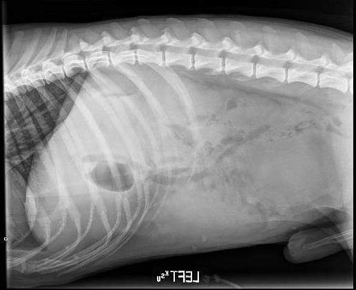
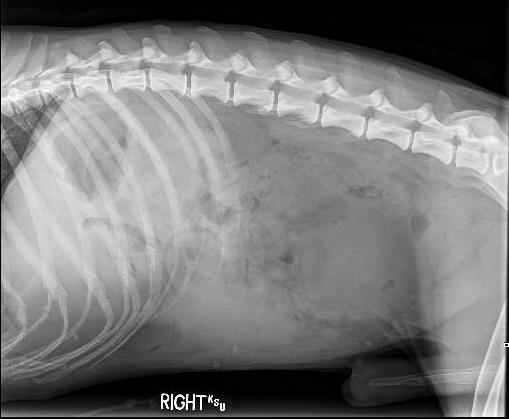
1 What s Your Diagnosis? Signalment: 5 year old MC Belgian Malinois Presenting Complaint: Perineal hernia as well as not eating or defecating History: The patient presented to the KSU VHC on 7/28/2018 for repair of a perineal hernia. Ram has been straining to defecate, was lethargic and losing weight. Previous veterinarians performed an enema to clear out feces. No blockage was observed on a contrast radiograph and after the surgery defecation was in small, hard pieces. Appetite was reduced after the surgery. Upon presentation at KSU VHC bilateral perineal hernias were diagnosed on rectal palpation. After blood work diagnostics were began to determine the full problem. Physical Exam Findings: Pulse = Strong HR = 110 RR = 30 Weight = 28.9 Kilograms Temperament = Bright, Alert and Responsive Abnormal oral cavity (canines were brown) Abnormal Digestive System (straining to defecate) Abnormal Urinary System (urinating in the house) Clinical Pathology Test Results: CBC Acute Inflammatory leukogram Peritoneal Fluid Analysis Marked neutrophilic inflammation with bacterial sepsis Culture Abundant E. coli Diagnostic Plan: Step 1 Abdominal Radiographs
2
3


4 3 View Abdominal Radiograph Findings: Extrathoracic soft tissue is normal. Musculoskeletal structures are normal. The abdominal contour is normal. Loss of serosal detail is seen diffusely throughout the entire abdomen. Nodules of heterogeneous soft tissue opacity are present in the ventral abdomen and positioned cranially to the bladder and along the left body wall lateral to the kidney. Noncontained gas opacities are scattered through the abdomen. Abdominal Radiographic Impressions: Free gas and peritoneal effusion is consistent with septic peritonitis. Nodular abdominal structures have differentials of loculated fluid accumulation, nodular reactive peritonitis, or carcinomatosis. Perianal hernia Post Radiograph Recommendation: Abdominal ultrasound


5 Step 2 Abdominal Ultrasound Ultrasound Findings: The bladder wall is diffusely moderately thickened with retention of normal wall layering. Wall thickness up to 6.1mm. The bladder is mildly distended with anechoic speckled content and there is a focal, irregularly ovoid hypoechoic luminal structure along the dependent aspect which is mobile on ballottement. The prostate retains a typical shape to a large degree with diffuse mild mottling throughout and multifocal to coalescing anechoic foci throughout the parenchyma. There are several peripheral wedge shaped hypoechoic foci noted throughout the parenchyma. Prostate measures 32.1x63.6mm. Arising from the left dorsolateral margin of the prostate there is a poorly marginated, ill defined, heterogenous region of tissue which extends intra-abdominally along the ventral and left ventrolateral abdominal cavity. Associated with this structure are multifocal discoid to linear hyperechoic foci, some of which show twinkling artifacts and some of which show subtle distal shadowing. On deep transducer pressure/ballottement, there is mobility of some of the linear echoes. There is a cluster of prominent vascularity associated with the aforementioned caudal abdominal pathology. The testis are bilaterally symmetrical with normal architecture however the epididymii appear plump and enlarged and this extends along the spermatic cord. Blood flow is patent throughout. The medial iliac lymph nodes are moderately enlarged and hypoechoic however they retain their normal S:L axis ratio. Mild perinodal hyperechogenicity noted. There is a moderate amount of highly echogenic free abdominal fluid. There are hyperechoic linear shadowing foci scattered along the uppermost peritoneal surface representing free abdominal gas. Ultrasound Impressions: Prostatopathy with changes most consistent with BPH and multifocal sub-acute prostatic infarcts. Suspect ruptured prostatic abscess or infected and rupture para-prostatic cyst. Cellular abdominal effusion and pneumoperitonium consistent with septic peritonitis. Mild cystopathy - however the lack of marked bladder distention may affect the perceived degree. Suspect associated testicular/spermatic cord edema.
6 Diagnosis After Surgery: Paraprostatic Cyst & Septic Peritonitis Outcome: The patient was recovering in the ICU and receiving treatments for septic peritonitis. Discussion: Paraprostatic cysts are embryonic remnants of the female reproductive tract that develop outside of the parenchyma in males and adhere to the capsule of the prostate. Treatment of a paraprostatic cyst would require surgical exploration and debulking of the cyst. A paraprostatic cyst is also likely to recur as long as the male patient is an intact specimen. Signs of a paraprostatic cyst occur once the cyst has become large enough to place pressure on surrounding organs or cause abdominal distension. Septic peritonitis is an inflammation of the serous membrane of the peritoneal cavity caused by bacterial growth. Septic peritonitis occurs due to contamination of the peritoneal cavity by bacteria which can be due a mass, perforation, infection or other disease processes. Septic peritonitis can be tested through culturing and testing of the infectious exudative effusion within the peritoneal cavity. Septic exudate is ideally caused by intracellular bacteria and degenerate neutrophils can be observed. Care must be taken when testing for septic peritonitis because an absence of organisms does not actually rule-out sepsis.
What s your diagnosis?
 What s your diagnosis? Signalment: 9 year old MC 2.7 kg Papillion Presenting Complaint: Presented for work up of anorexia and vomiting History: He had presented to cardiology for work up of a grad IV/VI
What s Your Diagnosis?
 What s Your Diagnosis? Courtney S. Wait Signalment: 11 year old FS Labrador Retriever Presenting Complaint/History: The patient presented to the referring DVM for inappetance, vomiting, lethargy, and anorexia.
What s Your Diagnosis??? Renée Fahrenholz, Class of 2012
 Renée Fahrenholz, Class of 2012 What s Your Diagnosis??? Signalment Emma, a 9 year old, Female, Spayed, Domestic Short Haired Feline Presenting Complaint Weight loss, vomited the morning of her visit,
What is Your Diagnosis?
 What is Your Diagnosis? Izabela Ragan, Class of 2014 Signalment Species: Canine Breed: English Bulldog Sex: Male castrated Date of birth: 04/14/11 Presenting Complaint Dog was presented for vomiting and
What s Your Diagnosis? Signalment: Species: Canine Breed: Golden Retriever Sex: Female (spayed) Date of Birth: 04/01/99
 Date of Birth: 04/01/99") What s Your Diagnosis? Signalment: Species: Canine Breed: Golden Retriever Sex: Female (spayed) Date of Birth: 04/01/99 Presenting Complaint: Acute onset of lethargy Vomited twice (partially digested food)
What s Your Diagnosis? Signalment: Species: Canine Breed: Golden Retriever Sex: Female (spayed) Date of Birth: 04/01/99 Presenting Complaint: Acute onset of lethargy Vomited twice (partially digested food)
Ultrasonographic and Clinical Studies on Benign Prostatic Hyperplasia in Dogs
 Theriogenology Insight: 6(1): 67-72, April, 2016 DOI Number: 10.5958/2277-3371.2016.00009.7 Ultrasonographic and Clinical Studies on Benign Prostatic Hyperplasia in Dogs K. Rajkumar* and C. Ansarkamran
What s your diagnosis? Malori Marotz. Squirt, an 8month old mix breed puppy. History:
 What s your diagnosis? Malori Marotz Squirt, an 8month old mix breed puppy History: The owner obtained squirt at 12 weeks of age. The owner reported that Squirt was passing soft stools lately and he is
Category Term Definition Comments 1 Major Categories 1a
 Working Lexicon Categories, Terms & Definitions Category Term Definition Comments 1 Major Categories 1a Physiologic Category (consistent with normal ovarian physiology) Follicle Simple 3 cm in premenopausal
What s Your Diagnosis? Sara Alves, Class of Signalment: 9-year-7-month old female spay American Miniature Eskimo dog
 What s Your Diagnosis? Sara Alves, Class of 2018 Signalment: 9-year-7-month old female spay American Miniature Eskimo dog Presenting Complaint: The patient presented on 5/30/17 with signs of lethargy and
What s Your Diagnosis? Allison Crow, Class of 2014
 What s Your Diagnosis? Allison Crow, Class of 2014 Signalment: 13 year old male castrated mixed breed dog History: The patient presented to the rdvm for pain in the hind end, weakness and neck stretching
GENERAL ABDOMINAL IMAGING PERITONEAL SPACE, PANCREAS, & SPLEEN. VMB 960 March 25, 2013
 GENERAL ABDOMINAL IMAGING PERITONEAL SPACE, PANCREAS, & SPLEEN VMB 960 March 25, 2013 REFERENCE Chapters 35-36 Pages 650-678 Chapter 37 Pages 694-701 Chapter 3 Pages 38-49 OBJECTIVES Radiography and Ultrasound
Signalment: Gidget, 12 year old, female spayed, Scottish Terrier, 10.7 kg
 Signalment: Gidget, 12 year old, female spayed, Scottish Terrier, 10.7 kg Presenting Complaint: Gidget presented after having elevated liver enzymes, patchy alopecia and PU/PD. History: Gidget had been
GENERAL ABDOMINAL IMAGING PERITONEAL SPACE, PANCREAS, & SPLEEN
 GENERAL ABDOMINAL IMAGING PERITONEAL SPACE, PANCREAS, & SPLEEN VMB 960 March 25, 2013 REFERENCE Chapters 35-36 Pages 650-678 Chapter 37 Pages 694-701 Chapter 3 Pages 38-49 OBJECTIVES Radiography and Ultrasound
Imaging the Urinary Tract
 Imaging the Urinary Tract Laura Armbrust, DVM, DACVR Gregory F. Grauer, DVM, MS, DACVIM Kansas State University Radiographic and ultrasound imaging in addition to history, physical examination, and clinicopathologic
Proceedings of the 34th World Small Animal Veterinary Congress WSAVA 2009
 www.ivis.org Proceedings of the 34th World Small Animal Veterinary Congress WSAVA 2009 São Paulo, Brazil - 2009 Next WSAVA Congress : Reprinted in IVIS with the permission of the Congress Organizers IMAGING
Imaging the Neonatal Foal. Equipment. Neonatal foals. 5 or 7.5 MHz linear array 10 MHz linear array. Abdomen Thorax Musculoskeletal system
 Imaging the Neonatal Foal Leanne Begg BVSc DipVetClinStud MS MACVSc Dip ACVIM RANDWICK EQUINE CENTRE Equipment 5 or 7.5 MHz linear array 10 MHz linear array Neonatal foals Abdomen Thorax Musculoskeletal
Ultrasonography of Peritoneal and Retroperitoneal Spaces and Abdominal Lymph Nodes
 IMAGING Ultrasonography of Peritoneal and Retroperitoneal Spaces and Abdominal Lymph Nodes Clifford R. Berry, DVM, DACVR; Elizabeth Huyhn, DVM; and Danielle Mauragis, CVT University of Florida Welcome
Abdominal Ultrasound
 Abdominal Ultrasound Imaging Control Buttons Depth The organ imaged should take up 3/4 of the screen Frequency = Penetration Use high frequencies (harmonics) for fluid filled and superficial structures
Guidelines, Policies and Statements D5 Statement on Abdominal Scanning
 Guidelines, Policies and Statements D5 Statement on Abdominal Scanning Disclaimer and Copyright The ASUM Standards of Practice Board have made every effort to ensure that this Guideline/Policy/Statement
US in non-traumatic acute abdomen. Lalita, M.D. Radiologist Department of radiology Faculty of Medicine ChiangMai university
 US in non-traumatic acute abdomen Lalita, M.D. Radiologist Department of radiology Faculty of Medicine ChiangMai university Sagittal Orientation Transverse (Axial) Orientation Coronal Orientation Intercostal
Describing and interpreting gross lesions. Prepared for VPM 4600, May 2018; Shannon Martinson
 Describing and interpreting gross lesions Prepared for VPM 4600, May 2018; Shannon Martinson How to Describe (and Interpret) Lesions Step 1 Step 2 Step 3 Step 4 Look at the specimen: Is it normal or abnormal
General Abdominal Radiography
 General Abdominal Radiography Tony Pease, DVM, MS Assistant Professor of Radiology North Carolina State University Objectives Acquisition of radiographs Abdominal radiographic anatomy Radiographic patterns
Scrotum Kacey Morrison Amanda Baxter Sabrina Tucker July 18, 2006 SCROTUM
 Scrotum Kacey Morrison Amanda Baxter Sabrina Tucker July 18, 2006 SCROTUM 1) Other Names: Scrotum None Testicles Testes (Curry Tempkin, p. 236, 2/3/2) Ductus deferens spermatic cord (Tempkin, p. 279, Anatomy
Inflammation of the Prostate (Prostatitis) and Prostatic Abscess
 and Prostatic Abscess") Customer Name, Street Address, City, State, Zip code Phone number, Alt. phone number, Fax number, e-mail address, web site Inflammation of the Prostate (Prostatitis) and Prostatic Abscess Basics OVERVIEW
Customer Name, Street Address, City, State, Zip code Phone number, Alt. phone number, Fax number, e-mail address, web site Inflammation of the Prostate (Prostatitis) and Prostatic Abscess Basics OVERVIEW
Inflammation Laboratory 1
 Inflammation Laboratory 1 Lab1 Emphasis: The exudates of acute inflammation Descriptions Morphologic Diagnoses Shannon Martinson: http://people.upei.ca/smartinson VPM 152: March 2013 Describing Lesions
Close window to return to IVIS. in collaborazione con RICHIESTO ACCREDITAMENTO. organizzato da certificata ISO 9001:2000
 in collaborazione con Close window to return to IVIS RICHIESTO ACCREDITAMENTO SOCIETÀ CULTURALE ITALIANA VETERINARI PER ANIMALI DA COMPAGNIA SOCIETÀ FEDERATA ANMVI organizzato da certificata ISO 9001:2000
Imaging the Urogenital System
 maging the Urogenital System Tony Pease, DVM, MS, DACVR Assistant Professor of Radiology North Carolina State University Reading Thrall Chapters 42-46 Prostate Gland Not visible radiographically in normal
Abdomen and Retroperitoneum Ultrasound Protocols
 Abdomen and Retroperitoneum Ultrasound Protocols Reviewed By: Anna Ellermeier, MD Last Reviewed: March 2018 Contact: (866) 761-4200, Option 1 **NOTE for all examinations: 1. If documenting possible flow
Acute flank pain in children: Imaging considerations
 Acute flank pain in children: Imaging considerations Carlos J. Sivit MD Rainbow Babies and Children s Hospital Case Western Reserve School of Medicine Flank pain Results from distention of ureter or renal
HISTOPATHOLOGY. Shannon Martinson
 HISTOPATHOLOGY Shannon Martinson March 2013 Case #1 History: 8 year old beagle Neck pain for the past couple of weeks Paresis, followed by paralysis developed over the past few days Gross Description courtesy
Abdominal ultrasound:
 Abdominal ultrasound: Non-traumatic acute abdomen Wittanee Na-ChiangMai, MD Department of Radiology ChiangMai University 26/04/2017 Contents Technique of examination Normal anatomy Emergency conditions
A Practical Approach to Adnexal Masses
 A Practical Approach to Adnexal Masses Darcy J. Wolfman, MD Section Chief of Genitourinary Imaging American Institute for Radiologic Pathology Clinical Associate Johns Hopkins Community Radiology Division
What s Your Diagnosis?
 Claire Legallet What s Your Diagnosis? Signalment: Species: Canine Breed: Catahoula Sex: Female Intact Age at presentation: 6 months Presenting Complaint: Chronic intermittent bloody diarrhea and vomiting
Policies, Standards, and Guidelines. Guidelines for Abdominal Ultrasound Examination
 Policies, Standards, and Guidelines Guidelines for Abdominal Ultrasound Examination Approved by Council Feb 2018 Disclaimer and Copyright The ASUM Standards of Practice Board have made every effort to
Pathology of the Alimentary Tract
 Pathology of the Alimentary Tract Lab 2: Lower alimentary tract SI, LI, cecum, and peritoneum GIST in the cecum of a dog Shannon Martinson: http://people.upei.ca/smartinson VPM 221: November, 2011 3 year
Prostate Disease in Dogs
 Prostate Disease in Dogs An essential component of a complete physical examination for every male dog is an evaluation of the prostate, a walnut-sized gland located between the urinary bladder and the
Chapter 3. Sonographic Image Interpretation
 Chapter 3 Sonographic Image Interpretation Sonograms are two-dimensional gray-scale images that allow assessment and diagnosis of many anatomic and pathologic changes that can occur in the human body.
FHS Appendicitis US Protocol
 FHS Appendicitis US Protocol Reviewed By: Shireen Khan, MD; Sarah Farley, MD; Anna Ellermeier, MD Last Reviewed: May 2018 Contact: (866) 761-4200 **NOTE for all examinations: 1. If documenting possible
Summary and conclusions
 Summary and conclusions 7 Chapter 7 68 Summary and conclusions Chapter 1 provides a general introduction to this thesis focused on the use of ultrasound (US) in children with abdominal problems. The literature
Cellular Pathology. Histopathology Lab #2 (web) Paul Hanna Jan 2018
 Paul Hanna Jan 2018") Cellular Pathology Histopathology Lab #2 (web) Paul Hanna Jan 2018 Slide #91 Clinical History: a necropsy was performed on an aged cat the gross pathological changes included: widespread subcutaneous edema
Cellular Pathology Histopathology Lab #2 (web) Paul Hanna Jan 2018 Slide #91 Clinical History: a necropsy was performed on an aged cat the gross pathological changes included: widespread subcutaneous edema
Proceedings of the World Small Animal Veterinary Association Sydney, Australia 2007
 Proceedings of the World Small Animal Sydney, Australia 2007 Hosted by: Next WSAVA Congress CANINE PROSTATIC DISORDERS Remo Lobetti BVSc MMedVet (Med) PhD Dipl ECVIM (Internal Medicine) Bryanston Veterinary
Inflammation Laboratory 1
 Inflammation Laboratory 1 Lab1 Emphasis: The exudates of acute inflammation Descriptions Morphologic Diagnoses Shannon Martinson: http://people.upei.ca/smartinson VPM 152: February 2012 Describing Lesions
DIAGNOSTIC ULTRASOUND D R. E R I C A J O H N S O N
 DIAGNOSTIC ULTRASOUND D R. E R I C A J O H N S O N ULTRASOUND BASICS Medical ultrasound machines generate and receive ultrasound waves Ultrasound waves are emitted from the peizolectric crystals of the
Proceedings of the American Association of Equine Practitioners - Focus Meeting. Focus on Colic. Indianapolis, IN, USA 2011
 www.ivis.org Proceedings of the American Association of Equine Practitioners - Focus Meeting Focus on Colic Indianapolis, IN, USA 2011 Next Focus Meetings: July 22-24, 2012 - Focus on Hind Limb Lameness
DIAGNOSTIC IMAGING: LIVER DISEASE
 Vet Times The website for the veterinary profession https://www.vettimes.co.uk DIAGNOSTIC IMAGING: LIVER DISEASE Author : Abby Caine Categories : Vets Date : February 1, 2010 ABBY CAINE reviews both established
Inflammation Laboratory 2. Shannon Martinson: VPM 152: March 2012
 Inflammation Laboratory 2 Shannon Martinson: http://people.upei.ca/smartinson VPM 152: March 2012 Reminder - Creating a Morphologic Diagnosis for Inflammatory Lesions Organ and Process Exudate Distribution
Imaging in breast cancer. Mammography and Ultrasound Donya Farrokh.MD Radiologist Mashhad University of Medical Since
 Imaging in breast cancer Mammography and Ultrasound Donya Farrokh.MD Radiologist Mashhad University of Medical Since A mammogram report is a key component of the breast cancer diagnostic process. A mammogram
WELCOME! Introduction to Bedside Ultrasound
 WELCOME! Introduction to Bedside Ultrasound TEACHERS University of California-Irvine School of Medicine Nathan Molina nathan.d.molina@gmail.com Trevor Plescia taplescia90@gmail.com Jack Silva jpsilva42@gmail.com
Swelling. Size: measure exact size in cm using a tape measure (measure longitudinal and transverse axis and if possible the depth)
") Swelling Inspection Site: exact anatomic position Number: single or multiple Shape: spherical, oval, kidney-shaped or irregular Size: measure exact size in cm using a tape measure (measure longitudinal
Swelling Inspection Site: exact anatomic position Number: single or multiple Shape: spherical, oval, kidney-shaped or irregular Size: measure exact size in cm using a tape measure (measure longitudinal
Why? Ultrasound of the Foot. Ultrasound of the Foot. General Rules. Plantar Fascia. Plantar Fasciitis 18/09/2018
 Ultrasound of the Foot Why? Ultrasound of the Foot Plantar fasciitis Plantar fascia fibromatosis Morton s neuroma Intermetatarsal bursitis Adventitial bursitis Plantar plate tears MTP joint synovitis Ganglia
Leonard M. Glassman MD
 BI-RADS The New BI-RADS Leonard M. Glassman MD FACR Former Chief of Breast Imaging American Institute for Radiologic Pathology Washington Radiology Associates, PC Breast Imaging Reporting and Data System
Normal Sonographic Anatomy
 hapter 2:The Liver DUNSTAN ABRAHAM Normal Sonographic Anatomy Homogeneous, echogenic texture (Figure 2-1) Measures approximately 15 cm in length and 10 12.5 cm anterior to posterior; measurement taken
Normal Morphology. Anatomic Considerations. Normal Urothelial Histology and Cytology
 1 Normal Morphology Anatomic Considerations The urinary tract can be divided into three regions: the kidney; the calyces, pelves and ureters (upper collecting system or upper tract); and the bladder and
Penis and Prostate. Holly White Jennifer Zang September 7, Penis and Prostate. 1) Other Names None
 Other Names None") Penis and Prostate Penis and Prostate Holly White Jennifer Zang September 7, 2006 1) Other Names None 2) Definition/ Location The prostate is a doughnut-like gland that lies inferior to the urinary bladder
Penis and Prostate Penis and Prostate Holly White Jennifer Zang September 7, 2006 1) Other Names None 2) Definition/ Location The prostate is a doughnut-like gland that lies inferior to the urinary bladder
Gastrointestinal Tract Imaging. Objectives. Reference. VMB 960 April 6, Stomach Small Intestine Colon. Radiography & Ultrasound
 Gastrointestinal Tract Imaging VMB 960 April 6, 2009 Stomach Small Intestine Colon Objectives Radiography & Ultrasound Contrast Examination of the Small Intestine Reference Chapters 45 47 Pages 750 805
What s Your Diagnosis? Jessica Eisenbarth. Signalment: Jazz is a female intact 2 year old German Shorthaired Pointer.
 What s Your Diagnosis? Jessica Eisenbarth Signalment: Jazz is a female intact 2 year old German Shorthaired Pointer. Presenting complaint: Jazz was presented to the K-State emergency service on August
Respiratory Pathology Lab 2: Lung. Shannon Martinson,
 Respiratory Pathology Lab 2: Lung Shannon Martinson, 2017 http://people.upei.ca/smartinson/ Case 1 Signalment: 9 month old DSH cat History: Poor doer with stunted growth One month of lethargy one day the
Pathology of the Hematopoietic System. Case studies
 Pathology of the Hematopoietic System Case studies Shannon Martinson, September 2015 Signalment: 9 yr-old MC cat Case Study 1 History: Cat had been anorexic and developed bleeding in the eyes Physical
A CASE OF HEPATIC CYST AND HEPATIC LOBE TORSION IN A CHOW-CHOW MALE
 Scientific Works. Series C. Veterinary Medicine. Vol. LXIII (1) ISSN 2065-1295; ISSN 2343-9394 (CD-ROM); ISSN 2067-3663 (Online); ISSN-L 2065-1295 A CASE OF HEPATIC CYST AND HEPATIC LOBE TORSION IN A CHOW-CHOW
Inflammation Laboratory 3 Emphasis: Chronic inflammation and healing. Shannon Martinson: VPM 152: April 2013
 Inflammation Laboratory 3 Emphasis: Chronic inflammation and healing Shannon Martinson: http://people.upei.ca/smartinson VPM 152: April 2013 Example A Reproductive tract and colon/rectum from a sheep Previous
Contents. Basic Ultrasound Principles and Terminology. Ultrasound Nodule Characteristics
 Contents Basic Ultrasound Principles and Terminology Basic Ultrasound Principles... 1 Ultrasound System... 2 Linear Transducer for Superficial Images and Ultrasound-Guided FNA... 3 Scanning Planes... 4
What s Your Diagnosis? Catherine Donewald, Class of 2016
 What s Your Diagnosis? Catherine Donewald, Class of 2016 Signalment: 9 ½ year old, male castrate Greyhound dog History: The patient presented to referring veterinarian with a history of decreased energy
The Adnexal Mass. Handout NCUS 3/18/2017 Suzanne Dixon, MD
 The Adnexal Mass Handout NCUS 3/18/2017 Suzanne Dixon, MD Objectives: Pelvic mass differential Characteristics of the normal ovary Standard terminology for ovarian masses Benign vs. malignant features
objectives Pitfalls and Pearls in PET/CT imaging Kevin Robinson, DO Assistant Professor Department of Radiology Michigan State University
 objectives Pitfalls and Pearls in PET/CT imaging Kevin Robinson, DO Assistant Professor Department of Radiology Michigan State University To determine the regions of physiologic activity To understand
4/7/2017. Ultrasound of the Urinary Bladder. Indications for Bladder Ultrasound. Patient Preparation. Transition Adjustments.
 Indications for Bladder Ultrasound Ultrasound of the Urinary Bladder Hematuria, pyuria, or other UA abnormality Abnormal transitional cells Pollakuria, dysuria, stranguria, periuria History of urinary
Radiology of hepatobiliary diseases
 GI cycle - Lecture 14 436 Teams Radiology of hepatobiliary diseases Objectives 1. To Interpret plan x-ray radiograph of abdomen with common pathologies. 2. To know the common pathologies presentation.
of Thyroid Lesions Comet Tail Crystals
 2 Ultrasound Features of Thyroid Lesions There are many different features indicating a certain benign or malignant tumor type, but many of these are overlapping signs. Combining several features is considered
The appendix is a small, tube-like structure attached to the first part of the large intestine, also called the colon. The appendix.
 The appendix is a small, tube-like structure attached to the first part of the large intestine, also called the colon. The appendix is located in the lower right portion of the abdomen. It has no known
Acute scrotum. Acute Epididymo-orchitis. Phyllis Yan, APDR (QEH)
") Acute scrotum Acute Epididymo-orchitis Phyllis Yan, APDR (QEH) Conditions leading to acute pain Torsion Acute Epididymitis / Epididymoorchitis Scrotal trauma Inguinal hernias Testicular tumors Epididymitis/epididymo
Acute scrotum Acute Epididymo-orchitis Phyllis Yan, APDR (QEH) Conditions leading to acute pain Torsion Acute Epididymitis / Epididymoorchitis Scrotal trauma Inguinal hernias Testicular tumors Epididymitis/epididymo
Musculoskeletal Ultrasound Fundamentals
 Fundamentals Benjamin D. Levine, M.D. Associate Professor of Radiology Musculoskeletal Imaging Dept. of Radiological Sciences UCLA Health System I. Image Optimization II. Image Interpretation Artifacts
Abdomen and Genitalia Injuries. Chapter 28
 Abdomen and Genitalia Injuries Chapter 28 Hollow Organs in the Abdominal Cavity Signs of Peritonitis Abdominal pain Tenderness Muscle spasm Diminished bowel sounds Nausea/vomiting Distention Solid Organs
RELATIONSHIP BETWEEN PROSTATOMEGALY, PROSTATIC MINERALIZATION, AND CYTOLOGIC DIAGNOSIS
 RELATIONSHIP BETWEEN PROSTATOMEGALY, PROSTATIC MINERALIZATION, AND CYTOLOGIC DIAGNOSIS CHRISTINA A. BRADBURY, JODI L. WESTROPP, RACHEL E. POLLARD Canine prostatic disease is commonly evaluated with abdominal
Abdominal Ultrasound : Aorta, Kidneys, Bladder
 Abdominal Ultrasound : Aorta, Kidneys, Bladder Nilam J. Soni, MD, MSc Associate Professor of Medicine Divisions of Hospital Medicine and Pulmonary/Critical Care Medicine Department of Medicine University
Development of pancreas and Small Intestine. ANATOMY DEPARTMENT DR.SANAA AL-AlSHAARAWY DR.ESSAM Eldin Salama
 Development of pancreas and Small Intestine ANATOMY DEPARTMENT DR.SANAA AL-AlSHAARAWY DR.ESSAM Eldin Salama OBJECTIVES At the end of the lecture, the students should be able to : Describe the development
Perforation of a Duodenal Diverticulum. Elective Student S. C.
 Perforation of a Duodenal Diverticulum 2008 4 Elective Student S. C. Case History An elderly male presented to the Emergency Department with abdominal pain. Chief Complaint: Worsening, diffuse abdominal
ABDOMINAL RADIOLOGY UNDERSTANDING
 ABDOMINAL RADIOLOGY UNDERSTANDING CACVT 2017 SPRING CONFERENCE - GREENWOOD VILLAGE, CO Amy Newfield, CVT, VTS (ECC) BluePearl Massachusetts - Waltham, MA INTRODUCTION As a technician you will likely be
The Essentials Tissue Characterization and Knobology
 The Essentials Tissue Characterization and Knobology Randy E. Moore, DC, RDMS RMSK No relevant financial relationships Ultrasound The New Standard of Care Musculoskeletal sonography has become the standard
1 yr old girl presented with Fever on and off 3 months H/o frequent semisolid bulky stools 3 months Progressive abdominal distension 3 months Failure
 Dr Rajasree S Dr Srinivas S, Dr Bagdi RK, Dr Satheesh C Apollo Childrens Hospital, Chennai 1 yr old girl presented with Fever on and off 3 months H/o frequent semisolid bulky stools 3 months Progressive
Anatomy Jessica Ferguson Ashley Dobos May 31, 2006 LIVER
 Anatomy Jessica Ferguson Ashley Dobos May 31, 2006 LIVER 1) Other Names: Reidel s Lobe normal anatomic variant; projection of the right lobe that can extend as far as the iliac crest (Tempkin, p.54, Anatomy).
Clinical summary. Male 30 year-old with past history of non-seminomous germ cell tumour. Presents with retroperitoneal lymphadenopathy on CT.
 Clinical summary Male 30 year-old with past history of non-seminomous germ cell tumour. Presents with retroperitoneal lymphadenopathy on CT. For restaging PET/CT. PET/CT findings No significant FDG uptake
EUROPEAN ASSOCIATION OF VETERINARY DIAGNOSTIC IMAGING EUROPEAN COLLEGE OF VETERINARY DIAGNOSTIC IMAGING
 EISAGOGIKO EUROPEAN ASSOCIATION OF VETERINARY DIAGNOSTIC IMAGING EUROPEAN COLLEGE OF VETERINARY DIAGNOSTIC IMAGING ARISTOTLE UNIVERSITY OF THESSALONIKI SCHOOL OF VETERINARY MEDICINE SECTION OF RADIOLOGY
The Focused Assessment with Sonography for Trauma, (FAST) procedure.
 procedure.") The Focused Assessment with Sonography for Trauma, (FAST) procedure. ROBERT H. WRIGLEY Professor Veterinary Diagnostic Imaging University of Sydney Veterinary Teaching Hospital Professor Emeritus Colorado
The Focused Assessment with Sonography for Trauma, (FAST) procedure. ROBERT H. WRIGLEY Professor Veterinary Diagnostic Imaging University of Sydney Veterinary Teaching Hospital Professor Emeritus Colorado
Case Presentation: Mr. S
 Case Presentation: Mr. S History Seen as inpatient in May, but has significant prior history and is a poor historian 53 y.o. Male no PMH, has been out of contact with medicine for years aside from hernia
Role of imaging (images) in my practice. Dr P Senthur Nambi Consultant Infectious Diseases
 in my practice. Dr P Senthur Nambi Consultant Infectious Diseases") Role of imaging (images) in my practice Dr P Senthur Nambi Consultant Infectious Diseases Medical images: My thoughts Images are just images Subject to the intellect of the interpreter View it in conjuction
Role of imaging (images) in my practice Dr P Senthur Nambi Consultant Infectious Diseases Medical images: My thoughts Images are just images Subject to the intellect of the interpreter View it in conjuction
Discussion of Complex Clinical Scenarios and Variable Review ACS NSQIP Clinical Support Team
 Discussion of Complex Clinical Scenarios and Variable Review CS NSQIP Clinical Support Team SCR Open Q& Calls The CS NSQIP Clinical Team is trialing Open format Q& calls for NSQIP SCRs Participation in
Urgent Cases and Foreign Bodies
 Urgent Cases and Foreign Bodies Catherine J. Brandon, MD, MS University of Michigan Ann Arbor, MI, USA Introduction: Patients added on to the schedule from the emergency department or as urgent add-on
Principles of Ultrasound. Cara C. Prideaux, M.D. University of Utah PM&R Sports Medicine Fellow March 14, 2012
 Principles of Ultrasound Cara C. Prideaux, M.D. University of Utah PM&R Sports Medicine Fellow March 14, 2012 None Disclosures Outline Introduction Benefits and Limitations of US Ultrasound (US) Physics
... Inflammatory disorder of the colon that occurs as a complication of antibiotic treatment.
 Definition Inflammatory disorder of the colon that occurs as a complication of antibiotic treatment. " Epidemiology Humans represent the main reservoir of Clostridium difficile, which is not part of the
ISUOG Basic Training. Distinguishing between Normal & Abnormal Appearances of the Urinary Tract. Seshadri Suresh, India
 ISUOG Basic Training Distinguishing between Normal & Abnormal Appearances of the Urinary Tract Seshadri Suresh, India Learning objectives 13 & 14 At the end of the lecture you will be able to: describe
Patient Information. Age: 8 y/o Sex: Female. Date of Admission: Date of Discharge:
 Patient Information Age: 8 y/o Sex: Female Date of Admission: 92-10-08 Date of Discharge: 92-10-18 Chief Complaint Severe admominal pain and vomiting with dysuria since last afternoon Present Illness Lower
Preamble (disclaimer)
") Preamble (disclaimer) PHYSICS AND PRINCIPLES OF HEAD/NECK ULTRASOUND Joseph C. Sniezek, MD FACS LTC, MC, USA Otolaryngology/H&N Surgery Tripler Army Medical Center 1. I am not a physicist 2. ACS has recommended
Preamble (disclaimer) PHYSICS AND PRINCIPLES OF HEAD/NECK ULTRASOUND Joseph C. Sniezek, MD FACS LTC, MC, USA Otolaryngology/H&N Surgery Tripler Army Medical Center 1. I am not a physicist 2. ACS has recommended
My Patient Has Abdominal Pain PoCUS of the Biliary Tract and the Urinary Tract
 My Patient Has Abdominal Pain PoCUS of the Biliary Tract and the Urinary Tract Objectives PoCUS for Biliary Disease PoCUS for Renal Colic PoCUS for Urinary Retention Biliary Disease A patient presents
2017 ATA Victoria Advanced Thyroid US
 2017 ATA Victoria Advanced Thyroid US DIFFUSE THYROID CONDITIONS Stephanie L. Lee, M.D., Ph.D. Director of the BMC Thyroid Nodule and Cancer Center Section of Endocrinology, Diabetes and Nutrition Boston
Sonographic Features of Thyroid Nodules & Guidelines for Management
 Sonographic Features of Thyroid Nodules & Guidelines for Management Mark A. Lupo, MD, FACE, ECNU Thyroid & Endocrine Center of Florida Assistant Clinical Professor of Medicine Florida State University,
Question 1 History. Likely Diagnosis Differential. Further Investigation or Management. Requires Paediatric Surgical referral for laparotomy
 Question 1 Male newborn spilling green tinged vomit day 1 of life Imaging Abdominal X-Rays performed on 03/05/2012 Upper and lower gastrointestinal contrast studies performed on 03/05/2012 Abdominal X-Rays
Radiographic Positioning. Small Animal Abdominal Radiography. Lecture Outline. Matthew Paek, VMD, MS, DACVR
 Small Animal Abdominal Radiography Matthew Paek, VMD, MS, DACVR Email: Matthew.Paek@SynergyVIP.com 7/30/2018 1 Lecture Outline Radiographic technique Introduction to systematic review and principles of
Ovarian Tumors. Andrea Hayes-Jordan MD FACS, FAAP Section Chief, Pediatric Surgery/Surgical Onc. UT MD Anderson Cancer Center
 Ovarian Tumors Andrea Hayes-Jordan MD FACS, FAAP Section Chief, Pediatric Surgery/Surgical Onc. UT MD Anderson Cancer Center Case 13yo female with abdominal pain Ultrasound shows huge ovarian mass Surgeon
Imaging of the Thoracolumbar Region and Pelvis
 Published in IVIS with the permission of the AAEP Close this window to return to IVIS Imaging of the Thoracolumbar Region and Pelvis Natasha M. Werpy, DVM, Diplomate ACVR Author s address: Equine Orthopaedic
CHAPTER 3. Ultrasonic diagnosis of clinical oesophagostomiasis: a novel cause of 'target' colonic lesions
 CHAPTER 3 Ultrasonic diagnosis of clinical oesophagostomiasis: a novel cause of 'target' colonic lesions Storey PA, Anemana S, Van Oostayen JA, Polderman AM, Magnussen P British Journal of Radiology 2000;73:328-332
42 yr old male with h/o Graves disease and prior I 131 treatment presents with hyperthyroidism and undetectable TSH. 2 hr uptake 20%, 24 hr uptake 50%
 Pinhole images of the neck are acquired in multiple projections, 24hrs after the oral administration of approximately 200 µci of I123. Usually, 24hr uptake value if also calculated (normal 24 hr uptake