Auditory system. Organ of hearing
|
|
|
- Alan Fitzgerald
- 5 years ago
- Views:
Transcription
1 Auditory system Organ of hearing
2 Auditory systems is an engineering masterpiece Size of a pea Detects vibrations as small as the size of an atom Responds 1000 times faster than the visual photoreceptors Much of human communications is mediated by the auditory system, including music
3 Role of Audition in Behavior Language Localization of a sound source in the absence of visual cues Species-specific specific vocalizations Prosody emotional content of language Appreciation of music

4 Periodic condensation and rarefaction of air molecules are detected by the auditory system


5 Complex sounds Complex sounds can be broken down into the fundamental frequency components
 Phase Amplitude (db) Frequency (Hz) spectrum 20 Hz to")
6 Sound = pressure waves generated by vibrating air molecules Waveform (amplitude/time) Phase Amplitude (db) Frequency (Hz) spectrum 20 Hz to 20 khz
7 Flow of information External and middle ears collect sound waves and amplify their pressure Sound energy in the air is transmitted to the fluid- filled cochlea of the inner ear In inner ear a series of biomechanical processes break up the signal into simpler sinusoidal components Frequency, amplitude, phase of the original signal are all faithfully transduced by the sensory cells and encoded by the electrical activity of nerves Several levels of central processing in CNS
8 Major components of the ear External (outer) ear, auris externa auricle (pinna) external acoustic meatus (ear canal) Middle ear, auris media tympanic membrane (ear drum) tympanic cavity auditory (eustachian) tube auditory ossicles Internal (inner) ear, auris interna - auditory and vestibular portions osseous labyrinth membranous labyrinth

9 Ear overview 3 components

10 Major components of the ear (Lat. auris)
 Ligaments of auricle - extrinsic (connect the auricle with the temporal bone) and intrinsic (connect individual auricular")
11 External ear auricle (pinna) Tuberculum Antihelix Helix Tragus Antitragus Lobulus Thin skin with fine hairs Elastic fibrocartilage Lobule of auricle Auricular tubercle (of Darwin) Ligaments of auricle - extrinsic (connect the auricle with the temporal bone) and intrinsic (connect individual auricular cartilages)

12 External ear muscles M.antitragicus M. auricularis sup. M. helicis major M. helicis minor M.tragicus M. auricularis ant. Extrinsic auricular muscles - auriculares anterior, superior and posterior Intrinsic auricular muscles - helicis major and minor, tragicus, antitragicus, transversus auriculae and obliquus auriculae Innervation - CN VII


13 External ear blood supply

14 External ear sensory innervation
, Osseous part inner (~16 mm long), Thin skin; the")

15 External ear external acoustic meatus Cartilaginous part outer (~8 mm long), Osseous part inner (~16 mm long), Thin skin; the thicker cerumen-producing ear canal Skin has fine hairs, tragi Sebaceous glands in the hair follicles Ceruminous glands ear wax, cerumen


16 Conductive hearing loss Occlusion of the auditory canal by cerumen The tip of a water-filled syringe is placed just inside the ear canal, and a stream of warm water is instilled into the canal to remove earwax.
 tube auditory")
17 Middle ear Function - sound conduction apparatus Components tympanic membrane tympanic cavity auditory (eustachian) tube auditory ossicles
 fibrous layer absent in pars flaccida radiate fibers circular fibers mucous layer - part of the mucosa of the tympanic")
18 Tympanic membrane Separates the tympanic cavity from the external acoustic meatus Diameter mm longest/8-9 mm shortest Parts pars flaccida (Schrapnell s membrane) pars tensa umbo Layers cuticular layer - continuous with the thin skin of the meatus (keratinized) fibrous layer absent in pars flaccida radiate fibers circular fibers mucous layer - part of the mucosa of the tympanic cavity (single layer of flat cells, no ciliated cells

19 Otoscopic appearance of the right tympanic membrane


20 Tympanic membrane Otoscope The inner surface of the membrane is convex and the point of greatest convexity is termed the umbo

21 Conductive hearing loss Damage to the tympanic membrane perforation of the tympanic membrane this ear drum has a 20% perforation in the posterior portion of this right ear drum this hole would usually be repaired with a graft placed under the ear drum...an operation called a tympanoplasty Mainly innervated by the auriculotemporal nerve, and appears to perceive only pain

22 Conductive hearing loss Otitis media inflammation of the middle ear cleft immobile tympanic membrane, which can be dull, opaque, red, bulging, or even show pus through it a demonstrable conductive hearing loss
 - above the level of the membrane, which contains the upper half of the malleus and most of the incus")
23 Tympanic cavity 1.5 cm 3, air-filled Epitympanic recess (the attic) - above the level of the membrane, which contains the upper half of the malleus and most of the incus Tympanic cavity proper (mesotympanum) - opposite the tympanic membrane Hypotympanum (hypotympanic recess) connected to the eustachian tube
24 Tympanic cavity 6 walls Lateral wall paries membranaceus membrana tympani et recessus epitympanicus Medial wall paries labyrinthicus Superior wall, roof paries tegmentalis tegmen tympani otogenic meningitis Inferior wall, floor paries jugularis canaliculus tympanicus Anterior wall paries caroticus Posterior wall paries mastoideus antrum mastoideum
25 Tympanic cavity

26 Course of facial nerve in the tympanic cavity medial Chorda tympani lateral

27 Course of chorda tympani nerve

28

29 Brain is in proximity to the cells of the temporal bone infection propagation
 cartilaginous part - 24 mm bony part - 12")
30 Auditory (Eustachian, pharyngotympanic) ) tube Connects middle ear with nasopharynx Ventilates middle ear and air cells Equalizes pressure between middle ear & atmosphere Drains middle ear spaces Creates a barrier to ascending infection (more horizontal and shorter in infants) cartilaginous part - 24 mm bony part - 12 mm
 body, corpus incudis long process, crus longum lenticular process short process, crus breve Stapes (Lat. Lat.")
31 Middle ear - ossicles Malleus (Lat., hammer) - the largest, mm long head, caput mallei neck, collum mallei handle, manubrium mallei anterior and lateral processes Incus (Lat. Lat., anvil) body, corpus incudis long process, crus longum lenticular process short process, crus breve Stapes (Lat. Lat., stirrup) head, caput stapedis limbs (crura) anterior & posterior base, basis stapedis

32 Middle ear ligaments & joints body of incus long limb

33 Conductive hearing loss Otosclerosis a bony ankylosis (knee) knits the bone of the middle ear to the stapes, preventing normal transmission of sound from the eardrum into the inner ear
34 Importance of middle ear for hearing External ear Middle Ear Inner ear airborne environment Avoid reflection aqueous environment Low impedance Air-fluid boundary High impedance Because of the marked difference in elasticity and density between en air and fluid, almost 99% of acoustic energy is reflected back at the air-fluid interface between the middle ear and inner ear Middle ear ensures transmission of the sound energy across the air fluid boundary by boosting the pressure measured at the tympanic membrane almost 200-fold by the time it reaches the inner ear
35
36 Middle ear - muscles Stapes - stapedius muscle CN VII Malleus - tensor tympani muscle CN V Reflexively activated by loud noise Protection against loud noise damage

37 Stapedius & tensor tympani muscles tendon of tensor tympani m. tensor tympani m. stapedius tendon of stapedius

38 Stapedius & tensor tympani muscles tendon of tensor tympani tendon of stapedius

39 The stapedius reflex
40 Hyperacusis Definition abnormal sensitivity to everyday sound levels or noises, often sensitivity to higher pitched sounds, in the presence of essentially normal hearing Cause damage to the nerve to the stapedius or the stapedius muscle, thereby causing faulty amplification of sounds
41 Note: innervated by VII, IX, and X Meneire s cerumen, foreign bodies otitis media, otosclerosis Meniere s disease
42 Inner ear Located within the petrous portion of the temporal bone Contains 2 systems of canals or cavities osseous (bony) labyrinth - protective capsule membranous labyrinth (within bony) - working part

43 Location of labyrinth within the petrous portion of the temporal bone anterior medial lateral posterior
44 Bony vs membranous labyrinth Bony labyrinth Semicircular canals Vestibulum Cochlea Perilymph Membranous labyrinth Semicircular ducts Sacculus (anterior( cavity) cochlear duct Utriculus (posterior cavity) semicircular ducts Cochlear duct Endolymph


45 bony Components of the labyrinth membranous sensory
46 Perilymph vs endolymph Bony labyrinth perilymph similar in ionic composition to cerebrospinal fluid and extracellular fluid of other tissues, but contains little protein emerges from the microvasculature of the periosteum and is drained by a perilymphatic duct into the adjoining subarachnoid space suspends and supports the closed membranous labyrinth, protecting it from the hard wall of the bony labyrinth Membranous labyrinth endolymph high K + (150 mm) and low Na + (16 mm) similar to intracellular fluid generated largely by capillaries in the stria vascularis in the wall of the cochlear duct drained from the vestibule into venous sinuses of the dura mater by the small endolymphatic duct
47 Inner ear fluids Na + meq/l K + meq/l Ca++ MM Protein g/l Perilymph Endolymph Plasma CSF
48 The membranous labyrinth includes the sense organs for equilibrium & hearing Organs for the sense of equilibrium and balance Saccule macula Utricle macula Semicircular ducts crista ampullaris in ampulla Organ for the sense of hearing Cochlea

49 Liquor of inner ear Endolymphatic duct Perilymphatic duct

50 Components of the labyrinth
51 Cochlea (L., snail) 35 mm in length Makes 2⅓ two-and and-one-half turns around a bony core modiolus Modiolus contains blood vessels and surrounds the cell bodies and processes of the acoustic branch of CN VIII in the cochlear (spiral) ganglion


52 Modiolus (Lat.,

53 Modiolus & cochlear nerve


54 Spaces of Cochlea Scala vestibuli - perilymph Scala media (cochlear duct) - endolymph Scala tympani - perilymph Scala vestibuli and scala tympani are in reality one long tube, beginning at the oval window and ending at the round window. They communicate at the apex of the cochlea via an opening known as the helicotrema.

55 2 membranes separate scala media from the other compartments (vestibular) basement membrane with simple squamous epithelium on each side Organ of hearing thick basal lamina zona arcuata zona pectinata

56 Although the cochlea narrows from base to apex, the basilar membrane widens toward the apex. The helicotrema is the continuity between the scala vestibuli and the scala tympani.

57 Cross-section through cochlea = Scala media

 Stria vascularis Spiral")
58 Stria vascularis Epithelium responsible for production and maintenance of the endolymph for the entire membranous labyrinth Encloses a network of capillaries Cells with many deep basal infoldings of their plasma membranes numerous mitochondria Fluid and K+ ions pumped from the capillaries by the epithelial cells are released in the cochlear duct Reissner s (vestibular membrane) Stria vascularis Spiral prominence

59 Stria vascularis & disease CONGENITAL HEARING LOSS Derived from ectoderm, neural crest cells and mesenchyme Failure of NC migration results in absence of endolymph production Hair cells don t t survive

")
60 Spiral organ (organ of Corti)
 The site of mechanochemical")
61 Organ of Corti = sensory + supporting cells (~150 µm m wide) The site of mechanochemical transduction


62 Hair cells ~16,000/cochlea Kandel, Schwartz, Jessell; Principles of Neural Science, 4 th ed.
 in 3 rows Inner hair cells (IHC) in 1")
63 Hair cells auditory receptor cells Outer hair cells (OHC) in 3 rows Inner hair cells (IHC) in 1 row


64 Hair cells 1 Kinocilium + 30 stereocilia K Functions of kinocilia are unclear, they disappear shortly after birth in mammals Kandel, Schwartz, Jessell; Principles of Neural Science, 4 th ed.

65 Links between stereocilia Current Opinion in Neurobiology Volume 12, Issue 4, 1 August 2002, Pages


66 Stereocilia contain actin filaments
 & Claudius cells (outer side) form external borders of the organ of")
67 Supporting cells of the organ of Corti Pillar cells - filled with tonofibrils; converge to form the fluid-filled filled tunnel of Corti Phalangeal (Deiters') cells support IHC & OHC Hensen s cells columnar cells, outer to the outer phalangeal cells; outer border of the organ of Corti Border cells (inner side) & Claudius cells (outer side) form external borders of the organ of Corti

68 Tectorial membrane Gelatinous structure with embedded filamentous elements. It extends over the free surface of the organ of Corti. The hairs of the hair cells are attached to it.

69 Relations between phalangeal p & hair cells Outer phalangeal cells have processes OHC Inner phalangeal cells do not have processes IHC

70

71 vestibulum Bony cochlea H helicotrema M modiolus SG spiral ganglion VN n.vestibulocochlearis FN facial nerve OC organ of Corti SV scala vestibuli ST scala tympani CD cochlear duct AO auditory ossicle А ampulla of a semicircular canal CA crista ampullaris

72 Bony cochlea limbus spiralis scala vestibuli tectorial membrane Organ of Corti cochlear duct vestibular membrane stria vascularis ganglion spirale Lamina spiralis ossea Fibers of cochlear nerve basilar membrane Lig. spirale prominentia spiralis scala tympani epithelium Bony cochlea
 Outer phalangeal cells (Deiters) cuniculus med. (Nuel) Tunnel of Corti Outer pillar cell sulcus spiralis int.")
73 Organ of Corti sulcus spiralis ext. Lig. spirale Cells of Claudius Cells of Boettcher Cells of Hansen cuniculus ext. basilar membrane Outer hair cells cuniculus medius (Nuel) Outer phalangeal cells (Deiters) cuniculus med. (Nuel) Tunnel of Corti Outer pillar cell sulcus spiralis int. tectorial membrane Inner hair cell Border cell Inner pillar cell Internal sulcus cells Inner phalangeal cells Fibers of cochlear nerve Lamina spiralis ossea limbus spiralis

")

74 Basilar membrane is displaced by energy (sound) waves from the stapes Basilar membrane

75 Basilar membrane displacement is converted into electrical signals by the organ of Corti
76 Mechanoelectrical transduction Changes in sound waves Basilar membrane vibrations Ion Channels open Depolarization Action potential


77 Mechanoelectrical transduction & hair cells

78
79 Drug-induced damage to hair cells Some antibiotics damage OHCs Hearing abnormalities produced by antibiotics is a consequence of damage to the cochlear amplifier

80 Hair cells are innervated by the spiral ganglion 30,000 bipolar neurons
81 Receptors of sensory systems - primary sensory neurons pseudounipolar bipolar part of PNS (except jaw proprioception) part of CNS
82 Nerve supply to hair cells 10% of spiral ganglion neurons (type II cells) innervate OHC each fiber diverges to innervate many OHC 90% of spiral ganglion neurons (type I cells) innervate IHC each IHC receives contacts from about 10 fibers; each fiber contacts only 1 IHC


83 Efferent fibers to hair cells originate in the contralateral superior olive in the pons and and terminate on OHC and the afferent terminal boutons innervating IHC. Efferent fibers have an inhibitory effect on auditory stimuli.
84 Types of hearing loss Conductive hearing loss - various problems in the middle ear which can reduce conduction of vibrations by the chain of ossicles from the tympanic membrane to the oval window Sensorineural hearing loss is due to defects in any structure or cell from the cochlea to auditory centers of the brain, but commonly involves loss of hair cells or nerve degeneration (can be congenital or acquired)


85 Patients with severe sensorineural deafness may be helped by cochlear implants prosthetic electrode array

86 Basilar membrane is the mechanical analyzer of sound - tonotopy

87 A traveling wave is shown at a given instant along the cochlea, which has been uncoiled. The graphs profile the amplitude of the traveling wave along the basilar membrane for different frequencies and show that the position where the traveling wave reaching its maximum amplitude varies directly with the frequency of stimulation. ion.

88
 BA 41,42 Laterality: Bilateral Inferior Colliculus Medial Geniculate Auditory")
89 Auditory system Spiral Ganglion Dorsal Cochlear Nuc Cranial Nerve VIII Cochlear Nucleus Superior Olivary Nuc Lateral Lemniscus Ventral Cochlear Nuc Origin: Spiral ganglion Course: CNVIII, auditory pathway, auditory cortex Termination: Auditory Cortex (Heschl s gyrus) BA 41,42 Laterality: Bilateral Inferior Colliculus Medial Geniculate Auditory Cortex
 BA 41,42 Laterality:")
90 Auditory system Superior Olivary Nuc Lateral Lemniscus Origin: Spiral ganglion Course: CNVIII, auditory pathway, auditory cortex Termination: Auditory Cortex (Heschl s gyrus) BA 41,42 Laterality: Bilateral

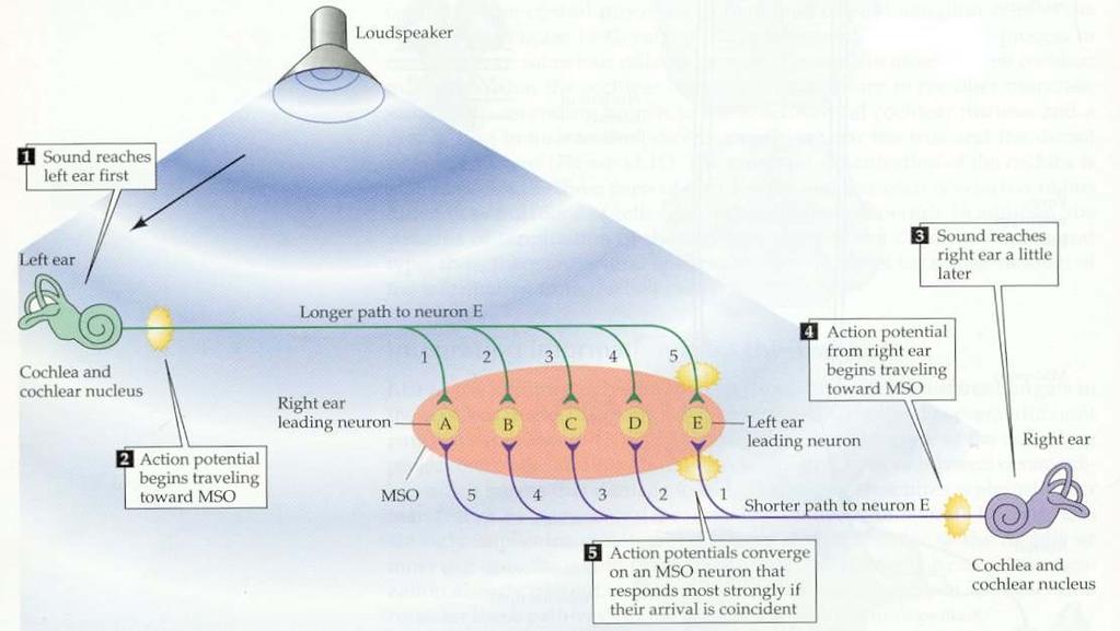
91 Superior olivary complex sound localization MSO = medial superior olive Purves, et al, Neuroscience, 3rd ed.
 BA 41,42 Laterality: Bilateral Lateral")
92 Auditory system Origin: Spiral ganglion Course: CNVIII, auditory pathway, auditory cortex Termination: Auditory Cortex (Heschl s gyrus) BA 41,42 Laterality: Bilateral Lateral Lemniscus
 BA 41,42 Laterality:")
93 Auditory system Inferior Colliculus Lateral Lemniscus Origin: Spiral ganglion Course: CNVIII, auditory pathway, auditory cortex Termination: Auditory Cortex (Heschl s gyrus) BA 41,42 Laterality: Bilateral
 BA 41,42 Laterality:")
94 Auditory system Brachium of Inferior Colliculus Origin: Spiral ganglion Course: CNVIII, auditory pathway, auditory cortex Termination: Auditory Cortex (Heschl s gyrus) BA 41,42 Laterality: Bilateral
 BA 41,42 Laterality:")
95 Auditory system Medial Geniculate Nucleus Origin: Spiral ganglion Course: CNVIII, auditory pathway, auditory cortex Termination: Auditory Cortex (Heschl s gyrus) BA 41,42 Laterality: Bilateral
 Transverse gyri of")
96 Auditory system area A1 (A-I) Transverse gyri of Heschl

97 Central auditory pathways - bilaterality Purves, et al, Neuroscience, 3rd ed.
98 Auditory system - overview Modality: Auditory Sensation (Hearing) Receptor: Organ of of Corti of of Cochlear Duct Cranial Nerve: VIII-cochlear 1st Neuron: Spiral Ganglion 2nd Neuron: Cochlear Nucleus, Ventral & Dorsal dorsal, intermediate and ventral acoustic striae trapezoid body lateral lemniscus 3rd Neuron: Inferior Colliculus brachium of of inferior colliculus 4th Neuron: Medial Geniculate Nucleus (MG) auditory radiation Termination: Primary Auditory Area (A (A I) I) Brodmann area 41, 41, 42 42
99 Auditory Pathways A. spiral ganglion B. dorsal and ventral cochlear nucleus C. inferior colliculus D. medial geniculate nucleus E. Primary auditory area F. superior olivary nucleus G. nucleus of lateral lemniscus 1. acoustic striae & trapezoid body 2. lateral lemniscus 3. brachium of inferior colliculus 4. auditory radiation 5. commissure of inferior colliculus VIIIc. cochlear division


100 Auditory pathways 3D

101 Primary auditory cortex A I

102 Purves, et al, Neuroscience, 3rd ed. The Auditory Cortex


103 Tonotopic organization is maintained along the entire auditory pathway

104 Tonotopic organization is maintained along the entire auditory pathway Purves, et al, Neuroscience, 3rd ed.
105 Auditory cortex Tonotopic (cochleotopic) organization Binaural properties of cortical neurons Wernicke s s area, next to A1, is for the comprehension of speech Probably as we move into auditory association areas - neurons that respond to complex sounds and species-specific specific vocalizations (Grandma s s voice )
106 Lesion localizing BECAUSE OF HEAVY CALLOSAL CONNECTIONS, A UNILATERAL LESION OF THE AUDITORY CORTEX PATHWAYS DISTAL TO THE COCHLEAR NUCLEI WILL RESULT IN VIRTUALLY NO LOSS OF HEARING THE PATIENT WILL MOST LIKELY EXPERIENCE AN IMPAIRMENT IN THE ABILITY TO LOCALIZE THE DIRECTION AND DISTANCE OF SOUNDS
107 Sound Localization 2 Cues Time cues Low frequency sounds (below 3 KHz) are localized due to interaural time differences, capitalizing on the phase-locking abilities of primary afferent fibers. Achieved by the medial superior olive (MSO). Intensity cues High frequency sounds (above 3 KHz) are localized due to interaural intensity differences. The head creates an acoustic shadow. Achieved by the lateral superior olive (LSO).
 If the sound comes from straight ahead, there is no interaural delay.")
108 INTERAURAL TIME DELAY AS A CUE TO THE LOCATION OF SOUND (a) Sound waves coming from the right side reach the right ear first, and there is a large interaural delay before the sound propagates to the left ear. (b) If the sound comes from straight ahead, there is no interaural delay. Delays for three directions are shown.
109
110 Interaural time differences - MSO Illustration of how the MSO computes the location of a sound by interaural time differences. A given MSO neuron responds most strongly when the two inputs arrive simultaneously, as occurs when the contralateral and ipsilateral inputs precisely compensate (via their different lengths) for differences in the time of arrival of a sound at the two ears. They systematic (and inverse) variation in the delay lengths of the two inputs creates a map of sound location: in this model E would be most sensitive to sound located to the left, and A to sound from the right; C would respond best to sounds coming from directly in front of the listener.

111 Higher-order cortical auditory areas Where pathway What pathway What & Where are processed separately

112 Inner ear development The rudiments of the internal ears appear shortly after those of the eyes as two patches of thickened surface epithelium, the otic placodes,, lateral to the hindbrain Carnegie stage 12 ~day 27


113 Late 4 th early 5 th week Otic pit By the time the anterior neuropore closes, the first and second pharyngeal arches are evident The regions between the pharyngeal arches are termed pharyngeal clefts. The indentation just dorsal to the second pharyngeal cleft is the developing inner ear, the otic pit. Otic pit

114 Later ear development Carnegie stage 21 ~day 53

115

116 Derivatives of the pharyngeal pouches

117 Some of the neural crest cells in each of the arches become cartilage

118 Otic vesicle & pinna development CRL


119 External ear development
AUDITORY APPARATUS. Mr. P Mazengenya. Tel 72204
 AUDITORY APPARATUS Mr. P Mazengenya Tel 72204 Describe the anatomical features of the external ear Describe the tympanic membrane (ear drum) Describe the walls of the middle ear Outline the structures
Anatomy of the Ear Region. External ear Middle ear Internal ear
 Ear Lecture Objectives Make a list of structures making the external, middle, and internal ear. Discuss the features of the external auditory meatus and tympanic membrane. Describe the shape, position,
Structure, Energy Transmission and Function. Gross Anatomy. Structure, Function & Process. External Auditory Meatus or Canal (EAM, EAC) Outer Ear
 Outer Ear") Gross Anatomy Structure, Energy Transmission and Function IE N O ME 1 Structure, Function & Process 4 External Auditory Meatus or Canal (EAM, EAC) Outer third is cartilaginous Inner 2/3 is osseous Junction
Gross Anatomy Structure, Energy Transmission and Function IE N O ME 1 Structure, Function & Process 4 External Auditory Meatus or Canal (EAM, EAC) Outer third is cartilaginous Inner 2/3 is osseous Junction
Unit VIII Problem 9 Anatomy of The Ear
 Unit VIII Problem 9 Anatomy of The Ear - The ear is an organ with 2 functions: Hearing. Maintenance of equilibrium/balance. - The ear is divided into 3 parts: External ear. Middle ear (which is also known
Chapter 17, Part 2! The Special Senses! Hearing and Equilibrium!
 Chapter 17, Part 2! The Special Senses! Hearing and Equilibrium! SECTION 17-5! Equilibrium sensations originate within the inner ear, while hearing involves the detection and interpretation of sound waves!
Chapter 17, Part 2! Chapter 17 Part 2 Special Senses! The Special Senses! Hearing and Equilibrium!
 Chapter 17, Part 2! The Special Senses! Hearing and Equilibrium! SECTION 17-5! Equilibrium sensations originate within the inner ear, while hearing involves the detection and interpretation of sound waves!
Auditory Physiology Richard M. Costanzo, Ph.D.
 Auditory Physiology Richard M. Costanzo, Ph.D. OBJECTIVES After studying the material of this lecture, the student should be able to: 1. Describe the morphology and function of the following structures:
The Ear. Dr. Heba Kalbouneh Assistant Professor of Anatomy and Histology
 The Ear Dr. Heba Kalbouneh Assistant Professor of Anatomy and Histology The Ear The ear consists of the external ear; the middle ear (tympanic cavity); and the internal ear (labyrinth), which contains
Unit VIII Problem 9 Physiology: Hearing
 Unit VIII Problem 9 Physiology: Hearing - We can hear a limited range of frequency between 20 Hz 20,000 Hz (human hearing acuity is between 1000 Hz 4000 Hz). - The ear is divided into 3 parts. Those are:
Deafness and hearing impairment
 Auditory Physiology Deafness and hearing impairment About one in every 10 Americans has some degree of hearing loss. The great majority develop hearing loss as they age. Hearing impairment in very early
Auditory System Feedback
 Feedback Auditory System Feedback Using all or a portion of the information from the output of a system to regulate or control the processes or inputs in order to modify the output. Central control of
Ear. Utricle & saccule in the vestibule Connected to each other and to the endolymphatic sac by a utriculosaccular duct
 Rahaf Jreisat *You don t have to go back to the slides. Ear Inner Ear Membranous Labyrinth It is a reflection of bony labyrinth but inside. Membranous labyrinth = set of membranous tubes containing sensory
Chapter 15 Hearing & Equilibrium
 Chapter 15 Hearing & Equilibrium ANATOMY OF THE OUTER EAR EAR PINNA is the outer ear it is thin skin covering elastic cartilage. It directs incoming sound waves to the EXTERNAL AUDITORY CANAL, which is
Auditory System. Barb Rohrer (SEI )
") Auditory System Barb Rohrer (SEI614 2-5086) Sounds arise from mechanical vibration (creating zones of compression and rarefaction; which ripple outwards) Transmitted through gaseous, aqueous or solid medium
Auditory System Barb Rohrer (SEI614 2-5086) Sounds arise from mechanical vibration (creating zones of compression and rarefaction; which ripple outwards) Transmitted through gaseous, aqueous or solid medium
THE COCHLEA AND AUDITORY PATHWAY
 Dental Neuroanatomy Suzanne S. Stensaas, PhD February 23, 2012 Reading: Waxman, Chapter 16, Review pictures in a Histology book Computer Resources: http://www.cochlea.org/ - Promenade around the Cochlea
Required Slide. Session Objectives
 Auditory Physiology Required Slide Session Objectives Auditory System: At the end of this session, students will be able to: 1. Characterize the range of normal human hearing. 2. Understand the components
THE COCHLEA AND AUDITORY PATHWAY
 Dental Neuroanatomy Suzanne S. Stensaas, PhD April 14, 2010 Reading: Waxman, Chapter 16, Review pictures in a Histology book Computer Resources: http://www.cochlea.org/ - Promenade around the Cochlea HyperBrain
The Ear The ear consists of : 1-THE EXTERNAL EAR 2-THE MIDDLE EAR, OR TYMPANIC CAVITY 3-THE INTERNAL EAR, OR LABYRINTH 1-THE EXTERNAL EAR.
 The Ear The ear consists of : 1-THE EXTERNAL EAR 2-THE MIDDLE EAR, OR TYMPANIC CAVITY 3-THE INTERNAL EAR, OR LABYRINTH 1-THE EXTERNAL EAR Made of A-AURICLE B-EXTERNAL AUDITORY MEATUS A-AURICLE It consists
Anatomy and Physiology of Hearing
 Anatomy and Physiology of Hearing The Human Ear Temporal Bone Found on each side of the skull and contains the organs for hearing and balance Divided into four major portions: - squamous - mastoid - tympanic
Receptors / physiology
 Hearing: physiology Receptors / physiology Energy transduction First goal of a sensory/perceptual system? Transduce environmental energy into neural energy (or energy that can be interpreted by perceptual
ENT 318 Artificial Organs Physiology of Ear
 ENT 318 Artificial Organs Physiology of Ear Lecturer: Ahmad Nasrul Norali The Ear The Ear Components of hearing mechanism - Outer Ear - Middle Ear - Inner Ear - Central Auditory Nervous System Major Divisions
SPECIAL SENSES: THE AUDITORY SYSTEM
 SPECIAL SENSES: THE AUDITORY SYSTEM REVISION OF PHYSICS: WAVES A wave is an oscillation of power, sound waves have two main characteristics: amplitude, which is the maximum displacement or the power of
20 The ear (Vestibulo-acoustic Organs)
") 20 The ear (Vestibulo-acoustic Organs) Median line Sella turcica Tuba auditiva Cavum tympani A. carotis int. Superior border of petrous part Membrana tympani Cochlea N. facialis Meatus acusticus internus
20 The ear (Vestibulo-acoustic Organs) Median line Sella turcica Tuba auditiva Cavum tympani A. carotis int. Superior border of petrous part Membrana tympani Cochlea N. facialis Meatus acusticus internus
to vibrate the fluid. The ossicles amplify the pressure. The surface area of the oval window is
 Page 1 of 6 Question 1: How is the conduction of sound to the cochlea facilitated by the ossicles of the middle ear? Answer: Sound waves traveling through air move the tympanic membrane, which, in turn,
Kingdom of Bahrain Arabian Gulf University College of Medicine and Medical Sciences Year 6 ENT SMC Otitis Media (Dr.
 Kingdom of Bahrain Arabian Gulf University College of Medicine and Medical Sciences Year 6 ENT SMC Otitis Media (Dr. Jalal Almarzooq) - Anatomy of the ear: The ear is divided into 3 parts: External ear.
MECHANISM OF HEARING
 MECHANISM OF HEARING Sound: Sound is a vibration that propagates as an audible wave of pressure, through a transmission medium such as gas, liquid or solid. Sound is produced from alternate compression
Hearing. By: Jimmy, Dana, and Karissa
 Hearing By: Jimmy, Dana, and Karissa Anatomy - The ear is divided up into three parts - Sound enters in through the outer ear and passes into the middle where the vibrations are received and sent to the
Anatomy of the ear: Lymphatics
 Anatomy of the ear: 1. External ear which consist of auricle and external auditory canal. The auricle has a framework of cartilage except the lobule, the skin is closely adherent to perichonderium at the
Otoconia: Calcium carbonate crystals Gelatinous mass. Cilia. Hair cells. Vestibular nerve. Vestibular ganglion
 VESTIBULAR SYSTEM (Balance/Equilibrium) The vestibular stimulus is provided by Earth s, and. Located in the of the inner ear, in two components: 1. Vestibular sacs - gravity & head direction 2. Semicircular
The cochlea: auditory sense. The cochlea: auditory sense
 Inner ear apparatus 1- Vestibule macula and sacculus sensing acceleration of the head and direction of gravity 2- Semicircular canals mainly for sensing direction of rotation of the head 1 3- cochlea in
Before we talk about the auditory system we will talk about the sound and waves
 The Auditory System PHYSIO: #3 DR.LOAI ZAGOUL 24/3/2014 Refer to the slides for some photos. Before we talk about the auditory system we will talk about the sound and waves All waves have basic characteristics:
The ear: some applied basic science
 Chapter 1 The ear: some applied basic science The pinna The external ear or pinna is composed of cartilage with closely adherent perichondrium and skin. It is developed from six tubercles of the first
Taste buds Gustatory cells extend taste hairs through a narrow taste pore
 The Special Senses Objectives Describe the sensory organs of smell, and olfaction. Identify the accessory and internal structures of the eye, and explain their function. Explain how light stimulates the
Histology of Ear. Histology Department Medical Faculty University of Sumatera Utara 2008
 Histology of Ear Histology Department Medical Faculty University of Sumatera Utara 2008 References: Gartner LP, Hiatt JL. Color textbook of histology. 2 nd ed. Philadelphia. WB Saunders company. 2001 Kierszenbaum
Gross Anatomy of the. TEMPORAL BONE, EXTERNAL EAR, and MIDDLE EAR
 Gross Anatomy of the TEMPORAL BONE, EXTERNAL EAR, and MIDDLE EAR M1 Gross and Developmental Anatomy 9:00 AM, December 11, 2008 Dr. Milton M. Sholley Professor of Anatomy and Neurobiology Assignment: Head
Gross Anatomy of the. TEMPORAL BONE, EXTERNAL EAR, and MIDDLE EAR. Assignment: Head to Toe Temporomandibular Joint (TMJ)
") Gross Anatomy the TEMPORAL BONE, EXTERNAL EAR, and MIDDLE EAR M1 Gross and Developmental Anatomy 9:00 AM, December 11, 2008 Dr. Milton M. Sholley Pressor Anatomy and Neurobiology Assignment: Head to Toe
Gross Anatomy the TEMPORAL BONE, EXTERNAL EAR, and MIDDLE EAR M1 Gross and Developmental Anatomy 9:00 AM, December 11, 2008 Dr. Milton M. Sholley Pressor Anatomy and Neurobiology Assignment: Head to Toe
What is the effect on the hair cell if the stereocilia are bent away from the kinocilium?
 CASE 44 A 53-year-old man presents to his primary care physician with complaints of feeling like the room is spinning, dizziness, decreased hearing, ringing in the ears, and fullness in both ears. He states
Intro to Audition & Hearing
 Intro to Audition & Hearing Lecture 16 Chapter 9, part II Jonathan Pillow Sensation & Perception (PSY 345 / NEU 325) Fall 2017 1 Sine wave: one of the simplest kinds of sounds: sound for which pressure
Printable version - Hearing - OpenLearn - The Open University
 Skip to content Accessibility Sign in Contact Search the OU The Open University Study at the OU Research at the OU OU Community About the OU Hearing Printable page generated Saturday, 12 November 2011,
Bony and membranous labyrinth. Vestibular system. János Hanics M.D.
 Bony and membranous labyrinth. Vestibular system. János Hanics M.D. The position of the inner ear The labyrinthes of the inner ear - Continuous cavity system in the petrous part of temporal bone - Cavity
THE EAR AND HEARING Be sure you have read and understand Chapter 16 before beginning this lab. INTRODUCTION: hair cells outer ear tympanic membrane
 BIOLOGY 211: HUMAN ANATOMY & PHYSIOLOGY ****************************************************************************************************** THE EAR AND HEARING ******************************************************************************************************
Dr. Sami Zaqout Faculty of Medicine IUG
 Auricle External Ear External auditory meatus The Ear Middle Ear (Tympanic Cavity) Auditory ossicles Internal Ear (Labyrinth) Bony labyrinth Membranous labyrinth External Ear Auricle External auditory
HEARING. Structure and Function
 HEARING Structure and Function Rory Attwood MBChB,FRCS Division of Otorhinolaryngology Faculty of Health Sciences Tygerberg Campus, University of Stellenbosch Analyse Function of auditory system Discriminate
Chapter 3: Anatomy and physiology of the sensory auditory mechanism
 Chapter 3: Anatomy and physiology of the sensory auditory mechanism Objectives (1) Anatomy of the inner ear Functions of the cochlear and vestibular systems Three compartments within the cochlea and membranes
Chapter 7. Audition, the Body Senses, and the Chemical Senses. Copyright Allyn & Bacon 2004
 Chapter 7 Audition, the Body Senses, and the Chemical Senses This multimedia product and its contents are protected under copyright law. The following are prohibited by law: any public performance or display,
Hearing. By Jack & Tori
 Hearing By Jack & Tori 3 Main Components of the Human Ear. Outer Ear. Middle Ear. Inner Ear Outer Ear Pinna: >Visible part of ear and ear canal -Acts as a funnel to direct sound Eardrum: >Airtight membrane
Chapter 11: Sound, The Auditory System, and Pitch Perception
 Chapter 11: Sound, The Auditory System, and Pitch Perception Overview of Questions What is it that makes sounds high pitched or low pitched? How do sound vibrations inside the ear lead to the perception
Sound. Audition. Physics of Sound. Properties of sound. Perception of sound works the same way as light.
 Sound Audition Perception of sound works the same way as light. Have receptors to convert a physical stimulus to action potentials Action potentials are organized in brain structures You apply some meaning
Audition. Sound. Physics of Sound. Perception of sound works the same way as light.
 Audition Sound Perception of sound works the same way as light. Have receptors to convert a physical stimulus to action potentials Action potentials are organized in brain structures You apply some meaning
Νευροφυσιολογία και Αισθήσεις
 Biomedical Imaging & Applied Optics University of Cyprus Νευροφυσιολογία και Αισθήσεις Διάλεξη 11 Ακουστικό και Αιθουσιαίο Σύστημα (Auditory and Vestibular Systems) Introduction Sensory Systems Sense of
Activity 1: Anatomy of the Eye and Ear Lab
 Activity 1: Anatomy of the Eye and Ear Lab 1. Launch the view! Launch Human Anatomy Atlas. Navigate to Quizzes/Lab Activities, find the Eye and Ear Lab section. Launch Augmented Reality mode and scan the
Systems Neuroscience Oct. 16, Auditory system. http:
 Systems Neuroscience Oct. 16, 2018 Auditory system http: www.ini.unizh.ch/~kiper/system_neurosci.html The physics of sound Measuring sound intensity We are sensitive to an enormous range of intensities,
PSY 215 Lecture 10 Topic: Hearing Chapter 7, pages
 PSY 215 Lecture 10 Topic: Hearing Chapter 7, pages 189-197 Corrections: NTC 09-1, page 3, the Superior Colliculus is in the midbrain (Mesencephalon). Announcements: Movie next Monday: Case of the frozen
A&P 1. Ear, Hearing & Equilibrium Lab. Basic Concepts. These notes follow Carl s Talk at the beginning of lab
 A&P 1 Ear, Hearing & Equilibrium Lab Basic Concepts These notes follow Carl s Talk at the beginning of lab In this "Lab Exercise Guide", we will be looking at the basics of hearing and equilibrium. NOTE:
SPECIAL SENSES PART I: OLFACTION & GUSTATION
 SPECIAL SENSES PART I: OLFACTION & GUSTATION 5 Special Senses Olfaction Gustation Vision Equilibrium Hearing Olfactory Nerves Extend through cribriform plate into nasal cavity on both sides of nasal septum
Auditory and vestibular system
 Auditory and vestibular system Sensory organs on the inner ear inner ear: audition (exteroceptor) and vestibular apparatus (proprioceptor) bony and membranous labyrinths within the temporal bone (os temporale)
Chapter Fourteen. The Hearing Mechanism. 1. Introduction.
 Chapter Fourteen The Hearing Mechanism 1. Introduction. 2. Hearing. 3. The Ear. 4. The External Ear. 5. The Inner Ear. 6. Frequency Discrimination. 7. The Organ of Corti. 8. Tests and Exrecises. 9. References.
Hearing: Physiology and Psychoacoustics
 9 Hearing: Physiology and Psychoacoustics Click Chapter to edit 9 Hearing: Master title Physiology style and Psychoacoustics The Function of Hearing What Is Sound? Basic Structure of the Mammalian Auditory
Auditory and Vestibular Systems
 Auditory and Vestibular Systems Objective To learn the functional organization of the auditory and vestibular systems To understand how one can use changes in auditory function following injury to localize
Presentation On SENSATION. Prof- Mrs.Kuldeep Kaur
 Presentation On SENSATION Prof- Mrs.Kuldeep Kaur INTRODUCTION:- Sensation is a specialty area within Psychology that works at understanding how are senses work and how we perceive stimuli in the environment.
College of Medicine Dept. of Medical physics Physics of ear and hearing /CH
 College of Medicine Dept. of Medical physics Physics of ear and hearing /CH 13 2017-2018 ***************************************************************** o Introduction : The ear is the organ that detects
Hearing: the function of the outer, the middle and inner ear. Hearing tests. The auditory pathways
 Hearing: the function of the outer, the middle and inner ear. Hearing tests. The auditory pathways Dr. Gabriella Kékesi 74. Hearing: the function of the outer, the middle and inner ear. Hearing tests.
SENSORY SYSTEM VII THE EAR PART 1
 SENSORY SYSTEM VII THE EAR PART 1 Waves Sound is a compression wave The Ear Ear Outer Ear Pinna Outer ear: - Made up of the pinna and the auditory canal Auditory Canal Outer Ear Pinna (also called the
9.01 Introduction to Neuroscience Fall 2007
 MIT OpenCourseWare http://ocw.mit.edu 9.01 Introduction to Neuroscience Fall 2007 For information about citing these materials or our Terms of Use, visit: http://ocw.mit.edu/terms. 9.01 Recitation (R02)
Chapter 13 Physics of the Ear and Hearing
 Hearing 100 times greater dynamic range than vision Wide frequency range (20 ~ 20,000 Hz) Sense of hearing Mechanical system that stimulates the hair cells in the cochlea Sensors that produce action potentials
Hearing Sound. The Human Auditory System. The Outer Ear. Music 170: The Ear
 Hearing Sound Music 170: The Ear Tamara Smyth, trsmyth@ucsd.edu Department of Music, University of California, San Diego (UCSD) November 17, 2016 Sound interpretation in the auditory system is done by
Music 170: The Ear. Tamara Smyth, Department of Music, University of California, San Diego (UCSD) November 17, 2016
 November 17, 2016") Music 170: The Ear Tamara Smyth, trsmyth@ucsd.edu Department of Music, University of California, San Diego (UCSD) November 17, 2016 1 Hearing Sound Sound interpretation in the auditory system is done by
Music 170: The Ear Tamara Smyth, trsmyth@ucsd.edu Department of Music, University of California, San Diego (UCSD) November 17, 2016 1 Hearing Sound Sound interpretation in the auditory system is done by
REVIEW/PREVIEW OF HEAD AND NECK ANATOMY FOR ENT EXAM
 REVIEW/PREVIEW OF HEAD AND NECK ANATOMY FOR ENT EXAM - 2017 PALPATE CAROTID ARTERY: AT LEVEL OF CAROTID BIFURCATION VERTEBRAL LEVEL C4 Sternocleidomastoid Muscle INTERNAL CAROTID EXTERNAL CAROTID COMMON
The Senses. Chapter 10 7/8/11. Introduction
 Chapter 10 The Senses Introduction A. Sensory receptors detect changes in the environment and stimulate neurons to send nerve impulses to the brain. B. A sensation is formed based on the sensory input.
For more information about how to cite these materials visit
 Author(s): Matthew Velkey, 2009 License: Unless otherwise noted, this material is made available under the terms of the Creative Commons Attribution Non-commercial Share Alike 3.0 License: http://creativecommons.org/licenses/by-nc-sa/3.0/
The Nervous System: General and Special Senses Pearson Education, Inc.
 18 The Nervous System: General and Special Senses Introduction Sensory information arrives at the CNS Information is picked up by sensory receptors Sensory receptors are the interface between the nervous
Sensory system. Dr. Carmen E. Rexach Anatomy 35 Mt San Antonio College
 Sensory system Dr. Carmen E. Rexach Anatomy 35 Mt San Antonio College Sensory receptors Detect stimuli Classified by structure Origin Distribution Modality Structural Classification naked nerve endings
Hearing. istockphoto/thinkstock
 Hearing istockphoto/thinkstock Audition The sense or act of hearing The Stimulus Input: Sound Waves Sound waves are composed of changes in air pressure unfolding over time. Acoustical transduction: Conversion
BCS 221: Auditory Perception BCS 521 & PSY 221
 BCS 221: Auditory Perception BCS 521 & PSY 221 Time: MW 10:25 11:40 AM Recitation: F 10:25 11:25 AM Room: Hutchinson 473 Lecturer: Dr. Kevin Davis Office: 303E Meliora Hall Office hours: M 1 3 PM kevin_davis@urmc.rochester.edu
COM3502/4502/6502 SPEECH PROCESSING
 COM3502/4502/6502 SPEECH PROCESSING Lecture 4 Hearing COM3502/4502/6502 Speech Processing: Lecture 4, slide 1 The Speech Chain SPEAKER Ear LISTENER Feedback Link Vocal Muscles Ear Sound Waves Taken from:
Chapter 1: Applied Anatomy, Physiology and Embryology of the Ear. Anatomy and Physiology. The Outer Ear. The Pinna. The External Ear Canal
 Chapter 1: Applied Anatomy, Physiology and Embryology of the Ear The ear contains two specialized sensory organs, the cochlea and the vestibular apparatus, enclosed in the extremely hard protective casing
Anatomy of External and Middle ear. Dr Sai Manohar
 Anatomy of External and Middle ear. Dr Sai Manohar 1 Human Ear For Anatomical description, Ear is divided into Auricle (or pinna) The external auditory canal The Middle Ear and its derivatives The Inner
Innervation of the Cochlea. Reading: Yost Ch. 8
 Innervation of the Cochlea Reading: Yost Ch. 8 Fine Structure of the Organ of Corti Auditory Nerve Auditory nerve (AN) is a branch of the VIII th cranial nerve (other branch is vestibular). AN is composed
Cochlear anatomy, function and pathology I. Professor Dave Furness Keele University
 Cochlear anatomy, function and pathology I Professor Dave Furness Keele University d.n.furness@keele.ac.uk Aims and objectives of these lectures Introduction to gross anatomy of the cochlea Focus (1) on
Vestibular/Auditory Systems
 Vestibular/Auditory Systems Jay Zenner on February 3, 2012 Dental Neuroanatomy Scott Rogers Office: SOM 2C132 Boney Labyrinth Vestibular Apparatus Two Major Divisions Cochlea (anterior) VIII VII Semicircular
Gathering information the sensory systems; Vision
 Visual System Gathering information the sensory systems; Vision The retina is the light-sensitive receptor layer at the back of the eye. - Light passes through the cornea, the aqueous chamber, the lens,
Week 5. Fall 2016 Part 2: Structure and Function of Auditory System 1
 This outline summarizes major points covered in lecture. It is not intended to replace your own lecture notes. Week 5 How sound is heard: EAR Mechanical energy reaches the eardrum, moves to the middle
A&P 1. Ear, Hearing & Equilibrium Lab. Basic Concepts. Pre-lab Exercises
 A&P 1 Ear, Hearing & Equilibrium Lab Basic Concepts Pre-lab Exercises In this "Lab Exercise Guide", we will be looking at the basics of hearing and equilibrium. NOTE: these notes do not follow the order
Cranial Nerves VII to XII
 Cranial Nerves VII to XII MSTN121 - Neurophysiology Session 13 Department of Myotherapy Cranial Nerve VIII: Vestibulocochlear Sensory nerve with two distinct branches. Vestibular branch transmits information
Carlson (7e) PowerPoint Lecture Outline Chapter 7: Audition, the Body Senses, and the Chemical Senses
 PowerPoint Lecture Outline Chapter 7: Audition, the Body Senses, and the Chemical Senses") Carlson (7e) PowerPoint Lecture Outline Chapter 7: Audition, the Body Senses, and the Chemical Senses This multimedia product and its contents are protected under copyright law. The following are prohibited
Carlson (7e) PowerPoint Lecture Outline Chapter 7: Audition, the Body Senses, and the Chemical Senses This multimedia product and its contents are protected under copyright law. The following are prohibited
HEARING AND COCHLEAR IMPLANTS
 HEARING AND COCHLEAR IMPLANTS FRANCIS CREIGHTON, MD NEUROTOLOGY & SKULL BASE SURGERY FELLOW JOHNS HOPKINS SCHOOL OF MEDICINE NOV 9 TH, 2017 THANKS TO: CHARLIE DELLA SANTINA, HEIDI NAKAJIMA AND DOUG MATTOX
SOLUTIONS Homework #3. Introduction to Engineering in Medicine and Biology ECEN 1001 Due Tues. 9/30/03
 SOLUTIONS Homework #3 Introduction to Engineering in Medicine and Biology ECEN 1001 Due Tues. 9/30/03 Problem 1: a) Where in the cochlea would you say the process of "fourier decomposition" of the incoming
ID# Exam 2 PS 325, Fall 2003
 ID# Exam 2 PS 325, Fall 2003 As always, the Honor Code is in effect and you ll need to write the code and sign it at the end of the exam. Read each question carefully and answer it completely. Although
The white of the eye and the part that maintains its shape is know n as the:
 Scrub In The white of the eye and the part that maintains its shape is know n as the: a. Cornea b. Pupil c. Retina d. Sclera The structure that is found in the ear and contains the organ of hearing is
For this lab you will use parts of Exercise #18 in your Wise lab manual. Please be sure to read those sections before coming to lab
 Bio 322 Human Anatomy Objectives for the laboratory exercise The Eye and Ear Required reading before beginning this lab: Saladin, KS: Human Anatomy 5 th ed (2017) Chapter 17 For this lab you will use parts
1. Axial view, left temporal bone. Plane through the upper antrum (A), superior semicircular canal (SSC) and IAC.
, superior semicircular canal (SSC) and IAC.") PA IAC SSC A 1. Axial view, left temporal bone. Plane through the upper antrum (A), superior semicircular canal (SSC) and IAC. IAC VII M I LSC Plane through the IAC, malleus head and incus and the lateral
PA IAC SSC A 1. Axial view, left temporal bone. Plane through the upper antrum (A), superior semicircular canal (SSC) and IAC. IAC VII M I LSC Plane through the IAC, malleus head and incus and the lateral
a) Central sulcus- shallow groove that runs across brain sagitally
 Central sulcus- shallow groove that runs across brain sagitally") KEY BRAIN Brain Gross Anatomy Terms 1) Explain each of the following in terms of structure of the brain a) Central sulcus- shallow groove that runs across brain sagitally b) Lateral fissure- deep groove
KEY BRAIN Brain Gross Anatomy Terms 1) Explain each of the following in terms of structure of the brain a) Central sulcus- shallow groove that runs across brain sagitally b) Lateral fissure- deep groove
Lectures on Medical Biophysics Department of Biophysics, Medical Faculty, Masaryk University in Brno
 Lectures on Medical Biophysics Department of Biophysics, Medical Faculty, Masaryk University in Brno Introduction to biophysics of receptors Biophysics of hearing and vestibular sense 1 Lecture outline
The speed at which it travels is a function of the density of the conducting medium.
 Sound is a compression wave which (unlike light) must have a medium to conduct it. If the medium is uniform in density, the sound will spread at as a uniform ring of circles (actually spheres). The speed
Copyright 2009 Pearson Education, Inc.
 Outline Nervous System Sensory Systems I. II. III. IV. V. VI. Biol 105 Lecture 11 Chapter 9 Senses Sensory receptors Touch Vision Hearing and balance Smell Senses Sensory receptor cells Sensory receptors
Assisting in Otolaryngology
 Assisting in Otolaryngology Learning Objectives Identify the structures and explain the functions of the external, middle, and internal ear. Describe the conditions that can lead to hearing loss, including
Cranial Nerve VIII (The Vestibulo-Cochlear Nerve)
") Cranial Nerve VIII (The Vestibulo-Cochlear Nerve) Please view our Editing File before studying this lecture to check for any changes. Color Code Important Doctors Notes Notes/Extra explanation Objectives
Cranial Nerve VIII (The Vestibulo-Cochlear Nerve) Please view our Editing File before studying this lecture to check for any changes. Color Code Important Doctors Notes Notes/Extra explanation Objectives
The Organs of Special Senses
 8 The Organs of Special Senses Special senses are those other than touch, pain, temperature, and proprioception. Vision, hearing, and equilibrium are the special senses discussed in this chapter. The Eye
General Sensory Pathways of the Face Area, Taste Pathways and Hearing Pathways
 General Sensory Pathways of the Face Area, Taste Pathways and Hearing Pathways Lecture Objectives Describe pathways for general sensations (pain, temperature, touch and proprioception) from the face area.
Sound and the auditory system
 978--521-68889-5 - Auditory Perception: An Analysis and Synthesis, Third Edition 1 Sound and the auditory system This chapter provides a brief introduction to the physical nature of sound, the manner in