Anatomy of External and Middle ear. Dr Sai Manohar
|
|
|
- Catherine Payne
- 5 years ago
- Views:
Transcription
1 Anatomy of External and Middle ear. Dr Sai Manohar 1
2 Human Ear For Anatomical description, Ear is divided into Auricle (or pinna) The external auditory canal The Middle Ear and its derivatives The Inner Ear. EXT Ear 2
3 Auricle 3
4 Auricle Single piece of Yellow elastic cartilage covered by skin. Incisura terminalis no cartilage b/w tragus & crus of helix endaural approach Several graft materials tragus, concha. 4

5 Nerve Supply Auricle 5
6 External Auditory Canal Bottom of concha TM 24mm. Outer part upwards, backwards & medially, Inner downwards, forwards & medially. To see TM pull pinna upwards, backwards & laterally. 6
7 Cartilaginous part Outer 1/3 rd 8mm continous with cartilage of pinna Fissures of Santorini parotid infections appear in meatus or vice cersa. Skin thick, ceruminous & pilosebaceous glands secrete wax. Hair confined to outer canal furuncles. 7
8 Bony part inner 2/3 rd 16mm. Skin thin, continuous over TM, devoid of glands & hair follicles 6mm lateral to TM narrowing - Isthmus. Ateroinferior part of deep meatus Anterior Recess Foramen of Huschke Anteroinferior in children infection to & fro from Parotid. 8
9 Relations of External Auditory Canal Superiorly middle cranial fossa. Posteriorly mastoid air cells & FN Inferiorly parotid gland. Anteriorly temporomandibular joint. Posterosuperior part of deep meatus related to mastoid antrum mastoiditis sagging. 9
10 Relations of External auditory canal 10
11 External Auditory Canal Nerve Supply Anterior wall & roof auriculotemporal N V3 Posterior wall & floor auricular branch of vagus X Arnolds N. Posterior wall sensory fibres of VII. 11

12 Tympanic Membrane 12
13 Tympanic Membrane Partition b/w EAC & ME. Oblique posterosuperior part is more lateral. 9-10mm tall, 8-9mm wide & 0.1mm thick. Pars Flaccida(Sharpnell s membrane) above lateral process of malleus & b/w notch of rivinus and anterior & posterior malleal folds. Not so taught, slightly pinkish. 13
14 Tympanic Membrane Pars Tensa - most of TM. Periphery thickened fibrocartilaginous ring Annulus Tympanicus. Central part tented inwards at tip of HOM Umbo. Cone of light tip of HOM to periphery in AI Quad. 14
15 Tympanic Membrane Three layers Outer epithelial layer Inner mucosal layer. Middle fibrous layer radial, circular & parabolic fibers. 15
16 Tympanic Membrane Nerve supply External surface of membrane corresponds to that of the meatus. Posterior half- Auricular branch of Xth cranial nerve Anterior half- Auriculotemporal branch of the Vth cranial nerve Medial surface Tympanic branch of CN IX Jacobson s N. 16
17 Middle Ear Cleft Vertical air containing cleft in the temporal bone Includes auditory tube, tympanic cavity & mastoid antrum and air cells. Lined by mucous membrane & filled with air. 17

18 Middle Ear Cross section of ear including the middle ear 18
19 Middle Ear Mesotympanum level of PT Epitympanum Attic above PT Hypotympanum below PT Six sided box. Roof thin plate of bone Tegmen Tympani also roof of aditus & antrum separates ME from middle cranial fossa. 19
20 Middle Ear The Floor thin plate of bone separates ME from Jugular bulb. sometimes deficient. The Anterior Wall thin plate of bone separates ME from Carotid artery. 2 openings lower ET, Upper canal for Tensor tympani. 20
21 The Posterior Wall Middle Ear Aditus Attic communicates with Antrum. bony projection Pyramid tendon of stapedius. FN runs behind pyramid. Facial recess FN, Chorda tympani, Fossa incudis. The Lateral Wall TM Bony outer Attic Wall Scutum. 21
22 Posterior wall of tympanic cavity 22
23 Middle Ear The Medial Wall By Labyrinth Bulge Promontory Basal coil of Cochlea Oval Window Foot plate of Stapes. Round Window Secondary TM. Canal for facial N. Prominance of lateral SCCanal. Processus cochleariformis Sinus tympani deep recess medial to pyramid Subiculum below & Ponticulus above. 23

24 Medial wall of Tympanic cavity 24
25 Mastoid Antrum Large air containing space in upper part of Mastoid, communicating with Attic through Aditus. Roof Tegmen antri Middle cranial fossa. Lateral wall plate of bone 1.5cm thick marked externally by suprameatal (Mac Ewen s) Triangle. 25
26 26
27 Aditus ad Antrum Attic communicates with Antrum. Prominance of Horizontal SCCon its medial side. Fossa incudis laterally. 27
28 Mastoid & its Air Cell System Mastoid bone cortex with a honeycomb of air cells underneath. Depending on air cell development Well pneumatised / cellular Diploetic marrow spaces & few air cells. Sclerotic / acellular 28
29 Mastoid Air Cell System Depending on location Zygomatic cells. - squamous cells Tegmen cells. Perisinus cells. Retrofacial cells. Perilabyrinthine cells. Peritubal cells. Tip cells 29
30 Development of Mastoid From Squamous & Petrous bones. Petrosquamal suture may persist as a bony plate Korner s Septum - Superficial squamous cells from deep petrous cells. Difficulty in locating antrum & deeper cells. 30
31 Contents of Middle Ear Chain of 3 small movable bones, 2 muscles, the chorda tympani and the tympanic plexus of nerves. Malleus 31
32 Ossicles The Malleus head, neck, handle, a lateral & an anterior process. Head & neck in Attic. Handle in fibrous layer of TM. Lateral process attachment to anterior & posterior malleal folds. 32
33 The Incus a body & a short process in attic, & a long process attaches to head of stapes. The Stapes head, neck, anterior & posterior crura & a footplate oval window. 33
34 Middle Ear Incus Stapes 34
35 Auditory Muscles Middle Ear Tensor Tympani - to neck of malleus tenses TM branch of mandibular N. Stapedius to neck of stapes dampen loud sounds Facial N 35
36 Chorda tympani N - Branch of FN enters ME through posterior canaliculus, runs on medial surface of TM b/w handle of malleus & long process of incus. Taste from anterior 2/3 rd of tongue, secretomotor to submandibular & sublingual glands. 36
37 Tympanic plexus On promontory & formed by 1.Tympanic branch of Glossopharyngeal nerve 2. sympathetic fibers from plexus of internal carotid artery Tympanic plexus supplies Lining membrane of tympanic cavity, mastoid air cells and auditory tube. The lesser superficial petrosal nerve otic ganglion auriculotemporal nerve - secretomotor to Parotid. 37
38 Lining of ME Cleft Mucous membrane of Nasopharynx is continuous with that of ME, Aditus, Antrum & air cells. Wraps ME structures Histologically ET ciliated pseudostratified columnar in cartilaginous part & columnar in bony part, mucus glands in submucosa Tympanic cavity ciliated clomnar in anterior & inferior parts, cuboidal inposterior part. Epitympanum & mastoid- flat nonciliated epithelium 38
39 Blood supply of ME Anterior tympanic branch of maxillary artery. Stylomastoid branch of posterior auricular artery. Petrosal branch of middle meningeal artery. Superior tympanic branch of middle meningeal artery. Branch of artery of pterygoid canal. Tympanic branch of internal carotid. 39
40 Lymphatic drainage ME retropharyngeal & parotid nodes. ET retropharyngeal nodes. 40
41 THANK YOU 41
The Ear The ear consists of : 1-THE EXTERNAL EAR 2-THE MIDDLE EAR, OR TYMPANIC CAVITY 3-THE INTERNAL EAR, OR LABYRINTH 1-THE EXTERNAL EAR.
 The Ear The ear consists of : 1-THE EXTERNAL EAR 2-THE MIDDLE EAR, OR TYMPANIC CAVITY 3-THE INTERNAL EAR, OR LABYRINTH 1-THE EXTERNAL EAR Made of A-AURICLE B-EXTERNAL AUDITORY MEATUS A-AURICLE It consists
The Ear. Dr. Heba Kalbouneh Assistant Professor of Anatomy and Histology
 The Ear Dr. Heba Kalbouneh Assistant Professor of Anatomy and Histology The Ear The ear consists of the external ear; the middle ear (tympanic cavity); and the internal ear (labyrinth), which contains
Gross Anatomy of the. TEMPORAL BONE, EXTERNAL EAR, and MIDDLE EAR
 Gross Anatomy of the TEMPORAL BONE, EXTERNAL EAR, and MIDDLE EAR M1 Gross and Developmental Anatomy 9:00 AM, December 11, 2008 Dr. Milton M. Sholley Professor of Anatomy and Neurobiology Assignment: Head
Gross Anatomy of the. TEMPORAL BONE, EXTERNAL EAR, and MIDDLE EAR. Assignment: Head to Toe Temporomandibular Joint (TMJ)
") Gross Anatomy the TEMPORAL BONE, EXTERNAL EAR, and MIDDLE EAR M1 Gross and Developmental Anatomy 9:00 AM, December 11, 2008 Dr. Milton M. Sholley Pressor Anatomy and Neurobiology Assignment: Head to Toe
Gross Anatomy the TEMPORAL BONE, EXTERNAL EAR, and MIDDLE EAR M1 Gross and Developmental Anatomy 9:00 AM, December 11, 2008 Dr. Milton M. Sholley Pressor Anatomy and Neurobiology Assignment: Head to Toe
Dr. Sami Zaqout Faculty of Medicine IUG
 Auricle External Ear External auditory meatus The Ear Middle Ear (Tympanic Cavity) Auditory ossicles Internal Ear (Labyrinth) Bony labyrinth Membranous labyrinth External Ear Auricle External auditory
AUDITORY APPARATUS. Mr. P Mazengenya. Tel 72204
 AUDITORY APPARATUS Mr. P Mazengenya Tel 72204 Describe the anatomical features of the external ear Describe the tympanic membrane (ear drum) Describe the walls of the middle ear Outline the structures
Unit VIII Problem 9 Anatomy of The Ear
 Unit VIII Problem 9 Anatomy of The Ear - The ear is an organ with 2 functions: Hearing. Maintenance of equilibrium/balance. - The ear is divided into 3 parts: External ear. Middle ear (which is also known
Anatomy of the Ear Region. External ear Middle ear Internal ear
 Ear Lecture Objectives Make a list of structures making the external, middle, and internal ear. Discuss the features of the external auditory meatus and tympanic membrane. Describe the shape, position,
Anatomy and Physiology of Hearing
 Anatomy and Physiology of Hearing The Human Ear Temporal Bone Found on each side of the skull and contains the organs for hearing and balance Divided into four major portions: - squamous - mastoid - tympanic
Anatomy of the ear: Lymphatics
 Anatomy of the ear: 1. External ear which consist of auricle and external auditory canal. The auricle has a framework of cartilage except the lobule, the skin is closely adherent to perichonderium at the
REVIEW/PREVIEW OF HEAD AND NECK ANATOMY FOR ENT EXAM
 REVIEW/PREVIEW OF HEAD AND NECK ANATOMY FOR ENT EXAM - 2017 PALPATE CAROTID ARTERY: AT LEVEL OF CAROTID BIFURCATION VERTEBRAL LEVEL C4 Sternocleidomastoid Muscle INTERNAL CAROTID EXTERNAL CAROTID COMMON
Lec [8]: Mandibular nerve:
![Lec [8]: Mandibular nerve:](/thumbs/94/121295776.jpg "Lec [8]: Mandibular nerve:") Lec [8]: Mandibular nerve: The mandibular branch from the trigeminal ganglion lies in the middle cranial fossa lateral to the cavernous sinus. With the motor root of the trigeminal nerve [motor roots lies
Lec [8]: Mandibular nerve: The mandibular branch from the trigeminal ganglion lies in the middle cranial fossa lateral to the cavernous sinus. With the motor root of the trigeminal nerve [motor roots lies
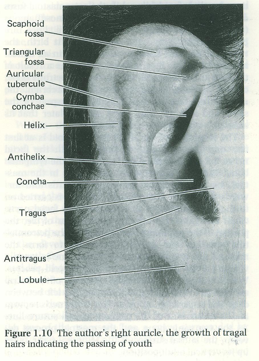
The ear: some applied basic science
 Chapter 1 The ear: some applied basic science The pinna The external ear or pinna is composed of cartilage with closely adherent perichondrium and skin. It is developed from six tubercles of the first
Dr.Ban I.S. head & neck anatomy 2 nd y. جامعة تكريت كلية طب االسنان املرحلة الثانية أ.م.د. بان امساعيل صديق 6102/6102
 جامعة تكريت كلية طب االسنان التشريح مادة املرحلة الثانية أ.م.د. بان امساعيل صديق 6102/6102 Parotid region The part of the face in front of the ear and below the zygomatic arch is the parotid region. The
For the following questions, indicate the letter that corresponds to the SINGLE MOST APPROPRIATE ANSWER
 GROSS ANATOMY EXAMINATION May 15, 2000 For the following questions, indicate the letter that corresponds to the SINGLE MOST APPROPRIATE ANSWER 1. Pain associated with an infection limited to the middle
Infratemporal fossa: Tikrit University college of Dentistry Dr.Ban I.S. head & neck Anatomy 2 nd y.
 Infratemporal fossa: This is a space lying beneath the base of the skull between the lateral wall of the pharynx and the ramus of the mandible. It is also referred to as the parapharyngeal or lateral pharyngeal
Parotid Gland, Temporomandibular Joint and Infratemporal Fossa
 M1 - Anatomy Parotid Gland, Temporomandibular Joint and Infratemporal Fossa Jeff Dupree Sanger 9-057 jldupree@vcu.edu Parotid gland: wraps around the mandible positioned between the mandible and the sphenoid
Cranial Nerve VII - Facial Nerve. The facial nerve has 3 main components with distinct functions
 Cranial Nerve VII - Facial Nerve The facial nerve has 3 main components with distinct functions Somatic motor efferent Supplies the muscles of facial expression; posterior belly of digastric muscle; stylohyoid,
Kingdom of Bahrain Arabian Gulf University College of Medicine and Medical Sciences Year 6 ENT SMC Otitis Media (Dr.
 Kingdom of Bahrain Arabian Gulf University College of Medicine and Medical Sciences Year 6 ENT SMC Otitis Media (Dr. Jalal Almarzooq) - Anatomy of the ear: The ear is divided into 3 parts: External ear.
Cone Beam CT Atlas of the Normal Suspensory Apparatus of the Middle Ear Ossicles
 Cone Beam CT Atlas of the Normal Suspensory Apparatus of the Middle Ear Ossicles Poster No.: C-2036 Congress: ECR 2013 Type: Authors: Educational Exhibit B. Smet 1, I. De Kock 2, P. Gillardin 2, M. Lemmerling
Dr.Ban I.S. head & neck anatomy 2 nd y. جامعة تكريت كلية طب االسنان املرحلة الثانية
 جامعة تكريت كلية طب االسنان التشريح مادة املرحلة الثانية أ.م.د. بان امساعيل صديق 6102-6102 1 The Palate The palate forms the roof of the mouth and the floor of the nasal cavity. It is divided into two
Temporal fossa Infratemporal fossa Pterygopalatine fossa Terminal branches of external carotid artery Pterygoid venous plexus
 Outline of content Temporal fossa Infratemporal fossa Pterygopalatine fossa Terminal branches of external carotid artery Pterygoid venous plexus Boundary Content Communication Mandibular division of trigeminal
Exposure of facial nerve and endolymphatic sac
 Exposure of facial nerve and endolymphatic sac 1 7 4 2 3 5 6 8 1 Vertical part of the facial nerve exposed 1 Second genu of facial nerve. 2 Vertical part of facial nerve. 3 Horizontal part of facial nerve.
Tikrit University collage of dentistry Dr.Ban I.S. head & neck anatomy 2 nd y. Lec [5] / Temporal fossa :
![Tikrit University collage of dentistry Dr.Ban I.S. head & neck anatomy 2 nd y. Lec [5] / Temporal fossa :](/thumbs/88/115294566.jpg "Tikrit University collage of dentistry Dr.Ban I.S. head & neck anatomy 2 nd y. Lec [5] / Temporal fossa :") Lec [5] / Temporal fossa : Borders of the Temporal Fossa: Superior: Superior temporal line. Inferior: gap between zygomatic arch and infratemporal crest of sphenoid bone. Anterior: Frontal process of the
Lec [5] / Temporal fossa : Borders of the Temporal Fossa: Superior: Superior temporal line. Inferior: gap between zygomatic arch and infratemporal crest of sphenoid bone. Anterior: Frontal process of the
Major Anatomic Components of the Orbit
 Major Anatomic Components of the Orbit 1. Osseous Framework 2. Globe 3. Optic nerve and sheath 4. Extraocular muscles Bony Orbit Seven Bones Frontal bone Zygomatic bone Maxillary bone Ethmoid bone Sphenoid
Dr. Sami Zaqout, IUG Medical School
 The skull The skull is composed of several separate bones united at immobile joints called sutures. Exceptions? Frontal bone Occipital bone Vault Cranium Sphenoid bone Zygomatic bones Base Ethmoid bone
Functional components
 Facial Nerve VII cranial nerve Emerges from Pons Two roots Functional components: 1. GSA (general somatic afferent) 2. SA (Somatic afferent) 3. GVE (general visceral efferent) 4. BE (Special visceral/branchial
The Pharynx. Dr. Nabil Khouri MD. MSc, Ph.D
 The Pharynx Dr. Nabil Khouri MD. MSc, Ph.D Introduction The pharynx is the Musculo-fascial halfcylinder that links the oral and nasal cavities in the head to the larynx and esophagus in the neck Common
Omran Saeed. Luma Taweel. Mohammad Almohtaseb. 1 P a g e
 2 Omran Saeed Luma Taweel Mohammad Almohtaseb 1 P a g e I didn t include all the photos in this sheet in order to keep it as small as possible so if you need more clarification please refer to slides In
Temporal region. temporal & infratemporal fossae. Zhou Hong Ying Dept. of Anatomy
 Temporal region temporal & infratemporal fossae Zhou Hong Ying Dept. of Anatomy Temporal region is divided by zygomatic arch into temporal & infratemporal fossae. Temporal Fossa Infratemporal fossa Temporal
1. Axial view, left temporal bone. Plane through the upper antrum (A), superior semicircular canal (SSC) and IAC.
, superior semicircular canal (SSC) and IAC.") PA IAC SSC A 1. Axial view, left temporal bone. Plane through the upper antrum (A), superior semicircular canal (SSC) and IAC. IAC VII M I LSC Plane through the IAC, malleus head and incus and the lateral
PA IAC SSC A 1. Axial view, left temporal bone. Plane through the upper antrum (A), superior semicircular canal (SSC) and IAC. IAC VII M I LSC Plane through the IAC, malleus head and incus and the lateral
Dr.Noor Hashem Mohammad Lecture (5)
") Dr.Noor Hashem Mohammad Lecture (5) 2016-2017 If the mandible is discarded, the anterior part of this aspect of the skull is seen to be formed by the hard palate. The palatal processes of the maxillae
Dr.Noor Hashem Mohammad Lecture (5) 2016-2017 If the mandible is discarded, the anterior part of this aspect of the skull is seen to be formed by the hard palate. The palatal processes of the maxillae
PTERYGOPALATINE FOSSA
 PTERYGOPALATINE FOSSA Outline Anatomical Structure and Boundaries Foramina and Communications with other spaces and cavities Contents Pterygopalatine Ganglion Especial emphasis on certain arteries and
Veins of the Face and the Neck
 Veins of the Face and the Neck Facial Vein The facial vein is formed at the medial angle of the eye by the union of the supraorbital and supratrochlear veins. connected through the ophthalmic veins with
The Skull and Temporomandibular joint II Prof. Abdulameer Al-Nuaimi. E. mail:
 The Skull and Temporomandibular joint II Prof. Abdulameer Al-Nuaimi E-mail: a.al-nuaimi@sheffield.ac.uk E. mail: abdulameerh@yahoo.com Temporal fossa The temporal fossa is a depression on the temporal
The Visible Ear Simulator Dissection Manual.
 The Visible Ear Simulator Dissection Manual. Stereoscopic Tutorialized Version 3.1, August 2017 Peter Trier Mikkelsen, the Alexandra Institute A/S, Aarhus, Denmark Mads Sølvsten Sørensen & Steven Andersen,
SCHOOL OF ANATOMICAL SCIENCES Mock Run Questions. 4 May 2012
 SCHOOL OF ANATOMICAL SCIENCES Mock Run Questions 4 May 2012 1. With regard to the muscles of the neck: a. the platysma muscle is supplied by the accessory nerve. b. the stylohyoid muscle is supplied by
Prevertebral Region, Pharynx and Soft Palate
 Unit 20: Prevertebral Region, Pharynx and Soft Palate Dissection Instructions: Step1 Step 2 Step 1: Insert your fingers posterior to the sternocleidomastoid muscle, vagus nerve, internal jugular vein,
Subdivided into Vestibule & Oral cavity proper
 Extends from the lips to the oropharyngeal isthmus The oropharyngeal isthmus: Is the junction of mouth and pharynx. Is bounded: Above by the soft palate and the palatoglossal folds Below by the dorsum
The Seventh Cranial Nerve The Facial By Prof. Dr. Muhammad Imran Qureshi
 The Seventh Cranial Nerve The Facial By Prof. Dr. Muhammad Imran Qureshi Functional Components: SVE: Fibers originate from nucleus of facial nerve, and supply facial muscles GVE: Fibers derived from superior
The sebaceous glands (glands of Zeis) open directly into the eyelash follicles, ciliary glands (glands of Moll) are modified sweat glands that open
 open directly into the eyelash follicles, ciliary glands (glands of Moll) are modified sweat glands that open") The Orbital Region The orbits are a pair of bony cavities that contain the eyeballs; their associated muscles, nerves, vessels, and fat; and most of the lacrimal apparatus upper eyelid is larger and more
The Orbital Region The orbits are a pair of bony cavities that contain the eyeballs; their associated muscles, nerves, vessels, and fat; and most of the lacrimal apparatus upper eyelid is larger and more
Skull Base Course. Dissection with fresh temporal bones and half heads
 Skull Base Course Dissection with fresh temporal bones and half heads 711 November 2016 Gruppo Otologico Via Emmanueli 42 Piacenza 29122 t +39 0523 754 362 fax +39 0523 453 708 www.gruppootologico.com
Mohammad Hisham Al-Mohtaseb. Lina Mansour. Reyad Jabiri. 0 P a g e
 2 Mohammad Hisham Al-Mohtaseb Lina Mansour Reyad Jabiri 0 P a g e This is only correction for the last year sheet according to our record. If you already studied this sheet just read the yellow notes which
Parotid Gland. Parotid Gland. Largest of 3 paired salivary glands (submandibular; sublingual) Ramus of Mandible. Medial pterygoid.
 Ramus of Mandible. Medial pterygoid.") Parotid region Parotid Gland Largest of 3 paired salivary glands (submandibular; sublingual) Ramus of Mandible Medial pterygoid Cross section of mandible Masseter D S SCM Parotid Gland Mastoid Process
Parotid region Parotid Gland Largest of 3 paired salivary glands (submandibular; sublingual) Ramus of Mandible Medial pterygoid Cross section of mandible Masseter D S SCM Parotid Gland Mastoid Process
By : Prof Saeed Abuel Makarem & Dr.Sanaa Alshaarawi
 By : Prof Saeed Abuel Makarem & Dr.Sanaa Alshaarawi OBJECTIVES By the end of the lecture, students shouldbe able to: List the nuclei of the deep origin of the trigeminal and facial nerves in the brain
Maxilla, ORBIT and infratemporal fossa. Neophytos C Demetriades MD, DDS, MSc Associate professor European University of Cyprus School of Medicine
 Maxilla, ORBIT and infratemporal fossa Neophytos C Demetriades MD, DDS, MSc Associate professor European University of Cyprus School of Medicine MAXILLA Superior, middle, and inferior meatus Frontal sinus
SKULL / CRANIUM BONES OF THE NEUROCRANIUM (7) Occipital bone (1) Sphenoid bone (1) Temporal bone (2) Frontal bone (1) Parietal bone (2)
 Occipital bone (1) Sphenoid bone (1) Temporal bone (2) Frontal bone (1) Parietal bone (2)") Important! 1. Memorizing these pages only does not guarantee the succesfull passing of the midterm test or the semifinal exam. 2. The handout has not been supervised, and I can not guarantee, that these
Important! 1. Memorizing these pages only does not guarantee the succesfull passing of the midterm test or the semifinal exam. 2. The handout has not been supervised, and I can not guarantee, that these
Anatomic Relations Summary. Done by: Sohayyla Yasin Dababseh
 Anatomic Relations Summary Done by: Sohayyla Yasin Dababseh Anatomic Relations Lecture 1 Part-1 - The medial wall of the nose is the septum. - The vestibule lies directly inside the nostrils (Nares). -
Bones of the skull & face
 Bones of the skull & face Cranium= brain case or helmet Copyright The McGraw-Hill Companies, Inc. Permission required for reproduction or display. The cranium is composed of eight bones : frontal Occipital
Bisection of Head & Nasal Cavity 頭部對切以及鼻腔. 解剖學科馮琮涵副教授 分機
 Bisection of Head & Nasal Cavity 頭部對切以及鼻腔 解剖學科馮琮涵副教授 分機 3250 E-mail: thfong@tmu.edu.tw Outline: The structure of nose The concha and meatus in nasal cavity The openings of paranasal sinuses Canals, foramens
Anatomy of Oral Cavity DR. MAAN AL-ABBASI
 Anatomy of Oral Cavity DR. MAAN AL-ABBASI By the end of this lecture you should be able to: 1. Differentiate different parts of the oral cavity 2. Describe the blood and nerve supply of mucosa and muscles
Tikrit University College of Dentistry Dr.Ban I.S. head & neck anatomy 2 nd y.
 Lec [3]/The scalp The scalp extends from the supraorbital margins anteriorly to the nuchal lines at the back of the skull and down to the temporal lines at the sides. The forehead, from eyebrows to hairline,
PCM1 Physical Exam Skills Session: Head and Neck FACILITATOR & STUDENT COPY
 PATIENT CENTERED MEDICINE - 1 GOALS & OUTCOMES: PCM1 Physical Exam Skills Session: Head and Neck FACILITATOR & STUDENT COPY 1. To introduce the applied anatomy relevant for the examination of the head
Skull-2. Norma Basalis Interna Norma Basalis Externa. Dr. Heba Kalbouneh Associate Professor of Anatomy and Histology
 Skull-2 Norma Basalis Interna Norma Basalis Externa Dr. Heba Kalbouneh Associate Professor of Anatomy and Histology Norma basalis interna Base of the skull- superior view The interior of the base of the
Cranial nerves.
 Cranial nerves eaglezhyxzy@163.com Key Points of Learning Name Components Passing through Peripheral distribution Central connection Function Cranial nerves Ⅰ olfactory Ⅱ optic Ⅲ occulomotor Ⅳ trochlear
Activity 1: Anatomy of the Eye and Ear Lab
 Activity 1: Anatomy of the Eye and Ear Lab 1. Launch the view! Launch Human Anatomy Atlas. Navigate to Quizzes/Lab Activities, find the Eye and Ear Lab section. Launch Augmented Reality mode and scan the
human anatomy 2016 lecture fifteen Dr meethak ali ahmed neurosurgeon
 Cranial Nerves Organization of the Cranial Nerves The cranial nerves are named as follows: I. Olfactory II. Optic III. Oculomotor IV. Trochlear V. Trigeminal VI. Abducent VII. Facial VIII. Vestibulocochlear
Dr.Ban I.S. head & neck anatomy 2 nd y جامعة تكريت كلية طب االسنان مادة التشريح املرحلة الثانية أ.م.د. بان امساعيل صديق 6102/6102
 جامعة تكريت كلية طب االسنان مادة التشريح املرحلة الثانية أ.م.د. بان امساعيل صديق 6102/6102 Pterygopalatine fossa: The pterygopalatine fossa is a cone-shaped depression, It is located between the maxilla,
Tympanic Bulla Temporal Bone. Digastric Muscle. Masseter Muscle
 Superior view Hyoid Bone The hyoid bone does not articulate with any other bones. It is held in place by ligaments to the styloid process of the temporal bone and the thyroid cartilage of the larynx. It
Primary Headache Prevalence % (95% CI) Migraine without aura 9 (7-9) Migraine with aura 6 (5-8)
 Migraine without aura 9 (7-9) Migraine with aura 6 (5-8)") Primary Headache Prevalence % (95% CI) Migraine without aura 9 (7-9) Migraine with aura 6 (5-8) Episodic tension-type headache 66 (62-69) Chronic tension-type headache 3 (2-5) Cluster headache 0.1 (0 1)
Primary Headache Prevalence % (95% CI) Migraine without aura 9 (7-9) Migraine with aura 6 (5-8) Episodic tension-type headache 66 (62-69) Chronic tension-type headache 3 (2-5) Cluster headache 0.1 (0 1)
Bony orbit Roof The orbital plate of the frontal bone Lateral wall: the zygomatic bone and the greater wing of the sphenoid
 Bony orbit Roof: Formed by: The orbital plate of the frontal bone, which separates the orbital cavity from the anterior cranial fossa and the frontal lobe of the cerebral hemisphere Lateral wall: Formed
Sensory system. Dr. Carmen E. Rexach Anatomy 35 Mt San Antonio College
 Sensory system Dr. Carmen E. Rexach Anatomy 35 Mt San Antonio College Sensory receptors Detect stimuli Classified by structure Origin Distribution Modality Structural Classification naked nerve endings
MECHANISM OF HEARING
 MECHANISM OF HEARING Sound: Sound is a vibration that propagates as an audible wave of pressure, through a transmission medium such as gas, liquid or solid. Sound is produced from alternate compression
THE EAR AND HEARING Be sure you have read and understand Chapter 16 before beginning this lab. INTRODUCTION: hair cells outer ear tympanic membrane
 BIOLOGY 211: HUMAN ANATOMY & PHYSIOLOGY ****************************************************************************************************** THE EAR AND HEARING ******************************************************************************************************
Chapter 1: Applied Anatomy, Physiology and Embryology of the Ear. Anatomy and Physiology. The Outer Ear. The Pinna. The External Ear Canal
 Chapter 1: Applied Anatomy, Physiology and Embryology of the Ear The ear contains two specialized sensory organs, the cochlea and the vestibular apparatus, enclosed in the extremely hard protective casing
Structure Location Function
 Frontal Bone Cranium forms the forehead and roof of the orbits Occipital Bone Cranium forms posterior and inferior portions of the cranium Temporal Bone Cranium inferior to the parietal bone forms the
ENT 318 Artificial Organs Physiology of Ear
 ENT 318 Artificial Organs Physiology of Ear Lecturer: Ahmad Nasrul Norali The Ear The Ear Components of hearing mechanism - Outer Ear - Middle Ear - Inner Ear - Central Auditory Nervous System Major Divisions
Essential questions. What are the structures of the sensory system? 3.03 Remember the structures of the sensory system 2
 Essential questions What are the structures of the sensory system? 3.03 Remember the structures of the sensory system 2 The Senses Eyes Sight Ears Hearing Nose Smell Tongue Taste Skin Touch 3.03 Remember
Tracing the Cranial Nerves Osteologically
 CN I II III IV V 1 Supra-orbital ethmoidal nn. Ext. nasal V 2 Tracing the Cranial Nerves Osteologically Nucleus of Origin Olfactory tracts of frontal lobe of cerebrum Optic tracts from optic chiasma and
The Neck the lower margin of the mandible above the suprasternal notch and the upper border of the clavicle
 The Neck is the region of the body that lies between the lower margin of the mandible above and the suprasternal notch and the upper border of the clavicle below Nerves of the neck Cervical Plexus Is formed
Chronic Ear Disease. Daekeun Joo Resident Lecture Series 11/18/09
 Chronic Ear Disease Daekeun Joo Resident Lecture Series 11/18/09 ETD URIs Viral-induced damage to ET lining resulting in decreased mucociliary clearance Viral invasion of ME mucosa results in inflamm Reflux
o A cushion of fat surrounds most of the eye
 Name Period SPECIAL SENSES The Senses of touch o Temperature o Pressure o Pain o Smell o Taste o Sight o Hearing o Equilibrium The Eye and Vision are in the eyes has over a o Most of the eye is enclosed
Nose & Mouth OUTLINE. Nose. - Nasal Cavity & Its Walls. - Paranasal Sinuses. - Neurovascular Structures. Mouth. - Oral Cavity & Its Contents
 Dept. of Human Anatomy, Si Chuan University Zhou hongying eaglezhyxzy@163.com Nose & Mouth OUTLINE Nose - Nasal Cavity & Its Walls - Paranasal Sinuses - Neurovascular Structures Mouth - Oral Cavity & Its
4 Cartilage Palisades in Underlay Tympanoplasty Techniques
 52 4 Cartilage Palisades in Underlay Tympanoplasty Techniques Definition Cartilage underlay palisade technique is the oldest and the most popular technique in cartilage tympanoplasty. As shown in Chapter
Anatomy #1; Respiratory Nose and the Nasal Cavity December 1st, 2013
 Note #1: the doctor skipped some slides in the lecture. Those slides are not included in this sheet and so you will have to review the slides to study them. The reason they were not included is because
Skull basic structures. Neurocranium
 Assoc. Prof. Květuše Lovásová, M.V.D., PhD. Skull basic structures Skull consists of two groups of bones: neurocranium (bones forming the brain box) splanchnocranium (bones forming the facial skeleton)
Temporomandibular Joint. Dr Noman ullah wazir
 Temporomandibular Joint Dr Noman ullah wazir Type of Joint TMJ is a Synovial joint between : The condylar head of the mandible. The mandibular fossa of squamous part of temporal bone. The joint cavity
AXIAL SKELETON SKULL
 AXIAL SKELETON SKULL CRANIAL BONES (8 total flat bones w/ 2 paired) 1. Frontal forms forehead & upper portion of eyesocket (orbital) 2. Parietal paired bones; form superior & lateral walls of cranium 3.
SPECIAL SENSES PART I: OLFACTION & GUSTATION
 SPECIAL SENSES PART I: OLFACTION & GUSTATION 5 Special Senses Olfaction Gustation Vision Equilibrium Hearing Olfactory Nerves Extend through cribriform plate into nasal cavity on both sides of nasal septum
*in general the blood supply of the nose comes from branches of the internal and external carotid arteries.
 In the previous lecture we talked about the anatomy of the nasal cavity, today we will talk about its blood supply, venous drainage, innervations, and finally about the paranasal sinuses. When we describe
THE PTERYGOPALATINE FOSSA MAXILLARY NERVE EAR VESTIBULOCOCHLEAR NERVE CN VIII
 THE PTERYGOPALATINE FOSSA MAXILLARY NERVE EAR VESTIBULOCOCHLEAR NERVE CN VIII 1 THE PTERYGOPALATINE FOSSA 2 THE PTERYGOPALATINE FOSSA The pterygopala/ne fossa lies between: the pterygoid process of the
View of a Skull, 1489 by Leonardo Da Vinci. Kaan Yücel M.D., Ph.D Tuesday
 View of a Skull, 1489 by Leonardo Da Vinci Kaan Yücel M.D., Ph.D. 26.11.2013 Tuesday 1.SKULL skeleton of the head cranium 22 bones excluding ossicles of the ear 1.SKULL Mandible Lower jaw bone Neurocranium
THIEME. Scalp and Superficial Temporal Region
 CHAPTER 2 Scalp and Superficial Temporal Region Scalp Learning Objectives At the end of the dissection of the scalp, you should be able to identify, understand and correlate the clinical aspects: Layers
MAXILLA, ORBIT & PTERYGOPALATINE FOSSA. Neophytos C Demetriades MD, DDS, MSc Associate professor European University of Cyprus School of Medicine
 MAXILLA, ORBIT & PTERYGOPALATINE FOSSA Neophytos C Demetriades MD, DDS, MSc Associate professor European University of Cyprus School of Medicine Maxilla MAXILLA Superior, middle, and inferior meatus Frontal
The white of the eye and the part that maintains its shape is know n as the:
 Scrub In The white of the eye and the part that maintains its shape is know n as the: a. Cornea b. Pupil c. Retina d. Sclera The structure that is found in the ear and contains the organ of hearing is
04 Development of the Face and Neck. Development of the Face Development of the neck
 04 Development of the Face and Neck Development of the Face Development of the neck Development of the face Overview of facial development The fourth week ~ the twelfth week of prenatal development Between
HBA THE BODY Head & Neck Written Examination October 23, 2014
 HBA 531 - THE BODY Head & Neck Written Examination October 23, 2014 Name: NOTE 2: When asked to trace nerve, artery, or vein pathways, do so by using arrows, e.g., structure a structure b structure c...
K. J. Lee: Essential Otolaryngology and Head and Neck Surgery (IIIrd Ed) Preface
 Preface") K. J. Lee: Essential Otolaryngology and Head and Neck Surgery (IIIrd Ed) Preface This book is written for the young otolaryngologist who is already acquainted with the field through recent formal residency
K. J. Lee: Essential Otolaryngology and Head and Neck Surgery (IIIrd Ed) Preface This book is written for the young otolaryngologist who is already acquainted with the field through recent formal residency
Biology 323 Human Anatomy for Biology Majors Week 10; Lecture 1; Tuesday Dr. Stuart S. Sumida. Cranial Nerves and Soft Tissues of the Skull
 Biology 323 Human Anatomy for Biology Majors Week 10; Lecture 1; Tuesday Dr. Stuart S. Sumida Cranial Nerves and Soft Tissues of the Skull FOREBRAIN MIDBRAIN HINDBRAIN Forebrain: Cerebrum Perception,
Neck-2. Dr. Heba Kalbouneh Associate Professor of Anatomy and Histology
 Neck-2 ` Dr. Heba Kalbouneh Associate Professor of Anatomy and Histology Triangles of the neck Side of the neck Midline Lower border of mandible Line between angle of mandible and mastoid Superior nuchal
UC SF. Safe Surgery Rule #1. Cholesteatoma. It s hard to have a surgical complication when you are not operating
 UC SF Cholesteatoma Chronic Ear Surgery: Staying Out of Trouble! Lawrence R. Lustig, MD Department of Oto-HNS University of California San Francisco Ligaments and folds Spaces NU Epitympanic Cholesteatoma
3-Deep fascia: is absent (except over the parotid gland & buccopharngeal fascia covering the buccinator muscle)
") The Face 1-Skin of the Face The skin of the face is: Elastic Vascular (bleed profusely however heal rapidly) Rich in sweat and sebaceous glands (can cause acne in adults) It is connected to the underlying
The Face 1-Skin of the Face The skin of the face is: Elastic Vascular (bleed profusely however heal rapidly) Rich in sweat and sebaceous glands (can cause acne in adults) It is connected to the underlying
-Ibrahim Al-Naser. -Dr Al- Muhtaseb. 1 P a g e
 -1 -Ibrahim Al-Naser - -Dr Al- Muhtaseb 1 P a g e The Digestive System The doctor started the lecture by talking about the class rules. The GI system is an organ system, it is divided into: The Alimentary
APRIL
 APRIL - 2003 OCTOBER - 2003 February 2009 [KU 652] Sub. Code : 4131 FIRST B.D.S DEGREE EXAMINATION (Modified Regulations III) Paper I HUMAN ANATOMY, HISTOLOGY AND EMBRYOLOGY Time : Three hours
Skull-2. Norma Basalis Interna. Dr. Heba Kalbouneh Assistant Professor of Anatomy and Histology
 Skull-2 Norma Basalis Interna Dr. Heba Kalbouneh Assistant Professor of Anatomy and Histology Norma basalis interna Base of the skull- superior view The interior of the base of the skull is divided into