sirona.com 3D imaging The Dental Company
|
|
|
- Clyde Burke
- 6 years ago
- Views:
Transcription


1 sirona.com A New Dimension in Diagnostics, Planning and Treatment 3D imaging with GALILEOS The Dental Company
2 Contents editorial 03 3D X-ray following implant complications 04 3D image identifies odontoma 06 Exact determination of the position: basis for minimally invasive orthodontic exposure 08 DVT facilitates the diagnosis of chronic rhinorrhea and postnasal secretion 09 Implant planning integrates CAD/CAM and DVT 11 Safety first for implantology 13 3D diagnostics for a computer-aided implant plan with external sinus lift 15 Benefits of integrated implant in at risk patients 17 CMD diagnosis and therapy in one appointment 19 Integrated FaceSCAN supports diagnosis and surgical planning 20 Today s integrated solutions give us a taste of tomorrow s innovative therapies 22 legal notice 23
3 2 3 Dear readers, The importance of 3D diagnostics has increased significantly in recent years. A growing number of dentists with different specializations are discovering the advantages of digital volume tomography (DVT) for diagnostics, and are aware of the numerous innovative options it presents for the planning and performing of treatments. As a result, new applications are continuously being developed for oral and maxillofacial surgery, orthodontics, implantology and the analysis of respiratory tract organs. By means of brief case studies, this brochure provides detailed information on the diverse applications of GALILEOS - the high-end digital imaging system from Sirona. Like all 3D X-ray units from Sirona, GALILEOS stands for the best possible image quality at the lowest dose and a perfect workflow. The careful coordination of all imaging elements and the loss-free interaction of resolution, noise suppression and filters for reducing metal artifacts, for example, are essential for achieving an excellent image quality. Users are also ensured maximum flexibility with regard to the use of image data: all of the treatment relevant X-ray images can be generated from a single scan. With the aid of findings-oriented software, GALILEOS offers users enhanced safety and reliability for difficult diagnoses as well as time-saving and efficient workflows. In combination with a surface scan from the optionally integrated FaceSCAN, the DVT also improves patient communication: patients have a better understanding of the diagnoses and opt for the proposed treatment more quickly and more frequently. Patients should also have the smallest possible exposure to radiation. This is why Sirona uses a state-of-the-art image intensifier for large scan volumes. GALILEOS satisfies even the most rigorous standards of general practices, specialists and clinics. Please enjoy reading, Jörg Haist Head of Product management Imaging systems
4 3D X-ray following implant complications Author Dr. Christian Scheifele, Hamburg (Germany) When complications arise despite a problemfree intervention, conventional imaging processes are often not able to supply enough information. Cases like this illustrate the added value of 3D X-rays. With GALILEOS, one scan is enough to capture the patient s entire jaw. As the system moves around the patient, the X-ray volume created provides dentists with all the relevant information they need. All anatomical structures can be analyzed in layers from virtually every perspective. The main advantage of the 3D X-ray scan is its exceptional informative value. A diagnosis can often be established for findings which conventional imaging techniques would not detect. Dentists receive a complete and comprehensive image of the jaw situation. This increases diagnostic accuracy and allows various oral and maxillofacial, orthodontic, ENT and implantology treatments (as illustrated by a specific patient case) to be planned exactly. A 66-year-old female patient who had suffered tooth loss in region 16 was treated with an implant following an internal sinus lift. Roughly six months later, after an initial symptom-free period, the patient reported recurrent infections and a white discharge from the nose. The post-operative OPT was normal (Fig. 1). Due to persistent peri-implantitis, the implant could no longer be retained and was removed (Fig. 2). Following detailed consultation, the patient opted for a new implant in region 16. In order to clarify possible sinusitis, a DVT was created using Galileos which had not been available to the implantologist at the time of the first implantation. This was done after the second implantation in region 16. Positioning of the window directly on the implant (Fig. 3) showed no unusual results. When the window was moved toward cranial, however, the result was surprising: the nasal cavity was located where the maxillary sinus would normally be found (Fig. 4). When questioned, the patient explained that as a child she had had a bad fall and landed on her nose, and that she also has quite often breathed through her nose. It was obvious that this trauma was the most likely cause for the pronounced septum deviation which, given her externally perceived straight nose, could not be initially clinically determined. The displacement of the maxillary sinus was the reason for the problematic implant position. The implantologist who performed the procedure is not at fault; given the 2D imaging available, he could not possibly have recognized the atypical position of the maxillary sinus. This is only possible with DVT. This case confirms the essential nature of 3D diagnostics


 Half-view panoramic x-ray following explantation.")

5 4 5 in implantological rehabilitation: the most important diagnostic information is available at a glance. Excellent image quality and functionality also play a role here. The essential qualities of GALILEOS X-ray images are their high resolution and excellent contrast relative to the exposure required. A further plus is the fact that the hardware is, in many ways, simpler to operate than conventional panoramic units. In particular, it is much easier to position the patient. 1 Postoperative half-view exposure with normal implant position. 2 (right) Half-view panoramic x-ray following explantation. The complete case study in German was published in: Scheifele C, 3D-Röntgen nach Implantat-Komplikationen. Dental Magazin, 5/ Both in the panoramic exposure and in the three transveral slices, a soft-tissue density opacity could be diagnosed which appeared to be delimited by air. 4 Upon moving the examination window cranially, it became evident that the implant position was partially located in the right nasal cavity.

6 3D image identifies odontoma author Dr. Fred Bergmann, Viernheim (Germany) A male patient presents a displaced tooth in region 43. The 3D image identified an odontoma, enabling treatment to be performed exactly as planned, without surprises. 1 Initial situation in the panoramic X-ray view. The mandibular canal can be marked in color on the DVT image. The main advantage of the DVT technique is the highly informative nature of the X-ray volume. It becomes possible to reach a comprehensive diagnosis, which makes both therapy planning and treatment easier for practitioners. Take, for example, the case of a young male patient with a displaced canine tooth (region 43).
 clearly shows that the displaced tooth is positioned vestibularly while the odontoma is positioned lingually. This information was of central importance when planning the intervention.")


7 6 7 Thanks to the 3D image, we discovered a complex odontoma in region 43 (Fig. 1). With this knowledge, we were able to tailor treatment precisely to these findings. The axial view (Fig. 2) clearly shows that the displaced tooth is positioned vestibularly while the odontoma is positioned lingually. This information was of central importance when planning the intervention. We were able to expose the tooth vestibularly, while the odontoma was removed from the lingual side. Exact pre-planning made the operation much simpler for the practitioner since there was no risk of surprises during the intervention. And the operation was also less traumatic for the patient. Displaced tooth 43 was then exposed vestibularly and inserted orthodontically. The odontoma was also removed lingually. 2 It is possible to determine the exact positioning of the odontoma and the displaced tooth by evaluating the axial layer. The complete case study in German was published in: Bergmann F; 3D-Aufnahme deckt Odontom auf. Oralchirurgie Journal, 4/ Exposed tooth 43. The odontoma is removed from lingual. 4 Following successful intervention: the odontoma has been removed. Exposed, displaced tooth 43 is integrated orthodontically.
 One of the most frequent cases of tooth retention is the palatinal or vestibular displacement of the remaining canine teeth in the upper jaw.")
.")
8 Exact determination of the position: basis for minimally invasive orthodontic exposure Author Dr. Fred Bergmann, Viernheim (Germany) One of the most frequent cases of tooth retention is the palatinal or vestibular displacement of the remaining canine teeth in the upper jaw. Spontaneous penetration does not always occur in such cases when the deciduous teeth are removed. In order to render the intervention as a traumatic as possible, the position of the canine teeth should be determined precisely by means of digital volume tomography (DVT). In the case of a 17-year-old male patient, the upper canine teeth were retained palatinally. The dentition had already been treated orthodontically, but space for the missing canine teeth had not yet been created. A DVT was produced to determine the correct surgical method and to establish the position of the canine teeth crowns as accurately as possible. The 3D image showed that the canine teeth crowns were located between the first and second incisors and that their labial surfaces were located on the palatal side. This allowed all risks to be weighed in advance, enabling the intervention to be performed as atraumatically as possible for the patient. Since the teeth were still covered by bone, the crowns were exposed by local osteotomy. The laser provided valuable assistance here. On the one hand, it reduced bleeding to a minimum a key prerequisite for adhering the brackets. On the other hand, as a result of the laser surgery, the patient had fewer postoperative complaints. The complete case study in German was published in: Bergmann F; Exakte Lageermittlung: Basis für minimalinvasive Kieferorthopädie. ZWR 12/ The position of the vestibular tooth surface is determined in the axial view. 2 State after adhesive fixation of brackets and retainers.
 Additionally, in the area of ENT, 3D imaging ensures more precise diagnostics, safer planning and more effective treatment in comparison to other imaging systems.")
9 8 9 DVT facilitates the diagnosis of chronic rhinorrhea and postnasal secretion Author Dr. Marco Capelli, Codogno (Italy) Additionally, in the area of ENT, 3D imaging ensures more precise diagnostics, safer planning and more effective treatment in comparison to other imaging systems. This improves communication with patients and increases overall satisfaction. An increasing number of mycoses have been diagnosed in the nasal and sinus areas over the past years. These parasitic infections are frequently caused by types of Aspergillus. In some cases, an aspergilloma (fungus ball) forms in the maxillary sinus, a common form 1 Coronal view of the affected maxillary sinus with hyperintense material due to a mycotic build-up.


10 2 Sagittal view of the maxillary sinus. 3 Axial view of the maxillary sinus. of chronic rhinosinusitis. The typical symptoms of acute rhinosinusitis cannot be determined until later. In many cases, this occurs together with bacterial superinfections as in the case of one patient who had been suffering from purulent rhinorrhea, postnasal drip syndrome and cacosmia for a number of months. He had already been treated with antibiotics, steroids and an aerosol. However, these treatments had not resulted in improvement. The patient was examined endoscopically at our practice, enabling us to diagnose congestion of the osteomeatal complex and a translucent polypoid formation in the central nasal passage. Using GALILEOS, we performed a DVT of the facial skull without using a contrast agent. We discovered an area with strong hyperintensity in the left maxillary sinus. The accumulation of material with a structure with the density of bone or metal led us to believe that this was an aspergilloma. On the basis of these findings, the focus of infection could be removed during a rhinosurgical intervention. Thanks to DVT, we were provided with valuable information which was not available endoscopically. As a result, we were able to plan and execute treatment successfully.

11 10 11 Implant planning integrates CAD/CAM and DVT Author Dr. Viktor Karapetian, Cologne (Germany) PD Dr. Dr. Lutz Ritter, PD Dr. Dr. Martin Scheer, Prof. Dr. Dr. Joachim E. Zöller The combination of various technologies in the world of dentistry opens up new treatment approaches. Thanks to integrated implantology, practitioners can now optimally plan implantation and, to a large extent, avoid risk even before the actual intervention. With integrated implantology, dentists specializing in implantology can superimpose the 3D X-ray data from the GALILEOS system onto the restoration proposal from CEREC to harmonize surgical and prosthetic planning. The combination of both technologies offers practitioners significant advantages. They acquire a precise image of the jaw and tooth situation prior to the actual intervention, are able to ensure the presence of sufficient bone material at the required points, and can select the right implant size. Using GALILEOS, we produced a 3D X-ray image of a 59-year-old male patient with anterior tooth trauma on teeth 21 and 11. We thereby determined that tooth 21 had to be removed due to a transverse fracture of the tooth root. Using the CEREC CAD/CAM restoration system, we created a restoration proposal and imported this into the DVT data with the GALILEOS Implant planning software. The practitioner marked corresponding points in the CAD/CAM data and in the 3D X-ray image 1 Integrated implant planning with GALILEOS and CEREC allows practitioners to create an ideal basis for treatment with just a few clicks.


12 to allow the software to superimpose both data sets precisely. During planning, it became evident that the lingual bone wall was not thick enough for an implantation, so we performed surgical socket preservation with granulate following removal of tooth 21. Tooth 11 was also restored with an acrylic filling and we splinted the entire anterior region in order to stabilize the teeth in position. We exposed the implant after three months and produced the final restoration with CEREC as follows: We adapted the restoration proposal - which we constructed virtually to the requirements of this case - and, using the CEREC MC XL milling machine, milled the restoration from multicolored layered feldspathetic ceramic. The implant crown was customized and finished with final firing and cemented onto the abutment. Virtual planning of the entire treatment process enables users to avoid potential complications. The practitioner s treatment proposal is also more transparent and easier for patients to understand. The complete case study in German was published in: Karapetian V, et al., Implantatplanung integriert CAD/CAM und DVT. Oralchirurgie Journal 2/2012. Integrated implantology provides practitioners with added value: it simplifies diagnostics and increases planning certainty. 2 Exposure three months after osseointegration: the gingiva has healed well. 3 Final clinical result: restored teeth 11 and 21 dovetail perfectly with the overall look.
13 12 13 Safety first for implantology Author Jochen Kusch, Executive Vice President Marketing and Sales of SICAT, Bonn (Germany) Many implantologists would like to see an increase in treatment safety. The use of digital technologies is all-important here, since the combination of DVT and CAD/CAM data facilitates the production of drill templates. The use of 3D X-ray images reduces the risk of unintentionally damaging anatomical structures and allows the bone situation to be assessed even before actual intervention. Using 3D X-ray and CAD data plus implant planning software, dentists can precisely plan where and at what angle the implant must be positioned and whether sufficient bone substance is available. The virtual plan can be used to produce drill templates with one of three options: CLASSICGUIDE, OPTIGUIDE and CEREC Guide. 1. CLASSICGUIDE method First, the dentist takes an impression using the conventional method. The dental technician waxes up the proposed restoration on the model and produces a deep-drawn tray into which the prosthetic proposal containing barium sulphate is integrated. This is fixed to an X-ray template. Alternatively, with more simple cases, the bite registration can be applied directly to the X-ray template. The patient wears this X-ray template during the DVT imaging process. The reference balls on the bite plate enable perfect positioning later during preparation of the drilling templates. After planning the implant, the dentist sends the 3D X-ray data, the plan and the X-ray template to SICAT. This workflow functions reliably both for edentulous jaws and jaws fully restored with metal crowns. SICAT analyzes the feasibility of each drilling template as a special service. Manufacturing accuracy is measured at the apical end of the implant and guaranteed to be under 0.5 mm. 2. OPTIGUIDE method When the implantologist uses CEREC, he or she can greatly simplify the process. The dentist captures the visual tooth situation with the CEREC camera and constructs the prosthetic recommendation using CEREC restoration software. He or she imports the CAD data into the implant planning software and merges it with the 3D X-ray data of a Sirona DVT. This process virtually matches the surgical and prosthetic planning. Next, he or she transfers the digital planning data simply by uploading them to the SICAT surgical fabrication template. This simplification of the workflow means considerable time savings for the dentist, since he can dispense with a radiographic guide.
 and implant crown (blue) are integrated into the X-ray data.")


14 1 X-ray template with proposed restoration made of barium sulfate. 2 SICAT OPTIGUIDE drilling templates with surgical sleeves in different sizes. 3 The virtual model with the implant (shown in orange) and implant crown (blue) are integrated into the X-ray data. There is sufficient distance to the mandibular canal (colored purple). 4 With the new CEREC 4.4 software, one can use CEREC Guide 2 to produce the drilling template for guided surgery quickly, easily and very inexpensively right in the dental practice. 3. CEREC Guide method CEREC users will be able to produce their own drilling templates (CEREC Guide 2) with a CEREC MC XL production unit. The impression formed by CEREC, the soft tissue information and the pre-planned prosthetics are merged to the GALILEOS system 3D data set. GALILEOS Implant does the planning. Next, the planning results are transmitted back to CEREC and milled from a PMMA plastic block. Neither a physical model nor a second X-ray scan with reference markers is required. Conclusion Each of the three methods leads to one goal: increased reliability through guided implantology. Depending on equipment and preferences, the practitioner decides on either the premium service of the drill template manufacturer with safety controls, or the fast and comfortable chairside procedure. Original German-language version of the article can be found in: Kusch J, Safety first in der Implantologie. Dentale Implantologie 11/2011.
 In edentulous regions of the jaw, bone material can recede rapidly due to lack of use.")

15 D diagnostics for a computer-aided implant plan with external sinus lift Author Dr. Fred Bergmann, Viernheim (Germany) In edentulous regions of the jaw, bone material can recede rapidly due to lack of use. If an implant restoration is planned, the remaining bone structure must first be measured. Digital volume tomography (DVT) is an invaluable aid for such an assessment. The advantages of DVT images include hard tissue structures being displayed with a high image quality, and patients being exposed to a comparatively low dose of radiation. This is especially important when planning implant treatment that involves insufficient supportive bone material,resulting in the dentist having to decide on a suitable augmentation method. For larger procedures such as an external sinus lift, precise findings from 3D X-ray images contribute considerably to reducing surgical trauma. In a 49-year-old patient, the missing teeth in region 24 to 27 were to be replaced with a fixed prosthesis on four implants. The computeraided planning of the implants in 3D volume showed that the maxillary bone required augmentation. Due to the large size of the implant treatment, only an external sinus lift was possible for bone augmentation. In this method, the insertion of bone and bone sub 1 Initial clinical situation of region 24 to X-ray template with the planned prosthesis. stitute material augmented the jaw enough to provide adequate support for the implants. The integrated and guided implantation gave the surgeon a high degree of security.


16 3 The reporter function allows for a clear overview of the planned treatment. 4 Precise drilling using the SICAT guide on a jaw that was previously augmented with bone substitute material. 5 The final restoration.

17 Benefits of integrated implant in at risk patients Author Dr. Martin Butz, Munich (Germany) Forward planning for implant surgery is no longer state of the art. Advanced practitioners start with the desired results when planning for the operation of the desired prosthetic treatment. Today, they can take advantage of innovative methods such as integrated implants and drilling templates methods which have proven themselves especially valuable with high-risk patients. Patient was a 63-year-old female who was a heavy smoker and very dissatisfied with her bite situation. She had seven missing teeth. We first made a DVT impression in order to in advance assess the conditions for successful implantation. It showed sufficient bone material for the required implants in the vertical and horizontal directions. Due to the anatomical conditions and the free end situation, a situation model was created in the practice laboratory with corresponding wax-ups of the intended crowns, and was then digitized using a CEREC Bluecam. Next, the digital data from the GALILEOS Implant planning software was imported using the OPEN GALILEOS interface. The software displayed the virtual crowns at the appropriate point in the 3D X-ray image. This facilitated surgical planning according to the desired prosthetic restoration. Based on this virtual, integrated implant planning, we ordered two drill guides one each for the upper and lower jaw from SICAT. Implants 16, 17, 26, 36 and 46 were inserted in accordance with the integrated implant plan. After a healing phase of four months, we exposed the implants and designed the emergence profile. Next, the implants were molded with Impregum using individual plastic partial trays, and the prosthetic restoration was made in the dental laboratory. The patient is now symptom-free and very satisfied with the new treatment. The complete case study in German was published in: Butz M, Vorteile der integrierten Implantologie bei Risikopatienten. Zahnarzt & Praxis, 5/ Integrated implant planning in the X-ray software.


18 2 DVT and prosthetic planning are merged in the x-ray software. 3 The drilling templates made by SICAT specified the position and di-rection during the procedure. 4 The drilling template is firmly seated in the patient s mouth. 5 The implant was stabilized after insertion through augmentation with autogenous bone. 6 The prosthetic restoration of teeth 16, 17 and 26.
, SICAT Function uses the DVT (GALILEOS) to record 3D image data, which is then combined with movement measurements (SICAT, JMT+) and digital surface data (CEREC).")

19 18 19 CMD diagnosis and therapy in one appointment Author Jochen Kusch, Executive Vice President Marketing and Sales of SICAT, Bonn (Germany) In order to diagnose and treat craniomandibular dysfunctions (CMD), SICAT Function uses the DVT (GALILEOS) to record 3D image data, which is then combined with movement measurements (SICAT, JMT+) and digital surface data (CEREC). As a result, an anatomically precise, actual dynamic patient situation is presented that can be comprehensively analyzed for the first time. Changes in the joint gaps when taking defined mandibular positions or in the course of mandibular movement can be displayed directly as measurements. During DVT recording and at the beginning of a JMT measurement, the patient bears a reference tray with radiopaque markers, the SICAT FusionBite. With SICAT JMT+, the jaw s real positions and movements are recorded with all six degrees of freedom. The movement paths can be visualized at any specific point on the mandible. In this way, the spatial relationship between the condyle and fossa in motion can be illustrated individually for the first time. Dynamic occlusion can also be tracked for each jaw position using the CEREC optical impression function. To accomplish this, the optical impressions acquired during the intraoral scanning procedure can be metrically superimposed on the DVT data with perfect accuracy. After diagnosis and analysis of the data at hand, the practitioner can decide whether or not to order a machine-made OPTIMOTION therapy track from SICAT. SICAT OPTIMOTION is based on the Michigan Principles and can be produced according to the practitioner s preferences. Original German-language version of the article can be found in: Kusch J; SICAT Function. Kieferorthopädie Nachrichten, Kompendium Dynamic occlusion with SICAT Function using the merged surface data. 2 Using the SICAT OPTIMOTION therapy track the dentist is able to treat temporomandibular joint problems.
 Face scanners may help orthodontists and oral and maxillofacial surgeons in treatment planning and patient communication.")
20 Integrated FaceSCAN supports diagnosis and surgical planning author PD Dr. med. Dr. med. dent. Lutz Ritter, Hennef (Germany) Face scanners may help orthodontists and oral and maxillofacial surgeons in treatment planning and patient communication. The prerequisite for such use of technology: the surface data of the patient s face must be precisely superimposed on the 3D X-ray data. In our practice, we predominantly use the GALILEOS DVT device with an integrated Facescan for patients with striking extra oral findings such as facial asymmetry. Facescan delivers an exact 3D reproduction of the surface, facilitating the analysis and evaluation of the facial proportions, including the nose, lips and chin configuration. On the basis of the 3D illustration of the face, we create the clinical findings and follow up with an orthodontic or orthognathic jaw surgery treatment plan. In the present case, after bimaxillary osteotomy and surgical correction of the remnant chin, the supramental scarred inclusion disturbed the patient. A preoperative DVT Facescan clearly displayed the remaining metal and incomplete osseous regeneration with the resulting inclusion (Fig. 1). We recommended that the patient undergo metal removal and bony formation of hard and soft tissue structures to reduce the supramental deficit. 1 Initial findings with supramental scarring after bimaxillary osteotomy. The use of the GALILEOS integrated facial scanner offered a number of advantages in the treatment. On the one hand, the practitioner was able to use the facial scans in treatment planning, implementation and


21 20 21 documentation. In addition, the 3D presentation provided excellent orientation shortly before and during the surgery. The image of the patient s face could also be used to supplement the documentation of the treatment course and in before-and-after comparisons of the clinical and aesthetic situations. It also helped the patient to understand the planned treatment. No additional radiation dose is applied and there is no increase in the time required for the system to rotate. The GALILEOS system with integrated Facescan is highly precise due to the surface and the 3D X-ray data being recorded simultaneously in the same coordinate system. As well, the computer can correctly classify them geometrically. The subsequent combination of face scans with separately created radiographs can not reach this level of precision. Since the images are calculated from the scanned 3D data, there is no distortion. 2 Preoperative situation with superimposed X-ray/ surface data. 3 Condition after scar resolution and augmentation of the supramental fold.

22 Today s integrated solutions give us a taste of tomorrow s innovative therapies Author Mark Bucher, Product manager 3D imaging, Bensheim (Germany) 1 Dentists analyze patient respiratory tracts and can order a protrusion prosthesis for treatment of obstructive sleep apnea. The future of our health care system depends on whether or not we are able to make treatments efficient enough so that they are able to overcome the impact of aging and increasingly higher standards. More and more innovative treatment approaches have emerged through the integration of proven digital solutions in recent years. With their efficient workflows, they demonstrate today how dentistry will be managed in the future. Digitization is now the basis for nearly all technological progress. The increasing integration of various digital technologies is becoming an increasingly important part of innovation. For example, Sirona systems aren t only standalone solutions, standalone solutions, but instead combine digital data to enable pioneering therapeutic concepts and efficient workflows in the areas of implantology, endodontics, orthodontics,
23 22 23 functional diagnostics and prosthetics. Digitization simplifies and accelerates many process steps, and system integration optimizes entire treatment processes. This includes networking many digital systems with one another, interconnected to patient management and controlled centrally. CAD/CAM and digital X-ray technology have evolved over the past decades to become key technologies in dentistry. DVTs are now an integral part of many treatments, as shown by the case studies in this brochure. And we are constantly developing new therapy concepts that go beyond these applications. Example, SICAT Air: For the first time, using this 3D software, we will present to the professional community at IDS 2015 the ability of dentists to analyze the upper airways of patients with obstructive apnea. Thanks to digital technology, we are now able to implement a customized therapy track in just two sessions. In addition, a 3D X-ray scan will provide anatomical information about the respiratory tract and jaw joints in the protruded position. These data provide dentists with information about whether the therapy is working without having an adverse affect on the jaw joints. In the next step, the dentist records the digital surface data of both jaws using CEREC and merges this data with the DVT data using the SICAT Air software. Next, users can then transfer the data from the software to SICAT and order the mandibular protrusion appliance (SICAT OPTISLEEP). The increasing interconnection of different systems is enabling efficient, reliable planning and the implementation of minimally invasive treatments. With our team of more than 300 scientists and engineers worldwide who work primarily in our Bensheimer Innovation Center and with our unique system expertise in the areas of CAD/CAM and imaging systems, treatment units, instruments and hygiene devices, we will continue pursuing these goals in the coming years. LEGAL Notice Publisher Sirona Dental GmbH Sirona Strasse 1 A 5071 Wals/Salzburg (Austria) contact@sirona.com Telephone: +43(0) Fax: +43(0) Responsible under german press law Vincent Kummer, Marion Weixlberger, Sirona Dental GmbH in Wals/Salzburg (Austria) Editorial and design ergo Unternehmenskommunikation GmbH & Co. KG Venloer Straße D Köln Special thanks also to: Dr. Fred Bergmann, Dr. Martin Butz, Dr. Marco Capelli, Dr. Gerd Frahsek, Dr. Viktor Karapetian, Jochen Kusch, PD Dr. Dr. Lutz Ritter, Dr. Christian Scheifele Printing Offset 5020 Druckerei & Verlag Ges.m.b.H. Bayernstraße 27 A-5072 Siezenheim Telephone: +43 (0) We reserve the right to make technical modifications, typographical errors and mistakes.
24 Order No. A91100-M47-B , Printed in Austria, Dispo-No
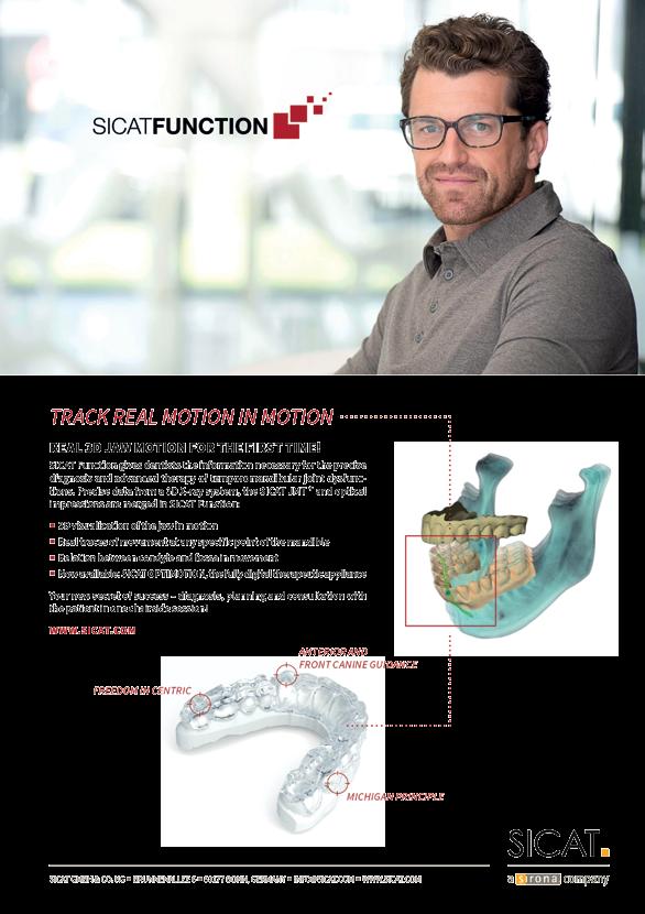
SICAT FUNCTION & OPTIMOTION. Individual functional treatment
 SICAT FUNCTION & OPTIMOTION Individual functional treatment REAL MOTION TIME FOR FACTS THE DIGITALIZATION of treatment processes based on 3D X-ray data is progressing quickly. What has been successfully
SICAT FUNCTION TRACK REAL MOTION IN MOTION. Real 3D jaw motion for the first time
 SICAT FUNCTION TRACK REAL MOTION IN MOTION Real 3D jaw motion for the first time REAL MOTION TIME FOR FACTS THE DIGITALIZATION of treatment procedures based on 3D X-ray data is progressing rapidly. What
IMPLANT MORE SUCCESSFULLY WITH SICAT SURGICAL GUIDES
 IMPLANT MORE SUCCESSFULLY WITH SICAT SURGICAL GUIDES MAKE EVERY CASE COUNT DIAGNOSTICS AND PLANNING IN 3D. In the field of implantology the use of surgical guides based on 3D X-ray data gives your practice
Full mouth rehabilitation with digital workflow
 Jung-plant dental office Dr. Jae-min, Lee D.D.S. Full mouth rehabilitation with digital workflow Solutions featured: 3Shape TRIOS 3Shape Dental System 3Shape Implant Studio Case information On first visit,
clinical articles management advice practice profiles technology reviews Implant PRACTICE US Volume 3 No 2
 clinical articles management advice practice profiles technology reviews Implant PRACTICE US Volume 3 No 2 Sirona Sirona tailors solutions to the needs of the different markets within dentistry to ensure
Digital Imaging from a new perspective
 TREATMENT CENTRES HANDPIECES HYGIENE SYSTEMS X-RAY SYSTEMS CEREC TREATMENT CENTRES HANDPIECES HYGIENE SYSTEMS X-RAY SYSTEMS CEREC SIRONA CREATING AND MAINTAINING VALUE. You are right to expect a great
T h e D e n t a l C o m p a n y FROM DIAGNOSTIC SCAN TO SURGERY, WE SHAPE THE FUTURE OF DENTISTRY.
 T h e D e n t a l C o m p a n y FROM DIAGNOSTIC SCAN TO SURGERY, WE SHAPE THE FUTURE OF DENTISTRY. SIDEXIS SOFTWARE ORTHOPHOS SL D/D SEAMLESS THE NEW STANDARD IN CLINICAL DIAGNOSIS AND PATIENT COMMUNICATION
Implant Studio Patient Case
 Jung-plant dental office Dr. Jae-min, Lee D.D.S. Implant Studio Patient Case Full mouth rehabilitation Case information On first visit, the patient was wearing a removable partial denture on lower jaw
Easy operation. Numerous diagnostic options. X-rays you can rely on: the ORTHOPHOS XG device family. All ORTHOPHOS XG 3Dready programs at a glance.
 CAD /CAM Systems Instruments Hygiene Systems Treatment Centers Imaging Systems Subject to technical changes and errors in the text, Order No. A91100-M47-B346-01-7600, Printed in Germany, Dispo-Nr. 04602,
From planning to surgery: a totally digital working flow for Leone implants placement
 Dr. Giancarlo Romagnuolo Roma, Italy From planning to surgery: a totally digital working flow for Leone implants placement Keywords guided surgery, 3D implant planning, single missing tooth, delayed immediate
For technical assistance: Tel Working time: 8.30 am pm
 I For technical assistance: Tel. +39 059 392827 Working time: 8.30 am - 5.30 pm Index JD Digital solutions for Intraoral Scanning JD Implant Libraries JD Guided Surgery Kit JD Surgical Guide and 3D Model
Safety and esthetics with
 Safety and esthetics with dental implants A guide for patients Dear reader, Implant restorations follow nature's example. You can have the functions of natural teeth completely restored and thus maintain
Guided immediate loading implant surgery planned with Implant Studio D.D.S. Jae-min, Lee
 Guided immediate loading implant surgery planned with Implant Studio D.D.S. Jae-min, Lee Jung-plant Dental office 1 PROLOGUE How can we deal with the immediate loading implant cases easier and more accurate
ZIRCONIA, SIMPLY FASTER. FULL CONTOUR ZIRCONIA RESTORATIONS IN A SINGLE VISIT ONLY WITH CEREC.
 ZIRCONIA, SIMPLY FASTER. FULL CONTOUR ZIRCONIA RESTORATIONS IN A SINGLE VISIT ONLY WITH CEREC. CEREC MAKES IT HAPPEN. What would you think about providing your patients full contour zirconia crowns in
STL. inside. ineos Blue: Precision, Speed and Control. THE SCANNER WITH Bluecam TECHNOLOGY
 CAD/CAM SYSTEMS INSTRUMENTS HYGIENE SYSTEMS TREATMENT CENTERS IMAGING SYSTEMS THE SCANNER WITH Bluecam TECHNOLOGY ineos Blue: Precision, Speed and Control. STL inside T h e D e n t a l C o m p a n y ineos
You design, we create.
 CAD/CAM SYSTEMS INSTRUMENTS HYGIENE SYSTEMS TREATMENT CENTRES IMAGING SYSTEMS infinident SIRONA S CENTRAL PRODUCTION You design, we create. www.infinident.gb.com T h e D e n t a l C o m p a n y infinident:
Immediate loading and implant surgery with digital workflow
 Mirero Dental Clinic Dr. Jaemin Lee Immediate loading and implant surgery with digital workflow Solutions featured: 3Shape TRIOS 3Shape Dental System 3Shape Implant Studio Case information Patient, 32-year
GuidedService. The ultimate guide for precise implantations
 GuidedService The ultimate guide for precise implantations ABGuidedService The ultimate guide for precise implantations At A.B. Dental we've brought implantology into the future with a 3D digitally planned
SICAT AIR & OPTISLEEP. The appliance-based 3D solution for the treatment of obstructive sleep apnea
 SICAT AIR & OPTISLEEP The appliance-based 3D solution for the treatment of obstructive sleep apnea DIGITAL & INTUITIVE with OPTISLEEP, the most advanced oral appliance straight from the dentist. THE DIGITAL
Screw retained implant crown restoration with digital workflow using scan body and surgical guide
 Dr. Anthony Mak W Dental Screw retained implant crown restoration with digital workflow using scan body and surgical guide Solutions featured: 3Shape TRIOS 3Shape Implant Studio 3Shape scan bodies 3Shape
The first 3D solution for obstructive sleep apnea therapy. MATRx plus with SICAT Air and OPTISLEEP
 The first 3D solution for obstructive sleep apnea therapy MATRx plus with SICAT Air and OPTISLEEP The first fully digital workflow for a more restful sleep Digital workflow with MATRx plus, SICAT Air,
THE 3D SOLUTION INVENTORS OPTIGUIDE
 THE 3D SOLUTION INVENTORS OPTIGUIDE 2012-01-20 1 SICAT Surgical Guides SICAT Surgical Guides Partially Edentulous Fully Edentulous Significant metal- or motion artifacts to be expected Only minor metal-
Simplant. Guided Surgery. delivering restorative driven implant treatment
 Simplant Guided Surgery delivering restorative driven implant treatment Simplant the key to unlocking digital potential As part of the Denstsply Sirona Implants Digital Solutions offering, Simplant delivers
SICAT Surgical Guides. Instructions for preparation of SICAT CLASSICGUIDE and SICAT OPTIGUIDE
 SICAT Surgical Guides Instructions for preparation of SICAT CLASSICGUIDE and SICAT OPTIGUIDE Table of contents 1. SICAT Surgical Guides 5 1.1 SICAT Surgical Guide types 6 1.2 Definition of terms 8 1.3
Instructions for use for the medical professional English
 English Page 2 of 32 TABLE OF CONTENTS TABLE OF CONTENTS 1 The OPTISLEEP therapeutic appliance... 4 1.1 Indications for use... 5 1.2 Contraindications... 5 2 OPTISLEEP Workflow... 6 3 Determining the treatment
Utilizing Digital Treatment Planning and Guided Surgery in Conjunction with Narrow Body Implants. by Timothy F. Kosinski, DDS, MAGD
 Utilizing Digital Treatment Planning and Guided Surgery in Conjunction with Narrow Body Implants by Timothy F. Kosinski, DDS, MAGD Implant dentistry is undergoing some amazing transformations. With the
TABLE OF CONTENTS P. 4-5 P. 6-7 P. 8-9 P P P P
 Full Smile Solution TABLE OF CONTENTS P. 4-5 P. 6-7 P. 8-9 P. 10-11 P. 12-13 P. 14-15 P. 16-17 FULL SMILE SOLUTION MGUIDE PROCESS ADVANTAGES CUSTOM SOLUTIONS MGUIDE SURGICAL SETS MCENTER REQUIRED SOURCES
Restorative Driven Implant Solutions Utilizing the Latest Technology
 Restorative Driven Implant Solutions Utilizing the Latest Technology Go online for in-depth content by Timothy F. Kosinski, DDS, MAGD As a general dentist who has placed nearly 7,000 dental implants, I
For a new dimension of a practice s success: The Sirona 3D family.
 CAD / CAM SYSTEMS INSTRUMENTS HYGIENE SYSTEMS TREATMENT CENTERS IMAGING SYSTEMS CAD/ CAM SYSTEMS INSTRUMENTS HYGIENE SYSTEMS TREATMENT CENTERS IMAGING SYSTEMS Subject to technical changes and errors in
inlab Software 18.0 New digital options dentsplysirona.com/inlab
 inlab Software 18.0 New digital options dentsplysirona.com/inlab 02 I 03 inlab CAD Software 18.0 Dental design requires good software The new inlab CAD Software 18.0 focuses even more closely on the requirements
Laser treatment doesn t have to be a big deal any more.
 CAD/CAM SYSTEMS I INSTRUMENTS I HYGIENE SYSTEMS I TREATMENT CENTERS I IMAGING SYSTEMS SIROLaser THE COMPACT DIODE LASER FOR YOUR PRACTICE Laser treatment doesn t have to be a big deal any more. T h e D
sironausa.com CAD/CAM for Sirona Dental, Inc Sirona Drive Charlotte, NC 28273
 sironausa.com CAD/CAM for Sirona Dental, Inc. 4835 Sirona Drive Charlotte, NC 28273 Everyone SIRONAUSA.COM WHY CAD/CAM? WHY NOW? Traditional dental procedures, like physical impressions and temporary bridges,
Devoted to the Advancement of Implant Dentistry
 Devoted to the Advancement of Implant Dentistry Devoted to the Advancement of Implant Dentistry Our ultimate goal is to provide you and your patients with the highest standards in implant case planning
Fear turns into satisfaction. PerioScan the gentle prophylaxis competence
 CAD/CAM SYSTEMS INSTRUMENTS HYGIENE SYSTEMS TREATMENT CENTERS IMAGING SYSTEMS PerioScan the gentle prophylaxis competence Fear turns into satisfaction. Gentle prophylaxis Prophylaxis without pain? That
Conus Concept: A Rewarding Complete Denture Treatment
 Conus Concept: A Rewarding Complete Denture Treatment Complete dentures have largely become the domain of the denturist due to the dissatisfaction general dentists feel with this treatment. Multiple visits,
Contents Graduate Diploma of Dental Implantology
 Graduate Diploma of Dental Implantology Information Brochure Contents Graduate Diploma of Dental Implantology DOH 551 Introduction to Dental Implants and Basic Restorative Implantology Module 1 Fundamentals
SIMPLANT Guided Surgery delivering restorative-driven implant treatment
 SIMPLANT Guided Surgery delivering restorative-driven implant treatment SIMPLANT the key to unlocking digital potential As part of the DENTSPLY Implants Digital Solutions portfolio, SIMPLANT digital implant
Where technology meets dentistry INNOVATION. at a GLANCE
 Where technology meets dentistry INNOVATION at a GLANCE ABOUT PALTOP Paltop is a premium manufacturer of dental implants that strives to provide highest quality products in the dental arena. Leveraging
SICAT Surgical Guides. Fabrication of Radiographic Templates Conebeam/CT scanning parameters 3-D Implant planning Ordering Surgical Guides
 SICAT Surgical Guides Fabrication of Radiographic Templates Conebeam/CT scanning parameters 3-D Implant planning Ordering Surgical Guides 2 Table of contents 1. SICAT Surgical Guides... 4 1.1 Important
Your stepping stones to success.
 CAD/CAM SYSTEMS INSTRUMENTS HYGIENE SYSTEMS TREATMENT CENTERS IMAGING SYSTEMS THE inlab SYSTEM AND ITS COMPONENT PARTS Your stepping stones to success. T h e D e n t a l C o m p a n y inlab YOUR FUTURE-ORIENTED
The mission of our company is the development of an individual approach in functional dentistry.
 The Prosystom company started up in 2013. It was created by Russian doctors and IT specialists for carrying out scientific projects and practical work in functional dentistry. The mission of our company
The 3D x-ray family. dentsplysirona.com
 The 3D x-ray family dentsplysirona.com 02 I 03 As versatile as practice life. The Dentsply Sirona 3D x-ray family offers 3 units, Galileos Comfort Plus, Orthophos SL 3D and Orthophos XG 3D, whose visual
Patient information booklet Orthognathic Surgery
 Patient information booklet Orthognathic Surgery 2 Table of contents This patient information booklet contains all the answers to your questions regarding orthognathic surgery. + + + + + + What is Orthognathic
Digital Smile Design using the M Proportions and GPS 2D to 3D Digital Facebow: Clinical Case 1
 Digital Smile Design using the M Proportions and GPS 2D to 3D Digital Facebow: Clinical Case 1 Dr Alain Méthot Dr Marco Delcorso A young female patient previously suffering from gastric reflux came to
Lava. Chairside Oral Scanner C.O.S. The Future in Motion
 Lava Chairside Oral Scanner C.O.S. The Future in Motion A Brilliant Impression of the Future. What if you could instantaneously view impressions in awe-inspiring detail and turn an unpleasant procedure
Keeping Up With the Technology of Restorative Dentistry
 Keeping Up With the Technology of Restorative Dentistry Keeping Up With the Technology of Restorative Dentistry -Training (learning curve) and continued education -Accuracy of digital scanning and
SCD Case Study. Implant-supported overdentures
 SCD Case Study Implant-supported overdentures An implant-retained overdenture may be indicated in patients with changed anatomy, neuromuscular disorders, significant gag reflex or considerable ridge resorption
Not just digital itero. The path to the perfect fit. 3 CAD modeling created by laboratory. 1 Digital scan by dentist. 2 Real-time chairside feedback
 Not just digital itero The path to the perfect fit The only digital impression system that allows for fabrication of all types of dental restorations eliminates the need for coating teeth utilizes single-use
There is one thing different. Everything.
 There is one thing different. Everything. THE MODEL MAKER THE SET-UP MODEL MAKER THE OCCLUSAL PLANE REFERENCE THE SURGICAL MODEL ACCURACY DEVICE Precise - Easy - Clean - Economical - Multi Purpose - Essential
Locator retained mandibular complete prosthesis (isy Implant System)
") Locator retained mandibular complete prosthesis (isy Implant System) Mucosa-supported complete prostheses with poor fit greatly reduce people's quality of life. This is why the importance of implant-supported
Locator retained mandibular complete prosthesis (isy Implant System) Mucosa-supported complete prostheses with poor fit greatly reduce people's quality of life. This is why the importance of implant-supported
Reinvent Your Practice and Your TEAM through 3D Guided Implantology By Patrick Hayden, M.Ed
 Reinvent Your Practice and Your TEAM through 3D Guided Implantology By Patrick Hayden, M.Ed The Dental Market In 2011 there were over half a billion missing teeth in adults 18-65*. Two million of those
CASE REPORT. CBCT-Assisted Treatment of the Failing Long Span Bridge with Staged and Immediate Load Implant Restoration
 Computer Aided Implantology Academy Newsletter - Newsletter 20 - July 2009 CASE REPORT CBCT-Assisted Treatment of the Failing Long Span Bridge with Staged and Immediate Load Implant Restoration Case Report
A new dimension of success in your practice
 3D Imaging Family A new dimension of success in your practice dentsplysirona.com 2/3 Good reasons for 3D With 3D imaging, you have the ideal basis for a new dimension of success in your practice. BETTER
SICAT SURGICAL GUIDES. Instructions for preparation of SICAT CLASSICGUIDE, SICAT OPTIGUIDE and SICAT DIGITALGUIDE
 SICAT SURGICAL GUIDES Instructions for preparation of SICAT CLASSICGUIDE, SICAT OPTIGUIDE and SICAT DIGITALGUIDE Table of contents Table of contents 1. SICAT Surgical Guides... 5 1.1. SICAT Surgical Guide
Everything. Supplying everything dental for your practice needs
 Everything Connect Dental Supplying everything dental for your practice needs henryschein.co.uk Highly accurate, durable and 3D Conebeam digital image ORTHOPHOS XG3D/SL Practice Management Software Secure
Neal S. Patel, DDS GALILEOS, THE M O D E R N D E N T I S T RY. Imagine the Possibilities
 GALILEOS, THE QUINTESSENCE OF M O D E R N D E N T I S T RY Imagine the Possibilities Neal S. Patel, DDS The new Cerec Bluecam and the CERECdoctors.com faculty members continue to push the envelope with
CPD Article ISSUE 59. Proud of our History, Looking Forward to the Future
 CPD Article ISSUE 59 Proud of our History, Looking Forward to the Future BIDST CPD This learning session has been judged as being equivalent to one hour of verifiable CPD. To claim your verifiable CPD
A new dimension of success in your practice
 3D Imaging Family A new dimension of success in your practice dentsplysirona.com CEREC Integration Diagnosis Treatment Plan Guided Implantology Endodontics Airway Analysis Functional Occlusal & TMD Orthodontics
CT Scanning Protocol For V2R Guided Surgery Solutions
 CT Scanning Protocol For V2R Guided Surgery Solutions 2 V2R CT Scanning Protocol \\ Contents Contents General requirements... 3 V2R Dual Scan Protocol... 5 V2R Single Scan Protocol... 8 Overview... 10
AMERICAN ACADEMY OF IMPLANT DENTISTRY
 AMERICAN ACADEMY OF IMPLANT DENTISTRY 211 East Chicago Ave., Suite 750, Chicago IL 60611-2616 312/335-1550 GUIDELINES FOR CASE REPORTS FOR FELLOW MEMBERSHIP* General Information Each Fellow candidate must
Guided implant placement using the trephine drill nonsleeve and immediate provisional crown or bridge in the esthetic zone
 Journal of Medicine and Medical Sciences Vol. 8(5) pp. 054-059, August 2017 DOI: http:/dx.doi.org/10.14303/jmms.2017.050 Available online http://www.interesjournals.org/jmms Copyright 2017 International
Immediate loading in heavy smokers
 case report Immediate loading in heavy smokers Dr Dr Branislav Fatori & Dr Inge Schmitz, Germany Today, numerous implant systems and many modifications thereof are available on the market, and it may be
OP 3D Vision The upgradable 3D X-ray system for the strictest demands.
 OP 3D Vision The upgradable 3D X-ray system for the strictest demands. The solution for every task: KaVo OP 3D Vision. Regardless of which dental query you may have, the KaVo ORTHOPANTOMOGRAPH OP 3D Vision
INNOVATING IN PERSONALISED SOLUTIONS
 INNOVATING IN PERSONALISED SOLUTIONS At the forefront of custom medical technologies 4 EXPERIENCE BUILT ON SUCCESS Innovation and research, AVINENT s twin foundations AVINENT is at the forefront of custom
An Introduction to Dental Implants
 An Introduction to Dental Implants Aims: This article provides an introduction to dental implants, outlining the categories of dental implants, the phases involved in implant dentistry and assessing a
and you can smile again.
 and you can smile again. Cerami c implan tooth 00% m ts the eta solutio l-free n missing for teeth Because your smile is worth it. Dear patients, There are many reasons for tooth loss, but whatever the
Contemporary Implant Dentistry
 Contemporary Implant Dentistry C H A P T ER 1 4 O F C O N T E M P OR A R Y O R A L A N D M A X I L L OFA C IA L S U R G E RY B Y : D R A R A S H K H O J A S T EH Dental implant is suitable for: completely
INTERNATIONAL MEDICAL COLLEGE
 INTERNATIONAL MEDICAL COLLEGE Joint Degree Master Program: Implantology and Dental Surgery (M.Sc.) Specialized Modules: List of individual modules Specialized Module 1 Basic principles of implantology
The 2D x-ray family. dentsplysirona.com
 The 2D x-ray family dentsplysirona.com 02 I 03 As versatile as practice life The Orthophos 2D X-ray family offers the right solution for every practice. From entry into digital radiography to the perfect
Reference to primary publication Properties of alginates in ZWP 4/2014 Xantalgin Crono - Introduction
 Reference to primary publication in ZWP 4/2014 Dr. Marcus Holzmeier, Wuerzburg Xantalgin Crono - Highly accurate alginate impressions. Introduction Even today when digital impression techniques are gaining
Implant Studio Patient Case
 Melbourne Bayside Dental Specialists Dr. Philip Tan Implant Studio Patient Case Benefits of guided surgery Case information Dr. Tan presents three different types of cases. Each case uses 3Shape Implant
Mini implants for Stabilization of partial dentures
 Systematically to the goal Mini implants for Stabilization of partial dentures Winfried Walzer Prostheses are still considered by many to be aesthetically pleasing, cost-effective, but often associated
Implant restoration in the aesthetic zone using guided surgery and immediate functional loading
 Prachatipat Hospital Prathumtani Province Dr. Nawakamon Suriyan Implant restoration in the aesthetic zone using guided surgery and immediate functional loading Digital Workflow: clinical patient information
Even faster thanks to ExpertEase Immediate restoration directly following implant placement. new
 Even faster thanks to ExpertEase Immediate restoration directly following implant placement new The Initial Situation: Patients want beautiful teeth quickly preferably directly after the implantation Patients
Element-Z Screw-Retained Hybrid
 Element-Z Screw-Retained Hybrid Implant-Level Restoration Step-by-Step Restorative Protocol The Element-Z Screw-Retained Hybrid offers a fixed, all-ceramic implant solution for edentulous patients desiring
Computer Guided Implant Placement - Why it s a Sound Investment For Dentists
 Computer Guided Implant Placement - Why it s a Sound Investment For Dentists Computer Guided Implant Placement 1 Table of Contents Overview Meeting the Challenges of Patient Demands 3 Free-hand Workflow
It is a flexible technology that can deal with a wide range of dental health problems.
 The Ultimate Guide to Dental Implants Dental implants are the most advanced technique for replacing teeth Dental implant technology has advanced to the stage where nobody except a dentist can tell the
FULL DENTURE SYSTEM THE INHOUSE MOVEMENT
 EN FULL DENTURE SYSTEM THE INHOUSE MOVEMENT 2 FULL DENTURE PROSTHETICS ACCORDING TO DENTAL TECHNOLOGY LOGIC. PRECISE, CUSTOMISED, AESTHETIC. Ceramill FDS (Full Denture System) describes a completely continuous
Basic information on the. Straumann Pro Arch TL. Straumann Pro Arch TL
 Basic information on the Straumann Pro Arch TL Straumann Pro Arch TL Contents 1. Introduction 2 1.1 Discover more treatment options with the 4 mm Short Implant 2 2. Technical information 3 3. Step-by-step
Case Presentation PROSTHETIC PHASE
 Case Presentation This gentleman presents with the Chief complaint I don't like the space between my teeth and fact that my teeth aren't visible when I smile. I am also concerned that my teeth are wearing
3D workflows in orthodontics, maxillofacial surgery and prosthodontics van der Meer, Wicher Jurjen
 University of Groningen 3D workflows in orthodontics, maxillofacial surgery and prosthodontics van der Meer, Wicher Jurjen IMPORTANT NOTE: You are advised to consult the publisher's version (publisher's
Evaluation of maxillary protrusion malocclusion treatment effects with prosth-orthodontic method in old adults
 Evaluation of maxillary protrusion malocclusion treatment effects with prosth-orthodontic method in old adults Peicheng Xu, DDS, MSD, a and Honghu Liu, DDS, PhD b a Shanghai Xuhui Dental Hospital and b
ident CT Guide Protocol
 ident CT Guide Protocol The ident computer planning and iguide production starts with the CT Guide. This is a simple device which can be made by a dental technician, but it is essential that it is made
A Clinician s Guide to Restoring Conus
 1994 A Clinician s Guide to Restoring Conus CONUS CONCEPT PROVIDED BY Dear Doctor, Absolute Dental has specialized in fixed prosthetics with an emphasis on Implant dentistry for more than two decades.
Introducing the Pala 3D-printed Denture
 ebook Create your best workflow and optimize production with cara 3D printing technology. Introducing the Pala 3D-printed Denture Powered by cara Print 4.0 and dima Print materials. < 2 hrs. Giving a hand
your talent. our technology. the perfect fit.
 your talent. our technology. the perfect fit. itero - exclusively distributed in Europe by The digital revolution Changing the Face of Dentistry Digital technology ensures a more accurate impression from
cara I-Butment regular/angled They fit like real teeth.
 Product Information max 25 cara I-Butment regular/angled They fit like real teeth. Giving a hand to oral health. cara I-Butment Zr regular/angled and Zr/Ti base Let yourself be amazed by the aesthetics.
Benefits of CBCT in Implant Planning
 10.5005/jp-journals-10012-1032 CLINICAL SCIENCE 1 Gregori M Kurtzman, 2 Douglas F Dompkowski 1 Private General Practice in Silver Spring, Maryland, USA 2 Private Periodontal Practice in Bethesda, Maryland,
Straight to the point with your diagnosis.
 CAD/CAM SYSTEMS INSTRUMENTS HYGIENE SYSTEMS TREATMENT CENTERS IMAGING SYSTEMS HELIODENT DS INTRAORAL X-RAY WITH EXCELLENT IMAGE QUALITY Straight to the point with your diagnosis. T h e D e n t a l C o
Very small abutment head easy and secure handling. Ankylos. The SmartFix concept. Prosthetic solution on angled implants
 Very small abutment head easy and secure handling Ankylos The SmartFix concept Prosthetic solution on angled implants Stable prosthetic fit The area supporting the prosthesis is extended distally by the
Full-coverage crown restorations on seven maxillary teeth and six mandibular teeth
 NYC Prosthodontics Dr. Jonathan L. Ferencz, D.D.S. DISCOVERY. ADOPTION. SUCCESS. dentalproductsreport.com Full-coverage crown restorations on seven maxillary teeth and six mandibular teeth Digital Workflow:
cara i500 Simply impress(ion)ing.
ing.") kulzer.com/cara-i500 Product Information cara i500 Simply impress(ion)ing. Giving a hand to oral health. cara i500 The I.D.E.A.L. choice for intraoral scanning. One precondition for perfectly fitting restorations
kulzer.com/cara-i500 Product Information cara i500 Simply impress(ion)ing. Giving a hand to oral health. cara i500 The I.D.E.A.L. choice for intraoral scanning. One precondition for perfectly fitting restorations
» Guided Surgery. by MEDENTiKA « MEDENTIGUIDE SURGERY MANUAL MICROCONE. IPS Implant systems
 » Guided Surgery by MEDENTiKA «IPS Implant systems MEDENTIGUIDE MICROCONE SURGERY MANUAL » MedentiGuide «MedentiGuide drill sleeves support the surgeon in preparing the implant bed for MEDENTiKA implants.
Case Report Total CAD/CAM Supported Method for Manufacturing Removable Complete Dentures
 Case Reports in Dentistry Volume 2016, Article ID 1259581, 5 pages http://dx.doi.org/10.1155/2016/1259581 Case Report Total CAD/CAM Supported Method for Manufacturing Removable Complete Dentures Arthur
Bone Reduction Surgical Guide for the Novum Implant Procedure: Technical Note
 Bone Reduction Surgical Guide for the Novum Implant Procedure: Technical Note Stephen M. Parel, DDS 1 /Steven L. Ruff, CDT 2 /R. Gilbert Triplett, DDS, PhD 3 /Sterling R. Schow, DMD 4 The Novum System
Reimagine dentures. Revitalize your lab.
 Digital Dentures Reimagine dentures. Revitalize your lab. There has never been a better time to go digital. The demand for dentures is growing, while the overall number of labs continues to decline. This
SLEEP APNEA DEVICES CATALOG
 SLEEP APNEA DEVICES CATALOG Engineered For Excellence Featured Inside! Custom Milled Devices Phone: 800.489.4020 Web: www.dynaflex.com Engineered For Excellence CAD/CAM Milled Devices 3 Year Warranty DynaFlex
INTRAORAL SCANNER TRANSFORMING CREATIVITY
 INTRAORAL SCANNER TRANSFORMING CREATIVITY COMING SOON! POWDER-FREE SCANNING* Increased efficiency, accuracy, and comfort for your patient. Gesture control module for infection control INTRAORAL SCANNER
Fixed Twin Blocks. Guidelines for case selection are similar to those for removable Twin Block appliances.
 Fixed Twin Blocks Development of Fixed Twin Blocks Dr Clark has enjoyed the cooperation of Dynaflex in developing the Fixed Twin Block. Six years of clinical testing has confirmed that this technique produces
SCANNER & SERVICES OVERVIEW
 SCANNER & SERVICES OVERVIEW Tim Mack VP BUSINESS DEVELOPMENT & GM ITERO 1 2014 Align Technology, Inc. 1 FORWARD LOOKING STATEMENT During this presentation and corresponding commentary we may make forwardlooking