Local anaesthetic techniques
|
|
|
- Baldwin Leonard
- 5 years ago
- Views:
Transcription
1 Vet Times The website for the veterinary profession Local anaesthetic techniques Author : CARL BRADBROOK Categories : Vets Date : June 30, 2014 CARL BRADBROOK BVSc, CertVA, DipECVAA, MRCVS discusses loco-regional anaesthesia techniques, when they are appropriate, any complications that may be observed and which drugs may be administered Summary Local anaesthesia is the technique of performing nerve blocks using local anaesthetic drugs; most commonly alone, but also in combination with other drugs to manipulate their onset, duration or add to their effect. The term loco-regional anaesthesia is now commonly used to describe this area of anaesthesia and analgesia. Loco-regional anaesthesia is relatively easy to perform with a good knowledge of anatomy and requires little in terms of drugs and equipment. Adding a nerve block into an anaesthetic protocol may aid in reducing intraoperative inhalational anaesthetic requirements, thus reducing the potential adverse effects associated with this class of drugs. This article discusses the techniques that may be performed, emphasising those most applicable to practice. Potential complications and guidelines on how to perform the blocks are highlighted, along with the commonly used drugs. Some advances in loco-regional anaesthesia are also discussed, highlighting those that have are increasingly popular in the veterinary anaesthesia community. Electro-neurolocation and ultrasound guidance are the two areas of clinical research that are considered to offer many advantages over the described blind techniques. 1 / 30
2 Key words anaesthesia, analgesia, loco-regional anaesthesia, local anaesthetics, pain THE term loco-regional anaesthesia is now used to describe the technique of performing nerve blocks using local anaesthetic drugs; most commonly alone, but also in combination with other drugs to manipulate their onset, duration or add to their effect The term is appropriate for describing both local anaesthesia and neuraxial (epidural and spinal) anaesthesia. The objective of loco-regional anaesthesia being to prevent or reduce perception of a painful stimulus (nociception). The techniques described in this article may be successfully incorporated into anaesthetic protocols for use on a daily basis in practice. Loco-regional anaesthesia is used extensively in human medicine to provide intra and postoperative analgesia. The techniques used in veterinary medicine have largely been adapted from those described in human medicine, but relevant species differences in regional anatomy are required for successful block performance. Clinical and cadaver studies now being carried out on a regular basis have allowed for a greater understanding of the most applicable techniques for our veterinary patients. Local anaesthetic techniques are relatively easy to perform with a good knowledge of anatomy and require very little in terms of drugs and equipment. With careful practice they can form an important part of a patient s anaesthetic management. Perhaps most importantly local anaesthetic techniques are the only part of the anaesthetic protocol that completely blocks peripheral nociceptor input, thereby aiding in reducing the development of altered or chronic pain states. Local anaesthesia can reduce intraoperative inhalational anaesthetic requirements, thereby reducing the adverse effects associated with this class of drugs, in particular vasodilation and subsequent hypotension. They can aid in providing a stable level of general anaesthesia and reduce the number of alterations that may be required to the vaporiser setting. They are particularly useful in patients where avoidance of significant hypotension and reduced cardiac contractility are desired. This may be, for example, in patients with cardiac, renal or hepatic disease. Incorporating a local anaesthetic technique into a patient s protocol (alongside the use of opioids and NSAIDs where appropriate) allows for the provision of multimodal analgesia, aids in improving postoperative analgesia and in reducing pain scores post-anaesthesia. Potential risks 2 / 30
3 A number of potential risks are associated with the local anaesthetic techniques described, but they are rare with appropriate training, the correct equipment and a good knowledge of species anatomy. Some of the risks include: inadvertent vascular injection; intra-neural injection; poor efficacy; poor technique; or abnormal anatomy. Commonly used local anaesthetics Local anaesthetics used in small animal practice include lidocaine, bupivacaine, levobupivacaine and ropivacaine. Bupivacaine and levobupivacaine are the most commonly used for loco-regional anaesthesia, primarily due to their longer duration of action compared to lidocaine. Levobupivacaine has superseded bupivacaine due to having a lower risk of cardiotoxicity, otherwise it is similar to bupivacaine. The clinical effect of a local anaesthetic depends on the dose, volume and route of administration. Careful calculation of the maximum dose to be used must be carried out as toxic levels are easily achieved, especially in smaller patients (Table 1). For example, care should be taken when performing a local block in a cat following use of lidocaine topically prior to endotracheal intubation, as the two doses used are cumulative and both must therefore be taken into account. Indicators of toxicity to be aware of include neurological signs such as seizures, which may be followed by cardiovascular signs including rhythm and electrocardiogram (ECG) abnormalities. Under general anaesthesia, neurological signs are not easily observed, therefore cardiovascular abnormalities are often the first sign of toxicity noted. Monitoring the ECG is very important if any suspicion of toxicity is present. Diluting local anaesthetics helps to reduce local tissue toxicity, assist in avoiding excess drug administration and increase the volume potentially facilitating better absorption. On the other hand, over-dilution should be avoided as it may reduce the clinical effect, with less local anaesthetic actually present in the required area. Other drugs may be combined with local anaesthetics for the provision of loco-regional anaesthesia, most commonly for epidural anaesthesia. The most commonly used adjunct with this 3 / 30
4 technique is the opioid analgesics, for example morphine or methadone. Techniques Head blocks There are four commonly performed head blocks. The mandibular nerve (Figure 1) is located on the medial aspect of the vertical ramus of the mandible and may be blocked either from an intraoral approach or through the skin on the ventral aspect of the mandible. To perform this block, the mandibular foramen should be located digitally within the oral cavity and used to direct the needle for correct placement of local anaesthetic. This block allows desensitisation of the entire mandible on the ipsilateral side and is suitable for dental, gingival and mandibular surgery. It is not advised to perform a bilateral block due to the potential for self-trauma to the tongue. The inferior alveolar (mental) nerve (Figure 1) is located on the lateral aspect of the rostral mandible and may be blocked where it exits from the mental foramen, which is easily palpable between the lower canine and first premolar. The needle may be advanced with care into the mental foramen prior to injection. The gingival fold often lies over this region and may make needle placement difficult. This block will only desensitise the most rostral aspect of the mandible and in most cases up to two thirds of the associated canine. It may be blocked bilaterally for rostral dental procedures. The infra-orbital nerve (Figure 2) is located where it exits from the infra-orbital canal on the lateral aspect of the maxilla. The block may be approached through the skin or the gingiva, aiming for the infra-orbital canal, which may be palpated, at a line drawn ventrally from the medial canthus of the orbit. The block may be performed using a hypodermic needle to deposit local anaesthetic at the entrance of the infra-orbital canal or by passage of a shielded-over needle-type cannula to the level of lateral canthus prior to deposition of local anaesthetic. The traditional technique only allows desensitisation to the level of the mid maxilla whereas the modified approach has been described to be an acceptable alternative to the maxillary nerve block. The maxillary nerve block (Figure 3) is performed by palpating the caudal aspect of the zygomatic arch and identifying the most dorsal aspect, this will denote the needle entry point. The needle should be inserted in a cranioventral direction and a spinal needle may be required for sufficient needle length in well-muscled patients until the caudal aspect of the infra-orbital canal is located. Careful aspiration prior to local anaesthetic injection is required as it is possible to puncture the maxillary artery in this location. Use of this block is indicated for dental treatment and surgery to the upper dental arcade, soft palate and maxilla. Ophthalmic blocks 4 / 30
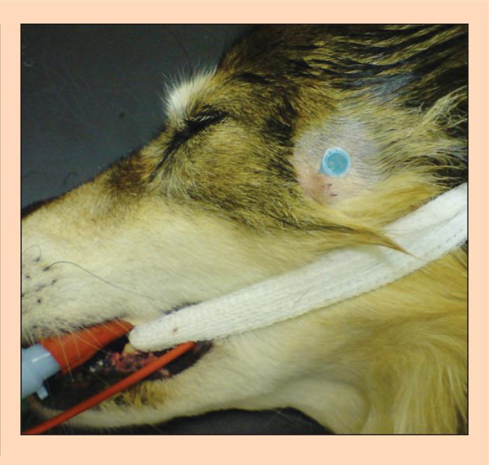
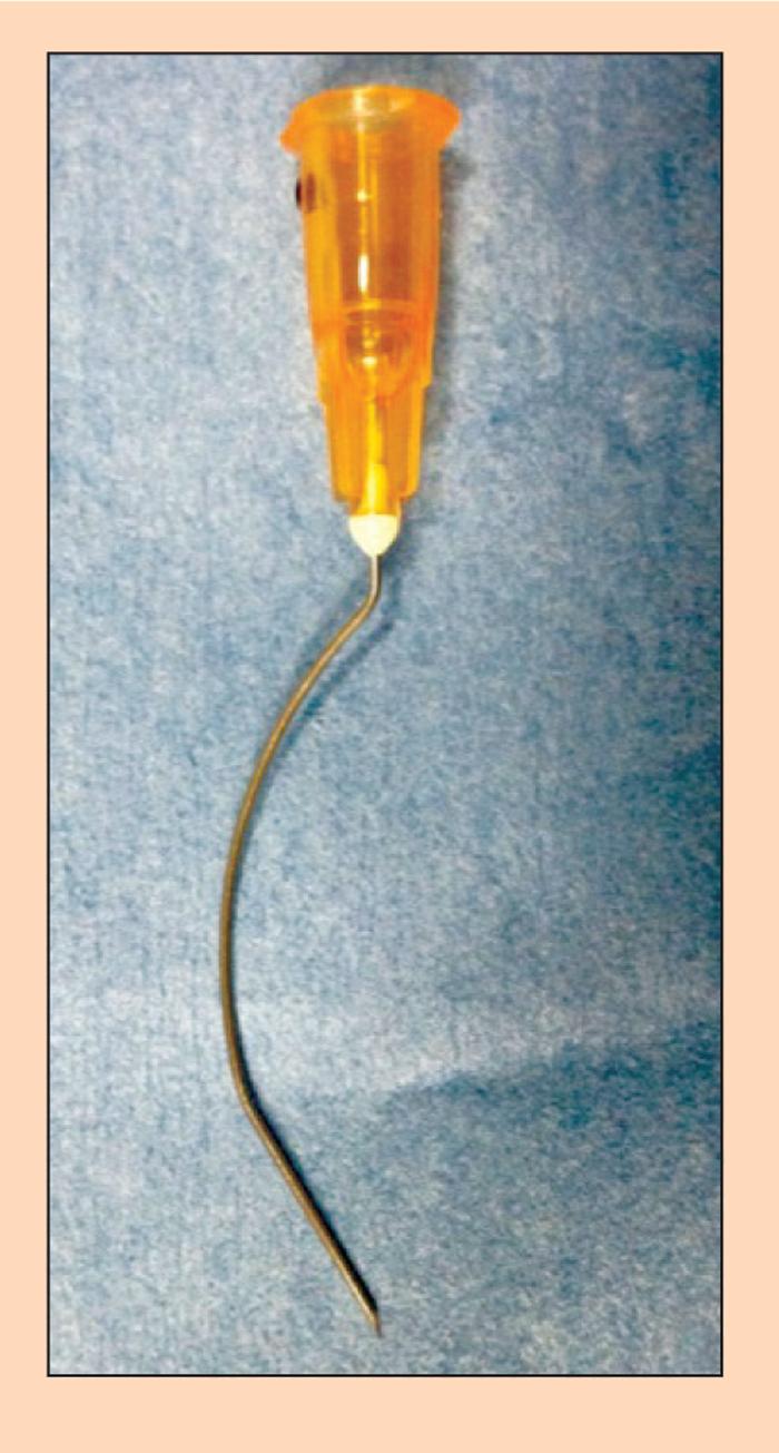
5 The retrobulbar nerve block (Figure 4) is approached dorsolateral to the globe in the orbit, passing through either the skin or conjunctiva. It is only advised for use before enucleation due to risk of trauma to the globe and associated structures. A pre-curved (Figure 5) or a self-curved needle should be walked off the orbital bone until the tip of the needle is caudal to the globe. Care should be taken to aspirate prior to injection and, if excess pressure is encountered during local anaesthetic injection, the needle should be repositioned and the injection repeated. Thoracic limb blocks The brachial plexus (BP) may be blocked by the traditional approach prior to any planned surgery to the forelimb, although it may be less effective for surgery proximal to the elbow. The BP block (Figure 6) may be performed blind or with the aid of electro-neurolocation. Care must be taken during this technique to avoid creating a pneumothorax and it should only be performed unilaterally due to potential for blocking of the phrenic nerve. The BP nerves arise from the spinal nerve roots of C6, C7, C8 and T1 and a successful technique will block the musculocutaneous, radial, ulnar and median nerves. The patient is positioned in lateral recumbency with the affected limb uppermost. A spinal needle is introduced parallel to the thoracic wall, cranial to the acromion and medial to the subscapularis muscle at the level of the scapulohumeral joint and directed in a caudodorsal direction. The first rib should be located, which will mark the caudal aspect of the brachial plexus. Local anaesthetic should be deposited as the needle is advanced, aspirating prior to each injection. Electro neurolocation may be used to identify each nerve individually prior to local anaesthetic injection. The radial, ulnar, median, musculocutaneous (RUMM) nerve block is indicated for surgery distal to the elbow and offers an alternative to the BP block. It requires three separate injection sites and may be approached blind, although electro neurolocation may improve accuracy. The radial nerve (Figure 7) may be palpated on the lateral aspect of the humerus at the junction between the middle and distal thirds. The ulnar nerve (Figure 8) is located on the medial aspect of the humerus and may be palpated over the region of the olecranon, but should be blocked at the level of the distal humerus. The median and musculocutaneous nerves are blocked together on the medial aspect of the distal humerus, proximal to the medial epicondyle of the humerus between the biceps brachii and medial head of the triceps. Pelvic limb blocks 5 / 30
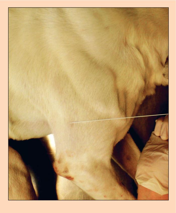
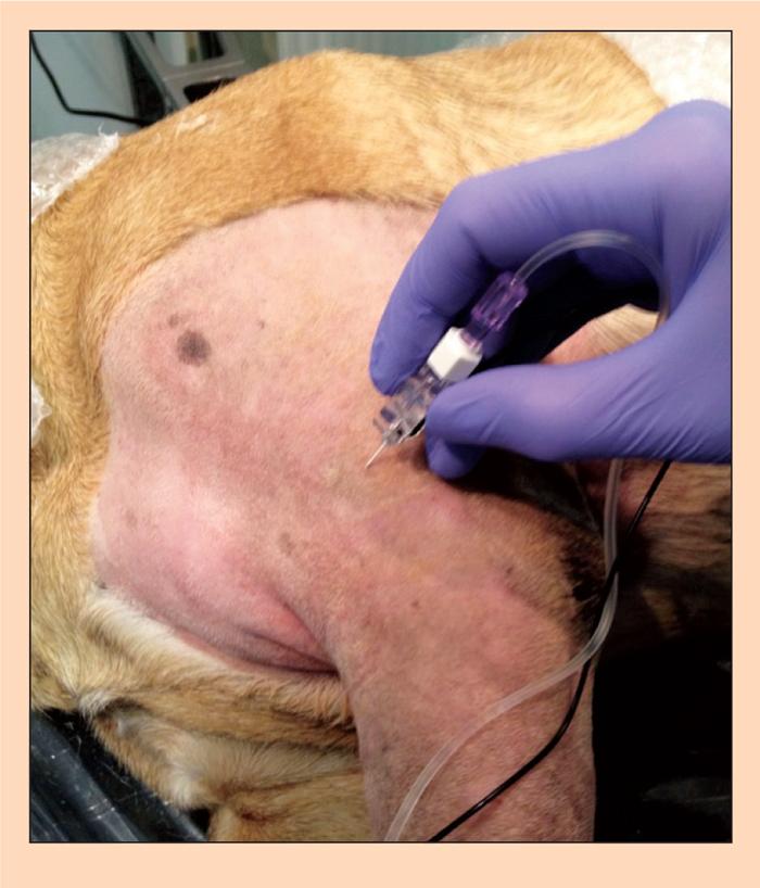
6 The sciatic nerve (SN) block may be combined with either a femoral nerve (FN) or lumbar plexus block (LPB) to provide analgesia for pelvic limb surgery. Electro-neurolocation is recommended for these techniques to improve accuracy, ensure safety and reduce the dose of local anaesthetic required. It is also unlikely satisfactory local anaesthesia will be gained without electroneurolocation. The SN (Figure 9) is blocked at its proximal location caudal to the greater trochanter of the femur. The puncture site is located at the junction of the cranial and middle thirds between a line drawn between the greater trochanter of the femur and the ischial tuberosity. The depth of needle insertion varies depending on the size of patient and may be up to 6cm to 8cm. The FN (Figure 10) is blocked at its location on the medial aspect of the pelvic limb in the femoral triangle. The femoral artery is palpated within the femoral triangle, held in place with light digital pressure and the FN is located and blocked cranial to the artery, usually in a superficial location. An alternative to the FN block is the LPB, which allows for the femoral nerve to be blocked more proximally and avoids the risk of missing the saphenous nerve high within the inguinal region. The saphenous nerve supplies the cutaneous innervation to the stifle and therefore if missed a patient may respond to skin incision. The LPB (Figure 11) is performed with the patient positioned in lateral recumbency with the side to be blocked uppermost. The dorsal process of L7 is palpated and from this the dorsal process of L5 identified. The needle is inserted lateral to L5 (approximately 2cm to 3cm) until the transverse process is contacted. The needle is then walked off bone caudally and a loss of resistance may be felt as it passes through the intervertebral ligament. Local anaesthetic is then injected after aspiration. Neuraxial blockade Epidural and spinal anaesthesia both come under the banner of neuraxial blockade, whereby local anaesthetic, sometimes in combination with other drugs is deposited in the bony vertebral canal. Epidural (extradural) anaesthesia is most commonly employed in veterinary anaesthesia, where the drug is deposited into the space outside of the dura. This technique is indicated for pelvic limb, perineal and abdominal surgery. Epidural injection (Figure 12) is most commonly performed at the lumbosacral junction (L7-S1), although caudal injection may be performed at Co1-Co2 for perineal surgery for example. Table 2 highlights the contraindications and complications of epidural anaesthesia. For epidural anaesthesia the patient should be positioned in either sternal (Figure 13) or lateral recumbency. In sternal recumbency the pelvic limbs should be extended cranially and the wings of 6 / 30
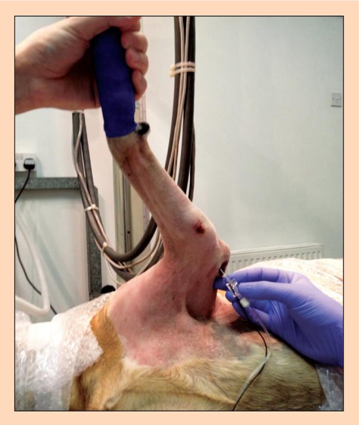
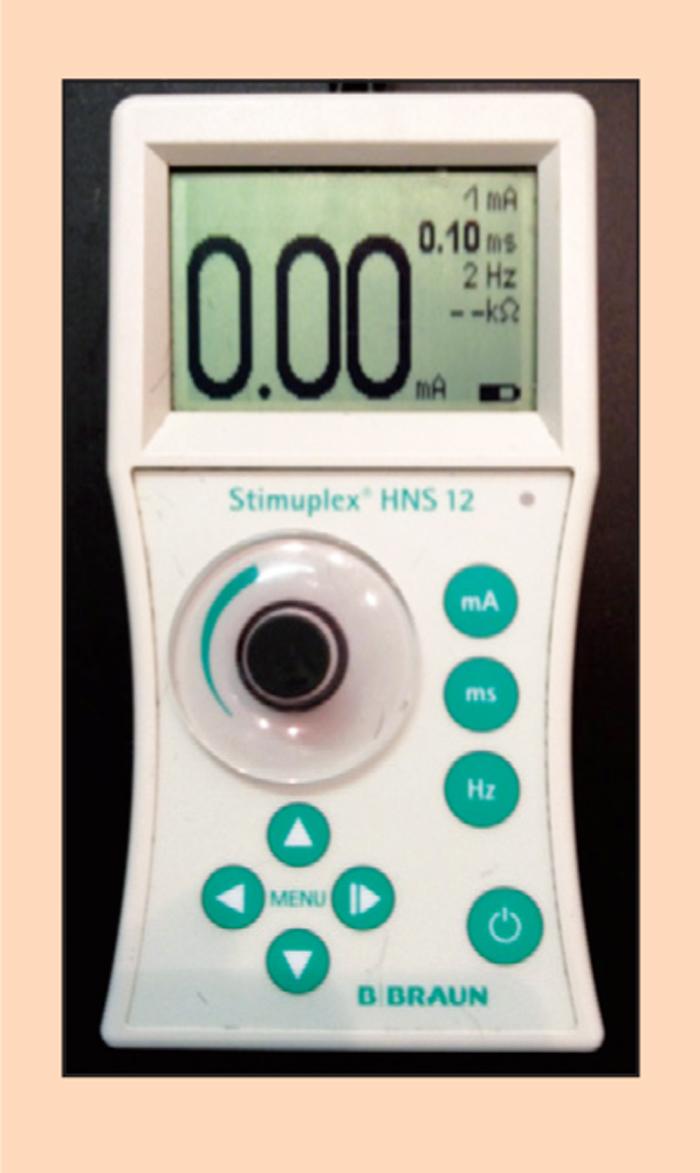
7 the ilium palpated with the thumb and second finger. The dorsal spinous process of L7 is palpated in the midline with the index finger, the lumbosacral space lies caudal to this. The spinal needle (Figure 14) is inserted in the midline perpendicular to the skin. The stylet is removed once in the subcutaneous tissue and saline applied to the needle hub. The needle is then advanced slowly until saline is aspirated into the needle due to the sub atmospheric pressure within the epidural space. The presence of cerebrospinal fluid (CSF) at this point should alert the clinician to withdraw the needle and reconfirm positioning within the epidural space. In the cat the more caudal extension of the spinal cord makes it more likely a spinal injection will be performed. Drug doses should be reduced by a half to three-quarters if a spinal injection is performed instead. A test injection may be performed before aspiration and slow injection of the selected drugs. Preservative-free drugs should be selected to minimise the risk of neurotoxicity. Advances in loco-regional anaesthesia Electro-neurolocation and ultrasound guidance are gaining popularity in veterinary medicine and offer many advantages over the described blind techniques. The use of peripheral nerve locators (Figure 15) is now common in the veterinary anaesthesia community and, once bought, the cost of consumables is not prohibitive if the technique is to be used regularly. Differences in anatomy between individuals and species allows the clinician to improve the accuracy of identifying the correct location for injection and to ensure local anaesthetic is deposited as close as possible to the desired nerve. Ultrasound is widely available in veterinary medicine and with some training and practice may be used to aid in neurolocation. There are a number of techniques now described in the dog and it is considered the gold standard in human anaesthetic practice. Summary Local anaesthetic techniques are easily employed in practice as part of a balanced, multimodal approach to anaesthesia and analgesia and should be considered where appropriate. With a good knowledge of anatomy and careful dose calculation, the various blocks can be carried out with excel- lent success. For the majority of the discussed techniques, no specialist equipment is necessary, requiring only items commonly utilised in practice. The use of local anaesthetic techniques can greatly improve patient analgesia both during anaesthesia and into the recovery period and beyond. There can also be a noticeable improvement 7 / 30
8 in post-operative pain scores and subsequent reduction in analgesic requirements during the recovery period. Further reading Campoy L and Read M R (2013). Small Animal Regional Anesthesia and Analgesia, WileyBlackwell. Dugdale A H A (2010). Veterinary Anaesthesia, Principles to Practice, Wiley-Blackwell. Lemke K A (2007). Pain management II: local and regional anaesthetic techniques. In Seymour C and Duke-Novakovski T (eds) BSAVA Manual of Canine and Feline Anaesthesia and Analgesia (2nd edn): / 30

9 Figure 1. Location of needle insertion site for the mandibular nerve block (top) and mental nerve block (bottom). 9 / 30

10 Figure 2. Location of needle insertion site for the infra-orbital nerve block. 10 / 30
11 11 / 30

12 Figure 3. Location of needle insertion site for the maxillary nerve block. 12 / 30
13 Figure 4. Needle insertion site for a retrobulbar nerve block. 13 / 30
14 14 / 30
15 Figure 5. Pre-curved needle for retrobulbar nerve block. 15 / 30
16 16 / 30
17 Figure 6. Landmarks for brachial plexus nerve block in the dog. 17 / 30
18 18 / 30
19 Figure 7. Location of needle insertion site for radial nerve block. 19 / 30
20 20 / 30
21 Figure 8. Location of needle insertion site for ulnar nerve block. 21 / 30

22 Figure 9. Landmarks for sciatic nerve block in the dog. 22 / 30

23 Figure 10. Landmarks for femoral nerve block in the dog. 23 / 30

24 Figure 11. Location of needle insertion site for lumbar plexus block. 24 / 30

25 Figure 12. Landmarks for epidural anaesthesia injection. 25 / 30

26 Figure 13. Identification of landmarks for epidural injection can be more challenging in obese patients. 26 / 30

27 Figure 14. Location of needle insertion site and connection of syringe prior to epidural injection. 27 / 30
28 28 / 30

29 Figure 15. Example of a peripheral nerve locator used for electro neurolocation techniques. Table 1. Doses of local anaesthetics used in small animal anaesthesia 29 / 30

30 Table 2. Absolute and relative contraindications of epidural anaesthesia 30 / 30 Powered by TCPDF (
Epidural anaesthesia and analgesia
 Vet Times The website for the veterinary profession https://www.vettimes.co.uk Epidural anaesthesia and analgesia Author : Matthew Gurney Categories : Vets Date : June 1, 2009 Matthew Gurney discusses
Regional Anaesthesia of the Thoracic Limb
 Regional Anaesthesia of the Thoracic Limb Trauma and inflammation cause sensitization of the peripheral nervous system and the subsequent barrage of nociceptive input (usually by surgery) produces sensitization
How and why to do an epidural in dogs and cats? Which Indications and which drugs?
 AMVAC/RoSAVA 2014 How and why to do an epidural in dogs and cats? Which Indications and which drugs? Prof. Yves Moens Dipl ECVAA Why do epidurals? A part of a balanced anesthesia A means to provide analgesia
Common Dosages** Fentanyl CRI Loading Dose 5 to 10 mcg/kg CRI 0.3 to 1.0mcg/kg/min (anes)
") ADVANCED ANESTHETIC AND ANALGESIC TECHNIQUES Jody Nugent-Deal, RVT, VTS (Anesthesia/Analgesia) (CP-Exotic Companion Animal) University of California Davis, William R. Pritchard Veterinary Medical Teaching
ADVANCED ANESTHETIC AND ANALGESIC TECHNIQUES Jody Nugent-Deal, RVT, VTS (Anesthesia/Analgesia) (CP-Exotic Companion Animal) University of California Davis, William R. Pritchard Veterinary Medical Teaching
Surgery Under Regional Anesthesia
 Surgery Under Regional Anesthesia Jean Daniel Eloy, MD Assistant Professor Residency Program Director Rutgers-New Jersey Medical School Rutgers The State University of New Jersey Peripheral Nerve Block
Block That Pain: Dental Pain Management Mary L, Berg, BS, RLATG, RVT, VTS(Dentistry) Beyond the Crown Veterinary Education Lawrence, KS
 Beyond the Crown Veterinary Education Lawrence, KS") Block That Pain: Dental Pain Management Mary L, Berg, BS, RLATG, RVT, VTS(Dentistry) Beyond the Crown Veterinary Education Lawrence, KS Pain management is more than the latest popular terminology. It is
Block That Pain: Dental Pain Management Mary L, Berg, BS, RLATG, RVT, VTS(Dentistry) Beyond the Crown Veterinary Education Lawrence, KS Pain management is more than the latest popular terminology. It is
Sensory fibers that innervate the bone, teeth, and soft tissue of the oral cavity. Regional Nerve Blocks for Oral Surgery in Companion Animals
 CE Vol. 24, No. 6 June 2002 V 439 Article #1 (1.5 contact hours) Refereed Peer Review Comments? Questions? Email: compendium@medimedia.com Web: VetLearn.com Fax: 800-556-3288 KEY FACTS Due to its long
Epidural analgesia technique
 Epidural analgesia technique Martin Pearson School of Veterinary Science University of Queensland, Gatton 0254601834 Peter Best Greencross South Tamworth Animal Hospital 88Duri Road, Tamworth 02 6765 4244
MAXILLARY INJECTION TECHNIQUE. Chinthamani Laser Dental Clinic
 MAXILLARY INJECTION TECHNIQUE Chinthamani Laser Dental Clinic Introduction A number of injection techniques are available to aid in providing clinically adequate anesthesia of the teeth and soft and hard
European Veterinary Dental College
 European Veterinary Dental College EVDC Training Support Document Preparation of Radiograph Sets (Cat and Dog) Document version : evdc-tsd-radiograph_positioning_(dog_and_cat)-20120121.docx page 1 of 13
Maxillary LA: Techniques. Ra ed Salma BDS, MSc, JBOMFS, MFDRCSI
 Maxillary LA: Techniques Ra ed Salma BDS, MSc, JBOMFS, MFDRCSI dr.raedsalma@riyadh.edu.sa https://sites.google.com/a/riyadh.edu.sa/raed/ LA Options for the Maxilla Infiltration Submucosal Supraperiosteal
Presentation Menu. Walk-in Slide. Full Presentation. Access. Site. Needle. Flush. Comfort. Monitor. Removing the EZ-IO catheter.
 Presentation Menu Walk-in Slide Full Presentation Access Site Needle Flush Comfort Monitor Removing the EZ-IO catheter Clinical Support Explore. Discover. Examine. Vidacare Workshop Programmes www.vidacare.com
Techniques of local anesthesia in the mandible
 Techniques of local anesthesia in the mandible The technique of choice for anesthesia of the mandible is the block injection and this is attributed to the absence of the advantages which are present in
Ultrasound Guided Lower Extremity Blocks
 Ultrasound Guided Lower Extremity Blocks CONTENTS: 1. Femoral Nerve Block 2. Popliteal Nerve Block Updated December 2017 1 1. Femoral Nerve Block Indications Surgery involving the knee, anterior thigh,
inerve Guide to Nerves 2009
 inerve Guide to Nerves 2009 A guide to self learning and self assessment Context: The following guide is intended to help interpret the sono-anatomy and follow a systematic stepwise approach to the practice
Thoracic Cooled-RF Training Presentation
 Thoracic Cooled-RF Training Presentation Patient Selection Anatomy Overview Neuroanatomy Lesion targets Technique Diagnostic Block Cooled-RF Precautions Summary Appendix AGENDA Patient Selection Thoracic
Human Anatomy. Spinal Cord and Spinal Nerves
 Human Anatomy Spinal Cord and Spinal Nerves 1 The Spinal Cord Link between the brain and the body. Exhibits some functional independence from the brain. The spinal cord and spinal nerves serve two functions:
Candidate s instructions Look at this cross-section taken at the level of C5. Answer the following questions.
 Section 1 Anatomy Chapter 1. Trachea 1 Candidate s instructions Look at this cross-section taken at the level of C5. Answer the following questions. Pretracheal fascia 1 2 5 3 4 Questions 1. Label the
ASA Closed Claims Project: Regional Anesthesia Claims 1990 or later Lorri A. Lee MD Department of Anesthesiology University of Washington, Seattle, WA
 ASA Closed Claims Project: Regional Anesthesia Claims 1990 or later Lorri A. Lee MD Department of Anesthesiology, Seattle, WA OVERVIEW 1. Closed Claims Project 2. Peripheral Nerve Blocks 3. Neuraxial Claims
Brachial plexus blockade within the interscalene groove involves local anesthetic
 Interscalene Brachial Plexus Block- How I do it. Part 1 of a 2 part discussion on technique. Stuart Grant Professor of Anesthesiology Duke University Medical Center Durham NC Brachial plexus blockade within
Neural Blocks in Pain Medicine D R M A R G A R E T E B O N E M B C H B F R C A F F P M R C A C O N S U LTA N T I N PA I N M E D I C I N E
 Neural Blocks in Pain Medicine D R M A R G A R E T E B O N E M B C H B F R C A F F P M R C A C O N S U LTA N T I N PA I N M E D I C I N E Stellate Ganglion Block Lumbar Sympathetic Block Requirements Diagnosis
region of the upper limb between the shoulder and the elbow Superiorly communicates with the axilla.
 1 region of the upper limb between the shoulder and the elbow Superiorly communicates with the axilla. Inferiorly, a number of important structures pass between arm & forearm through cubital fossa. 2 medial
ANAESTHESIA EDY SUWARSO
 ANAESTHESIA EDY SUWARSO GENERAL REGIONAL LOCAL ANAESTHESIA WHAT DOES ANESTHESIA MEAN? The word anaesthesia is derived from the Greek: meaning insensible or without feeling. The adjective will be ANAESTHETIC.
MUSCLES. Anconeus Muscle
 LAB 7 UPPER LIMBS MUSCLES Anconeus Muscle anconeus origin: distal end of dorsal surface of humerus insertion: lateral surface of ulna from distal margin of the semilunar notch to proximal end of the olecranon
BRACHIAL PLEXUS 11/12/2014 كيف تتكون الضفيرة FORMATION ENLARGEMENT (INTUMESCENCE) OF THE SPINAL CORD. Grey matter. Cervical intumescence - C 6 - T 2
 OF THE SPINAL CORD. Grey matter. Cervical intumescence - C 6 - T 2") BRACHIAL PLEXUS Prof. Fawzy Elnady ENLARGEMENT (INTUMESCENCE) OF THE SPINAL CORD Grey matter Cervical intumescence - C 6 - T 2 Lumbar intumescence - L 4 S 2 كيف تتكون الضفيرة FORMATION The ventral rami
BRACHIAL PLEXUS Prof. Fawzy Elnady ENLARGEMENT (INTUMESCENCE) OF THE SPINAL CORD Grey matter Cervical intumescence - C 6 - T 2 Lumbar intumescence - L 4 S 2 كيف تتكون الضفيرة FORMATION The ventral rami
Chapter 7: Skeletal System: Gross Anatomy
 Chapter 7: Skeletal System: Gross Anatomy I. General Considerations A. How many bones in an average adult skeleton? B. Anatomic features of bones are based on II. Axial Skeleton A. Skull 1. Functionally
Human Anatomy and Physiology - Problem Drill 07: The Skeletal System Axial Skeleton
 Human Anatomy and Physiology - Problem Drill 07: The Skeletal System Axial Skeleton Question No. 1 of 10 Which of the following statements about the axial skeleton is correct? Question #01 A. The axial
Important Parts of Bones
 Important Parts of Bones For 2015 Know: Humerus (posterior) Clavical Femur (Anterior) Foot Hand Mandible Os Coxa Scapula Skull (Anterior, Inferior, Lateral) Sternum Humerus (posterior) A. olecranon fossa
Anatomy of the Musculoskeletal System
 Anatomy of the Musculoskeletal System Kyle E. Rarey, Ph.D. Department of Anatomy & Cell Biology and Otolaryngology University of Florida College of Medicine Outline of Presentation Vertebral Column Upper
213: HUMAN FUNCTIONAL ANATOMY: PRACTICAL CLASS 1: Proximal bones, plexuses and patterns
 213: HUMAN FUNCTIONAL ANATOMY: PRACTICAL CLASS 1: Proximal bones, plexuses and patterns CLAVICLE Examine an isolated clavicle and compare it with a clavicle on an articulated skeleton. Viewed from above,
Regional Anesthesia. procedure if required. However, many patients prefer to receive sedation either during the
 1 Regional Anesthesia Regional anaesthesia (or regional anesthesia) is anesthesia affecting only a large part of the body, such as a limb or the lower half of the body. Regional anaesthetic techniques
Paraspinal Blocks a new paradigm in truncal analgesia
 Paraspinal Blocks a new paradigm in truncal analgesia Ki Jinn Chin, MBBS (Hons), MMed, FRCPC Associate Professor Toronto Western Hospital University of Toronto Online Resources https://youtu.be/lockhd
Lower Extremity Ultrasound-Guided Regional Anesthesia. Stephanie Duffy, CRNA Regional Anesthesia Faculty Acute Pain Service NMCSD
 Lower Extremity Ultrasound-Guided Regional Anesthesia Stephanie Duffy, CRNA Regional Anesthesia Faculty Acute Pain Service NMCSD Objectives Review anatomy of lumbosacral plexus Lumbar plexus blocks Psoas
Sign up to receive ATOTW weekly -
 1 SUBCLAVIAN PERIVASCULAR BRACHIAL PLEXUS BLOCK ANAESTHESIA TUTORIAL OF THE WEEK 156 19 th OCTOBER 2009 Dr. Martin Herrick Department of Anaesthesia, Addenbrooke s Hospital, Cambridge, U.K. Correspondence
ADVANCED PATIENT MONITORING DURING ANAESTHESIA: PART TWO
 Vet Times The website for the veterinary profession https://www.vettimes.co.uk ADVANCED PATIENT MONITORING DURING ANAESTHESIA: PART TWO Author : CARL BRADBROOK Categories : Vets Date : October 14, 2013
STRUCTURAL BASIS OF MEDICAL PRACTICE EXAMINATION 5. September 30, 2011
 STRUCTURAL BASIS OF MEDICAL PRACTICE EXAMINATION 5 September 30, 2011 PART l. Answer in the space provided. (12 pts) 1. Identify the structures. (2 pts) EXAM NUMBER A. Suprascapular nerve B. Axillary nerve
Forelimb Amputation. Indications. Pre operatively 11/12/2008. Technique Preparing for surgery. Bone neoplasia. Severe trauma/paralysis
 Forelimb Amputation January 10, 2007 Amy Bringardner Becky Pentecost Indications Bone neoplasia Osteosarcoma Fibrosarcoma Chondrosarcoma Severe trauma/paralysis Brachial plexus avulsion Infection Congenital
External Acoustic Meatus. Mastoid Process. Zygomatic Process. Temporal Bone
 Bone lab review 1. Frontal Bone 2. Supra-Orbital Foramen 3. Orbit (Orbital Cavity) 4. Superior Orbital Fissure 5. Inferior Orbital Fissure 6. Zygomatic Bone 7. Infra-Orbital Foramen 8. Maxilla 9. Mandible
Fundamentals of technique Types of local anaesthesia Topical or surface anaesthesia
 Fundamentals of technique The importance of a quiet, confident, and friendly manner towards all patients so physical comfort is also essential for the co-operation of the patient and the ease of operation
Human Anatomy Biology 351
 nnnnn 1 Human Anatomy Biology 351 Exam #2 Please place your name on the back of the last page of this exam. You must answer all questions on this exam. Because statistics demonstrate that, on average,
LOCAL ANESTHESIA IN PEDIATRIC DENTISTRY
 Disclaimer This movie is an educational resource only and should not be used to manage your health. All decisions about the management of local anesthesia in pediatric dentistry must be made in conjunction
With other members of your lab group, discuss the following questions: - The spinal cord connects directly to which part of the brain?
 BIOLOGY 211: HUMAN ANATOMY & PHYSIOLOGY ************************************************************************************************************************* SPINAL CORD, SPINAL NERVES, AND REFLEXES
Chapter 1: Introduction to the Human Body Test Bank
 Chapter 1: Introduction to the Human Body Test Bank MULTIPLE CHOICE 1. What is the branch of science that studies how the body functions? a. Anatomy b. Histology c. Pathology d. Physiology 2. Which word
Sacral, ilioinguinal, and vasal nerve stimulation for treatment of pelvic, sacral, inguinal and testicular Pain.
 Chapter 14 Sacral, ilioinguinal, and vasal nerve stimulation for treatment of pelvic, sacral, inguinal and testicular Pain. Introduction Sacral nerve root stimulation has been recognized as a treatment
Chapter 14. The Nervous System. The Spinal Cord and Spinal Nerves. Lecture Presentation by Steven Bassett Southeast Community College
 Chapter 14 The Nervous System The Spinal Cord and Spinal Nerves Lecture Presentation by Steven Bassett Southeast Community College Introduction The Central Nervous System (CNS) consists of: The spinal
CARPAL TUNNEL RELEASE BLOCK Author John Hyndman
 Questions CARPAL TUNNEL RELEASE BLOCK Author John Hyndman Web Editor Kirsten Fehrmann - kirstenfehrmann@hotmail.com 1) Why does a LA solution containing Epinephrine cause pain on injection and what can
Oral Surgery. Basic Techniques of Dental Local Anesthesia. A variety of techniques used in administration and deposition of local anesthesia:
 Oral Surgery Lecture: 9 Dr. Saif Saadedeen Basic Techniques of Dental Local Anesthesia A variety of techniques used in administration and deposition of local anesthesia: 1. Topical anesthesia 2. Infiltration
Biology 2401 The Skeletal System
 Biology 2401 The Skeletal System Purpose: The lab will describe the microscopic and gross anatomy of bone, identify bones of the body, and identify important bone markings. I. Overview of the Skeleton
Interscalene brachial plexus blocks in the management of shoulder dislocations
 Archives of Emergency Medicine, 1989, 6, 199-204 Interscalene brachial plexus blocks in the management of shoulder dislocations T. J. UNDERHILL, A. WAN & M. MORRICE Accident and Emergency Department, Derbyshire
The arm: *For images refer back to the slides
 The arm: *For images refer back to the slides Muscles of the arm: deltoid, triceps (which is located at the back of the arm), biceps and brachialis (it lies under the biceps), brachioradialis (it lies
Diana Mathioudakis DEAA EDIC AFRCA. consultant paediatric cardiac anaesthetist Intensivist(D/NL) emergency physician(d)
 emergency physician(d)") & Diana Mathioudakis DEAA EDIC AFRCA consultant paediatric cardiac anaesthetist Intensivist(D/NL) emergency physician(d) Anatomy Probe handling Sonoanatomy Tips and Tricks Literature For ultrasound guided
& Diana Mathioudakis DEAA EDIC AFRCA consultant paediatric cardiac anaesthetist Intensivist(D/NL) emergency physician(d) Anatomy Probe handling Sonoanatomy Tips and Tricks Literature For ultrasound guided
Upper arch. 1Prosthodontics. Dr.Bassam Ali Al-Turaihi. Basic anatomy & & landmark of denture & mouth
 1Prosthodontics Lecture 2 Dr.Bassam Ali Al-Turaihi Basic anatomy & & landmark of denture & mouth Upper arch Palatine process of maxilla: it form the anterior three quarter of the hard palate. Horizontal
Proceeding of the SEVC Southern European Veterinary Conference
 www.ivis.org Proceeding of the SEVC Southern European Veterinary Conference Oct. 17-19, 2008 Barcelona, Spain http://www.sevc.info Reprinted in the IVIS website with the permission of the SEVC www.ivis.org
Ultrasound Guided Regional Nerve Blocks
 Ultrasound Guided Regional Nerve Blocks In the country of the blind the one eyed man is King -Deciderius Erasmus (1466-1536) Objectives Benefits of Regional Anesthesia Benefits of US guidance Role of ultrasound
General Anatomy p. 1 Organization of the Human Body p. 1 Skeleton of the Human Body p. 4 Ossification of the Bones p. 6 Bone Structure p. 8 Joints p.
 General Anatomy p. 1 Organization of the Human Body p. 1 Skeleton of the Human Body p. 4 Ossification of the Bones p. 6 Bone Structure p. 8 Joints p. 10 Principal Joints (Immovable) p. 12 Synovial Joints
2018 Learning Outcomes
 I. Pain Physiology and Anatomy (20%) A. Describe the basic anatomy of the nervous system. B. Describe the physiological mechanisms of neuronal function (eg- action potentials). C. Review the nociceptive
Mandibular and Maxillary Anesthesia
 Mandibular and Maxillary Anesthesia Uses of the Conduction Technique JACK H. SELTSAM, D.D.S., M.D., Los Angeles THE ARMAMENTARIUM of a surgeon who operates on the head and neck should include the ability
SPINAL INJECTIONS SECTION 5 SPINAL INJECTION GUIDELINES 219
 SECTION 5 SPINAL INJECTIONS SPINAL INJECTION GUIDELINES 219 Overview 219 Safety 219 Accuracy 220 Efficacy 220 Indications for spinal injection 221 Summary 222 EXAMINATION OF THE SPINE 223 CAUDAL EPIDURAL
NEUROLOGICAL EXAMINATIONS: LOCALISATION AND GRADING
 Vet Times The website for the veterinary profession https://www.vettimes.co.uk NEUROLOGICAL EXAMINATIONS: LOCALISATION AND GRADING Author : MARK LOWRIE Categories : Vets Date : June 16, 2014 MARK LOWRIE
Proceedings of the World Small Animal Veterinary Association Sydney, Australia 2007
 Proceedings of the World Small Animal Sydney, Australia 2007 Hosted by: Next WSAVA Congress REDUCING THE PAIN FACTOR AN UPDATE ON PERI-OPERATIVE ANALGESIA Sandra Forysth, BVSc DipACVA Institute of Veterinary,
The Upper Limb III. The Brachial Plexus. Anatomy RHS 241 Lecture 12 Dr. Einas Al-Eisa
 The Upper Limb III The Brachial Plexus Anatomy RHS 241 Lecture 12 Dr. Einas Al-Eisa Brachial plexus Network of nerves supplying the upper limb Compression of the plexus results in motor & sensory changes
Perpendicular Plate Zygomatic Bone. Mental Foramen Mandible
 Glabella Frontal Middle Nasal Concha Nasal Lacrimal Perpendicular Plate Zygomatic Inferior Nasal Concha Maxilla Mental Mandible Skull (anterior view) Squamosal Suture Coronal Suture Frontal Parietal Nasal
GASTROCNEMIUS TENDON REPAIR VETLIG USING THE STIF CAT 30 SOFT TISSUE INTERNAL FIXATION VETLIG
 VETLIG SOFT TISSUE INTERNAL FIXATION GASTROCNEMIUS TENDON REPAIR USING THE STIF CAT 30 VETLIG A R T I F I C I A L L I G A M E N T S F O R V E T E R I N A R Y U S E VETLIG MANAGEMENT OF CHRONIC GASTROCNEMIUS
*the Arm* -the arm extends from the shoulder joint (proximal), to the elbow joint (distal) - it has one bone ; the humerus which is a long bone
, to the elbow joint (distal) - it has one bone ; the humerus which is a long bone") *the Arm* -the arm extends from the shoulder joint (proximal), to the elbow joint (distal) - it has one bone ; the humerus which is a long bone - muscles in the arm : *brachialis muscle *Biceps brachii
*the Arm* -the arm extends from the shoulder joint (proximal), to the elbow joint (distal) - it has one bone ; the humerus which is a long bone - muscles in the arm : *brachialis muscle *Biceps brachii
Temporal fossa Infratemporal fossa Pterygopalatine fossa Terminal branches of external carotid artery Pterygoid venous plexus
 Outline of content Temporal fossa Infratemporal fossa Pterygopalatine fossa Terminal branches of external carotid artery Pterygoid venous plexus Boundary Content Communication Mandibular division of trigeminal
Ultrasound-guided transversus abdominis plane block in the dog: an anatomical evaluation
 Veterinary Anaesthesia and Analgesia, 2011, 38, 267 271 doi:10.1111/j.1467-2995.2011.00612.x RESEARCH PAPER Ultrasound-guided transversus abdominis plane block in the dog: an anatomical evaluation Carrie
Lecture 14: The Spinal Cord
 Lecture 14: The Spinal Cord M/O Chapters 16 69. Describe the relationship(s) between the following structures: root, nerve, ramus, plexus, tract, nucleus, and ganglion. 70. Trace the path of information
PERIPHERAL REGIONAL BLOCKS. by Mike DeBroeck, DNP, CRNA
 PERIPHERAL REGIONAL BLOCKS by Mike DeBroeck, DNP, CRNA Why am I bothering with this topic at all? Do CRNAs REALLY even do peripheral regional anesthetics? YES!!!!!!! TOPICS GENERAL INFO SUCCESS RATES
Dr.Israa H. Mohsen. Lecture 5. The vertebral column
 Anatomy Lecture 5 Dr.Israa H. Mohsen The vertebral column The vertebral column a flexible structure consisting of 33 vertebrae holds the head and torso upright, serves as an attachment point for the legs,
Types of Anaesthesia for dermal and lip fillers at Simply Fox
 Types of Anaesthesia for dermal and lip fillers at Simply Fox The Juvederm range we use contains lidocaine- a local anaesthetic, however this does not work instantly as the needle is inserted, it is mixed
Synapse Homework. Back page last question not counted. 4 pts total, each question worth 0.18pts. 26/34 students answered correctly!
 Synapse Homework Back page last question not counted 26/34 students answered correctly! 4 pts total, each question worth 0.18pts Business TASS hours extended! MWF 1-2pm, Willamette 204 T and Th 9:30-10:30am,
Overview of the Skeleton: Bone Markings
 Name Overview of the Skeleton: Bone Markings Match the terms in column B with the appropriate description in column A. Column A 1. sharp, slender process* 2. small rounded projection* 3. narrow ridge of
Chapter 13: The Spinal Cord and Spinal Nerves
 Chapter 13: The Spinal Cord and Spinal Nerves Spinal Cord Anatomy Protective structures: Vertebral column and the meninges protect the spinal cord and provide physical stability. a. Dura mater, b. Arachnoid,
Axilla and Brachial Region
 L 4 A B O R A T O R Y Axilla and Brachial Region BRACHIAL PLEXUS 5 Roots/Rami (ventral rami C5 T1) 3 Trunks Superior (C5, C6) Middle (C7) Inferior (C8, T1) 3 Cords Lateral Cord (Anterior Superior and Anterior
Nerves of the upper limb Prof. Abdulameer Al-Nuaimi. E. mail:
 Nerves of the upper limb Prof. Abdulameer Al-Nuaimi E-mail: a.al-nuaimi@sheffield.ac.uk E. mail: abdulameerh@yahoo.com Brachial plexus Median nerve After originating from the brachial plexus in the axilla,
Human Anatomy and Physiology I Laboratory Spinal and Peripheral Nerves and Reflexes
 Human Anatomy and Physiology I Laboratory Spinal and Peripheral Nerves and Reflexes 1 This lab involves the second section of the exercise Spinal Cord, Spinal Nerves, and the Autonomic Nervous System,
Day 5 Respiratory & Cardiovascular: Respiratory System
 Day 5 Respiratory & Cardiovascular: Respiratory System Be very careful not to damage the heart and lungs while separating the ribs! Analysis Questions-Respiratory & Cardiovascular Log into QUIA using your
ANATYOMY OF The thigh
 ANATYOMY OF The thigh 1- Lateral cutaneous nerve of the thigh Ι) Skin of the thigh Anterior view 2- Femoral branch of the genitofemoral nerve 5- Intermediate cutaneous nerve of the thigh 1, 2 and 3 are
Axial skeleton bones and markings
 Axial skeleton bones and markings Skull Cranial bones Frontal x 1 Supraorbital foramen Occipital x 1 Foramen magnum Occipital condyles Superior nuchal line Inferior nuchal line Anterior cranial fossa External
Regional Anesthesia. Fatiş Altındaş Dept. of Anesthesiology
 Regional Anesthesia Fatiş Altındaş Dept. of Anesthesiology Regional anesthesia - Definition Renders a specific area of the body, e.g. foot, arm, lower extremities insensating to stimulus of surgery or
Biology 218 Human Anatomy. Adapted from Martini Human Anatomy 7th ed. Chapter 6 The Skeletal System: Axial Division
 Adapted from Martini Human Anatomy 7th ed. Chapter 6 The Skeletal System: Axial Division Introduction The axial skeleton: Composed of bones along the central axis of the body Divided into three regions:
ORTHOPAEDIC INJECTION AND ASPIRATION TECHNIQUES
 ORTHOPAEDIC INJECTION AND ASPIRATION TECHNIQUES OAAPN October 20, 2016 David H. Sohn, JD MD Chief, Shoulder and Sports Medicine University of Toledo Medical Center When to aspirate? To rule out infection
Upper limb Arm & Cubital region 黃敏銓
 Upper limb Arm & Cubital region 黃敏銓 1 Arm Lateral intermuscular septum Anterior (flexor) compartment: stronger Medial intermuscular septum Posterior (extensor) compartment 2 Coracobrachialis Origin: coracoid
Using methadone alongside other opioids. Dr. Jo Murrell BVSc. (hons), PhD, DiplECVAA, MRCVS
, PhD, DiplECVAA, MRCVS") Using methadone alongside other opioids Dr. Jo Murrell BVSc. (hons), PhD, DiplECVAA, MRCVS Why might we want to use methadone alongside other opioids? 1. Multi-modal analgesia strategies e.g. using methadone
Using methadone alongside other opioids Dr. Jo Murrell BVSc. (hons), PhD, DiplECVAA, MRCVS Why might we want to use methadone alongside other opioids? 1. Multi-modal analgesia strategies e.g. using methadone
PERIPHERAL NERVOUS SYSTEM
 CHAPTER 13 PERIPHERAL NERVOUS SYSTEM Functional division of nervous system = afferent info to the CNS ascending spinal cord = efferent info from CNS descending spinal cord somatic skin, muscles visceral
BRACHIAL PLEXUS. DORSAL SCAPULAR NERVE (C5) supraclavicular branch innervates rhomboids (major and minor) and levator scapulae
 supraclavicular branch innervates rhomboids (major and minor) and levator scapulae") THE BRACHIAL PLEXUS DORSAL SCAPULAR NERVE (C5) supraclavicular branch innervates rhomboids (major and minor) and levator scapulae SCHEMA OF THE BRACHIAL PLEXUS THE BRACHIAL PLEXUS PHRENIC NERVE supraclavicular
THE BRACHIAL PLEXUS DORSAL SCAPULAR NERVE (C5) supraclavicular branch innervates rhomboids (major and minor) and levator scapulae SCHEMA OF THE BRACHIAL PLEXUS THE BRACHIAL PLEXUS PHRENIC NERVE supraclavicular
J. 0. AKINOSI, B.D.s., F.D.S.R.C.S.
 British Journal of Oral Surgery 15 (1977-78) 83-87 A NEW APPROACH TO THE MANDIBULAR NERVE BLOCK J. 0. AKINOSI, B.D.s., F.D.S.R.C.S. Department of Oral Surgery and Pathology, College of Medicine, Lagos
Practical 2 Worksheet
 Practical 2 Worksheet Upper Extremity BONES 1. Which end of the clavicle is on the lateral side (acromial or sternal)? 2. Describe the difference in the appearance of the acromial and sternal ends of the
ADVANCED PATIENT MONITORING DURING ANAESTHESIA: PART ONE
 Vet Times The website for the veterinary profession https://www.vettimes.co.uk ADVANCED PATIENT MONITORING DURING ANAESTHESIA: PART ONE Author : CARL BRADBROOK Categories : Vets Date : October 7, 2013
INDIANA HEALTH COVERAGE PROGRAMS
 INDIANA HEALTH COVERAGE PROGRAMS PROVIDER CODE TABLES Note: Due to possible changes in Indiana Health Coverage Programs (IHCP) policy or national coding updates, inclusion of a code on the code tables
ARROW EZ-IO Intraosseous Vascular Access System Procedure Template
 ARROW EZ-IO Intraosseous Vascular Access System Procedure Template PURPOSE To provide procedural guidance for establishment of intraosseous vascular access using the ARROW EZ-IO Intraosseous Vascular Access
Fascia Iliaca Compartment Block. Angela Stewart ANP 22/08/17
 Fascia Iliaca Compartment Block Angela Stewart ANP 22/08/17 Motivation Anaesthetist Dr Joellene Mitchell from acute pain service Ayr hospital produced a guideline to allow Non-medical prescribers (NMP)
Lab no 1 Structural organization of the human body
 Physiology Lab Manual Page 1 of 6 Lab no 1 Structural organization of the human body Physiology is the science which deals with functions of the body parts, and how they work. Since function cannot be
bio4165 lab quiz 1 Posterior View Anterior View Lateral View Anterior View bio fall.quarter lab.quiz.1...page.1 of 6
 B A Posterior View D C E Lateral View bio.4165...fall.quarter.2005...lab.quiz.1...page.1 of 6 F I G 35 Posterior View H bio.4165...fall.quarter.2005...lab.quiz.1...page.2 of 6 J Posterior View L K Inferior
GUIDELINES FOR PERIPHERAL NERVE / PLEXUS BLOCK CATHETER MANAGEMENT DEPARTMENT OF ANAESTHESIOLOGY AND INTENSIVE CARE HOSPITAL KUALA LUMPUR
 GUIDELINES FOR PERIPHERAL NERVE / PLEXUS BLOCK CATHETER MANAGEMENT DEPARTMENT OF ANAESTHESIOLOGY AND INTENSIVE CARE HOSPITAL KUALA LUMPUR INTRODUCTION Regional block provides superior pain relief, compared
First BHMS Anatomy Question Papers Calicut University
 First BHMS Anatomy Question Papers Calicut University 1996-2000 FIRST B.H.M.S. DEGREE EXAMINATION, DECEMBER 1996 Time: Three Hours Maximum: 100 Marks Answer any five questions. Draw diagrams wherever needed.
The thigh. Prof. Oluwadiya KS
 The thigh Prof. Oluwadiya KS www.oluwadiya.com The Thigh: Boundaries The thigh is the region of the lower limb that is approximately between the hip and knee joints Anteriorly, it is separated from the
Spring Written By: J. E. Sutton. Contents: I. Overview of the Skeleton: II. Appendicular Skeleton III. Axial Skeleton IV.
 Spring 2012 Written By: J. E. Sutton Contents: I. Overview of the Skeleton: II. Appendicular Skeleton III. Axial Skeleton IV. Articulations Overview of the Skeleton: I. Orientation to Human Skeleton: a.
Copyright 2003 Pearson Education, Inc. publishing as Benjamin Cummings. Dr. Nabil khouri
 Dr. Nabil khouri Appendicular Skeleton The appendicular skeleton is made up of the bones of the upper and lower limbs and their girdles Two girdles: Pectoral girdles attach the upper limbs to the body
Bone Flashcards for 10a
 Bone Flashcards for 0a CLAVICLE (collar bone). Sternal extremity (end) flat end. Acromial extremity (end) rounded end. SCAPULA (shoulder blade). Right or left scapula?. Superior border (superior margin).