Postilla 30 November 1 980
|
|
|
- Amie Posy Spencer
- 5 years ago
- Views:
Transcription
1 Peabody Museum of Natural History Yale University New Haven, CT Postilla 30 November Number 181 Miocene Hominoids David R. Pilbeam Michael D. Rose Catherine Badgley Bonnie Lipschutz (Received 13 September 1980) Abstract Remains of hominoid primates collected by Yale Peabody Museum - Geological Survey of Pakistan expeditions to the Siwalik Group rocks of the Potwar Plateau, Pakistan, are described. They consist of facial, gnathic, dental, and postcranial remains of Ramapithecus, Sivapithecus, and Gigantopithecus. They are discussed anatomically and without precise taxonomic attributions. The hominoids come from 24 localities, the majority being around eight million years old. The depositional environments of 21 hominoid localities are documented in the form of microstratigraphic sections. These sections depict depositional and postdepositional features that are necessary for interpreting the facies of fossiliferous horizons. Within the predominantly fluvial Siwalik Group sediments, a three-fold division of facies is convenient for distinguishing certain taphonomic influences on hominoid and other vertebrate fossils. These facies are 1) channel, 2) channel margin, and 3) floodplain. A locality consists of one or more fossiliferous horizons, and thus one or more facies may be represented. Interpretations of the facies represented at each locality accompany the microstratigraphic sections. Copyright 1980 by the Peabody Museum of Natural History, Yale University. All rights reserved. No part of this publication, except brief quotations for scholarly purposes, may be produced without the written permission of the Director, Peabody Museum of Natural History. Introduction The hominoids described here were recovered during five seasons of field work ( to ) by expeditions of the joint Yale Peabody Museum - Geological Survey of Pakistan (YPM-GSP) research project led by David Pilbeam and Dr. S. M. Ibrahim Shah. Initial descriptions and discussions of some of the material have appeared in Pilbeam et al. (1977b), and Pilbeam (1978a, 1978b, 1979a, 1979b). The specimens are sampled from at least three species of hominoids: Ramapithecus punjabicus, Sivapithecus indicus, and Gigantopithecus bilaspurensis. These, and other middle and late Miocene species have been variously classified, sometimes in Pongidae and at other times in both Pongidae and Hominidae. Most recently it has been suggested that they be included in a family Ramapithecidae, separate from archaic apes (Dryopithecidae), Pongidae, and Hominidae (Pilbeam, 1979a; Pilbeam etal. 1977b). This proposal was made for two main reasons. First, to emphasize the point that these genera shared a number of features in common which are probably of adaptive importance and which separate them as a group from other hominoids. For example, all have thick occlusal enamel like hominids, and an anterior dentition that, although resembling those of apes, exhibits different occlusal relationships and amounts of wear than found in dryopithecids or pongids. Their postcranial skeletons show features advanced over dryopithecids, and more resemble those of living pongids. Second, in our view the hominoid fossil record is not suffi-

2 2 Miocene Hominoids Postilla 181 ciently complete for us to decide which (if any) Miocene forms are ancestral to living hominoids. All that can be plausibly argued at this stage is that ramapithecids can be interpreted as being broadly ancestral to both pongids and hominids. Classifying them in a distinct family emphasizes this point and also, it is hoped, will deemphasize arguments about phylogeny that cannot now be settled. The sample greatly expands our knowledge of this important group. Previously unknown parts are now represented, in particular postcranial remains. In earlier accounts (for example, Pilbeam et al. (1977b)) specimens were assigned to taxa. During the course of preparation of these detailed descriptions it became clear that, although at least three hominoid species are represented in the Siwaliks, and perhaps more, they are very similarto each other morphologically and they overlap in size. Given our ignorance of the nature of sexual dimorphism in these species, and the fact that most specimens are worn, incomplete, or broken, many specimens cannot now be assigned with confidence to particular taxa. It is possible that further, "non-morphological" work, such as studies of enamel histology, will provide us with better sorting criteria for dental remains. Of course, the recovery of more complete material, especially associated postcranial remains, may clarify these matters. However, it is possible that even quite complete specimens may not allow us to clear up all taxonomic issues. (Such is the case, for example, for Plio-Pleistocene Hominidae.) Indeed, as more specimens have been recovered from Pakistan the problems of assigning specimens to species and of associating skull and postcranial groups have grown more complex. This of course does not render the hominoids devoid of biological interest. These detailed descriptions, written in as "neutral" a tone as possible, are offered as the essential background to further work, mainly of a functional or biomechanical nature. We believe that a wealth of information concerning diet, masticatory function, and positional behaviors waits to be revealed. For the first step though, such descriptions are necessary. The primates come from the Potwar Plateau, near Rawalpindi, mainly from the area around Khaur, although one locality, 311, is at Sethi Nagri: see maps and discussion in Pilbeam et al. (1977a) and Pilbeam et al. (1979). Figure 1 is a simplified stratigraphic diagram showing the distribution of hominoid localities in the Khaur area. Of the 24 localities, 21 are very close to the level of sandstone U, a prominent marker horizon. The total stratigraphic range of the Khaur area primate localities is approximately 2000 m. A variety of data discussed in Pilbeam, Behrensmeyer, Barry, and Shah (1979) together with new data of Gary Johnson and Lisa Tauxe (personal communications) suggest that the U-level deposits are around 8 million years old, while the total time range represented by the hominoids is from a little over 10 m.y. to a little under 7 m.y. Locality 311 at Sethi Nagri falls within this span and is perhaps a little over 9 m.y. old. The geological context of the fossils is discussed in more detail by Badgley in the next section. Stratigraphic diagrams for each locality are given in the text. The hominoids are discussed in two sections. First, dental, gnathic, and cranial remains are reviewed by locality, by Pilbeam and Lipschutz. Locality descriptions and sections by Badgley are given in this section. Postcranial remains from particular localities are just listed. The skull remains fall into three size groups, and probably at least four morphological groups. Second, postcranial material is reviewed by Rose with specimens grouped into size categories. Measurements are in millimeters, and are defined in Pilbeam (1969) or are self-explanatory. Abbreviations are as follows: B Bu D Di H L Lab breadth buccal depth distal height length labial Li M Max Tal Th Trans Tri lingual mesial maximum talon or talonid thickness transverse trigonortrigonid
3 3 Miocene Hominoids Postilla 181 MALHUWALA JABBI KAULIAL KAS KAS I CHOUKTRIWALI KAS BORA(HASAL) KAS GANDA KAS RATHA KAS MY UTO DHOK MILA KAS, ,211,22 DINGA KAS 409,463,414 f^-4*3 1^^261 BHIANWAU KAS Fig.1 Simplified stratigraphic chart of Khaur area hominoid localities showing approximate relative placements and ages. Depositional Environments One of the aims of the YPM-GSP project is to understand patterns of fossil accumulation in Siwalik deposits. This has involved mainly microstratigraphic and taphonomic analyses of selected localities in the Khaur area, with supplementary information from similar work at a large locality in type Nagri area (311) and from a biostratigraphic field survey. The purpose of these studies is to determine the depositional environments in which fossils occur and the taphonomic processes that affected the original Miocene death assemblages. In conjunction with Behrensmeyer's study of lithofacies variation at a larger scale [see Pilbeam et al. (1979) and Badgley and Behrensmeyer (1980)], the results of this study form the basis for reconstructing fluvial regimes, paleoenvironments, and the mammal community within the restricted stratigraphic interval which is the source of most of the hominoid fossils. The interpretation of depositional environments involves detailed field studies of sediments at selected fossil localities which span vertical m. The final interpretation of a sedimentary environment rests upon both the characteristics of the sediments in question and the distributional relationships of different sediment-types to each other. Siwalik sedimentation is predominantly fluvial, consisting of vertical alternations of sandstone, sandy silt, silt and silty clay. Conglomerate lenses occur within or marginal to sandstone units. Erosional features and contacts between strata indicate very low local relief. The composition of conglomerate clasts shows that prior deposits from the same system were regularly reworked; the clasts consist of silt and clay fragments and nodular precipitates of calcium carbonate or limonite which formed postdepositionally within some floodplain deposits. At any stratigraphic horizon there was a network of channels weaving across a broad

4 4 Miocene Hominoids Postilla 181 alluvial plain. Within a 200 m. stratigraphic interval studied in the Khaur area, the variation and distribution of major sandstone units indicate characteristics of both braided and meandering fluvial regimes (and possibly of two rivers one of each mode). Lateral to the channels were extensive sandy-silty, bioturbated margins with ponds and swales and varied floodolain subenvironments. At present, a threefold subdivision of depositional environments is convenient for the interpretation of major taphonomic influences on vertebrate fossils (excluding microfauna). These environments are: (I) channel base or bar, (II) channel margin, (III) floodplain. They are distinguished on the basis of several features, including lithology (mineralogy, color, sorting, texture), bedding, lateral and vertical variation, extent of bioturbation, geometry, and relationship to adjacent (lateral and vertical) strata. Figure 2 shows the main field characters used to describe fossil localities and interpret depositional environments. No single feature is entirely diagnostic of overall depositional environment. Some features may occur in all three environments to different degrees. A single lithology, e.g., silty clay, may be present in all three depositional settings, but in Type I localities silty clay occurs in the form of clay drapes over predominantly sandy or gravelly surfaces while in Type III localities it is often the exclusive sediment. Thus, there is some geological variation among the localities of each major setting. Wherever possible these variations are highlighted to refine the interpretation of the burial and postburial history of fossils. As work progresses this scheme may give way to a more elaborate subdivision. A brief description of the major depositional environment follows: Type I The fossiliferous lithology of channel localities is commonly conglomerate, composed of clasts of silt, clay, and nodular precipitates of calcium carbonate or limonite all derived from earlier Siwalik deposits. Onkolites and onkolitic crusts on other clasts are also common elements. The conglomerate lenses are less than 1 m thick and occur within or at the margins of sandstone units which range from 2-20 m in thickness. The conglomeratesandstone complex exhibits abundant crossbedding, erosional bases cut-and-fill features, and occasional clay or silt drapes. While most such units contain a scatter of root or burrow traces, the depositional features (bedding, cross-bedding) of these deposits are commonly intact. In most other Siwalik sediments, bedding features are absent. In the spectrum of Siwalik sediments, the cross-bedded sandstones and conglomerate lenses represent the highest flow energies. These Type I sediments and their associated fossils are interpreted as channel base or bar lag deposits. The distribution of in situ fossils indicates that concentrations of bones occur more commonly in the conglomerate lenses than in the associated sandstone. Crossbedded sandstones typically contain a lowdensity scatter of fossils, even when no conglomerate lenses are present. Type II Channel margin localities are characterized by heterogeneous silty-sandy lithologies in proximity to channel sand units. The lithological heterogeneity reflects a combination of the following: 1) deposition of poorly sorted sediment typically a mixture of fine sand and silt; 2) rapid changes in depositional mode, evidenced by vertical successions of thin beds of varying lithology, clay and silt drapes, and lateral interfingering of different lithologies; and 3) postdepositional, organic mixing of sediment. Bioturbation is a prevalent feature of these deposits in the form of root and burrow traces, physical mixing of sediments, footprints, and mottling. Bedding surfaces are often present but are typically disturbed. Sometimes, it is clear that these sediments have been deposited within a topographic depression at an edge of a cross-bedded sandstone body. These deposits are interpreted as low-relief levees, swales behind or within levees, orthe fill material of some abandoned channelways in proximity to a more persistent channel.

5 5 Miocene Hominoids Postifla 181 Type III Floodplain localities occur in predominantly silty or clayey units within a vertical sequence of similarly fine-grained units. The lithologies include silt, clayey silt, and silty clay with variable sand content. The colors include graybrown, red-brown, yellow-brown, maroonbrown, and gray-black. Typically, no bedding features are present; their virtual absence through meters and meters of fine-grained sediment presumably reflects both deposition on irregular surfaces (e.g., vegetated land surfaces) and postdepositional mixing by numerous organic agents. Root and burrow traces are common. Extensive color mottling occurs along some horizons. Nodular precipitates of calcium carbonate and, less frequently, limonite occur at varying densities in these units. Some horizons of especially marked organic and inorganic modification have been tentatively identified as soils. These deposits are interpreted as aggradational land surfaces with the most extensive subaerial modification of primary depositional features. Most fossils in the Siwaliks occur as surface finds, and a locality consists of the spatial distribution of the eroded material. Most fossil concentrations are discretely bounded laterally and vertically by expanses of nonfossiliferous sediment. Within a locality, usually one or more sedimentary units are present with bones in situ. The assignment of surface finds to source horizons is based on: 1) matrix on surface finds in relation to lithologies present at a locality; 2) the relation of surface material to local topography; 3) associations among skeletal parts believed to have come from the same skeleton. Usually source of surface material is unambiguous; ambiguity results when more than one fossil-bearing level is present or when no fossils can be found in situ. In both instances, potential source strata are indicated. The YPM-GSP hominoid fossils come from 25 of the 485 localities known to the expedition at the end of the 1979 field season. Of these, 21 localities are described here. The hominoid localities occur in all three depositional environments described above. Within the Khaur hominoid interval, hominoid fossils are persistent but uncommon, as are fossils representing certain other "uncommon" taxa such as anthracotheres and some carnivore families. The relative abundance of hominoid fossils in comparison to fossils of other mammals of similar body size probably reflects the pattern of relative abundance in the original Siwalik community (Badgley and Behrensmeyer, 1980). Each microstratigraphic section represents the geologic context of a single fossil'locality whose number is shown to the right of the diagram. The information presented in the sections is considered essential to the interpretation of depositional environment(s) of fossiliferous sediments. Solid lines mark the stratigraphic interval with bones in situ; these deposits are considered the most likely source for any surface finds (hominoids and nonhominoid) unless otherwise noted. Dashed lines indicate uncertainty of provenance. Hominoid fossils found in situ are marked with an H. The lithology of each unit is indicated by the grain-sized symbols and the distance each unit projects toward the left. The scale is in meters; the base of each section is m 0. Skull Remains All localities except 311 are in the Khaur area. Locality 182 (Ganda Kas) GSP 4635 (Fig. 24K). Collector: J. Barry. The specimen consists of the crown and root of a right Mi with a small amount of adhering alveolar bone. A small chip of enamel is missing from the mesial buccal corner of the crown, and the roots have been gnawed. The buccal cusps are worn flat and dentine is exposed at the protoconid (2.5 x 2) and hypoconid (0.5 diameter). Large mesial and distal contact facets shorten the tooth. The metaconid, which is still prominent, contacts the hypoconid so that the grooves form a Y-pattern. The hypoconulid lies in the midline. MDiLwom 11.5 est unworn 12.5 TriB 10.3 TalB 10.5
6 6 Miocene Hominoids Postilla 181 LITHOLOGY clay --^_-_-_1 silt ±;^-.- : -^\ silty-clayey sand silty-clay, sandy-clayey. A silt sand sandy-silty clay Krfeferl sandy silt fegg$ ^ gravelly sand clayey silt silty sand CONTACTS sharp gradational distinct gradational indistinct interfingering SEDIMENTARY AND POST-DEPOSITIONAL STRUCTURES pml cross-bedding thinly laminated silt or clayey silt <i -<r roots or burrows nodules *»%*«, M mottling slickensides soil cracks FOSSILS stromatolites, onkolites 6\ & ro gastropods range of fossiliferous sediments bivalves f T f plant fragments I probable range of fossiliferous sediments H in situ position of hominoid fossils Fig.2 Key for interpretation of locality stratigraphic diagrams. GSP 4735 (Fig. 24L). Collector: H. Thomas. The specimen is a right Ma crown without roots, exhibiting some chemical weathering. Moderate wear on the buccal cusps has reduced their height but has not exposed dentine. Occlusal wrinkling and moderate anterior and posterior foveas are apparent. Occlusal grooves form a Y-pattern. The metaconid, which is partially bifid, is the dominant cusp. The hypoconulid is almost as far buccal as the other buccal cusps. Strong grooves separate the metaconid, protoconid, hypo-
7 7 Miocene Hominoids Postilla 181 GSP 5019 (Fig. 24B). Collector: M. Pickford. The specimen is a right M 1 crown lacking roots and most of the enamel at the cervix. Preservation is excellent; there is very little occlusal wear, only a tiny mesial contact facet and no distal facet. The crown outline is rhomboidal. The protocone is the largest cusp, the hypocone smallest, with the paracone and metacone subequal. The occlusal faces of the cusps are pyramidal or sharply angulated, while the external faces are rounded. Occlusal grooves are clearly marked, crossing the low trigon crests. A small mesial fovea is marked lingually by a protoconule; the distal fovea is larger. A small Carabelli groove at the mesiolingual corner runs between the protoconule and protocone into the occlusal fovea. MDiL 9.7 BLi B 9.8 I i 182 Fig. 3 Locality 182 Fossils come from a sequence of thin, sandygravelly layers interbedded with fine-grained, bioturbated units. The fine-grained, silt, and silty clay units are more laterally persistent than the sand layers. Features of both channel (Type I) and floodplain (Type III) environments are present. The sandy-gravelly layers represent small episodic channelways lateral to a large channel, some distance away. Very little material occurs in situ. Surface positions and matrix of the fossils suggest that they derive from the sandy-gravelly layers, which represent channel deposits (Type I). conid, and hypoconulid. The lingual surface of the tooth is steep; the buccal flare varies, but is most marked on the hypoconid. MDiL 15.1 TriB 11.8 TalB 12.0 GSP 5020 (Fig. 24H). Collector: M. Pickford. The specimen is a left P* crown without roots. A small enamel flake is lost distally. The tooth is moderately worn, with clear mesial (3x1.5) and distal (5.5 x 2) contact facets. A small but distinct mesial fovea is connected to the talonid by a groove that crosses the metaconid-protoconid ridge. The lingual surface is vertical and straight mesiodistally. The buccal surface exhibits more flare and is curved at the mesiolingual and especially the distolingual corners, giving the crown an asymmetric appearance in occlusal view. MDiL worn 7.0 est unworn 7.5 BLi B 8.0 GSP 5067 (Fig. 24D). Collector: J. Barry. The specimen consists of a left M 2 crown with roots broken distolingually just above and mesiobuccally just below the cervix. Wear has flattened the cusps without exposing dentine, and chemical weathering has especially affected the mesial half of the crown. Thus virtually all surface detail has been lost, although traces of a small distal fovea remain. The protocone is the largest cusp; the distal cusps are markedly reduced, giving the crown an abbreviated appearance. MDiL worn 8.3 est unworn 8.7 BLiB 11.3 GSP 5464 (Fig. 24G). Collector: J. P. Conroy. The specimen is a right IT with roots broken just below the cervix. Occlusal wear is slight and there are small mesial and distal contact facets. The cervical line is higher mesially and

8 8 Miocene Hominoids Postilla 181 distally than labially and lingually. The buccal surface tapers from the occlusal surface to the cervix and is gently convex buccally. The lingual surface also tapers, but is longer and more strongly curved. Labial and lingual faces are virtually smooth. MDiL 5.3 BLi B 6.3 GSP Collector: H. French. The specimen consists of portions of the distolingual half of a left Pa crown, lacking roots. The distobuccal surface is intact, including much of a distal contact facet. An oblique break running from distobuccal to mesiolingual cuts through the protoconid. A small but distinct metaconid is present; enamel has been chipped from the metaconid along the lingual surface. The protoconid was significantly larger and higher than the metaconid and was connected to it by a high ridge. A mesial fovea tapers mesioinferiorly along the sloping mesial face; it is connected by a groove crossing the protoconid-metaconid crest to the talonid basin, which itself is subdivided into three parts by two distal-running small crests between the protoconid and metaconid. GSP 8679 (Fig. 24F). Collector: J. Damuth. The specimen consists of a worn right C with the inferior third of the root absent. The root and crown are somewhat compressed mesiodistally and there is a pronounced mesial and more modest distal groove running along the root and, in the case of the mesial groove, onto the crown. The convex anterior or mesiobuccal surface of the crown preserves the lower portions of a broad groove. There is some vertical wrinkling on the mesiolingual, mesial, and mesiobuccal surfaces. Occlusal wear is considerable and there are broad distobuccal and occlusal facets that are continuous. The pulp cavity is just perforated. The occlusal facet slopes slightly interiorly towards the distal side, and gives the crown a truncated appearance. MaxL 12.5 Trans B 9.9 GSP 8928 (Fig. 24A). Collector: J. Barry. The specimen is a right Hacking only the superior fifth of the root. The specimen has been chemically weathered. There are small mesial and distal contact facets, and an occlusal facet showing a long narrow band of enamel (6.9 x 0.6). The root and long axis of the crown form a straight line. In cross section the root is a rounded triangle with the apex lingual. Atthe cervix, the cross section is more oval with the long axis mesiodistal; root dimensions there are 6.5 x 6.0. Lingually the crown has a smooth basal portion that continues the lingual surface of the root for some 4, and from there the lingual surface slopes at about 45 to the occlusal surface. This portion of the lingual surface is very gently concave transversely and has numerous small vertical grooves and crests. The labial surface expands slightly occlusally and is transversely gently convex. MDi L 8.2 BLi B 6.5 GSP 8927 (Fig. 241). Collector: E. Lindsay. The specimen consists of a relatively wellpreserved left Ms crown lacking roots. The buccal cusps are worn almost flat, although no dentine is exposed. There is a mesial contact facet (4.7 x 3). The lingual cusps, especially the metaconid, are still prominent. The occlusal grooves form a Y-pattem with both the hypoconid and hypoconulid contacting the metaconid. The hypoconulid is barely lingual to the two other buccal cusps. The largest cusp is the metaconid, then the protoconid, hypoconulid, hypoconid, and entoconid. The metaconid is nicked by a tiny groove distally, setting off a cuspule which has two smaller grooves crossing it. A cuspule is present on the distal crest between the hypoconulid and entoconid, closing a small distal fovea. A moderate mesial fovea is present. The lingual surface is relatively steep; the buccal surface exhibits more flare, especially in the talonid region. MDiL worn 11.9 est unworn 12.0 Tri B 9.5 Tal B 9.3
9 9 Miocene Hominoids Postilla 181 GSP 8926 (Fig. 24J). Collector: M. Pickford. The specimen consists of a right M crown, lacking roots and with enamel lost from the cervical margin of the distal half. The tooth is unworn, with strong coarse occlusal wrinkling, is lacking a contact facet, and is probably unerupted. There are marked mesial and distal foveas. The occlusal grooves form a Y-pattern, with the metaconid dominant, followed in size by the protoconid; the remaining cusps are subequal. The hypoconulid is almost as buccal as the protoconid and hypoconid. The enamel is thick; close to the tip of the entoconid it is a little over 2 thick. MDiLest 11.9 Tri B 9.5 GSP 8702 (Fig. 24C). Collector: G. Meyer. The specimen consists of a well preserved left M 3 crown with roots broken 3 below the cervix. A sliver of enamel is gone from the cervical region around the mesiolingual corner. There is a 5.3 x 2.6 mesial contact facet. Some of the coarse occlusal wrinkling has been removed by wear, especially on the protocone and mesial paracone. Well developed mesial and distal foveas are present. The protocone and paracone are the largest cusps, with the metacone and hypocone reduced so that the tooth narrows distally. The lingual face is flared, and there is a faint groove between the protocone and hypocone. MDiLworn 9.6 est unworn 9.8 BLiB 11.3 GSP 8925 (Fig. 24E). Collector: M. Pickford. The specimen consists of a complete right C. Buccally enamel has been removed near the cervix by weathering. The root is robust and curved in the mesiodistal plane and also buccally towards the tip. It has moderate grooves buccally and lingually. The crown has a small distolingual cingulum. There is a mesial groove running down the crown, the edges of which have been flattened by wear. Distolingually three ridges descend the crown, demarcating shallower lingual and deeper distal grooves. Wear has begun to flatten these ridges. Wear has removed the apex of the crown, exposing dentine, and is confluent with mesial and distal wear. MaxL 12.7 Trans B 10.0 Lab H est worn 15.7 est unworn 18.5 RootL 16.5 Root midpoint Max L 12.0 Trans B 10.0 GSP Collector: M. Pickford. The specimen consists of the buccal half of a left M? crown, with roots broken just below the cervix. Part of the paracone is also gone. Occlusal wear is moderate; slight chemical weathering on the paracone has obliterated the mesial contact facet. The cusps have a pyramidal rather than rounded appearance, although this could partly be due to wear. The buccal surface is relatively vertical. GSP 4230 (Fig. 25). Collector: H. French. The specimen consists of part of a right mandible including the corpus and ascending ramus, M2 crown, and Ms root sockets. The bone is weathered and sandy matrix is still adherent, so much fine surface detail is lost. The lower 23 of the anterior third of the ascending ramus is present, descending at first vertically then curving to form the lateral prominence on the external surface of the corpus. This continues as a thickening to the inferior border, where there is a bulge below Ms. The M2 is moderately worn, with buccal cusps reduced in height. The prominent metaconid is the largest cusp followed by the protoconid and entoconid, then the hypoconid, with the hypoconulid smallest. There is a Y-pattern to the occlusal grooves, the metaconid contacting the hypoconid. The hypoconulid is set toward the buccal side. Mesial and distal foveas are well developed. There is a moderate (~4 wide) mesial contact facet and a smaller distal facet. The buccal surface flares more than the lingual. At Ms D 30.5 Th 22.8 Mf MDiLworn 14.9 est unworn 15.3 Tri B 12.6 TalB 11.7
10 10 Miocene Hominoids Postilla 181 GSP 4622 (Fig. 26). Collectors: M. Pickford, W. Barry. The specimen consists of a mandible lacking ascending rami, with the left molars and right M intact. The right corpus is cracked in such a way as to cause it to diverge from the sagittal plane a little more than it did in life. Internally the mandibular surface is complete to the alveolar margin, although externally bone has been removed to reveal sockets of anterior premolars, canines, and incisors. The inferior 10 of the anterior 10 of the right ascending ramus is preserved, descending vertically before curving to form the lateral prominence which continues to the inferior border where there is a bulge below Ms. The alveolar and inferior mandibular borders are subparallel. The mental foramina are below P*. some 8 or 9 above the inferior border. The alveolar margin is slightly everted at the level of Pa since the mesial roots are external to the canines; thus the anterior dentition canines and incisors is "wedged" between the premolars. The symphyseal contour in the midline is rounded, with a long inferior sweep that merges into the inferior transverse torus. Slight depressions on the inferior part of the external surface are separated by a raised median keel. Internally there is a moderate planum alveolare sloping at an angle of about 45 to the alveolar border. A moderate superior transverse torus projects to the level of distal Pa; the inferior transverse torus projects to the level of distal Pi. The tori are separated by a genioglossal fossa in which there is a small but deep pit. The fossa passes laterally into shallow sulci, eventually merging into the internal surface below M2. The tooth rows diverge posteriorly; due to minor distortion the divergence of the left row is a little high. Canines and incisors, judging from sockets, were small. The Pa crowns were set obliquely, judging from the roots, with mesiobuccal roots clearly lateral to canine roots. The long axis of the cheek teeth is concave buccally. The molars are well worn, all cusps except the metaconids being essentally flattened. Dentine is exposed in a single wear pit 7x2.5 on the buccal cusps of the left MT, and as a pit 1.5 in diameter on the M2 protoconid. There is a Y-pattern of the occlusal grooves, and the hypoconulids are relatively central on all molars. The third molars are tapered distally. Symphysis P* Ma BT BC LP4--Ms L Pa--Ma Mi Ms- Ma (left) Ma (right) D Th D Th D Th Postcranial Remains GSP MDi Lworn est unworn TriB TalB MDi Lworn unworn TriB TalB MDi Lworn unworn TriB TalB MDi Lworn unworn TriB TalB Locality 260 (Kaulial Kas) est 12.0 est GSP 9565 (Fig. 28B). Collector: M. Pickford. The specimen consists of part of a left C crown with up to 6 of the root preserved. Approximately the lingual half of the crown remains, including the apex. Wear has flattened the mesial groove and adjacent crests, and there are additional horizontal grooves worn across the mesial facet halfway up the crown. Distally, dentine is exposed in a broad facet. The distal and mesial wear facets are linked by an apical facet, on which there is a dentine pit. GSP 9895 (Fig. 270). Collector: M. Pickford. The specimen consists of a left M 3, imbedded in alveolar bone. The roots are ~17 long, the
11 11 Miocene Hominoids Postilla 181 GSP Collector: J. Damuth. The specimen consists of a small broken portion of left maxilla. Roots of P*, and sockets for M 1 and M 2 are preserved. The alveolar processes are eroded away. Part of the palatine process is present with both palatal and nasal surfaces. The external maxillary surface is gone exposing the inferior part of a maxillary sinus. Anterior to this is the eroded part of a canine jugum. 260 Fig. 4 Locality 260 Fossils have been collected both from the surface and in excavation from poorly sorted, bioturbated silts and sands. All hominoid fossils in situ come from one silty sand layer, extensively mottled in gray-green and red-brown. The 1.5 m of fossiliferous sediments occupy a topographic depression in the top of a thick ctiannel sandstone and grade upward into a thick sequence of silts and clays. The fossiliferous level represents a channel-margin environment (Type II). lingual diverging at about 20 to the buccal roots. The crown is greatly weathered, but little worn. The mesial fovea is present, as are distinct occlusal surface grooves. The protocone is a little larger than the paracone, and both distal cusps are reduced; the crown tapers distally. There is marked flare on the lingual surface, less on the buccal. MDiL 11.8 BLiB 12.6 GSP 9896 (Fig. 27K). Collector: M. Pickford. The specimen is a left M? crown with buccal roots gone and the lingual root broken 3 above the cervix. The crown is heavily worn and weathered. Mesial and distal surfaces are flattened by contact with adjacent teeth. The cusps are flattened, dentine being exposed on the protocone and in the mesial fovea. The crown tapers distally. MDiL est 9.9 BLiB est 11.7 GSP 9898 (Fig. 27B). Collector: M. Pickford. The specimen consists of a left I 1 lacking the superior quarter of the root. Root and crown form a straight line. The root is subcircular, at the cervix measuring MDi 7.3, BLi 7.4. It does not taper greatly. The buccal crown surface expands slightly from the cervix to the occlusal surface and longitudinally is gently convex buccally. The basal portion of the lingual surface runs vertically for 6.1 from the cervix, then becomes strongly concave lingually, where it is crossed by many vertical grooves. There is a mesial contact facet, and an occlusal strip ~8 long along which dentine is intermittently present. The distal edge of the occlusal surface is bevelled. MDiL 9.4 BLiB 7.7 BuHest 10.6 Li H est 12.1 GSP 9899 (Fig. 28C). Collector: H. Thomas. The specimen consists of a right M with mesial roots broken ~4 below the cervix and distal roots broken ~3 below to a little above the cervix. Enamel is gone from the distobuccal cervical margin. There is a moderate mesial contact facet, and occlusal wear has flattened all cusps except the metaconid, without exposing dentine. There is a Y-pattern of grooves with the metaconid contacting the hypoconid. The metaconid is the largest cusp, followed by the protoconid, entoconid, hypoconid, and hypoconulid, which is located toward the buccal side of the crown. There is a small sixth cusp distal to the distal fovea. A small mesial fovea is also present. MDiLworn 12.0 est unworn 12.5 Tri B 9.8 TalBest 9.9

12 12 Miocene Hominoids Postilla181 GSP 9900 (Fig. 27L). Collector: M. Pickford. The specimen consists of a right M 3 crown without roots. The crown is weathered and most surface detail is lost. Occlusal wear is not marked. The protocone, the largest cusp, is somewhat flattened by wear, while the smaller paracone is still slightly projecting. The distal cusps are greatly reduced, the hypocone being little more than the mesial extension of the distal marginal crest. MDiLest 9.2 BLiBest 10.9 GSP 9901 (Fig. 27F). Collector: M. Pickford. The specimen is a right l? with root broken 6 above the cervix. It is possibly part of GSP The crown is asymmetric and caniniform, with a less steeply sloping mesial margin joining a vertical enamel ridge 2 below the cervix. The root is grooved mesially and distally. There is a moderate mesial contact facet, and a very large distolingual occlusal facet running from the apex onto the root. The facet is concave distolingually but has two additional superimposed major grooves, one running horizontally, the other obliquely interiorly from distal to mesial. GSP 9905 (Fig. 28A). Collector: M. Pickford. The specimen is a right C lacking the superior half of the crown. The root is straight, not particularly tapered, and grooved mesially and distally. On the crown, the inferior portions of the mesial groove and adjacent crests, and of the lingual grooves, are preserved. Distally, the lower portion of a 3 to 4 broad occlusal facet reaches to the cervix where there is a small cingular bulge. The facet is crossed by a small transverse groove. MaxL 12.3 Tran B 9.8 Root L 26.0 GSP 9903 (Fig. 27E). Collector: M. Pickford. The specimen consists of a right I 1 with root broken 3 above the cervix. The crown is moderately weathered and quite worn, with a broad occlusal facet (7 x 1 to 1.5). Lingually the crown is vertical for 3 below the cervix, before turning at ~45 to run 5 to the occlusal facet. This portion of the crown has some longitudinal grooves remaining. Crown MDiLworn 8.8 BLi B 6.4 La H worn 6.6 Root at cervix MDiL 6.3 BLi B 5.6 GSP 9906 (Fig. 27G). Collector: M. Pickford. The specimen consists of a left P- with roots broken at the cervix lingually and 3 below the cervix buccally. The crown is weathered, but otherwise relatively unworn. The paracone and protocone are subequal, and are connected by two transverse grooves. The central fovea so formed is connected by a small groove to the distal fovea, which is closed by a distal marginal crest. Both lingual and buccal surfaces are flared, and the cusp tips are rather close together (intercuspal diameter of 5.4 compared to BLi B of 10.2). MDi L 6.9 BLiB 10.2 GSP 9969 (Fig. 27N). Collector: L. Tauxe. The specimen consists of a left M i with alveolar bone adherent to the roots. The lingual root is grooved lingually; the buccal roots are separate. The buccal and lingual roots diverge at about 30. The crown is moderately worn, the cusps being barely (lingual) to slightly (buccal) projecting without dentine exposure. There are moderate (3.5 x 2.5) mesial and distal contact facets. The protocone is the largest cusp, followed closely by the paracone, with the distal cusps somewhat smaller; so there is a slight distal tapering of the crown. Trigon crests are still apparent though worn. Mesial and distal foveas are present, the distal less obscured by wear and connected with a strong groove running first between the metacone and hypocone, then the protocone and hypocone. The lingual surface is more strongly flared than the buccal. There is a hypoplastic groove that circles the crown about 2.5 above the cervix. MDiLworn 12.4 est unworn 12.9 BLiB 13.4
13 13 Miocene Hominoids Postilla 181 GSP 9972 (Fig. 27M). Collector: J. Barry. The specimen consists of a right M lacking the superior third of the lingual root, the tip of the distobuccal root and the mesiobuccal corner of the crown. Lingual and buccal roots diverge at about 30. The crown is very worn; all cusps are flattened and there is a large excavation of dentine on the protocone and a smaller (1 x 1) pit on the hypocone. The distal contact facet, almost 6 broad, has reduced the distal bulge. BLiB 13.7 GSP (Fig. 27J). Collector: J. Barry. The specimen consists of most of the crown of an upper molar, either M 1 or dp-. The crown is heavily weathered. In most places, except mesiolingually and lingually, it is broken above the cervix. The trigon cusps are subequal, the hypocone a little smaller. Mesial and distal foveas are clearly demarcated, as are the trigon crests. The lingual surface is more strongly flared than the buccal. MDiLest 9.2 BLi B est 9.4 GSP (Fig. 27H). Collector: M. Pickford. The specimen consists of a left P* with the lingual root broken 6.5 above the cervix and the buccal root broken at the cervix. The crown is quite weathered. The paracone and protocone have moderate occlusal wear; they are subequal and are connected by two transverse crests, leaving a small central fovea. There is a small mesial and large distal fovea. Lingual flare is more marked than buccal. MDiLworn 8.1 est unworn 8.3 BLiB 11.8 GSP (Fig. 271). Collector: B. MacFadden. The specimen consists of a left C with the crown broken a few millimeters below the apex, and with the superior one-third of the root missing. The convex mesial surface has a deep mesial groove, the adjacent crests being moderately worn. The cervical region is beaded, and longitudinal grooves run from it on most parts of the crown. The distal occlusal facet is well developed, and runs almost to the cervix where it is 3.5 wide. MaxL 13.7 Trans B 11.6 GSP and GSP (Fig. 27D and C). Collectors: M. W. Hassan; Party. The specimens are, respectively, left and right V-s of the same individual, complete except for a few chips. The roots are tapering, rounded triangles in cross section with the apices lingual. The roots curve lingually in their apical fifth. The crowns are moderately worn, with clear mesial contact facets, slight distal facets, and thin strips of occlusal dentine. A longitudinal groove is present on the adjacent mesial one third of each crown, centered on the contact facet. The buccal surfaces are gently convex vertically and horizontally. Lingually the crown is vertical for 4.3 below the cervix, then turns at about 45 and is concave, crossed by longitudinal grooves, to the occlusal edge. There is a slight bevel to the distal corner of the occlusal edge Crown MDiL BLi B Root at cervix MDiL BLiB BuL 15.7 est 15.6 GSP (Fig. 28D). Collector: Party The specimen consists of the root of a left C. There is a moderate lingual and very shallow superior buccal groove. The root is straight, pear-shaped in cross section, with the apex distal. Root at cervix Max L 11.9 Trans B 9.4 L GSP (Fig. 27A). Collector: Party The specimen consists of a left I 1. Enamel has been chipped from much of the buccal and distal surfaces. The root is stout, rounded triangular in cross section, and curves posteriorly at the tip. The crown shows moderate occlusal

14 14 Miocene Hominoids Postilla 181 wear, with a dentine strip 0.5 to 1.5 along the occlusal surface. The buccal surface is gently concave longitudinally and transversely. Lingually, the crown is vertical for~6 below the cervix, then turns at 45 and is slightly concave to the occlusal edge. In this portion some longitudinal grooves remain unworn. There is a moderate mesial contact facet. Two items of especial interest are, first, a broad groove running across the occlusal surface anteroposterior^ in the mesial third ofthe crown; and second, the remains of a deep notch worn into the distal corner of the occlusal surface which has removed approximately the distal fifth of the occlusal surface and the inferior half ofthe distal surface. Crown BLi B est 8.3 Root at cervix MDiL 7.8 BLi B 7.9 H 18.0 GSP 9563 (Fig. 29A). Collector: M. Pickford. The specimen consists of a partial mandible in two pieces, contacting in the region ofthe right Pg. One piece includes the superior part of the right corpus with roots of Ms and P*. and crowns of MT and M. The other is a symphyseal portion running to the inferior border, including the crown of Pa and the roots or sockets ofthe left P*. C, Is, IT, right IT and k Alveoli or roots of the right C and Ps are lost. The symphyseaf region is narrow and the tooth rows diverge only slightly posteriorly. Mesial P roots are lateral to the midpoints of the C sockets, canines and incisors being arranged in a gently curved line between the anterior parts ofthe Pss. Externally, the vertical symphyseal contour is strongly curved, in its lower half especially. The lower quarter exhibits a raised keel. Below the alveolar margin, the external surface curves abruptly medially around the strong jugum for the mesiobuccal root of P, behind which is a depression. Internally, there is a long planum alveolare, sloping at ~45 to the plane of the alveolar margin, running to a superior transverse torus. This is above a genioglossal fossa with two pits, below which is an inferior transverse torus projecting to the level of distal P*. The left Pa is damaged somewhat in the cervical region, particularly distobuccally. However, basic crown features are clear. The crown is moderately worn, with what was probably a quite pronounced distal contact facet. There is a distinct metaconid, lower than the large protoconid but separated by a groove running between the mesial and distal foveas. The distal fovea is well developed, the mesial less so, it being shallow and broad on the long mesiobuccal crown extension. There is a moderate amount of wear on this part of the crown, although dentine is not exposed. The left canine socket is no larger than the P root area; the incisor sockets, although sectioned low down, would have been very small at the alveolar margin. The right MT has lost much of its enamel, some remaining on the lingual half of the occlusal surface and on the distolingual corner. The crown is worn, with cusps flattened and dentine exposed on the buccal cusps and hypoconulid. The M2 is also heavily worn, with dentine exposure on all cusps but the metaconid essentially flat. Enamel is lost distally and mesiobuccally. There is a Y-pattem of occlusal grooves and the hypoconulid is centrally placed. The broken roots of M suggest that the crown probably tapered distally. Left P3 MDi L 8.7 BLi B 9.4 MaxL 11.5 Trans B 6.3 Right M2 MDiL worn 12.0 est unworn 12.4 TalB 10.3 Symphysis L est 33.0 Th 17.0 GSP (Fig. 28E). Collector: C. Badgley. The specimen consists of an infant mandible in four major pieces, plus isolated teeth or tooth fragments probably ofthe same individual. This specimen will be described in greater detail elsewhere. Preserved is much of the right ascending ramus and right corpus; missing are the condyle and condylar process, coronoid process, mandibular notch, posterior margin of the ascending ramus, and mandibular angle. The symphysis is complete, though a little distorted with the right

15 15 Miocene Hominoids Postilla181 side being displaced forward along the midline. On the left side the superior portion of the corpus is complete to the level of dps; posterior to this, portions of the inferior border and the anterior part of the ascending ramus are preserved in two parts. The crown of the right dps is in the jaw; most of the crown of the right dc is known, although not adjoining the root. Parts of what are probably distal segments of the left and right dps are also known. Present in the jaw are roots of all other deciduous teeth. Visible in the mandible are the unerupted partial crowns of the left MT, PA, right C, P, P*, and MT. The jaw is very small; symphyseal length ~24 and thickness ~15, projected distance between the anterior symphyseal border and the anterior border of ascending ramus ~34. The right dps crown is cracked, slightly displacing a few of the pieces, and there are one or two chips; otherwise it is intact. The mesial cusps a large protoconid and somewhat smaller metaconid are well developed and clearly separate, the protoconid lying mesial to the metaconid. There is a marked mesial fovea, extending mesiobuccally. A modest occlusal wear facet is present on the mesiobuccal face of the protoconid. Distally there is a well-developed talonid on which the hypoconid is distinct. The talonid is narrower and lower than the trigonid. The right dc crown is little worn. There is a modest lingual cingulum which is vertical for some ~4 at the mesiolingual corner. From there the mesiolingual ridge ascends to the apex. The distal ridges, enclosing a groove, descend more steeply to the cervix which is lower at the distal border. dc Max L 6.4 Trans B 5.0 Bu H est 9.0 dps MDiLest 9.4 TriBest 7.1 TalBest 5.7 GSP 9977 (Fig. 30A). Collector: M. Pickford. The specimen consists_of right and left maxillae and premaxillae, with C through Ms crowns and incisor roots or sockets preserved on both sides. The two sides may contact in at least one place, although the areas of possible contact are damaged. Zygomatic processes are present, projecting more (some 20) on the left side. Superiorly, parts of the nasal cavity and, more clearly, the maxillary sinuses are preserved. Parts of the face of what is probably the same individual are also present. In particular, two portions are identifiable: a piece of left orbital margin, especially the lateroinferior corner, with the maxillary-frontal suture; and a fragment of right nasal, with a small part of the left medial-inferior corner of the. orbit and the superior portion of the anterior wall of the lacrimal canal. Externally (laterally, labially, buccally) the alveolar processes are well preserved. The left maxillary tuberosity is damaged, exposing the sinus. Up to 20 of the left zygomatic process projects laterally at the level of the mesial half of M. The anterior surface of the process would have been relatively vertical. Anterior to this is a shallow canine fossa situated behind a well developed canine jugum which becomes the lateral border of the nasal aperture. The nasal alveolar clivus is continuous with the floor of the nasal cavity. It is horizontal for about 20 anterior to the nasal aperture; anterior to this it is broken, exposing the alveoli forthe central incisors. The right side is essentially the same as the left. Superiorly the floor of the nasal cavity runs along the superior portion of the palatine processes. It is damaged or absent. Anteriorly parts of the incisive canals and palatine foramina are preserved. Maxillary sinuses are present running above the molars, the floors being at about the level of the root apices. There is one major partition at the level of distal M 3, and several minor ones. The sinus extends into the zygomatic process. The palate is moderately arched, being some 8 deep at M-; it shallows from the canines anteriorly. Alveolar length is estimated to have been a little under 80, breadth at C about 52, and at M? about 54 (these are all approximate). Damaged sockets of the right incisors and the left I 1 are present, along with the root of the left I 2 broken just above the alveolar margin. There is a diastema of 4 between the canine and the lateral incisor. The crowns of the remaining teeth are in moderate to good con-

16 16 Miocene Hominoids Postilla181 dition. The right teeth will be described first since they are generally in better condition than those on the left. The C, which has its long axis oriented buccolingually, is heavily worn mesially, apically, and distally, the three facets being confluent. There is a deep transverse groove cut mesially just above the cervix into the root. The P- is triangular in outline, with a mesiobuccal extension. The paracone is still prominent although wear has flattened the protocone, on which dentine is just beginning to appear. The P* has a lower paracone than P 3 - and a larger (~1 diameter) protocone dentine pit. Faint traces of the original transverse crests and grooves remain over both premolars. The M 1 has lost its mesiobuccal and distolingual corners; the latter was probably chipped before death since the dentine at the distolingual corner appears to show wear. The occlusal surface is worn essentially flat, and has a large (5 x 2 to 1) dentine area on the protocone. The M i is similarly worn, and has a modest protocone dentine area. The metacone and hypocone are a little smaller than the mesial cusps. This produces a distal taper to the crown, which is even more pronounced on M 3 -, which otherwise is similar to M. On all cheek teeth lingual surfaces are more flared than buccal, and lingual roots diverge at angles of around 30 to buccal roots. The molars form one straight line, slightly convergent anteriorly, while the premolars form another, which is slightly divergent to the canines. Thus in occlusal view each tooth row is concave buccally. No features of significance in addition to those discussed appear on the left side. Left Right C MaxL Trans B est 11.2 est 10.5 P 3 - MDiLworn 8.9 est unworn BLiB P* MDiLworn est unworn BLiB 12.2 M 1 MDiLworn est unworn BLiB 13.3 est 13.3 M* MDiLworn est unworn BLiB M 3 - MDiLworn est unworn BLiB GSP 9564 (Figs. 31 and 32). Collector: M. Pickford. The specimen consists of a mandible lacking the ascending rami, with the crowns of the left P* through Ms and the roots or sockets of all other teeth. It comes from the same individual as GSP The left corpus is slightly displaced in a counterclockwise direction (Ms is shifted toward the sagittal plane). The lowermost parts of the anterior borders of the ascending rami are preserved, and descend vertically to pass into lateral prominences opposite M2 and Ma, and then merge into the bulge in the inferior border below M2. Anteriorly the external surface is depressed below P* and behind the P and C juga. The mental foramina lie below mesial MT some 14 above the inferior border. The symphyseal region is slightly concave in the midline below the alveolar margin; about halfway down the contour curves back, and the surface merges interiorly with the inferior transverse torus. There is a median keel separating two flattened areas on the inferior quarter of this anterior surface. Internally there is a long planum alveolare sloping at about 35 to the alveolar margin to a superior transverse torus that projects to the midpoint of the P*s. A shallow genioglossal fossa lies below this, above a thick inferior torus that projects to the midpoint of the MTS. The incisor region projects anteriorly to the large canines, known only from their sockets. The cheek teeth diverge posteriorly. They are known from roots only on the right side; the left P is also known only from its roots. The P roots are oriented at about 60 to the long axes of the tooth rows. On the left side the crowns of P4 through M are preserved, although they are worn, weathered, and, in some cases, damaged. They were worn essentially flat without dentine exposure. Of interest is the Ma which, although broken longitudinally with loss of the lingual half, had a buccally placed
17 17 Miocene Hominoids Postilla 181 hypoconulid and was parallel sided, rather than distally tapering. Symphysis D 52.0 Th 20.0 P4 D 43.0 Th 16.0 Ma D 40.0 Th 26.0 L Pa-Ma est 60.0 Pi MDiLworn 9.0 est unworn 9.7 BLiB 10.6 MT MDiLworn 12.2 est unworn 13.0 M2 MDiLworn 13.5 est unworn 14.5 TriBest 12.5 TalBest 12.7 Ms MDiLest 15.5 GSP (Fig. 30B). Collector: J. Damuth. The specimen consists of a right mandibular corpus with P$ root and crowns of all three molars. The inferior margin is present from a little mesial to P* to the mesial part of Ms. The inferior border is beginning to turn towards the symphysis at its most anterior edge. At MT, the mandible is ~30 deep and ~15 thick. The MT has lost enamel from the distal parts of the buccal and lingual and from the distal surfaces. The crown is weathered. Occlusal wear has almost flattened the buccal cusps, but the metaconid is still prominent. The mesial fovea and grooves are still apparent, there being a Y-pattern of occlusal grooves. The metaconid is the largest cusp, its distal third being demarcated by a small transverse groove. The entoconid and hypoconulid are the smallest cusps. The hypoconulid lies toward the buccal side of the tooth. The other molars are quite similar to the first, although less worn. Breaks in the enamel reveal a considerable thickness: e.g., on Ms close to the metaconid tip enamel thickness is at least 2 and near the apex of the entoconid more than 2.5. MT MDiLworn 11.9 est unworn 13.0 TriB 10.4 M2 Ma MDi Lworn est unworn Tri B est MDi Lest unworn GSP (Fig. 29B). Collector: B. Lipschutz. The specimen consists of an edentulous mandible with right corpus and symphysis to left distal Pa. The right ascending ramus is gone. The roots of the right Pa through M2 and of the left Pa, and the sockets of the canines and incisors are present. The mandible is shallow and robust, with subparallel alveolar and inferior margins. The lateral prominence is thick opposite M, and descends to form a bulge at the inferior border below MT. The anterior symphyseal contour is curved, and sweeps back markedly interiorly to merge with the inferior transverse torus. Internally, there is a moderate planum alveolare sloping back to 45 to the alveolar margin to a superior transverse torus that projects to the level of distal Pa. Below this is a genioglossal fossa, above an inferior transverse torus that projects to distal P4. The incisor region is small, as are the canines, judging from their roots. The mesiobuccal roots of the Pas are lateral to the canines, and this makes the anterior dentition appear "wedged" between the premolars. The cheek teeth diverge posteriorly, with their long axes concave buccally. Symphysis D 31.5 Th 15.0 P4 D 26.0 Th 13.5 Ma distal Dest 23.0 Th 19.0 LP3-M Postcranial Remains GSP 9894, 12654, 13168, 13420, 13506, 15782, 15783, and Locality 311 (Sethi Nagri) GSP 7144 (Fig. 33K). Collector: T. Shuja. The specimen consists of the mesial half of a left lower molar, probably MT though possibly M2, broken through the tip of the hypoconid and through the metaconid-entoconid
18 18 Miocene Hominoids Postilla 181 Fig. 5 Locality 311 In numbers of specimens both in situ and on the surface, Locality 311 is the richest of all Y-GSP localities known thus far. The main fossiliferous zone is 5.0 m thick and contains three distinct levels. The lowest level is a sandstone-conglomerate complex, distinctive for the presence of large stromatolites and smaller onkolites (layered algal growths that are not attached to the substrate, e.g., algal growths around pebbles). Many of the conglomerate clasts are onkolites. This level represents channel bar and lag deposits (Type I). The second fossiliferous level is also a sandstone-conglomerate complex. The clasts of conglomerate lenses at this level are silt, clay, and carbonate- and iron-rich nodules. Stromatolites and onkolites are absent. The iron-rich nodules have imparted a dark, redbrown stain to many of the conglomerate lenses. This level represents a shallow channel complex (Type I), whose axes shifted laterally through time over about 1 km at the locality. In part of the locality, the lowest and middle levels are separated by nonfossiliferous silt. The bedding orientations suggest a channel fill. The middle level grades upward directly into the highest fossiliferous level. This level includes layers of interfingering red-brown clayey silt, and gray-green sandy silt. The sediments are extremely bioturbated. Molluscs and a few leaf impressions are present. In some parts of this level, there are autochthonous, dense nodule horizons. This level represents a channel-margin environment (Type II). \^B : Lj ju&^cfa 'Jr^ts: M *! /? " ; A«t;?5L 311 groove. The roots are broken off entirely at the cervix. The occlusal surface is either unworn or only very slightly worn, although detail is obscured by chemical weathering. Intercuspal grooves are prominent, that between the protoconid and hypoconid running halfway down the buccal surface, and that between the metaconid and protoconid cutting a moderate intercuspal ridge to join the mesial fovea, which is clearly demarcated. The lingual surface is steep; buccal flare is moderate. Enamel thickness as exposed by the break is considerable; close to the tip of the hypoconid unworn enamel thickness is around 3. MDi Lest at least 18.0 TriB 14.7 Metaconid H 9.2 GSP 9986 (Fig. 33E). Collector: N. Johnson. The specimen is a right M crown (not M 1 as stated in Pilbeam etal., 1977b). Buccal roots are broken 3 to 6 above the cervix, while the lingual root is absent. The crown is markedly worn, with the occlusal surface essentially flattened. The surface is rhomboidal in outline, and mesiodistally shortened by well-developed mesial and distal contact facets (8.4 x 3.0 and 8.7 x 4.0 respectively). Dentine is about to be exposed on the metacone; there is a slight pit on the paracone, and larger
19 19 Miocene Hominoids Postilla 181 dentine rings on the protocone (3.2 x 2.5) and hypocone (2.5 x 2.0). MDiLworn 11.2 est unworn 13.0 BLiB 14.0 GSP10493(Fig. 33F). Collectors: M. Pickford andw. Bishop. The specimen consists of the root and partial crown of a left C, reconstructed from several fragments. Substantial portions of the distal, mesial, and apical surfaces are missing. The root is slightly curved and gently tapering, and in cross section is elongated mesiodistally. The crown would have been high, but was truncated in life by wear. A very large distal occlusal facet is confluent apically with a moderate mesial facet. Max Lest 15.0 Trans B 13.3 GSP (Fig. 33C). Collectors: M. Pickford and W. Bishop. The specimen consists of the buccal half of a worn left M. The buccal roots are broken from 5 to just above the cervix. The buccal cusps are worn almost flat without dentine exposure; the break runs through what were evidently marked lingual cusp wear facets (4.5 long on protocone). The tooth is shortened by marked mesial and distal contact facets. fyidilworn 13.5 est unworn 15.0 GSP (Fig. 33B). Collector: M. Pickford. The specimen consists of most of the buccal half of a worn right M 2. Enamel is almost gone from what remains of the distal surface, and roots are lacking. The buccal cusps are worn almost flat without dentine exposure. GSP (Fig. 33A). Collector: M. Pickford. The specimen consists of the buccal half of a worn left P 2 crown (not P* as stated in Pilbeam etal., 1977b). Enamel is gone from the mesiobuccal corner, and roots are broken off at the cervix. The crown is worn, with dentine exposed on the distal face of the paracone. There is a marked distal contact facet. MDiLworn 8.4 GSP (Fig. 33M). Collectors: M. Pickford and D. Stiles. The specimen consists of much of the left mandibular body of an infant, from the symphysis to the level of the MT alveolus. The inferior margin is intact to below dp*. A lower incisor (IT?) is exposed on the symphyseal face of the fragment, and an unerupted P* is also partially exposed. An unworn, rootless, presumably unerupted M found close to the mandible probably comes from the same individual. The symphysis has superior and inferior transverse tori, the inferior projecting further posteriorly. The planum alveolare slopes gently. Sockets for deciduous incisors and canines are present, along with gubernacular canal openings for the permanent teeth internal to the sockets. The mandibular corpus shallows markedly posteriorly from the symphysis (height at symphysis 27, at dp* 22). Laterally there is a marked bulge below dps forthe permanent canine, below which lies the mental foramen. The deciduous premolars are moderately worn. The dps is a well molarized tooth, the trigonid being slightly broader than the talonid. Both the protoconid and metaconid are well developed, and connected by a prominent ridge that crosses the crown about midway along its length. The mesial fovea expands mesially from this ridge, and is slightly lingual and parallel to the midline. The hypoconid and, especially, the entoconid are smaller and lower. There is some dentine exposure on the buccal cusps, and there is a marked (4 x 2.5) canine wear facet on the mesi "buccal corner of the occlusal and adjacent surfaces. The tooth has mesial and buccal roots. The dps resembles a permanent molar, all five major cusps being present together with a small sixth one between the metaconid and entoconid. The grooves follow a Y-pattem with contact between the metaconid and hypoconid. Three transverse crests between the protoconid and metaconid demarcate two foveas. The metaconid is the largest and

20 20 Miocene Hominoids Postilla 181 highest cusp, followed in decreasing area by the hypoconid, entoconid, hypoconulid, and the sixth cusp. Wear has flattened the buccal cusps, and dentine is just exposed on the protoconid. The lingual cusps are still somewhat elevated. Buccal and lingual surfaces are steep. There is a moderate mesial contact facet, but no discernible distal facet. No MT is known and presumably it had not erupted. The isolated and probably associated unerupted left Ms is rootless. Cusp tips are prominent rather than rounded and the occlusal surface is coarsely wrinkled. The buccal surface is strongly flared. Accessory cuspules are present between the metaconid and entoconid. Occlusal grooves form a Y-pattern. The metaconid is the largest and highest cusp, followed in decreasing area by the protoconid, hypoconid, entoconid, and hypoconulid. Dimensions, presumably minima for eventual size, are: MDi L 16, Tri B 14, TalB13.4. The talonid of a large P* (probably ~11 long and ~10 broad) is exposed in the jaw close to the inferior border. The mesial half of a large incisor crown, probably a first, is exposed at the symphyseal surface. The crown is approximately 7 labiolingually at the cervix; labial height is approximately 14. MDiL Trans B MDi Lworn est unworn TriB TalB GSP (Fig. 33I). Collector: M. Pickford. The specimen consists of the crown and broken roots of a moderately worn right M. Roots are broken 4 to 11 below the cervix. Much of the occlusal surface detail has been removed by wear, the buccal half of the crown being flattened while the lingual cusps remain sharp and prominent. In mesial view, the occlusal surface is concave, from the lingual side sloping first steeply then more gently towards the buccal margin. Occlusal grooves form a Y-pattern. The hypoconulid is in the midline. There are marked mesial (7 x 4) and distal (5 x 3.5) contact facets; dentine is exposed in small pits on the protoconid (2 diameters) and hypoconid (1 diameter). MDi Lworn 13.1 est unworn 14.0 TriB 12.6 TalB 11.8 GSP (Fig. 33D). Collector: M. Pickford. The specimen is a right M 1 crown, lacking roots. There are few parts of the crown that preserve the cervix, so height estimates are difficult. Wear is slight; so surface detail is good. The crown is rhomboidal; the paracone, metacone, and protocone are subequal, the hypocone a little smaller. The cusps are pyramidal, separated by marked grooves. There are well developed mesial and distal foveas, and a small protoconule just lingual to the mesiodistal midline of the tooth. The lingual surface is moderately flared, the buccal surface less so. There is a small mesial contact facet and a tiny distal one. MDiL 12.6 BLiBest 14.0 GSP Collector: M. Pickford. The specimen consists of the mesial twothirds of the crown of a right M 1, lacking roots. This, together with the absence of occlusal or contact facets, suggests that the tooth was unerupted. There is moderate lingual flare, while the buccal surface is more vertical. Trigon crests are clearly marked, and there is a well-demarcated mesial fovea, at the lingual end of which is a small protoconule. There is a modest Carabelli groove at the mesiolingual corner. The cusps are pyramidal on their occlusal aspects but those faces directed toward the outside of the tooth are more rounded. The break runs through the center of the metacone and somewhat mesial to the hypocone tip. Enamel thickness is ~2 near the hypocone tip and a little less than 2 near the metacone tip. GSP (Fig. 33G). Collector: C. Badgley. The specimen consists of the heavily worn crown and partial roots, broken 8.5 below the cervix, of a right lateral T. Dentine is exposed on the occlusal surface in a facet 5.7 by about 3.5. Mesiolingually the occlusal facet dips
21 21 Miocene Hominoids Postilla 181 towards the cervix. There are broad mesial and distal contact facets. The lingual surface is almost featureless, although remnants of mesial and distal vertical grooves remain. MDi L approx 7 above labial cervix, worn est 6.0 BLiB 7.6 GSP (Fig. 33L). Collector: G. Meyer. The specimen consists of the heavily worn and broken lingual half of a first or (more probably) second right lower molar crown, lacking roots. Originally listed as a right M 1 fragment in Pilbeam et al. (1977b), the addition of a small tooth fragment, GSP 13163, has metamorphosed the specimen into a lower molar. Although the buccal cusps are gone, large dentine exposures were present on all these cusps and were probably confluent. A small dentine pit is present on the almost flattened entoconid. The metaconid is still prominent. Although enamel is broken from the buccal surface and a large mesial contact facet shortens the tooth, the mesiodistal length is still considerable. The hypoconulid lies in the midline. MDiL 16.0 est unworn 17.5 GSP (Fig. 33H). Collector: C. Badgley. The specimen consists of the heavily worn crown and partial roots, broken 10 below the cervix, of a left central IT. The distal margin is chipped. Dentine is exposed on the occlusal surface in a facet 6x6. The facet slopes mesially slightly. BLiB 7.7 GSP (Fig. 33J). Collector: N. Hacking. The specimen consists of a right lower molar, probably first, with roots broken 4 below the cervix. The crown is weathered, but surface details are still clear. The buccal cusps have been somewhat reduced by wear, the lingual cusps are still prominent, especially the metaconid, which is the largest cusp. There is a moderate mesial fovea connected to the occlusal grooves, which form a Y-pattem. The hypoconulid is placed centrally. There is a small buccal cingulum between the protoconid and hypoconid. MDiL worn 13.3 est unworn 13.8 TriB 12.0 TalB 11.7 Postcranial Remains GSP 6454, 6664, 6665, 6666, and Locality 317 (Chouktriwali Kas) GSP Collector: R. Raza. The specimen is an unerupted upper molar crown, M 1 or more probably M?. Enamel is broken away near the cervix and from the distal surface. The trigon cusps are subequal and linked by well developed crests. The hypocone is similar in size to the other cusps. The mesial fovea is well developed. The occlusal surface is coarsely wrinkled. In outline, its shape is rhomboidal. Just distal to its apex, the metacone enamel is almost 2.5 thick. MDi est 10.5 BLiest 11.5 GSP 7619 (Fig. 34D). Collector: M. Raza. The specimen consists of a fragment of left corpus with the broken roots of dp*, the exposed distal surface of an unerupted P* embedded in the corpus, and the entire crown of an unerupted Ps. The Pa is above the level of P*. Laterally there is a small mental foramen. The Ps crown is either fully formed or close to being formed and is erupting. It is broad buccolingually, with two cusps. The metaconid is distinct from a large and higher protoconid. Distally there is a distal fovea closed by a distal marginal crest. Lingually the metaconid bears a small cingulum. The crown is elongated mesiobuccally, the protoconid having a long, sloping mesial crest running to the mesiobuccal corner. There the crest hooks lingual and distal, enclosing a sloping mesial fovea with a number of mesial grooves. MDi L 8.8 BLiB 10.3 Max L 11.4 Trans B 7.0
22 22 Miocene Hominoids Postilla 181 H I y -2 E':.vV^#j 317 Fig. 6 Locality 317 The fossiliferous zone includes a conglomeratesandstone complex at m and the interbedded silts and silty clays directly above at m This sequence represents interfingering channel (Type I) and levee or channel fill (Type II) deposits, respectively. Atthis location the main conglomerate lenses are lateral to, rather than within, the major channel sand bodies. One of the hominoid postcranial specimens, the radius, was found in a thin, silty clay lens within a conglomerate layer. GSP (Fig. 34E). Collector: M. Pickford. The specimen consists of a fragment of right corpus, broken interiorly, with the roots of I2 and C, the crown and roots of Pg, and the crown of P*. Both crowns show some weathering, and enamel is lostfrom the cervical region lingually in P and distally on P*. Judging from the positions of the premolar crowns relative to the alveolar bone, these teeth were not fully erupted. There is only a small amount of occlusal wear on P*. The crown of Ps is damaged by weathering, but there is also little evidence of wear there. Further, the mesiobuccal root of Pa can be seen on the lateral surface of the corpus and appears to be open. The l root is strongly compressed mesiodistally. It is broken probably just below the cervix, and approximate diameters would have been MDi 3.7, BLi 7.0. The canine root is small. A tiny piece of enamel remains at the distobuccal corner. Diameters are MDi (Trans) 6.0, BLi (Max) 8.5. The P3 crown and roots are rotated some 30 from the long axis of the tooth row. On the crown there is a prominent protoconid with a long mesiobuccal extension sloping toward the cervix. The crest on this surface hooks distolingually to enclose a sloping mesial fovea transversed by mesial grooves. Lingually there is a small but distinct metaconid, connected by a crest to the protoconid. This crest is cut by a groove connecting the mesial and distal foveas. There is a small distal fovea distally. The P* is bicuspid with a clear-cut mesial fovea and a strong talonid demarcated by a distal ridge. A groove cuts the protoconid-metaconid crest and connects the mesial and distal foveas. The buccal surface is moderately flared, the lingual more vertical. P? MDiL BLiB Max L Trans B MDi Lest BLiB 8.2 GSP and GSP 7619 come from the same area of Locality 317. Their Pas are also very similar, and there is a strong possibility that they represent the same individual. GSP 13444(Fig. 34G). Collector: M. Pickford. The specimen consists of a portion of left mandibular corpus with the roots of MT, M, and the distal root of either P* or dp*. The corpus is shallow and thick. At the level of MT, the height to the alveolar margin is estimated at ~30, thickness at The corpus widens

23 23 Miocene Hominoids Postilla 181 opposite M, as the anterior margin of the ascending ramus merges with the corpus. At its maximum, opposite distal Ms, the corpus is ~21 thick. The shallowness of the corpus suggests a juvenile individual. GSP comes from the same area of Locality 317 as GSP and GSP It, too, may represent the same individual. GSP 9930 (Fig. 34C). Collector: M. Pickford. The specimen consists of a left Ma with roots broken some 2 below the cervix. The crown is heavily weathered, and this combined with occlusal wear has obliterated much detail. The buccal cusps are worn flat without exposing dentine. The metaconid, and, to a lesser extent, entoconid are still prominent. The hypoconulid is probably placed buccally, since the crown narrows little distally. The mesial fovea is small, the mesial surface having a marked contact facet. MDiLworn 11.9 unworn 12.4 Tri B 9.5 Tal B 9.0 GSP (Fig. 34F). Collector: M. Pickford. The specimen is a weathered superior portion of a left edentulous corpus with roots of P through M. Approximate length from the mesiobuccal root of Ps to the distal root of Ms is 48. Of note are the Pa roots, with broad distal and smaller mesiobuccal entities; the long axis of the roots (and crown) is some 30 to the long axis of the tooth row. GSP 11786(Fig. 34A). Collectors: M. Pickford and H. French. The specimen is a portion of right maxillary alveolar process with P 3 through M g ; superiorly the inferior floor of a portion of maxillary sinus is preserved above M 1 and M?. Lingually the lingual roots of M 1 and M? are exposed, each with a longitudinal groove. Laterally roots of all teeth are exposed, except for the distal buccal root of P- (which has two buccal roots). Crowns of M and P- are intact while M 1 is damaged buccally and P 3 has lost the distobuccal corner. Gnaw marks are present on the M 1 roots and the exposed dentine. The specimen is moderately weathered. If the midline of M- and M 1 is taken as a reference, the P- and P 3 crowns are rotated so that their midlines are directed anterior and buccal. The P 3 crown is roughly triangular, with a mesiobuccal extension supported by a stout mesiobuccal root. Enamel extends higher in this area than elsewhere. The paracone is larger than the protocone; they are connected by two crests. Most surface detail is lost. P- has cusps of subequal size, again connected by two crests demarcating a central fovea as well as mesial and distal foveas. The distal surface is more curved (distally convex) while the mesial is flatter. Both buccal and lingual surfaces slope slightly occlusally. As noted, M 1 is damaged. The lingual cusps are worn almost flat without exposing dentine. Where the enamel is sectioned close to the apices of paracone and metacone it is up to 2 thick. The crown outline would have been slightly rhomboidal with straight mesial and distal surfaces, The lingual surface is moderately flared. The M? is well preserved and similar to M 1. The paracone and protocone are subequal, the metacone and hypocone subequal and slightly smaller so that the crown tapers slightly distally. Cusps, especially the lingual, are flattened and crests are rounded. Lingual flare is moderate, the buccal surface is more vertical. Overall the tooth has a squared, "blocky" appearance. P 3 Max Lest 8.0 MDiLest 7.0 BLiBest 11.0 P 4 - MDiLest 8.3 BLiB 11.9 M 1 MDiLworn 11.1 M* MDiLest 11.9 BLiBest 13.5 GSP (Fig. 34B). Collector: M. Pickford. The specimen is a badly weathered right M 3 with roots broken 1 above the cervix. The tooth is worn so that the cusps are almost flat. The mesial surface is also flattened by wear. The hypocone and metacone are greatly reduced, shortening the crown and giving it a tapered appearance. The buccal surface shows moderate flare, while lingual flare is more marked. MDiLworn 8.6 est unworn 8.9 BLiB 10.5

24 24 Miocene Hominoids Postilla181 GSP (Fig. 35A). Collector: M. Pickford. The specimen consists of a right mandibular corpus with molar roots; a tiny portion of Ma enamel remains at the mesiobuccal corner. Laterally the base of the anterior portion of the ascending ramus passes into a lateral prominence, producing a very thick corpus opposite Ma. Interiorly the prominence is present as a thickening below M. Internally the inferior margin reaches almosfto the symphysis, and forms an inferior transverse torus; above this region is a shallow sulcus running back to the level of M. At MT depth is ~34 and thickness ~17; equivalent measurements at Ma are -34 and -24. GSP (Fig. 35B). Collector: M. Pickford. The specimen consists of the superior portions of a mandible running from the right Ma to the region of the left Pa roots. The mandibular bone is generally weathered or damaged so that the region mesial to the right P* roots yields no useful data. Internally, parts of the planum alveolare remain down to the superior transverse torus. From the mesial root of P* to the distal surface of Ma is~50. The right Macrown is well preserved. It is moderately worn with no dentine exposed. The metaconid is the largest and most prominent cusp, and is crossed in its distal half by several small transverse grooves. The protoconid is the next largest cusp, the remaining cusps being smaller and subequal. The hypoconulid is situated buccally. There is a Y-pattern to the occlusal grooves, a modest mesial fovea and larger distal fovea. The buccal surface, especially the hypoconid, is somewhat more flared than the essentially vertical lingual surface. MDiLworn 14.9 est unworn 15.6 TriB 11.8 TalB 12.0 GSP (Fig. 36A). Collector: M. Pickford. The specimen consists of part of a right premaxilla and maxilla with the socket of I 1 filled with matrix, the root of \ g broken ~3 below the alveolar margin, the broken crown of C, and the crowns of P 3, P 4, and M 1. The fragment is broken obliquely behind M 1. Externally, the zygomatic process is present above M 1, beginning some 20 above the alveolar margin. The anterior face is steep. Anterior to the process is a shallow canine fossa, bounded anteriorly by a strong canine jugum. The alveolar margin turns rather sharply medially at the canine. The nasoalveolar clivus is at first horizontal and smoothly continuous with the floor of the nasal cavity, then slopes over the I 2 root. It is broken away above the I 1 root. Superiorly the floor of the nasal cavity is preserved, separated by its lateral wall from the floor of the maxillary sinus, above M 1 and M 2. The sinus extends into the zygomatic arch. The palatine process may reach to the midline, although its edge is weathered. The incisive canal and palatine foramen are present in cross section. The palate is ~8 or~9 deep at the level of M 1, shallowing anteriorly. The tooth rows would have been subparallel, the teeth posterior to MT diverging, those anterior to MT converging posteriorly. There is a small diastema~4 in length between the \- and C roots. The long axis of the C is oriented buccolingually. It is broken close to the cervix, although remnants of a distal facet remain as a dentine strip 2.5 wide. The P- is triangular, with the cervical line reaching higher on the mesiobuccal root than the distobuccal. The crown is worn, though no dentine is exposed. The paracone is still prominent and is connected to the protocone by two transverse ridges. The P* paracone and protocone are subequal in area, although the paracone projects more. Crowns and crests are almost obliterated, although remnants of mesial, central, and distal foveas remain. The lingual cusps of M 1 are worn flat, with dentine exposed on the protocone (1 diameter) and hypocone (just). The buccal cusps are a little more prominent. Lingual surfaces of all cheek teeth flare more than buccal, and the lingual roots diverge from the buccal about 30. C MaxL 14.8 Trans B 10.8 P 3 - MDi L 9.4 BLiB 11.4 P 4 - MDiLworn 7.6 est unworn 8.1 BLiB 11.6 M 1 MDiLworn 10.5 est unworn 11.5 BLiB 12.7
25 25 Miocene Hominoids Postilla 181 GSP (Fig. 36B). Collector: M. Pickford. The specimen consists of a right premaxilla and maxilla, containing crowns of P- through M 2. Externally, the specimen reaches superiorly as far as the zygomatic arch which is beginning to evert ~20 above the alveolar margin. A shallow canine fossa lies posterior to the canine jugum. The nasoalveolar clivus, damaged above the roots of the incisors, is relatively horizontal where it merges with the nasal cavity. The alveolar process in this region is crushed and distorted. The inferior part of a maxillary sinus is present above P- and the molars, and is deepest above M 1 and M. It extends into the zygomatic arch. The lateral wall of the nasal cavity is broken and part is displaced into the sinus. The palatine processes are broken considerably short of the midline. The teeth are in good condition although the M? is displaced buccally. All teeth are moderately to well worn. The P- is slightly damaged buccally. The paracone is prominent, its enamel extending higher on the root mesially and mesiobuccally, and extends mesiobuccally. There is a small mesial fovea, mesial to the first of two transverse crests, distal to which is a distal fovea. The protocone is almost flattened. The P- protocone has a large area of dentine exposed 2 in diameter. The paracone is a little more prominent. Most crown detail is gone. M 1 and M g are similar, although M 1 is rhomboidal while M i tapers distally. The crowns are virtually flattened by wear, though the buccal cusps still project a little. There is a dentine pit on the protocone of M 1. The crown of M 3 tapers more markedly than that of M? but is otherwise similar. Crests and grooves are almost obliterated. The lingual surfaces flare more than the buccal, and the lingual roots diverge at some 30 from the buccal. P 3 - MDi L 9.4 BLiB 12.0 P* MDi L worn 8.0 est unworn 8.6 BLiB 12.0 M^ MDi L worn 12.2 est unworn 12.9 K/\ 2 - M 3 - MDi Lwom est unworn BLiB MDi Lwom est unworn BLiB Postcranial Remains GSP 7611, Locality 137 (Ganda Kas) GSP 3293 (Fig. 37A). Collector: W. Barry. The specimen consists of a left!, with the root broken 5 above the cervix. The specimen is weathered, with loss of surface detail, and the root has been gnawed. The root would have been oval near the cervical margin, with its long axis mesiodistal and dimensions approximately 9.5 x 8. There was a small mesial T 137 Fig. 7 Locality 137 The hominoid specimen occurred in float below the sandstone at m 5.2 in the figure. Neither fossils in situ nor a surface concentration were present. The surface position of the specimen suggests that it weathered out from afloodplain sequence (Type III) or a channel sandstone (Type I).
26 26 Miocene Hominoids Postilla 181 contact facet and a small amount of occlusal wear, although dentine is only exposed in a tiny strip near the mesial margin. The labial surface is convex buccally, and expands slightly from the cervix to the occlusal surface (MDi L's at cervix and occlusal surface respectively 9.5 and 12.0). Lingually there is a basal pillar running vertically for 6.5 below the cervix. For the remaining half of the lingual surface the crown is strongly concave lingually, crossed by numerous vertical grooves and ridges. Mesial and distal borders of the lower lingual surface are slightly raised. MDiL 12.0 BLi B 8.7 BuHest 12.1 Li H est 13.1 Locality 191 (Ganda Kas) I I GSP12568 (Fig. 37B). Collector: I. Cheema. The specimen consists of a left C with the tip of the root and the apical third of the crown missing. The crown is badly weathered and much surface detail is lost. The root is curved distally in the mesiodistal plane and also buccally toward the tip. There is a broad shallow buccal groove. Remnants of a mesial groove are left on the crown, along with the superior portions of a well-developed distal occlusal facet that reaches the cervix. Max Lest 12.5 Trans Best 10.3 Locality 207 (Malhuwala Kas) GSP 5001 (Fig. 37C). Collector: H. Thomas. The specimen is a right M crown without roots. Weathering has slightly altered the metaconid. The occlusal surface is moderately worn, without dentine exposure, and there are mesial (5 x 3) and distal (5 x 1.5) contact facets. The buccal surface is moderately flared, while the lingual surface is more vertical. The occlusal grooves form a Y-pattem. The metaconid is the largest and most prominent cusp, followed by the hypoconid, protoconid, entoconid, and a small hypoconulid set two-thirds towards the buccal margin. A small Fig. 8 Locality 191 The provenance of the hominoid specimen is the weathering dip slope of an extensive, crossbedded sandstone with numerous conglomerate lenses. The surface positions of fossils suggest that they derived from the conglomerate lenses. These sediments are channel lag deposits (Type I). sixth cusp has developed on the hypoconulidentoconid crest that closes off the distal fovea. There is a moderate mesial fovea. MDiLworn 14.1 est unworn 14.9 YriBest 12.3 TalBest 12.5 Locality 211 (Dinga Kas) GSP 5260 (Fig. 37D). Collector: J. Barry. The specimen consists of the enamel cap of a right upper molar, probably M 1. Parts of all surfaces are preserved. There are small mesial and distal contact facets. The occlusal surface is moderately worn, the lingual cusps being essentially flattened without dentine exposure. (A flake of enamel has been chipped from the protocone postfossilization.) The trigon cusps are subequal in size and the hypocone barely smaller. The trigon cusps are linked by still distinct crests. Mesial and
27 27 Miocene Hominoids Postilla 181 Vfl/y^x.: : /Strv^J T I I I! 211 _fl^_o 207 Fig. 9 Locality 207 Fossils occur in float over a 6.0 m zone representing channel and channel-margin environments (Types I and II). The channel sediments consist of crossbedded sandstones with cross-bedded conglomerate lenses; about 20 m east of the locality, this level thickens fivefold. From m 6.3 upward, the sediments assume a channel-margin, levee character. Nonhomogeneous mixtures of sand, silty sand, and clayey-sandy silt interfinger with thin beds of alternating sand and silty clay. Silty-clayey sand layers, at the top of the sequence, have root casts, nodules, and mottling. The provenance of the hominoid specimen is unknown. Fig. 10 Locality 211 Most fossil material derives from a conglomerate sheet within cross-bedded sandstone (Type I). Some fossils occur in the sandstone as well. The conglomerate layer cuts into two interfingering lithologies: cross-bedded sandstone and brown mottled silt with root casts, nodules, and thin, sandy layers. In places, the conglomerate interfingers with sandy-clayey silt. The conglomerate represents a cut-and-fill episode over earlier channel and channel-margin sediments. distal foveas are clearly present, and there is a small protoconule at the lingual end of the mesial fovea. MDiLworn 12.2 est unworn 12.6 BLiBest 13.5 Locality 221 (Dinga Kas) GSP 6758 (Fig. 37E). Collector: M. Pickford. The specimen consists of a left M 2 with the lingual root, lacking only the tip, and fused
28 28 Miocene Hominoids Postilla 181 Hs.^l:" HIT -^ *&\ 221 GSP 6759 (Fig. 37F). Collector: M. Pickford. The specimen consists of a left M crown without roots, which probably represents the same individual as GSP Enamel is broken from much of the cervical margin, particularly along the distal buccal surface. The specimen is weathered. There is a moderate mesial contact facet, and moderate occlusal wear that has flattened the buccal cusps. The metaconid and, to a lesser extent, entoconid are still prominent. The occlusal grooves form a Y-pattem, with the hypoconid contacting the metaconid. The metaconid and protoconid are the largest cusps, followed by the hypoconid, entoconid, and hypoconulid. There is a modest mesial fovea. Although broken, the crown probably tapers distally. The hypoconulid is centrally placed. Close to the hypoconid tip, enamel thickness is at least 2 after wear. MDiLworn 13.0 est unworn 13.4 TriB 11.1 TalBest 10.0 Postcranial Remains Fig. 11 Locality 221 The fossiliferous zone extends from m Two conglomerate layers, at m 3.3 and at m 6.5, represent sites of especial concentration of fossils in situ. Altogether, the sequence represents a complex of small channels with laterally shifting axes. While both channel and channel-margin environments are present, fossils appear to derive mainly from the channel-lag conglomerates and the associated sandstones (Type I). buccal roots broken 3.5 above the cervix. The lingual root is strongly grooved internally and is dissected some 15 to the vertical. The crown is weathered. There is a small mesial facet, and moderate occlusal wear that has not completely obliterated occlusal wrinkles. The protocone and paracone are the largest cusps, the distal cusps being reduced somewhat. The lingual surface is moderately flared, as is the buccal paracone surface. MDiLworn 9.4 est unworn 9.5 BLiB 10.4 GSP Locality 224 (Dinga Kas) GSP 7308 (Fig. 37H). Collector: M. Pickford. The specimen consists of a right M 1 crown lacking roots; the hypocone is broken off, essentially along the grooves demarcating trigon and talon. Enamel is also lost from the protocone distally. No mesial contact facet can be seen nor is there any occlusal wear. The crown is rhomboidal in occlusal view. The trigon cusps are subequal, rounded on their external surfaces, connected by prominent crests, and marked by coarse occlusal wrinkles. The mesial marginal crest mesial to the fovea is lower, terminated lingually by a small but distinct protoconule. The lingual surface is markedly flared, the buccal less so. The tips of the protocone and paracone are, in the mesiodistal plane, 4.8 apart; thus the occlusal surface is quite constricted (BLi B is 11.6). MDiLest 11 BLiB 11.6
29 29 Miocene Hominoids Postilla Fig. 12 Locality 224 Fossils occur in sandy-clayey silt and the overlying silty sand unit. The sediments of both units are very heterogeneous in color and texture. Bedding surfaces and contacts are irregular. Pelecypods and small fish bone fragments are abundant. The orien tation of bedding suggests that these sediments filled a depression, such as a pond or abandoned channel. This is a channel-margin environment (Type II). GSP (Fig. 37G). Collector: B. Lipschutz. The specimen consists of a right upper molar, probably M 1, lacking buccal roots; the lingual root is broken 3 above the cervix and slopes some 20 to the vertical. The occlusal surface is rhomboidal in outline. The specimen is moderately weathered and quite worn. Contact facets have flattened the mesial and distal surfaces, although remnants of mesial and distal foveas remain. Lingual cusps are flattened and a small dentine pit 1 in diameter is present on the protocone; buccal cusps are almost flattened. The lingual surface is less vertical than the buccal. MDiLworn 9.4 est unworn 10.2 BLiB 10.6 GSP (Fig. 371). Collector: B. Lipschutz. The specimen consists of four mandibular fragments, probably from one individual. One piece consists of the inferior portion of a mandibular corpus, representing some 14 of the inferior border. It probably came from a specimen with a shallow robust corpus. Another piece preserves superior parts of the right corpus, and has parts of the crowns of P and MT, and the broken roots of P*. The corpus is intact 20 below MT, and would have been thick (est maximum thickness 20). MT is very worn, with deep excavations into the dentine on all cusps except the entoconid and part of the centrally placed hypoconulid. There is a large distal contact facet. The lingual surface is intact and vertical; enamel extends around the hypoconid but is broken away from the remaining buccal and mesial surfaces. The Ps long axis is oriented at some 35 to the long axis of the tooth row. Weathering and polishing obscures some detail on this tooth but it too is heavily worn down to the dentine. Enamel is preserved only at the distolingual surface. MT TalBest 10.2 GSP 6153 (Fig. 38A). Collector: G. Conroy. The specimen consists of approximately the superior 20 of a left mandibular corpus with the crowns of P- through M 3. The teeth are quite heavily worn. The metaconids remain projecting but other cusps are flattened; and though the molars clearly had a Y-pattem, virtually all other details of groove patterns have been obliterated. The mesiolingual corner of Ma was lost, probably in life since the exposed dentine and enamel edge are polished. The P? protoconid has a small dentine pit. On MT a crescentic dentine area runs from the hypoconid, through the protoconid and onto the metaconid. M2 has a small dentine area (~2 in diameter) on the protoconid and a tiny pit on the hypoconid. The hypoconid of Ms has a 3.5 x 2 dentine area. P? MDiLworn 7.5 est unworn 8.2 BLiB 9.5
30 30 Miocene Hominoids Postilla 181 Fig. 13 Locality 226 The provenance of fossils is uncertain. A lowdensity scatter of bones occurs on the weathering surfaces of m This zone includes all three major depositional environments: floodplain (Type III), m ; channel-margin (Type II), m ; and channel (Type I), m Bones in situ are found in the sandstone-conglomerate unit at m and in float blocks of sandstone from this level. However, there is not a distinct zone of concentration. The hominoid specimen could have come from any of the three depositional environments. MT M2 Ma MDi Lwom est unworn TriB TalB MDi Lwom est unworn TriB TalB MDi Lwom est unworn TalB ^~ Postcranial Remains GSP6178. Locality 226 (Dinga Kas) GSP 6160 (Fig. 38B). Collector: J. P. Conroy. The specimen consists of the superior portion of a right mandibular corpus with the crowns of P through Ms. Posteriorly the roots of Ms are exposed; anteriorly the fragment reaches to the midline at the superior transverse torus. The P crown is unweathered, whereas the rest are weathered. The orientation of P can be determined by its contact with P*. since a segment of its root is missing; the long axis of the crown is at about 60 to that of the tooth row. Some enamel is lost mesially near the cervix. The protoconid is prominent, and has a long mesial extension. The mesiobuccal face has a faint area of occlusal wear on the whole surface. The mesiolingual surface slopes toward the cingulum and is crossed by several longitudinal grooves. There is a distal fovea. There was a distinct small metaconid, & although this has been broken off. The remaining teeth are heavily weathered, and on MTthe enamel is lost from about half of the occlusal and buccal surfaces and from the entire lingual surface. Other than chemical damage these teeth are relatively unworn: they have Y-pattems of occlusal grooves, and the M hypoconulid is placed buccally. The P* lingual and buccal surfaces are oriented lingually from mesial to distal.
31 31 Miocene Hominoids Postilla181 P4 MT M 2- M3 MaxL Trans B MDiL BLiB MDiL BLiB MDi Lest unworn MDi Lwom est unworn TriB TalB MDi Lest unworn Locality 227 (Dinga Kas) GSP 6206 (Fig. 37J). Collector: J. Barry. The specimen consists of a right M? crown lacking roots, with enamel broken above the cervix. The specimen is weathered and relatively unworn, with a modest mesial contact facet and little occlusal wear. Occlusal wrinkles are visible as are mesial and distal foveas. A small protoconule occurs lingual to the mesial fovea. The protoconule is the largest cusp, followed by the paracone, metacone, and hypocone. The trigon crests are still prominent. In occlusal view the crown is rhomboidal and tapers distally. The lingual surface flares, while the buccal surface is more vertical. MDi Lwom 12.0 est unworn 12.3 BLiB est 13.0 GSP (Fig. 37K). Collector: M. Pickford. The specimen consists of a left I 1, lacking the root tip. The crown and root form a straight line, with lingual surfaces straight and buccal surfaces slightly convex buccally. At the cervix, root dimensions are MDi 6.5, BLi 6.8. The crown is moderately worn, with a small mesial contact facet and occlusal wear that exposes dentine in a strip 8 x 0.5. Buccally the crown expands occlusally, its lengths at the cervix and occlusal surface being respectively 7 and 9.7. Lingually the crown is vertical for 3.8 above the cervix, then is concave towards the tip, this portion being marked by well developed surface wrinkles. MDiL 9.7 BLi B 7.3 BuH est 9.3 LiHest 10.4 Locality 230 (Dinga Kas) GSP 6999 (Fig. 37L). Collector: J. Barry. The specimen is a right I 1, with the superior third of the root unformed. The crown is unworn and the tooth presumably unerupted. The root in cross section is almost circular, with buccal flattening. At the cervix dimensions are MDi 7.3, BLi 7.1. The buccal crown surface is convex buccally; lingually the surface is vertical 6.6 above the cervix, then strongly concave lingually to the occlusal surface. This portion is heavily wrinkled. The occlusal surface is slightly bevelled distally, and is also notched in two places centrally. MDiL 10.3 BLi B 7.6 BuH 11.5 Li H 13.0 Locality 251 (Ratha Kas) GSP 8836 (Fig. 37M). Collector: E. Lindsay. The specimen consists of an upper molar, probably M 1 (not M 2 as listed in Pilbeam et al., 1977b), with roots broken 3.5 above the cervix. Buccal roots were separate. The grooved lingual root diverges at about 20 to the vertical. The specimen is weathered and also worn: there are marked mesial and distal contact facets, and occlusal wear has flattened the lingual cusps and exposed dentine (1.3 diameter on the protocone and 0.7 diameter on the hypocone). The lingual cusps still project a little. Mesial and distal surfaces are flattened by wear and foveas are g reatly red uced. The cusps are subequal in size and the occlusal outline is rhomboidal. MDi Lwom 10.0 est unworn 10.7 BLiB 10.8
32 32 Miocene Hominoids Postilla181 EAST END WEST END
33 33 Miocene Hominoids Postilla 181 Fig. 14 Locality 227 Locality 227 has two main collecting areas, the "east end" and the "west end,'' separated by a grassy barren patch. The fossiliferous levels are different in the two areas, although the same sedimentary environment is represented at each. At the east end, bones occur in float from m The greatest concentration is on the dip slope of the prominent cross-bedded sandstone. These sediments represent mainly channel bar and lag deposits (Type I), with channel-margin character at m At the west end fossils derive from the weathering surface of fine-grained, cross-bedded sandstone, m Silty sand, gravelly sand, and gravel layers are minor components of this unit. These sediments are channel deposits (Type I). Locality 259 (Kaulial Kas) GSP (Fig. 37R). Collector: Party. The specimens consist of most of the crown of a left dp 2 with a few millimeters of the lingual root and a fragment of a distobuccal root. The middle third of the mesial surface is lost. The paracone is prominent; the protocone has a large (3.5 x 2) dentine area exposed. Two crests link the two cusps, distal to which is a broad and shallow distal fovea. The lingual surface is flared. 'BLi B 8.4 Locality 261 (Kaulial Kas) GSP 9987 (Fig. 37N). Collector: A. Behrensmeyer. The specimen is a right P- with the lingual root broken at the cervix and the buccal root broken about 3.5 above the cervix. There are marked mesial and distal contact facets, although the distal surface still bulges distally while the mesial surface is almost flattened. Occlusal wear has exposed a dentine pit on the paracone tip and a large (5.3 x 3) hemicircular facet on the protocone. MDiLworn 8.0 est unworn 8.8 BLi Best 13.8 I I Fig. 15 Locality 251 Most of the fossil bone occurs on an extensive sandstone dipslope, equivalent to m on the section. The most fossiliferous areas are the conglomerate lenses, on the basis of material in situ. A particularly rich conglomerate horizon occurs at the top of the sequence, m The conglomerates are very poorly sorted. Some layers are a "slurry" of sand and angular silt and clay fragments suggesting erosion of silt-clay banks. Carbonate nodules are a minor component. These features suggest little winnowing of the lag material, relative to conglomerates from other localities. Above m 6.0 is a sequence of interbedded sand, "slurry," sandyclayey silt, and lenses of silty clay with roots and gastropods. At m are pond deposits, consisting of thinly laminated, gray-brown silt with root and burrow traces. The overall sequence represents mainly channel-bar and lag deposits (Type I) with some pond or channel fills in the upper part.
34 34 Miocene Hominoids Postilla 181 ;-Tfc&jisjC/ im 261 $ Fig. 16 Locality 261 The fossiliferous zone is a conglomerate sheet at the base of a cross-bedded sandstone unit. Fossils occur in situ and on the weathering slopes of the conglomerate, which caps a small knoll. The conglomerate has an erosional base and many crossbedded trough structures. It represents a channel lag deposit (Type I). l ri-\ Locality 309 (Castle Rock Area) GSP (Fig. 370). Collector: M. Pickford. The specimen consists of a left C with the crown apex broken off and roots broken 8 above the cervix. The specimen is weathered. There is a broad buccal groove on the root. The superior portion of the mesial groove and its adjacent crests are present, with the crests flattened by wear. Distally the superior part of a prominent occlusal facet reaches to the cervix. Max Lest 14.0 Trans B 10.7 Locality 310 (Castle Rock Area) GSP 10785a (Fig. 37P). Collector: M. Pickford. The specimen consists of a left M with roots broken 4 below the cervix. The specimen is weathered. Occlusal wear has flattened the buccal cusps without exposing dentine. There W&M 309 Fig. 17 Locality 309 Fossils occur in surface concentrations at several levels, representing channel, channel-margin, and floodplain depositional environments. The greatest density of bones occurs in association with the thick, cross-bedded sandstone at m and the conglomerate lenses within it. This unit.is a channel deposit (Type I). is a marked mesial contact facet. The metaconid and, to a lesser extent, entoconid remain projecting. There is a Y-pattern of occlusal grooves; the metaconid is a little larger than the protoconid, with the hypoconid and entoconid subequal. The hypoconulid is
35 35 Miocene Hominoids Postilla 181 m T I I X *-'** *:""-""-«314 I 1 (-0 ~~0 7 -*~ 310 Fig. 18 Locality 310 Fossils in situ occur at three levels at this locality: 1) within a conglomerate lens at m 1.7; 2) within a thicker, more laterally extensive conglomerate horizon at m 7.0; and 3) at m , where parts of an elephant skeleton are embedded in find sandstone. The major bone concentration, including the hominoid talar fragment, is from the weathered surface of the conglomerate at level 2. Abundant ironrich nodule-clasts have imparted a red-brown color to this unit. In addition to vertebrate fossils, pelecypod casts are numerous. This lag concentrate occurs at the top of a thick channel sandstone (Type I). The conglomerate horizon grades upward into sediments that are smaller grained, more variable, and more bioturbated than below. This change represents the transition to a channelmargin environment. Fig. 19 Locality 314 Fossils come from thin conglomerate lenses within sandstone. The conglomerates represent lag deposits (Type I) from a relatively small channel. One conglomerate lens sits on a thin silt band full of root casts suggesting lateral channel migration or periods of nondeposition. smallest, and is centrally placed. The crown tapers distally. MDiLworn 12.7 est unworn 13.1 TriBest 10.2 TalBest 9.1 Postcranial Remains GSP 10785b. Locality 314 (Castle Rock Area) GSP (Fig. 37Q). Collector: M. Pickford. The specimen consists of a complete, well preserved, unweathered left C. The root is curved distally in the vertical plane, and turns buccally towards the apex. It is suboval in cross section with the long axis mesiodistal and the buccal surface flattened. The crown has slight mesial and apical wear. Distal wear has exposed a thin dentine strip 17 x 0.5 to 1
36 36 Miocene Hominoids Postilla 181 that runs from the apex to the cervix. The surface of the entire crown is covered with coarse vertical wrinkles. The buccal surface is convex buccally and vertically, whereas the lingual surface is gently concave. There is a deep mesial groove running three-quarters of the way to the apex. MaxL 14.5 Trans B 11.7 Bu H est 20.5 Li H est 22.0 RootL 17.5 Locality 327 (Utran Kas) GSP (Fig. 39A). Collector: Party The specimen consists of the distobuccal half of an upper molar, probably M 1, crown with 5 of the distobuccal root. The crown is broken through the hypocone and the paracone. The paracone and metacone are damaged. The enamel is at least 1.5 thick over the hypocone. A distinct distal fovea is present. Locality 350 (Bhagwala Kas) GSP (Fig. 39B). Col lector^ Party The specimen consists of a right C lacking the inferior quarter of the root; it is rather weathered. The root is compressed, and is ovoid in cross section with the long axis mesiodistal (11 x 7.5 at the cervix). The lingual surface is gently curved while the buccal is fiat with a shallow groove running the length of the root. The crown is compressed transversely and is rather straight. There is a shallow mesial groove. Lingual to this a segment of enamel 6 x 2 is missing. A distal occlusal facet runs from the apex, which is chipped, about 5 interiorly. The crown has numerous vertically running wrinkles. There is a small distal cingulum some of which has been broken away. MaxL 12.5 Trans Best 8.7 BuHest 19 Li H est 17.5 GSP (Fig. 38C). Collector: C. Badgley. The specimen consists of a portion of right mandibular corpus with M2 and Ms. The lateral I 327 Fig. 20 Locality 327 Small fossiliferous areas occur at several levels within a predominantly fine-grained sequence of silts with variable clay and sand content. The hominoid specimen comes from a poorly sorted, sandyclayey silt with small root casts, charred plant fragments, and carbonate nodules. This sequence, including the hominoid level, is a floodplain environment (Type III). prominence is present and runs towards the inferior border which is thickened under M2. At Ma the corpus is 24.5 deep and 19.5thick. The molar crowns are flattened by wear although dentine is exposed in tiny facets only on the Mi protoconid and hypoconid. Both teeth have Y-pattems of occlusal grooves, and a small transverse groove marking off the distal quarter of the metaconid. Enamel is lost from the mesial surface and adjacent parts of the occlusal surface of M and the mesiolingual corner of M is broken off, revealing that, although worn, occlusal enamel is thick. M2 MDi Lest unworn 14 TalB 11.8 Ma MDi L worn 15.4 est unworn 16.4 TriBest 11.8 TalB 11.3
37 37 Miocene Hominoids Postilla Fig. 22 Locality 409 Provenance of the hominoid specimen is uncertain. The surface position suggests that it came from either cross-bedded, silty sandstone or a conglomerate lens within the sandstone. The sandstone-conglomerate sequence represents channel bar and lag deposits (Type I). Locality 409 (Kaulial Kas) H v 350 Fig. 21 Locality 350 Hominoid fossils occurred both in situ and on the surface. Two fossiliferous levels were recognized on the basis of material in situ and the distribution of surface fossils. One hominoid specimen was in a small conglomerate lens within an extensive sandstone conglomerate sequence. The other, as well as most of the other fossils from the lower level, was found in float on the weathered surface of the same sandstone-conglomerate complex. This unit and the sandstone and conglomerate of the upper fossiliferous level both represent channel deposits (Type I). GSP (Fig. 39C). Collector: Party The specimen consists of the superior ~20 of a right mandibular corpus with MT roots, and crowns of P*. Mi and Ms. The specimen is weathered, and much of the surface detail is lost. Of note is the parallel-sided Ms, with the hypoconulid oriented buccally. P 4 - M 2 - Ms MDiLest BLi B est MDi Lest unworn TriB MDi L est unworn TriB TalB Locality 414 (Kaulial Kas) GSP (Fig. 39D). Collector: M. Raza. The specimen consists of a right mandibular corpus with roots of MT through Ms. It is extremely weathered. Of note is the small size. Mesially the corpus is eroded, exposing the roots of MT. Molar length is 30. At Ms the corpus depth is 21.5 and thickness is GSP (Fig. 39E). Collector: M. Raza. The specimen consists of a left C, moderately weathered. The root is curved posteriorly and
38 38 Miocene Hominoids Postilla 181 buccally, and has a shallow groove on the buccal side. The crown is strongly worn, the mesial occlusal facet joining a broad apical facet, which then runs to a wide distal facet (8 broad) that interiorly crosses the cervix. MaxL 14.5 Trans B 11.2 Locality 416 (KaulialKas) GSP15629 (Fig. 39G). Collector: A. Behrensmeyer. The specimen consists of a symphyseal fragment with the sockets of the canines, incisors, and right Pg, and the right P* root. The alveolar border is eroded and weathered, although in the midline only a little has been lost. The incisor region is very narrow, probably no more than 14B at the alveolar margin, and is set between the canines. The mesial root of Pa is lateral to the canine socket. The planum alveolare slopes at about 45 to the alveolar plane to an unobtrusive superior transverse torus. Below this is a shallow genioglossal fossa, above a thick rounded inferior transverse torus. The inferior margin is flattened. Symphysis D ~35 Th -18 GSP (Fig. 39F). Collector: Party The specimen consists of a right Ms with the mesial root broken 3.7 below and distal root at the cervix. The crown is worn so that all cusps except the metaconid are almost flat. Dentine is exposed on the protoconid in an area 2.5 in diameter. The metaconid has a small transverse groove marking off its distal quarter. The occlusal grooves form a Y-pattem, with the metaconid, the largest cusp, contacting the hypoconid; the protoconid is next largest, then the hypoconid and hypoconulid. The hypoconulid is set toward the buccal side of the tooth. The lingual surface curves buccally in its distal half. Mesial and distal foveae are present. MDiLworn 12.4 est unworn 13.0 Tri B 9.9 Tal B 9.8 i 463 Fig. 23 Locality 463 Fossils occur in situ in a conglomerate lens and on its weathering slopes. The hominoid specimen had weathered out and was below the conglomerate lens. This lens is at the base of a massively bedded sandstone, along an erosional contact, and represents a channel-lag deposit (Type I). Locality 442 (Kaulial Kas) GSP (Fig. 39H). Collector: Party The specimen is an edentulous left mandibular corpus, broken in the region of Is. Details of the symphyseal region are lost, and there is damage to the alveolar region. Internally there is a faint shallow sulcus which extends back from the genioglossal area. Laterally the lowest part of the anterior border of the ascending ramus is present, continuing as a modest lateral prominence. The mental foramen is low, ~10 above the inferior border, and below P*.
39 39 Miocene Hominoids Postilla 181 AtP* Dest 29.0 Thest 13.5 At Ma Dest 28.5 Thest 14.0 Locality 463 (Kaulial Kas) of the size of the fossil specimens. All measurements in the tables are in millimeters. It is hoped that the qualitative assessment will suggest where further studies of a more detailed quantitative nature using larger comparative samples might be useful. GSP15557 (Fig. 39I). Collector: M. Raza. The specimen consists of a left P- and M 1 with adherent parts of the alveolar process. The socket for P 3 is also present. The crowns show some weathering but are in generally good condition. The lingual cusps are moderately worn, without exposing dentine. Lingual flare is more marked than buccal on M 1, subequal on P*. On P- the paracone and protocone are subequal, joined by two transverse crests that are separated by a small central fovea. Mesial and distal foveae are still present. The trigon cusps of M 1 are subequal, and the hypocone barely smaller. This gives the crown a rhomboidal outline. P* MDiLyvorn 7.5 est unworn 7.9 BLiB 11.4 W- MDiLworn 11.0 est unworn 11.6 BLiB 12.3 Postcranial Remains Introduction In the following section the major anatomical features of the postcranial specimens are described. Comparisons with other primates and functional implications of some features are included in the comments after the description of each specimen or group of specimens. Comparisons have been made with small samples of material from extant higher primates, and with available casts of fossil species. For this reason only major points of similarity or difference are mentioned in the comments. The postcranial specimens fall into three size categories and are described under the headings of large-, medium-, and small-sized specimens. All descriptions and comparisons are of a qualitative nature; tables are included only to give a fairly precise idea Large-Sized Specimens GSP (Fig. 40). Locality 311. Collector H. Thomas. GSP is a partial distal right humerus (see Table 1). The capitular articular surface is almost complete. The lateral trochlear margin is present, together with small areas of the adjacent trochlear surface anteriorly, and posteriorly where it extends into the olecranon fossa. The lateral epicondyle is complete, together with about 30 of the lateral supracondylar ridge. On the anterior surface the radial fossa is complete and the inferolateral part of the coronoid fossa is also present. About 40 of shaft is present superior to the articular surfaces. Posteriorly the lateral third of the olecranon fossa is present, together with the posterior surface of the shaft between the olecranon fossa and the lateral epicondyle and supracondylar ridge which runs for just over 10 superior to the fossa. The capitulum is well rounded and anteriorly is maximally curved just inferior to the radial fossa. The surface is also widest mediolaterally at this point. The articular surface extends fairly far posteriorly and is separated from the lateral trochlear ridge by a well developed groove. The lateral trochlear ridge is well defined and continues posteriorly to form the lateral margin of the olecranon fossa. A part of the trochlear surface lying medial to the lateral ridge is present anteriorly, where it extends up to the lateral part of the coronoid fossa. The orientation of the lateral limb of the notch in the trochlear surface, forming the inferior margin of the coronoid fossa, indicates that the notch was relatively shallow. Posteriorly the lateral part of the trochlear surface is present where it extends deeply into the inferior part of the olecranon fossa. Although in posterior view the trochlear surface passes slightly laterally (radially) as it extends superiorly, this screw-

40 4U Miocene Hominoids Postilla 181 like orientation of the surface is by no means marked. The lateral surface of the olecranon fossa is mostly articular and is aligned posteroanteriorly to form an almost vertical, minimally curved surface set at right angles to the inferior margin of the fossa. The inferior part of the shaft, where it is present laterally, is deep anteroposteriorly, so that the olecranon fossa is also relatively deep. The radial fossa and the partial coronoid fossa are both relatively shallow. The most inferior parts of the areas of attachment of M. brachialis anteriorly on the shaft, and of the medial head of M. triceps posteriorly are just visible. The lateral epicondyle appears as a laterally directed, swollen tuberosity. A well defined area for the common extensor origin is present on the most superior part of the epicondyle. Immediately inferior to this is a pit for the attachment of the radial collateral ligament. Between this pit and the lateral margin of the capitulum is a narrow area for a humeral origin of M. supinator. A relatively extensive area for the attachment of M. anconeus is present posterior to the common extensor and collateral ligament areas. An area for the attachment of M. extensor capri radialis longus is present on the anterior surface, adjacent to the lateral epicondyle, and a more limited area for the origin of M. brachioradialis is present on the lateral supracondylar ridge. Comments on GSP The outstanding feature of GSP is its remarkable similarity in both size and morphology to the distal humerus of Gorilla gorilla. As in G. gorilla the form of the capitulum would allow extensive pronation-supination of the forearm to take place over a wide range of positions of flexion-extension at the elbow. The presence of considerable articular surface within the olecranon fossa indicates that a position of full extension or even hyperextension was possible at the elbow, although the proximal ulna would be necessary to confirm this. The extension of the capitular surface fairly far posteriorly adds to the impression that the elbow joint may have been frequently held in a working position of full or near full extension. This would be consistent with a gorilla-like use of the forelimb as a strut during the quadrupedal positional activities, or as a tension-bearing element during the use of the forelimb in overhead positions. The heavy buttressing of the shaft between the olecranon fossa and the lateral epicondyle is consistent with this supposition. Distinctive features of GSP suggest that the elbow may even have been "hypergorilloid" with respect to the use of the elbow in extension. These include the alignment of the articular surface on the lateral wall of the olecranon fossa, which would presumably reduce the range of adjunct and conjunct movements as the joint moved into full extension, and the predominant alignment of the trochlear surface anteroposteriorly rather than as the segment of a screw. In this latter respect GSP resembles Pongo pygmaeus rather than G. gorilla (at least as far as the radial side of the trochlea is concerned). The orientation of the lateral epicondyle and the positioning of M. anconeus would also allow a favorable extending force to act across the elbow once it was close to a position of full extension. Given the resemblance of GSP to G. gorilla, it is possible that this similarity might also be evident in articular features associated with the lateral part of the ulnar trochlear notch, the proximal radial head, and the superior and inferior radioulnar joints. However, without additional specimens it would be unwise to extend this reasoning to elements lying either proximal or distal to this complex. GSP 6664 (Fig. 41A and B). Locality 311. Collectors: H. Thomas and M. Pickford. GSP 6664 consists of 15 of a proximal pollicial phalanx (stated by M.D.R. as being a hallucial phalanx in the text but not in the table of Pilbeametal., 1977b). The terminal part of the shaft is present, together with the distal articular surface. There is some abrasion of the surface around the margins of the articular surface, and to a small extent on the surface itself. The articular surface is confined to the anterior and distal parts of the specimen. No part of the articular surface is visible in posterior view. The surface is relatively wide and only modestly curved transversely. Well
41 Table 1 Humeral Measurements anteroposterior diameter capitulum transverse diameter capitulum distance lateral epicondyleolecranon fossa anter shaft super olecr mean range mean range mean range mean Gorilla gorilla (male) Gorilla gorilla (female) Pongo pygmaeus Pan troglodytes Pan paniscus Mandrillus leucophaeus GSP GSP GSP n=5 for comparative samples used in this and subsequent tables.

42 42 Miocene Hominoids Postilla 181 defined depressed areas for the attachment of the collateral ligaments are present on either side of the head. These areas are relatively large and extend posteriorly to form ridges on the posterior surface of the shaft. A relatively narrow strip of posterior surface separates these ridges. The shaft is relatively compressed anteroposteriorly. Ridges for the attachment of the fibrous flexor sheath are present at the borders of the shaft. Comments on GSP A number of features of this specimen point towards the same functional conclusion. The positioning of the articular surface indicates that the midpoint of the range of flexionextension at the interphalangeal joint was at a slightly more flexed position than is usual in living hominoids. The relative shallowness of the transverse curvature of the surface suggests that the amount of medial rotation normally conjunct to flexion at this joint may have been relatively large. For the same reason appreciable adjunct movements may also have been possible. The ability of the terminal segment of the thumb to assume a stable semiflexed and partially medially rotated position would be advantageous for manipulatory movements involving the opposition of the pulp surface of the pollex, either against the side of the second finger (as in pongids) or against the pulp surfaces of the second to fourth fingers (as in humans). The collateral ligaments of interphalangeal joints become tight in flexion. The presence of well-developed collateral ligaments in GSP 6664 suggests that although the joint was not quite as restricted in its movements as most interphalangeal hinge joints, it could nevertheless be stabilized by tight collateral ligaments in one of its important working positions, even if this position was not a close-packed one. Medium-Sized Specimens GSP (Fig. 42). Collector: C. Badgley. GSP 6663 (Fig. 41C and D). Locality 311. Collectors: H. Thomas and M. Pickford. Together these two specimens provide an almost complete picture of the morphology of the distal half of the humerus (see Table 1). GSP 6663 consists of parts of the capitulum and trochlea of a right humerus. The capitulum is complete apart from a small area of erosion posteriorly, and some gnaw marks on its surface anteriorly. Its surface is strongly curved anteroposteriorly. Anteriorly the surface is continuous with a small portion of what must have been a very shallow radial fossa. The surface extends fairly far posteriorly. A very narrow strip of the most inferior part of the lateral epicondyle is present adjacent to the lateral edge of the capitulum. A shallow depression on this surface may represent part of the site of attachment of the radial collateral ligament. A well-defined groove separates the capitulum from the lateral trochlear ridge. Most of the anterior part of the trochlear surface is missing. Parts of a pronounced lateral trochlear ridge are present anteriorly, and almost all of the ridge is present posteriorly. The trochlear groove is present posteriorly and the most posterosuperior part of the medial trochlear ridge is just present, so that the margin of the trochlear surface that forms the inferior margin of the olecranon fossa is almost completely present. Enough of the trochlea is present to indicate that it was clearly spool-shaped. In anterior view the lateral trochlear ridge inclines medially as it passes interiorly, and in posterior view the ridge and the adjacent part of the articular surface pass laterally towards the region of the olecranon fossa. GSP consists of parts of the distal half of a right humerus. The specimen has been reconstructed from a number of fragments of various sizes. The articular surfaces and the epicondyles are missing, but most of the supracondylar ridges and the adjacent parts of the shaft, including most of the olecranon fossa and parts of the coronoid fossa, are present. Interiorly the distal part of the shaft widens first gradually and then appreciably, especially on the medial side, towards the region of the epicondyles. The lateral border is complete for about half the length of the shaft, as is the posterior surface. The medial border is present for about a third of the length of the shaft, but much of the anterior surface is missing, especially on the medial side. There are
43 Table 2 Pollicial Proximal Phalanx Measurements transverse head anteroposterior head transverse distal shaf mean range mean range mean range Gorilla gorilla (male) Gorilla gorilla (female) Pongo pygmaeus Pan troglodytes GSP

44 44 Miocene Hominoids Postilla 181 also some missing fragments on the lateral side of the anterior surface. The distal part of the shaft is compressed anteroposteriorly, although the olecranon fossa is relatively quite deep. The margins of the olecranon fossa are roughly triangular. The inferior part of the margin is missing for about 10 about the midline, but the remaining portions indicate that interiorly it was somewhat concavely curved. The medial superior margin forms an angle of about 45 with the horizontal, whereas the lateral superior margin forms an angle of about 80 with the horizontal. The lateral wall of the fossa is articular and is anteroposteriorly more extensive than the medial wall, so that the fossa is deeper and more buttressed laterally than medially. The angulation of the margins of the olecranon fossa are a reflection of the orientation of the supracondylar buttresses. The acutely angulated medial buttress is robust and flares slightly distally towards the region of the medial epicondyle. Although the epicondyle itself is missing it almost certainly projected directly medially. A poorly defined areaforthe origin of the humeral head of M. pronator teres is present on the anterior surface of the buttress. The medial supracondylar ridge is rounded, but becomes sharper superiorly, where the buttress becomes continuous with the medial border of the shaft. Anteriorly the superior part of the coronoid fossa is present medial to the medial supracondylar buttress. The fossa is relatively shallow and ovalshaped. Although most of the floor of the fossa is missing, it is present superiorly as a very thin plate separating the coronoid fossa from the olecranon fossa. The lateral supracondylar buttress does not diverge much from the axis of the shaft. It is relatively robust medially, but narrows to a thin supracondylar ridge laterally. Impressions for the origins of MM. extensor carpi radialis longus and brachioradialis are present on the anterior surface of the buttress and on the supracondylar ridge respectively. The more distal part of the shaft is triangular in outline. The anterolateral surface is more extensive than the anteromedial surface. As with the lateral buttress, the lateral supracondylar ridge is set close to the vertical. On the more proximal part of the shaft the ridge is continuous with a distinct border separating a lateral surface from a posterior surface. This border extends proximally to the region of the radial groove, although the groove itself is not well defined. Comments on GSP13606 and GSP These specimens show a number of features that clearly indicate their hominoid nature. These include a relatively wide articular region (inferred in part from the width of the most inferior part of GSP 13606); the clear separation of the capitulum and trochlea and the presence of a distinct lateral trochlear ridge; the degree of inflation of the capitulum and the extent of its articular surface; the morphology of the olecranon fossa; and the (probable) orientation of the medial epicondyle. The specimens exhibit a number of features similar to those of other Miocene hominoids, and to those of living hominoids. Among Miocene hominoid humeri the Rudabanya 53 humerus is strikingly similar to GSP and GSP 6663 in most comparable features. The GSP specimens also show similarities with larger dryopithecine humeri such as Proconsul major (KNM-SO 1007), P. nyanzae (BMNH M16334), the St. Gaudens humerus (D. fontani), and the Austriacopithecus humerus. Features of similarity include the anteroposterior compression and triangular shape of the distal shaft; the sharply defined and fairly vertical lateral supracondylar ridge; a relatively well developed and splayed out medial supracondylar buttress; a more vertically aligned lateral supracondylar buttress; and a triangular olecranon fossa with articular surface well represented on its lateral wall. This last feature is also found in some smaller Miocene humeri such as P. africanus (KNM-RU 2036), Dendropithecus macinnesi (KNM-SO 967), and the Fort Ternan humerus (KNM-FT 2751). In addition to the general features mentioned above, some of these more detailed features are also shared with some living hominoids. Thus great ape and human humeri tend to have a well developed medial supracondylar buttress and a compressed distal shaft. In P. pygmaeus this last feature is even more pronounced than in GSP Some

45 45 Miocene Hominoids Postilla 181 other features are more variably developed in living hominoids. Thus the morphology of the lateral supracondylar ridge in Pan (both species) and G. gorilla is similar to that of GSP In Pan the distal shaft tends toward a triangular outline, and the lateral supracondylar buttress is fairly vertical. The presence of an articular surface in the lateral wall of the olecranon fossa is variably developed in pongids and humans, but is a distinctive feature of hylobatids (and also of groundadapted cercopithecines such as Papio). In view of these fairly eclectic but variable similarities it is perhaps unwise to be too specific about the functional aspects of the elbow region of the fossil specimens. Clearly, the humero-ulnar and humeroradial joints are at least partially functionally separated, movement into full or even hyperextension was possible at the humero-ulnar joint, and a relatively wide range of pronation-supination of the forearm was possible throughout most of the range of flexion-extension of the humero-ulnar articulation. The spool-shaped trochlea (at least on its lateral side) implies a relative stability of the humero-ulnar joint throughout a fairly wide range of movement. The transverse robusticity of the supracondylar and distal shaft regions and the elaboration of the lateral wall of the olecranon fossa imply that these regions may have effectively withstood transversely directed forces, especially those acting in a mediolateral direction as the humero-ulnar joint approached a position of full extension. However, as some of these features are found in animals as different as Hylobates and Papio, presumably in association with activities as diverse as, for example, locomotor suspension and quadrupedal running, caution is advisable in speculations concerning the ultimate functional significance of these features in GSP and GSP GSP (Fig. 43E-G). Locality 260. Collector: A. Behrensmeyer GSP (Fig. 43B) and GSP (Fig. 44A and B). Locality 260. Collector: Party GSP (Fig. 43D). Locality 317. Collector: M. Pickford GSP (Fig. 43C). Locality 221. Collector: M. Pickford GSP 6178 (Fig. 43A). Locality 224. Collector: J. P. Conroy These specimens are all femoral fragments (see Table 3). GSP 11867, GSP 13929, GSP 6178, and GSP are all femoral head and neck fragments. GSP consists of the femoral head together with up to 15 of the proximal part of the femoral neck, GSP consists of the head with only the most proximal part of the neck, and the other two specimens are partial heads. GSP 11867, GSP 13929, and probably GSP are from the left side (as indicated by the eccentric placement of the pit for the ligament of the head of the femur). The heads are all roughly of the same size. The articular surface is evenly curved to form part of a sphere and the head is least covered by articular surface superiorly and interiorly, where nonarticular surface flares in towards the neck. The articular surface encroaches onto the neck anteriorly and posteriorly (GSP and GSP 13929). A well defined pit for the ligament of the head is present on all four specimens. In both GSP and GSP the neck is compressed anteroposteriorly so that in cross section it presents flattened anterior and posterior surfaces with rounded superior and inferior borders. GSP consists of the proximal part of a left femur, lacking the head and part of the neck, the superior and posterior parts of the greater trochanter, the lesser trochanter, and the shaft. The main part of the specimen is markedly flattened anteroposteriorly, as is the neck. Although it is impossible to orient the specimen along the original long axis of the shaft, it is likely that the neck/shaft angle was relatively obtuse. At its eroded proximal end the neck flares slightly. Interiorly this flaring is continued as a ridge, indicating the junction of the head and neck. Virtually the complete length of the neck is therefore represented. The superoinferior and anteroposterior neck dimensions are almost identical to those of GSP and GSP The base of the greater trochanter is present anteriorly, medially, and laterally. A small part of the area of attachment of M. gluteus minimus tendon is present on the anterolateral part of the greater

46 46 Miocene Hominoids Postilla 181 trochanter. A ridge posteroinferior to this may mark the most superior part of the attachment of M. gluteus maximus onto the femoral shaft. A well defined and relatively deep trochanteric fossa is present posteriorly, and is divided into superior and inferior halves by a horizontal ridge. Due to the marked flattening of the whole ofthis region the fossa is most evident in posterior view rather than in superior view. A well defined and fairly vertical intertrochanteric crest runs interiorly from the base of the greater trochanter. The base of the superior part of the lesser trochanter is indicated by a slightly raised ridge of bone adjacent to the distal end of the specimen. GSP consists of two pieces of a right femoral shaft. The proximal fragment is approximately 60 long, and is unevenly broken proximally at the level of the lesser trochanter and slightly more distally. In cross section the proximal end of the specimen is roughly triangular, with a relatively narrow and flat medial surface and more extensive anterior and posterior surfaces that meet relatively acutely at a distinct lateral border. The distal end of the fragment is oval in cross section, with its greatest diameter aligned mediolaterally. There is a small area of roughening and slight flaring at the most proximal part of the junction of the medial and posterior surfaces, indicating the most inferior part of the lesser trochanter. The most proximal part of the posterior surface flares slightly towards the end of the specimen, in the region where the shaft swells out towards the greater trochanter, just lateral to the inferior part of the intertrochanteric crest. There is a line of roughening on the border between the medial and posterior surfaces. This is part of the spiral line of attachment of M. vastus medialis. A wider area of roughening on the posterior aspect of the lateral border represents part of the gluteal tuberosity for the attachment of M. gluteus maximus. These two lines of roughening converge slightly on the posterior surface as they pass distally, but the specimen terminates before they join to form a linea aspera. A small nutrient foramen is present distally on the posterior surface. The distal fragment is also approximately 60 long. Interiorly the medial and lateral borders flare out towards the epicondylar regions. Medially this flare reaches almost as far as the adductor tubercle, whereas on the lateral side the specimen terminates proximal to the epicondylar region. Although the epicondyles are not present, the degree of flaring of the supracondylar borders indicates that the biepicondylar width may have been relatively narrow. In cross section the shaft is flattened posteriorly, is well rounded laterally, and not so strongly curved medially, so that there is a fairly distinct posteromedial border and a not so distinct posterolateral border. The medial two-thirds of the most distal part of the posterior surface reaches as far as the line of capsular attachment of the knee joint, as indicated by numerous pits and ridges. On the posterior surface close to the medial border there is a small tubercle indicating the site of attachment of the medial head of M. gastrocnemius. Comments on the femoral fragments. A major feature of these specimens is the degree of anteroposterior flattening found in the neck, proximal region and proximal shaft region. In the neck and upper shaft regions this flattening is greater than occurs in most other living or comparable fossil primates. The degree of flattening of these various regions is variable among living and fossil primates. The only species that shows a comparable degree of flattening in all these regions is P. pygmaeus. Both Miocene and Plio-Pleistocene hominoid femora tend to be more anteroposteriorly robust than the GSP specimens. The tendency for the articular surface of the head to encroach on the neck both anteriorly and posteriorly is a feature shared with a number of large and medium-sized dryopithecine species such as that from Maboko (BNMH M16331), Proconsulnyanzaetrom Rusinga (KNM-RU 1753), and P. africanus (KNM-SO 399) from Songhor, and among living hominoids with hylobatids and some P. pygmaeus specimens. The common primate pattern is for the articular surface to encroach on the neck posteriorly. In living and Plio- Pleistocene hominids the pattern is of either an anterior encroachment or an equal encroachment anteriorly, superiorly, and posteriorly.
47 Table 3 Femoral Measurements head diameter superoinferior neck anteroposterior neck transve proxima mean range mean range mean range mean r Pan troglodytes A Pongo pygmaeus Pan paniscus Mandrillus leucophaeus Macaca nemestrina GSP GSP GSP GSP GSP GSP

48 48 Miocene Hominoids Postilla 181 A relatively highly inclined neck (inferred for GSP12654) is a feature of most Miocene hominoid femora, and occurs to a variable degree in living hominoids, being most evident in P. pygmaeus. A laterally facing trochanteric fossa is evident in the Maboko femur and to a lessetextent in some living platyrrhine monkeys, and in P. pygmaeus. The anterior positioning of the lesser trochanter is evident in most comparable Miocene specimens, but is not a marked feature of living hominoids except hylobatids and P. pygmaeus. The narrow biepicondylar width inferred for GSP is a feature found in larger dryopithecines. This feature is also found in nonhominoid living higher primates. Among comparable fossil specimens the femoral material shares a number of similarities with the large dryopithecine femur from Maboko, although there are some distinct differences between the two forms, for example in the degree of neck and shaft flattening. Among living hominoids a relatively large number of features are shared with P. pygmaeus, although again there are also marked differences. Implications for function are therefore best made by direct reference to the Pakistan specimens themselves rather than by comparisons with living species. The flattening of the whole of the proximal femur suggests a means of resisting laterally directed forces, possibly occurring during a wide range of abduction-adduction movements at the hip. The conformation of the articular surface on the head also suggests that positions of abduction and flexion, and adduction and extension at the hip may both have been common. The (inferred) high neck/shaft angle would also be compatible with a fairly wide range of abduction. Climbing and postural or locomotor suspension with active participation of the hindlimbs are obvious positional activities for which this type of morphology might be useful. GSP 6454 (Fig. 44C-F). Locality 311. Collector: M. Pickford. GSP 6454 is a complete left intermediate cuneiform, with erosion along some of its borders (see Table 4). It is roughly wedge-shaped, with dorsal, anterior, posterior, medial, and lateral surfaces, and a ventral keel. The dorsal surface is rectangular and longer anteroposteriorly than transversely. Its posteromedial part slopes interiorly so that this part of the surface is visible in medial view. In medial view the superoinferior length of the specimen is a little longer than the anteroposterior length. The anterior and posterior borders of the medial surface are directed slightly inferoposteriorly, so that the medial surface is rhomboidal in shape. A plane articular surface for articulation with the medial cuneiform is present anterosuperiorly. The specimen is eroded posterior to this so that the extent of the rest of the articular area on the medial surface, also for articulation with the medial cuneiform, is not clear. There was probably a hiatus between the two articular surfaces. Anteroinferiorly, a roughened area on the medial surface represents the site of attachment of the interosseous ligament between the medial and intermediate cuneiforms. The lateral surface is more squared off and not so long anteroposteriorly as the medial surface. A small plane articular surface for articulation with the lateral cuneiform occupies the posterosuperior corner of the lateral surface. The rest of the surface is irregularly roughened over the area where the interosseous ligament between the intermediate and lateral cuneiforms was attached. The anterior surface for articulation with the base of the second metatarsal is roughly triangular in outline. The articular surface is saddle-shaped, with the concavity running from the superolateral corner to a point halfway down the medial border, and the convexity running from the superomedial corner to the inferior part of the lateral border. There is a shallow notch on the lateral border of the anterior surface, and a more clearly defined notch on the medial border. The posterior surface, for articulation with the navicular, is roughly comma-shaped, with the lateral border being concave interiorly. The articular surface is concave, with a maximum curvature interiorly. Comments on GSP Many features of the intermediate cuneiform are variable in most primate species. Never-
49 Table 4 Intermediate Cuneiform Measurements maximum anteroposterior mean range Pan troglodytes Pongo pygmaeus Pan paniscus Mandrillus leucophaeus GSP maximum transverse mean range

50 50 Miocene Hominoids Postilla 181 theless, in most of its features GSP 6454 is similartothe intermediate cuneiforms of larger living hominoids, particularly pongids. In nonhominoid higher primates the intermediate cuneiform is usually appreciably longer than it is wide. In hominoids such as Pan and G. gorilla it is frequently wider than it is long. In the proportions of its superior surface GSP 6454 is most similar to P. pygmaeus (and also to the Olduvai hominid, OH8). GSP 6454 is also similar to OH8 in its proportions in side view. However, the relationship between anteroposterior length and superoinferior length does not follow a clear pattern in higher primates, and is also variable within species. The proportions of the wedge shape seen in anterior or posterior view are also variable, but again GSP 6454 and OH8 are similar. However, GSP 6454 differs from OH8 in its slightly rhomboidal shape evident in medial view. This feature is seen in a more pronounced form in many cercopithecids, and is also a feature of some P. pygmaeus intermediate cuneiforms. GSP 6454 also differs from OH8 in the shape of its anterior articular surface. This surface is more or less plane in Homo sapiens and OH8, and shows a varying pattern of curvature in other higher primates. The roughly triangular outline of the anterior surface is similar to the condition found in pongids. GSP 6454 resembles most living hominoids in the shape and curvature of the posterior articular surface. In GSP 6454 the anterior and posterior articular surfaces are roughly parallel to each other. This is again a feature of larger living hominoids as opposed to other higher primates. The intermediate cuneiform serves together with the other cuneiforms as a unit in at least three functional complexes. Anteriorly (distally), the cuneiforms form more or less of a socket for articulation with the base of the second metatarsal. Posteriorly the cuneiforms articulate with the navicular, and together with the cuboid they form a transverse arch across the forefoot, of which the intermediate cuneiform is the keystone. The presence on the intermediate cuneiform of a curved anterior articular facet, which may approximate a sellar surface in some hominoids, is associated with a number of other features relating to the presence of a mobile hallux and of some mobility of the second metatarsal. This mobility of the second metatarsal involves a small amount of flexion-extension with a conjunct medial rotation accompanying extension. These metatarsal movements are maximal when the line of flexion-extension parallels the articular surface between the medial cuneiform and the medial side of the base of the second metatarsal, when these articular surfaces are slightly curved rather than being plane, and when there is minimal or absent contact between the lateral cuneiform and the lateral side of the base of the second metatarsal. These features are evident to a variable extent in individual pongid specimens. The loss of contact between the lateral cuneiform and the second metatarsal is especially evident in P. pygmaeus and G. gorilla specimens. All these features are associated with a medial torsion of the distal part of the second metatarsal with respect to its base. The whole complex is associated with the use of the medial side of the forefoot as a grasping pincer. While only a single feature of this complex is evident in the medial cuneiform, it is a feature that is crucial to the complex as a whole. Its presence in GSP 6454 at least indicates that some mobility of the second metatarsal was possible, in the same way as in living pongids. The articulation between the cuneiforms and the navicular is associated principally with supination-pronation (as distinct from eversion-inversion) of the forefoot. In this movementthe medial and intermediate cuneiforms are particularly closely associated. The joint between the lateral cuneiform and the navicular is partly isolated (in terms of its orientation) in many higher primates. The cuneiform-navicular composite joint surface in hominoids shows a relatively shallow curvature, both transversely and superoinferiorly, possibly in association with a relatively small range of movement and relative stability. Again, only one element of the complex is available in GSP 6454, but it does resemble the hominoid condition. Features of the cuneiforms related to the transverse arch are variable in higher primates, especially as reflected in the morphology of their medial and lateral surfaces (for example, the degree of wedging
. Locality 260. Collector: W. Keene. GSP 14046 is a partial right hallux, consisting of the distal part of the first metatarsal, and the two phalanges (see Table 5).")
51 51 Miocene Hominoids Postilla 181 of the medial cuneiform). Intercuneiform articular surfaces are virtually plane, and intercuneiform ligaments well developed in virtually all species. GSP (Fig. 45A and B). Locality 260. Collector: W. Keene. GSP is a partial right hallux, consisting of the distal part of the first metatarsal, and the two phalanges (see Table 5). The head of the metatarsal is complete, and approximately the distal 10 of the shaft is present dorsally, with about 20 of the shaft present ventrally. The central part of the articular surface is a strongly curved ellipsoid that extends over the dorsal and ventral parts of the head to approximately the same extent. The dorsal extension of the articular area is restricted as it extends between the areas for the attachment of the collateral ligaments. The central articular area does not extend as far proximally on the ventral surface in the midline as it does laterally, so that in ventral view the margin of the surface is V-shaped with the apex pointing distally. The main articular surface is separated by ventrally situated shallow grooves from smaller ventrolateral^ situated areas. The central area serves for articulation with the base of the proximal phalanx, and projects somewhat distal to the lateral areas, which serve for articulation with the sesamoid bones associated with the tendons of insertion of M. flexor hallucis brevis. Pits for the attachment of the collateral ligaments are situated on the lateral surface of the head, immediately dorsal to the ventrolateral articular areas. Each pit is surmounted by a dorsal tubercle. The most distal part of the shaft is compressed dorsoventrally and is relatively flat and featureless dorsally. The more extensively represented ventral part of the shaft is transversely curved convex ventrally and proximodistally curved concave dorsally. The proximal phalanx is almost complete. Postfossilization breakage due to torsion about the long axis of the bone has resulted in there being two main fragments, with small fragments of bone missing between them. However, the two main portions can be reunited with good contact in places where there are no missing fragments. The proximal articular surface is approximately oval in outline and has a moderate and even curvature. A well-formed tubercle for the insertion of the tendon of M. adductor hallucis brevis and the lateral tendon of M. flexor hallucis brevis is present on the proximal part of the shaft, ventrolateral to the proximal articular facet. An area of roughening on the dorsal aspect of the base of the shaft corresponds to the capsular attachment of the metatarsophalangeal joint, and the region of insertion of the hallucial tendon of M. extensor digitorum brevis. The whole of the head is tilted medially with respecttothe long axis of the bone. The trochlear distal articular surface is relatively broad and gently curved transversely, and extends onto the dorsal surface around the midline. There are relatively extensive areas on the sides of the head for the attachment of the collateral ligaments. These are surmounted dorsally by irregular tubercles. On the lateral side a particularly well developed tubercle is present immediately proximal to the ventral part of the area for the collateral ligament attachment. This tubercle is continuous with the distal part of the ridge for the attachment of the fibrous flexor sheath. The shaft is robust. While it does taper to a small extent between its proximal and distal ends, it does not show any waisting in the midshaft region. On the medial border this lack of waisting is reinforced by the presence of a well-marked ridge for the attachment of the fibrous flexor sheath. Much of the corresponding ridge on the lateral border is missing as a result of breakage, but its most proximal and distal parts are still present. The shaft is somewhat compressed dorsoventrally and the dorsal surface is more strongly curved transversely than the ventral surface. The terminal phalanx is complete except for small areas missing from the ventral surface of the unguicular tuberosity and from the articular surface. The bone is relatively broad transversely and compressed dorsoventrally. The articular surface is divided for articulation with the two sides of the trochlear surface of the head of the proximal phalanx. The two parts of the surface are set at an angle to each oher so that in dorsal view the base of the bone is V-shaped with the apex of the V pointing

52 52 Miocene Hominoids Postilla 181 proximally. The base bears tubercles on each side, level with the most distal part of the articular surface, for the attachment of the collateral ligaments of the interphalangeal joint. An irregular area of roughening on the ventral surface of the base, between the tubercles, indicates the area of insertion of the tendon of M. flexor hallucis longus. This area extends slightly more distally on the right (fibular) side of the base than on the left side. The shaft tapers markedly towards the head. The shaft shows a valgus (fibular) deviation, so that the right border of the shaft is shorter than the left border. There is no axial torsion to the shaft. The unguicular tuberosity forms a moderately developed swelling at the distal end of the shaft. Comments on GSP While they do show a number of unique features, the three elements of GSP all show features of similiarity with pongids. Pongidlike features of the metatarsal include the shape and degree of curvature of the main articular surface of the head, and the extent of the areas for articulation with the sesamoid bones. The robusticity of the proximal phalanx, its lack ofwaisting in the midshaft region, and its relative flatness resemble the condition found in G. gorilla. While a slightly tilted head of the proximal phalanx occurs in some Pan and G. gorilla specimens, this feature is never as pronounced as it is in GSP It is possible that this feature may represent an abnormality of the specimen. The eccentricities of the terminal phalanx compensate for the tilting of the head of the proximal phalanx. The V-shaped base, the eccentric insertion of the tendon of M. flexor hallucis longus, and the deviated shaft all resemble the condition found in H. sapiens. However, in humans these features are not associated with a tilted proximal phalangeal head, and are associated with an axial torsion of the terminal phalanx that is not a feature of GSP The degree of expansion of the unguicular tuberosity resembles that found in Pan and in G. gorilla. Various features of the three hallucial elements indicate that the hallux was strongly built and capable of a relatively wide range of movement, presumably in association with powerful gripping. The form of the metatarsophalangeal joint suggests that a relatively wide range of abduction-adduction movement, as well as flexion-extension, was possible. While flexion-extension is the main movement allowed at the interphalangeal joint, any conjunct movements would probably have been maximal due to the shallowness of the trochlear distal surface of the proximal phalanx. GSP (Fig. 45F and J). Locality 260. Collector: M. Pickford. GSP (Fig. 45D and H) and GSP (Fig. 45C and G). Locality 260. Collector: Party. GSP 6666 (Fig. 45E and I). Locality 311. Collectors: H. Thomas and M. Pickford. These specimens are all distal parts of phalanges. It is not possible on qualitative morphological grounds to assign any of these specimens to hands or feet, to the proximal or intermediate phalangeal row, or to any of the second to fifth digits in particular. On the basis of their size, they are most likely to belong with the medium-sized group of specimens. GSP and GSP 6666 both have their heads preserved, although in both specimens there is some erosion on the articular surface. The transverse width of the head of GSP 6666 is less than that of the shaft. The trochlear articular surface is not markedly curved transversely, but is more strongly curved dorsoventrally. The articular surface extends onto the dorsal surface of the head, and more extensively onto the ventral surface. Pits for the attachment of the collateral ligaments are present on the sides of the head. The head of GSP 6666 is bent ventrally with respect to the shaft. The head of GSP shows a similar morphology except that the articular suface is more markedly curved transversely and the areas for the attachment of the collateral ligaments are more extensive. On one side the pit extends for about 5 proximally onto the shaft, and there are three distinct pits for the attachment of different parts of the ligament. The dorsal tubercles associated with the most proximal of these pits make the specimen widest at this point, from which the head and that part of the shaft that is present taper away. The shaft is most extensively represented in
53 Table 5 Hallucial Measurements metatarsal head proximal ph maximum transverse maximum dorsoventral maximum length maximum transver mean range mean range mean range mean ra Gorilla gorilla (female) Pan troglodytes Pongo pygmaeus Pan paniscus GSP distal phalanx maximum length mean range maximum transverse mean range maxi dors mean Gorilla gorilla (female) Pan troglodytes Pongo pygmaeus Pan paniscus GSP

54 54 Miocene Hominoids Postilla 181 GSP 6666, and is only minimally present in GSP In all the specimens the ventral surface is relatively flat, while the dorsal surface is more strongly curved transversely. In GSP 6666 the shaft tapers slightly towards its broken proximal end. Ridges for the attachment of the fibrous flexor sheath are present on GSP 6666, and GSP is more cylindrical in cross section. Its lateral ridges have been eroded off. There is not enough of the shaft present on any of the specimens to indicate the extent of any longitudinal curvature. Comments on phalangeal specimens. The transversely narrow heads, indications of relatively well-developed collateral ligaments, and the general robusticity of the shafts of these specimens are similar to the condition found in large living hominoids. Ventral bending of the head on the shaft is found on some pongid phalanges. The extent and curvature of the articular surface is more similar to that of pongid rather than human phalanges. There was probably a good range of flexion-extension possible at the interphalangeal joints, and the well developed collateral ligaments would have allowed stable semiflexed positions of the joints to be achieved. Small-sized Specimens GSP 7611 (Fig. 46). Locality 317. Collector: D. Pilbeam. GSP 7611 is a partial left radius from a juvenile individual (see Table 6). The epiphyses are missing and the shaft is broken into four main fragments. Displacement in situ of the proximal end of the distal fragment shattered and forced apart the distal end of the adjacent fragment. A triangular area of the distal end of the anterior surface of the distal fragment was fractured and depressed during fossilization. The shaft is relatively gracile and surface markings are not generally evident. The transverse width is relatively constant in the proximal third of the specimen, increases slightly in the middle third, and then increases appreciably in the distal third. However, the distal end of the specimen is only modestly flared, and most of the flare takes place laterally, along the line of the anterior border. The neck and shaft incline medially between the proximal end and the bicipital tuberosity. Distal to the bicipital tuberosity the shaft is moderately bowed laterally. The most proximal part of the diaphysis adjacent to the epiphysis for the head is preserved, although there is erosion around parts of its border. However, enough is preserved to indicate that this region, and presumably the head also, was oval in outline, with the long axis directed anterolateral^ to posteromedially. Because of the inclination of the neck region the proximal end is tilted with respect to the long axis of the specimen as a whole, so that it is higher medially than laterally. It is impossible to tell whether the head was additionally tilted with respect to the neck. The distal epiphysis is also missing and there has been some erosion of the distal surface. The anterior border is also somewhat disturbed due to the deformation of the distal part of the anterior surface. The outline of the distal surface is roughly triangular with a slightly concave anterior side, a sharp medial angle, and rounded posterior and lateral angles separating more or less straight posteromedial (shorter) and posterolateral (longer) sides. There is a well-marked, elongated bicipital tuberosity on the superomedial part of the shaft. A sharp interosseous border commences at the inferior end of the tuberosity and extends to the inferior end of the specimen. At the junction of the proximal and middle thirds of the specimen the sharpness of this border is accentuated by a shallow groove running for about 30 along the adjacent part of the posterior surface. The anterior border is distinct and fairly sharp in the lower third but is much less distinct in the proximal third of the specimen. The posterior border is rounded throughout its course and terminates interiorly as the proximal part of a modestly developed dorsal tubercle. The anterior surface is relatively flat throughout its extent, and at no point becomes concave. It is roughened for about 80 inferior to the level of the bicipital tuberosity. There is no evidence of an anterior oblique line, or of the nutrient foramen usually present on this surface just about the midpoint. The posterior surface is relatively flat proximally but becomes more rounded distally. Its inferior

55 55 Miocene Hominoids Postilla 181 part does not show grooves for the extensor tendons. The lateral surface is the most rounded of the three surfaces proximally, but becomes less rounded inferiorly. A very slight depression about halfway along the surface is the only indication of the usually well defined area of roughening associated with M. pronator teres attachment. The most distal part of the lateral surface is somewhat eroded. Distally there is a small triangular-shaped medial surface immediately superior to the position of the ulnar notch. Comments on GSP Because GSP 7611 is from a juvenile individual there are a number of features, apart from the lack of epiphyses, that make it difficult to assess function. The degree of bowing of the shaft, its robusticity, the sharpness and degree of development of some of the borders, and the distinctness of muscular marking are all likely to have been different in an adult animal. However, there are some features, particularly of the proximal end, that are worthy of comment. GSP 7611 does appear to be relatively gracile, even when compared with juvenile specimens of larger living higher primates. In this respect it resembles other small Miocene species such as Proconsul africanus (KNM-RU 2036, also a juvenile specimen) and Pliopithecus vindobonensis. The combination of an inclined neck and probably a tilted head, together with an oval head shape is a complex found among some larger platyrrhine monkeys (particularly atelines) and contrasts with the condition found in hominoids, where the usual pattern includes a nearly circular nontilted head on an appreciably inclined neck, and cercopithecids, where an oval tilted head surmounts a neck that does not incline markedly with respect to the shaft. The smaller Miocene species mentioned above are again similar to GSP 7611 in these features. The pattern suggests an elbow and forearm region in which the demands of stability and mobility were fairly nicely balanced. Stability would thus be maximal as the radiohumeral and proximal radioulnar joints were neartheir close-packed positions in semiflexion and mid to full pronation respectively, as in the stance phase of quadruped activities. There was probably a relatively wide range of pronation-supination, especially with the radiohumeral joint more extended. This mobility may have been utilized during manipulative activities or during more complex locomotor activities. GSP 9894 (Fig. 47A). Locality 260. Collector: M. Pickford. GSP 9894 is a partial femoral head (see Table 3). The articular surface has a fairly even curvature and includes the region of the pit for the ligament of the head of the femur. There are few comments that can be made about as fragmentary a specimen as this. Its size is not disproportionate when compared with the other specimens in the small-sized category. GSP 10785b (Fig. 47B and C). Locality 310. Collector: M. Pickford. GSP 10785b consists of most of the body and associated articular surfaces of a right talus (see Table 7). Only the most anteromedial part of the trochlear surface is missing. In dorsal view this surface is narrower posteriorly than it is anteriorly, with most of the narrowing taking place in the posterior half of the surface. The margin to the surface is present anterolateral^, where a very small part of the dorsolateral part of the neck lies anterior to it. Posteriorly the margin is not distinct and is separated from the posterior process by a shallow groove for the posterior talocalcaneal ligament. The transverse concavity of the trochlear surface is relatively deep. The trochlear lips are subequal in height, but the lateral part of the trochlear surface is more sharply inclined, and narrower transversely than the medial part. In lateral view the anterior part of the lateral trochlear margin exhibits a fairly pronounced and even curvature convex superiorly, while immediately posterior to this the curvature is less pronounced, and flattens out even more towards the posterior extent of the margin. The anterior part of the medial margin is missing, but the margin also seems to have been more strongly curved anteriorly than posteriorly, although the change in curvature is less marked than on the lateral side. The lateral part of the posterior process is present. Most of the oblique groove for the
56 Table 6 Radial Measurements length excluding epiphyses transverse neck transv midsha mean range mean range mean Macaca nemestrina Ateles spp * GSP Comparative samples are of adult individuals.

57 57 Miocene Hominoids Postilla 181 tendon of M. flexor hallucis longus is present, together with a part of the lateral tubercle for the attachment of the posterior talofibular ligament. The lateral surface, for articulation with the lower end of the fibula, is extensive and extends onto the lateral process of the body. A small part of this surface is missing anteriorly. The dorsal part of the surface is placed vertically and then inclines inferolaterally as it passes onto the lateral process. There is a well defined pit posteroinferiortothe lateral articular surface, indicating the main site of attachment of the posterior talofibular ligament. A large part of the medial surface of the body is missing, but a part of the medial articular surface is present superiorly. In anterior view the surface forms an angle of about 45 with the vertical. The superoinferior width of this surface narrows markedly as it passes posteriorly and lies superior to a small part of a roughened area for the insertion of the deep part of the deltoid ligament. Of the structures forming the inferior surface of the body only the articular surface taking part in the posterior talocalcaneal joint is present. This surface is aligned at an angle of approximately 20 to the long axis of the trochlear surface. The surface is fairly evenly curved transversely, and along its long axis. In anterior view the most posterior part of the surface is slightly twisted anticlockwise with respect to the more anterior part of the surface. Comments on GSP 10785b. GSP10785b shares a number of features with other Miocene hominoids, including Proconsul africanus (KNM-RU 2036), P. nyanzae (KNM-RU 1743), P. major (KNM-SO 389), Rudabanya 27, Pliopithecus vindobonensis, and Dendropithecus macinnesi. Some of these features are also shared with various living higher primates. The wedging of the superior surface is greater than that seen in most living higher primates, but equivalent to that occurring in the Miocene species mentioned above. Similarly, the greater width of the medial side of the trochlea, and the greater inclination of the lateral side is a feature of the Miocene species. The angulation of the trochlear surfaces is variable in living higher primates, but the lateral trochlear surface is either slightly wider (hominoids, larger platyrrhine monkeys), or appreciably wider (cercopithecids) than the medial surface. The pronounced transverse curvature of the trochlear surface with pronounced medial and lateral lips is a feature shared by GSP 10785, Pronconsul africanus, Rudabanya 27, Pliopithecus vindobonensis, D. macinnesi, and cercopithecids. The longitudinal curvatures of the trochlear margin as seen in lateral or medial view are not regular, but it seems likely that the axis for dorsiflexion at the talocrural joint was approximately horizontal, and that the plantarflexion axis passed transversely superolateral^ to inferomedially. This is consistent with the wedging of the trochlear surface and implies an adduction of the foot as a whole resulting from a medial rotation of the talus during plantarflexion. The articular surface on the inferior part of the body is part of the posterior portion of the subtalar joint complex where the inversioneversion movements of the foot take place. The long axis of the talar articular surface does not diverge markedly from the long axis of the trochlear surface (and by implication, from the long axis of the foot as a whole). This is again a feature shared with most Miocene hominoids (with the puzzling exception of Proconsul africanus). In living higher primates the angulation is more pronounced, although the feature is highly variable. The axis for inversioneversion, which passes through the head of the talus to the lateral side of the heel, runs at slightly less than a right angle to the long axis of the articular surfaces of the posterior talocalcaneal joint. The medial deviation of the neck of the talus from the long axis of the trochlear surface is similar to the deviation of the inversion-eversion axis from the long axis of the foot. Thus applying a rather elongated chain of logic to the situation found in GSP 10785b, it is possible that in superior view both the inversion-eversion axis and the talar neck may have formed a relatively large angle with the long axis of the foot. This combination of features, found to a varying degree in most nonhuman higher primates, is indicative of a foot in which a wide range of inversion is asso-
58 Table 7 Talar Measurements maximum transverse trochlea anteroposterior body dorso body mean range mean range mean Mandrillus leucophaeus Symphalangus syndactylus Ateles spp. Macaca nemestrina GSP 10785b

59 59 Miocene Hominoids Postilla 181 ciated with plantar-flexion and adduction. As mentioned above, the latter two movements were also probably associated together at the talocrural joint of GSP 10785b. This combination of movements is in turn associated with the use of the foot as a grasping organ, and with the transfer of weight to the medial side of the foot during quadrupedal locomotion. The inferior facet of GSP 10785b is relatively narrow. Once again this feature is similar to the situation found in most Miocene hominoids and contrasts with the cercopithecid condition. Some living hominoids, such as hylobatids and P. pygmaeus, and the larger platyrrhines also exhibit this feature. GSP 10785b also differs from cercopithecids and resembles other higher primates in the curvature of the inferior facet, which is evenly curved along its long axis. In cercopithecids this surface is usually much more strongly curved at its posteromedial end. GSP 4664 (Fig. 47D-F). Locality 182. Collector: J. Barry. GSP 4664 is a partial right calcaneus (see Table 8). The posterior process is missing and most of the remaining surfaces show varying amounts of erosion. The posterior third of the dorsal surface is missing. The posterior articular facet for articulation with the talus is present anteriorly, but posteriorly and medially it has been eroded from the tuberosity on which it is situated. It appears to have been fairly strongly but evenly curved convex dorsally, narrow transversely, and aligned so that its long axis forms a relatively small angle with the long axis of the bone. The tuberosity upon which this surface is placed is only modestly elevated above the more anterior parts of the bone and is fairly centrally placed on the transverse width of the specimen. The anterior, mostly nonarticular third of the dorsal surface, is relatively wide transversely and shows well marked areas for the attachment of the extensor retinaculum posteriorly, M. extensor digitorum brevis anterolateral^, and the cervical ligament anteromedially. In side view the dorsal surface curves strongly dorsally in its most anterior part, in the region of the anterior tuberosity of the bone. The dorsal surface is continuous medially with the dorsal surface of the sustentaculum tali. The sustentacular surface is separated from the posterior articular surface by a nearly anteroposteriorly aligned sulcus calcanei. The dorsal surface of the sustentaculum has been partly eroded, and there has been considerable erosion of the anterior and posterior parts of its medial surface. The middle of the anterior facets for articulation with the talus are confluent. Although it is difficult to discern the margins of this articular area anteriorly there seems to be only a limited encroachment of the surface onto the anteromedial part of the main dorsal surface of the bone. There is no marked notch between the anterior and posterior parts of the sustentaculum, and as with the posterior facet and the sulcus calcanei, it is aligned close to the long axis of the bone. The medial surface bears a well-defined groove for the tendon of M. flexor hallucis longus, bounded superiorly by the ventral surface of the sustentaculum and interiorly by a longitudinal ridge. This ridge together with an area of roughening posterior and superior to its posterior end indicate the regions of attachment of the fibrous sheath for the tendon of M. flexor hallucis longus, and for the medial part of M. flexor digitorum accesorius. On the lateral surface a partly eroded peroneal tubercle is present inferior to where the anterior articular facet is situated. A groove for the tendon of M. peroneus brevis is present superior to the tubercle. The inferior surface is flattened towards the midline. There is a welldefined border between it and the lateral surface, and a more rounded border on the medial side. Anteriorly there is a modestly developed anterior tubercle for attachment of the short plantar ligament. The anterior surface has been extensively eroded. A part of the surface for articulation with the cuboid is present superiorly, together with a depression inferomedially for articulation with the beaked part of the posterior articular surface of the cuboid. In posterior view the eroded base of the posterior process is roughly oval in outline, with its long axis passing superolateral^ to inferomedially. Comments on GSP Although GSP 4664 is a partial specimen and
60 Table 8 Calcaneal Measurements minimum transverse body and sus tentaculum an t er o po s t er io r anterior to superior facet mean range mean range Mandrillus leucophaeus Ateles spp. Symphalangus syndactylus GSP

61 61 Miocene Hominoids Postilla 181 shows considerable erosion, it does possess a number of morphological features worth comment. On the superior surface the tuberosity bearing the posterior facet resembles that of nonhominoid higher primates in being relatively low (although hylobatids and P. vindobonensis also show this feature). In the central placement of this tuberosity, and in the narrow and nearly anteroposteriorly aligned posterior articular surface, GSP 4664 resembles larger platyrrhine monkeys, Proconsul africanus (KNM-RU 2036), P. major (KNM-SO 390), and Pliopithecus vindobonensis. A fairly even curvature of this facet is found in most higher primates other than cercopithecids. While there is considerable erosion along the margin of the sustentaculum it is clear that there was an appreciable width of shelf between the anterior and posterior expansions and that the articular surface of these parts was confluent. These features are variable in higher primates, but GSP 4664 seems to be intermediate between the dumbbell-shaped articular surface found in hylobatids and most available Miocene hominoids, and the waisted striplike surface characteristic of other living hominoids. GSP 4664 is intermediate between hominoid and nonhominoid living higher primates in the angulation of the sustentacular facets as seen in medial view, and in the robusticity of the anterior part of the body as seen in superior view. Proconsul species also share this intermediate position. GSP 4664 resembles the hominoid condition in: the form of the anterior surface for articulation with the cuboid; the modest development of the anterior tubercle on the inferior surface; the shape and orientation of the base of the posterior process; the well-developed groove for the tendon of M. flexor hallucis longus; and the positioning of the peroneal tubercle. Most of these features are also shown by the Proconsul specimens. None of the distinctive features of GSP 4664 have functional implications that contradict those mentioned for the talus, GSP In general, both inversion-eversion at the subtalar joints, and supination-pronation at the calcaneo-cuboid joint were probably rather more like that occurring in living hominoids than in other living higher primates. Acknowledgments We would like to thank M. Raza, G. Meyer, H. French, J. Barry, M. Pickford, A. K. Behrensmeyer, S. M. Ibrahim Shah, A. Walker, and R. Smith for help with various phases of the research. The work was supported by grants from the National Science Foundation and Smithsonian Foreign Currency Program (current grants are NSF BNS and SFCPFC ). Literature Cited Badgley, C. and A.K. Behrensmeyer, Paleoecology of Middle Miocene sediments and faunas, northern Pakistan. Palaeogeogr. Palaeoclimatol. Palaeoecol. 30: Pilbeam, D Tertiary Pongidae of East Africa: Evolutionary relationships and taxonomy. Bull. Peabody Mus. Nat. Hist. (Yale Univ.) No. 31, 185 pp. 1978a. Rearranging our family tree. Human Nature 1 (6): b. Rethinking human origins. Discovery (Peabody Mus. Nat. Hist., Yale Univ.) 13: a. Recent finds and interpretations on Miocene hominoids. Annu. Rev. Anthropol. 8: b. Major trends in human evolution. In L.K. Konigsson [ed.] Current argument on early man. Pergamon, Elmsford, New York. Pilbeam, D., J. Barry, G. E. Meyer, S. M. Ibrahim Shah, M. H. L. Pickford, W. W. Bishop, H. Thomas, and L.L. Jacobs. 1977a. Geology and Paleontology of Neogene strata of Pakistan. Nature (London) 270: Pilbeam, D., G.E. Meyer, C. Badgley, M.D. Rose, M.H.L. Pickford, A.K. Behrensmeyer, and S.M. Ibrahim Shah. 1977b. New hominoid primates from the Siwaliks of Pakistan and their bearing on hominoid evolution. Nature (London) 270: Pilbeam, D., A.K. Behrensmeyer, J.C. Barry, and S.M. Ibrahim Shah 1979 Miocene sediments and faunas of Pakistan. Postilla (Yale Peabody Mus. Nat. Hist.) No. 179, 45 pp.


62 82 Miocene Hominoids Postilla 181 Figures $ ' :% '4 9 j 8 K %* ^jih!ji{ {ll!ljlffi!!ll!jifl!!ii!jfil{ji{hjimi ifilh Fig. 24 Teeth from Locality 182, Views are occlusal or lingual, except for F which is distal, A, GSP 8928; 8, GSP 5019; C, GSP 8702; D, GSP 5067; E, GSP 8925; F, GSP 8679; G, GSP 5464; H, GSP 5020; /, GSP 8927; J, GSP 8926; K, GSP4635; L, GSP4735,


63 63 Miocene Hominoids Postilla181 * t >A' * W B KTKt t %J m Fig. 25 GSP 4230 from Locality 182, A is an occlusal stereo view, fl a lateral view.


64 64 Miocene Hominoids Postilla181 * V *& <


65 &';>'.**? B It 111 M 11i11II11 i M M 111 i 111 i 11 i i i11i11111 i t f 1 i i f H i ii! i 11 i» H j1111j M II11111 i 1111 j Fig. 26 GSP 4622 from Locality 182, A is an occlusal stereo view, B a lateral view, and C an inferior view, 40


66 ivv J 1 ' Fig. 27 Teeth from Locality 280, Views are occlusal or lingual. A, GSP 13930, B, GSP9898; C, GSP 13931; D, GSP 13558; E, GSP 9903; F, GSP 9901, G, GSP 9908; H, GSP 13168; /, GSP 13187; J, GSP 12847; K» GSP 9898; L, GSP 9900; M, GSP 9972; N, GSP 9989; O, GSP 9895.


67 67 Miocene Hominoids Postilla 181 Fig. 28 Specimens from Locality 260, A to D are occlusal or lingual views. A, GSP 9905; B. GSP 9565; C, GSP 9899; D, GSP E is an occlusal stereo view of GSP * '4FJ* *j$ 8?Ml,« 'V.. X


68 68 Miocene Hominoids Postilla 181 Fig. 29 Specimens from Locality 260; both are occlusal stereo views, A of GSP 9583, B of GSP 13875,


69 83 Miocene Honninoids Postilla 181 t


70 70 Miocene Hominoids Postilla181. v '.:*'?!^*P- * V. ' *"*"* ' ' V - J& ^W MS 1 %J - [ %.-I A ' B lllt tllljlh 3 10 m 50 f f} I! f i {! i! i J1 i f f j f 1! 1J i f 111 f I! 111 f f Fig. 30 Specimens from Locality 260; both are occlusal stereo views, A of GSP 9377, B of GSP 13165,


71 71 Miocene Hominoids Postilla 181 k ;<* f i J it.-*.->* 4 ^ *** «^ vi ^.^«r! f IJ f If!' t J11!!1111i H!; 1111 itniii!!!! i i i i I i Fig. 31 GSP 3564 from Locality 260. A is an occlusal stereo view, B a lateral view.


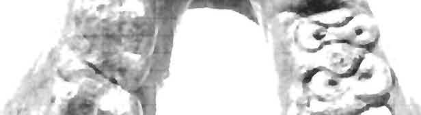


72 72 Miocene Hominoids Postilla181 s. J * y A Fig* 32 GSP 9584 from Locality 260, A is an occlusal view, B an inferior view,


73 73 Miocene Hominoids Postilla181 / > * * ' - ^ / / If lifil t i l I!i till I IMittrif tiff HIIMtitlfHltlftMilHIH III!


74 74 Miocene Hominoids Postilla 181 B!/ *j^'*y G H D * V F i 11 f 11 f I f 11!! i 11IIIJ11 f i IIIU i! f! I? 1 Si! I; Fig. 33 Specimens from Locality 311. A through L show occlusal or lingual views. A GSP 11534; B, GSP 11533; C, GSP 10500; O, GSP 11999; E, GSP 9986; F, GSP 10493; G, GSP 12648; H, GSP 13164; /, GSP 11998; J, GSP 15255; K, GSP 7144; L, GSP 13162, M is an occlusal view of GSP I. jr M


75 75 Miocene Hominoids Postilla181 <*** c \*J* * ^ "-***' & & #&,.,*-«** %** Fig, 34 Specimens from Locality 317, all are occlusal views. A, GSP 11786; B, GSP 13460; C, GSP 9930; O, GSP 7619; E, GSP 13445; f, GSP 11705; G, GSP 13444,


76 78 Miocene Hominoids Postiila 181 -W. % J- **&' 'V^; >» **-J# tf111iijt»!i111!i1111f *iii iiii i!imm<,[;;*., i 11! f 50 ' if /f-v 1 B v >l J3 ^0 50 Fig. 35 Specimens from Locality 317; both are occlusal stereo views, A of GSP 11706, B of GSP 11707,


")


77 77 Miocene Hominoids Postilia 181 w \ 1*&% >*;.: *&%. j '.io ~, >w: -<*& B ) Fig. 36 Specimens from Locality 317; both are occlusal stereo views, A of GSP 11704, B of GSP


78 78 Miocene Hominoids Postilla 181 B,. C /.> o =' ' H % 11 j f f i I {I i I f j * - s» f j i 11 f «; j 11, - f - f -. ; ;, -= S..;-, ;, ; : :in ' an * ^n «n : < Fig. 37 Occlusal or lingual views of specimens from various localities. A, GSP 3293, Loc, 137; B, GSP 12588, Loc. 191; C, GSP5001, Loc, 207; D, GSP 5260, Loc, 211: E, GSP8758, Loc. 221; f, GSP 8759, Loc, 221; G, GSP 13810, Loc. 224; H, GSP 7308, Loc. 224; /, GSP 13808, Loc. 224; J, GSP 8206, Loc. 227; K, GSP 13171, Loc, 227; L, GSP 6999, Loc. 236; M, GSP 8838, Loc. 251; N, GSP9987, Loc. 281; O, GSP 10232, Loc, 309; P, GSP 10785a, Loc. 310; O, GSP 11003, Loc. 314; R, GSP 14822, Loc, 259,


79 -. * 79 Miocene Hominoids Postilla181 K fcj >' *3> / IM M I if! : i* :! :


80 80 Miocene Hominoids Postilla 181 Fig. 38 Occlusal stereo views of: A GSP 6153, Loc. 224 B. GSP 6160, Loc. 226: C, GSP 13566, Loc, 350.


81 81 Miocene Hominoids Postilla181?> B t 'S^T SO 2C '>.Q 20 40


82 82 Miocene Hominoids Postilla181 a JPk: A.-. */ B I r 'A ** >> Fig, 39 Occlusal or lingual views, except H which is lateral, of specimens from various localities. A, GSP 14843, Loc. 327; 8, GSP 13622, Loc. 350; C, GSP 15556, Loc. 409; D, GSP 14951, Loc. 414; E, GSP 14997, Loc. 414; f, GSP 15030, Loc. 416; G.GSP 15629, Loc. 416, H, GSP 15397, Loc. 442; /, GSP 15557, Loc. 463.


83 83 Miocene Hominoids Postilla181 F f* A /,. \ - iif11! 1 j iii f jif it«i I It? f i!! ii5 i < I f iii i f! f! i i


84 84 Miocene Hominoids Postilla181.*TJ V-fr&r ) $ QQ m o " % ^ CM CM fe.l «~ CO P w O ^ H - il Q o.


85 85 Miocene Hominoids Postilla181 s B f fc*frj *$y, 11 f I! i h l il:ih ; I n s j 11 m 11 H M H i 11U! 1111 it m 11! n J11 n " Fig. 41 Pollicial phalanx, GSP 6664, Loc. 311:4, anterior; 8, posterior. Humeral distal articular surface, GSP 6663, Loc. 311: C, anterior; D, posterior.


86 86 Miocene Hominoids Postilla 181 I: O.* J Fig. 42 Humerus, GSP 13606, Loc. 260; A, anterior; B, posterior,


87 87 Miocene Hominoids Postilla 181 : \ A $!*»** Jh 0 M


88 88 Miocene Hominoids Postilla181 Iff -:* m >» J *. \ o


89 yf v >.. llli illl ll)l l»h[ll! i 10 20!tii ilii il!l Ml Fig. 43 Femoral heads: A, GSP 6178, Loc. 224; 8, GSP 15782, Loc. 260; C, GSP 13929, Loc. 221; 0, GSP 11867, Loc Proximal femur, GSP 12654, Loc. 260: E, anterior; F, posterior; G, medial.


90 90 Miocene Hominoids Postilla 181 i J-'"* i : \?A S m B I * i 111J111 jif t i11 i UJ0 o 0 10 F0 ryt if{jillli{ fltilif!fi!tfhii ff!ljflfhfif f n!ii:-- - i f 11 f 111 f * 111 i I i f i f 111 f l f 11! f I s; i i 111 s! l I i i I! i I i i i! i t i f ) Fig. 44 Femoral shaft, GSP 13420, Loc. 260; A. anterior; posterior. Cuneiform, GSP Loc. 311; C. medial; D, lateral; E, anterior; F, posterior.


91 31 Miocene Hominoids Postilla 181 o o r *% ft ; 5f, ^Ipi # <* m\ f V 4- -< j B 1 7* H L : Fig. 45 Hallux, GSP 14046, Loc. 260: A, ventral; B, dorsal. Phalanges. GSP 15784, Loc. 260: C, dorsal; G, ventral. GSP 15783, Loc. 260; D, dorsal; H, ventral. GSP 6666, Loc. 311: E, dorsal; /, ventral. GSP 13168, Loc. 260: F, dorsal; J, ventral.


92 92 Miocene Hominoids Postilla181 o Fig. 46 Radius, GSP 7611, Loc, 317: A, posterior; 8, anterior. B


93 93 Miocene Hominoids Postilla181 A B i - & Fig. 47 Femoral head, GSP 3834, Loc. 280; A, Talus, GSP 10785b, Loc. 310: B, dorsal; C, ventral. Calcaneus, GSP4684, Loc, 182: D, dorsal; E, ventral; F, medial.
Key points for starting off
 Key points for starting off First off, the five questions to ask yourself about a loose tooth before identifying it are: 1. 2. 3. 4. 5. Category (incisor, canine, premolar or molar)? Permanent or deciduous?
1. What is the highest and sharpest cusp on the lower first deciduous molar? 2. Which of the following is NOT the correct location of an embrasure?
 1 1. What is the highest and sharpest cusp on the lower first deciduous molar? a. mesiobuccal b. distobuccal c. distolingual d.mesiolingual 2. Which of the following is NOT the correct location of an embrasure?
Morphology of an Anatomic Crown. By: Assistant Professor Dr. Baydaa Ali Al - Rawi
 Morphology of an Anatomic Crown By: Assistant Professor Dr. Baydaa Ali Al - Rawi October 4, 2009 Elevated landmarks Depressed landmarks A) Elevated landmarks : 1. Dental lobe : is one of the primary centers
Fig. 12 Trigonias?osborni Lucas, SMNH PI637.2, left maxillary fragment with pi to MI; occlusal view, x I. DESCRIPTION
 Fig. 12 Trigonias?osborni Lucas, SMNH PI637.2, left maxillary fragment with pi to MI; occlusal view, x I. DESCRIPTION SMNH P1637.1 has the teeth somewhat cracked but otherwise well preserved. p 2 is very
Primary Teeth Chapter 18. Dental Anatomy 2016
 Primary Teeth Chapter 18 Dental Anatomy 2016 Primary Teeth - Introduction Synonyms deciduous teeth, baby teeth, temporary teeth, milk teeth. There are 20 primary teeth, designated as A thru T in the Universal
P3. (Figs. 3A-F, J-K, 4O-Q, 9C) P3 of P. gaoi bears three roots, the mesial and lingual
 P3 of P. gaoi bears three roots, the mesial and lingual") Boyer, Scott, and Fox, 2011: Supplementary File 2 Description of dentition of Pronothodectes gaoi P3. (Figs. 3A-F, J-K, 4O-Q, 9C) P3 of P. gaoi bears three roots, the mesial and lingual of which are subequal
Boyer, Scott, and Fox, 2011: Supplementary File 2 Description of dentition of Pronothodectes gaoi P3. (Figs. 3A-F, J-K, 4O-Q, 9C) P3 of P. gaoi bears three roots, the mesial and lingual of which are subequal
Fundamental & Preventive Curvatures of Teeth and Tooth Development. Lecture Three Chapter 15 Continued; Chapter 6 (parts) Dr. Margaret L.
 Dr. Margaret L.") Fundamental & Preventive Curvatures of Teeth and Tooth Development Lecture Three Chapter 15 Continued; Chapter 6 (parts) Dr. Margaret L. Dennis Proximal contact areas Contact areas are on the mesial and
Fundamental & Preventive Curvatures of Teeth and Tooth Development Lecture Three Chapter 15 Continued; Chapter 6 (parts) Dr. Margaret L. Dennis Proximal contact areas Contact areas are on the mesial and
Arrangement of the artificial teeth:
 Lecture Prosthodontic Dr. Osama Arrangement of the artificial teeth: It s the placement of the teeth on a denture with definite objective in mind or it s the setting of teeth on temporary bases. Rules
Lecture 2 Maxillary central incisor
 Lecture 2 Maxillary central incisor Generally The deciduous tooth appears in the mouth at 3 18 months of age, with 6 months being the average and is replaced by the permanent tooth around 7 8 years of
Central Incisor DR.Ahmed Al-Jobory B.D.S.,M.Sc. Conservative Department
 Dental Anatomy Lecture 3 Central Incisor DR.Ahmed Al-Jobory B.D.S.,M.Sc. Conservative Department The permanent maxillary Incisors Maxillary incisor are four in number. The maxillary central incisor is
Lecture. Permanent maxillary premolars
 Lecture Permanent maxillary premolars Permanent premolars The maxillary premolars are four in number: two in the right and two in the left. They are posterior to the canines and anterior to the molars.
Upper arch. 1Prosthodontics. Dr.Bassam Ali Al-Turaihi. Basic anatomy & & landmark of denture & mouth
 1Prosthodontics Lecture 2 Dr.Bassam Ali Al-Turaihi Basic anatomy & & landmark of denture & mouth Upper arch Palatine process of maxilla: it form the anterior three quarter of the hard palate. Horizontal
PERMANENT MANDIBULAR INCISORS
 PERMANENT MANDIBULAR INCISORS (Central and Lateral) DR.AHMED AL-JOBORY LEC. 5 PERMANENT MANDIBULAR INCISORS ARE 4 IN NUMBER : 2 CENTRAL (RIGHT &LEFT) AND 2 LATERAL INCISORS (RIGHT &LEFT). CHARACTERISTIC
Dr.Sepideh Falah-kooshki
 Dr.Sepideh Falah-kooshki MAXILLA Premaxillary/median palatal suture (radiolucent). Incisive fossa and foramen (radiolucent). Nasal passages (radiolucent). Nasal septum (radiopaque). Anterior nasal spine
The Premolars. Chapter 17 Permanent Posterior Teeth (p )
") The Premolars Chapter 17 Permanent Posterior Teeth (p. 230-244) General Information Function: u Hold and grind food u Work with molars in mastication. u Even without molars one may be able to chew well
The Premolars Chapter 17 Permanent Posterior Teeth (p. 230-244) General Information Function: u Hold and grind food u Work with molars in mastication. u Even without molars one may be able to chew well
Human Male Asian Skeleton, Robust
 Human Male Asian Skeleton, Robust Product Number: Specimen Evaluated: Skeletal Inventory: SC-287 Original Specimen Near-complete human skeleton with 28 teeth. Osteological Observations: This is a clean,
Bones Ethmoid bone Inferior nasal concha Lacrimal bone Maxilla Nasal bone Palatine bone Vomer Zygomatic bone Mandible
 splanchnocranium - Consists of part of skull that is derived from branchial arches - The facial bones are the bones of the anterior and lower human skull Bones Ethmoid bone Inferior nasal concha Lacrimal
Lec. 3-4 Dr. Saif Alarab Clinical Technique for Class I Amalgam Restorations The outline form
 Lec. 3-4 Dr. Saif Alarab Clinical Technique for Class I Amalgam Restorations Class I refers to -Restorations on the occlusal surfaces of posterior teeth, - The occlusal two thirds of facial and lingual
Concepts of occlusion Balanced occlusion. Monoplane occlusion. Lingualized occlusion. Figure (10-1)
") Any contact between teeth of opposing dental arches; usually, referring to contact between the occlusal surface. The static relationship between the incising or masticatory surfaces of the maxillary or
Any contact between teeth of opposing dental arches; usually, referring to contact between the occlusal surface. The static relationship between the incising or masticatory surfaces of the maxillary or
Morphological Scoring of Dental Casts Using the Arizona State University Dental Anthropology System
 University of Tennessee, Knoxville Trace: Tennessee Research and Creative Exchange University of Tennessee Honors Thesis Projects University of Tennessee Honors Program 5-2004 Morphological Scoring of
Medical NBDE-II. Dental Board Exams Part I.
 Medical NBDE-II Dental Board Exams Part I http://killexams.com/exam-detail/nbde-ii Question: 149 Anatomically, the term "clinical root" can be defined as which of the following: A. The space in the tooth
Arrangement of posterior artificial teeth Standardized parameters Curve of Wilson Curve of Spee
 . Arrangement of posterior artificial teeth Posterior teeth are set up in tight centric occlusion. The mandibular teeth are set in the wax occlusion rim over the residual ridge in their ideal buccolingual
6610 NE 181st Street, Suite #1, Kenmore, WA
 660 NE 8st Street, Suite #, Kenmore, WA 9808 www.northshoredentalacademy.com.08.900 READ CHAPTER The Professional Dental Assistant (p.-9) No Key Terms Recall Questions:,,,, and 6 CLASS SYLLABUS DAY READ
Permanent 2 nd Maxillary Molars
 Permanent 2 nd Maxillary Molars In comparison to the first max molar First molars appears in the oral cavity at the age of 6 years old.. While 2 nd molar 3 rd molar Max. 2 nd molar have long roots (sometimes
Characters.-M1 more like that in Procynodictis than like that of Uintacyon. Prodaphcenus (?) robustus in known features of jaw and lower dentition,
 robustus in known features of jaw and lower dentition,") VOL. 20, 1934 GEOLOGY: C. STOCK NEW CREODONTA FROM THE SESPE UPPER EOCENE, CALIFORNIA By CHESTER STOCK BALCH GRADUATE SCHOOL OF THE GEOLOGICAL SCIENCES, CALIFORNIA INSTITUTE OF TECHNOLOGY Communicated
VOL. 20, 1934 GEOLOGY: C. STOCK NEW CREODONTA FROM THE SESPE UPPER EOCENE, CALIFORNIA By CHESTER STOCK BALCH GRADUATE SCHOOL OF THE GEOLOGICAL SCIENCES, CALIFORNIA INSTITUTE OF TECHNOLOGY Communicated
Human Healed Trauma Skull
 Human Healed Trauma Skull Product Number: Specimen Evaluated: BC-303 Original Specimen Skeletal Inventory: 1 Cranium with full dentition (teeth ##1-16) 1 Mandible with full dentition (teeth ##17-32) Osteological
PERISSODACTYLA OF THE SESPE EOCENE, CALIFORNIA
 260 PALEONTOLOGY: C. STOCK PROC. N. A. S. PERISSODACTYLA OF THE SESPE EOCENE, CALIFORNIA BY CHESTER STOCK BALCH GRADUATE SCHOOL OF THE GEOLOGICAL SCIENCES, CALIFORNIA INSTITUTE OF TECHNOLOGY Communicated
Figure (2-6): Labial frenum and labial notch.
: Labial frenum and labial notch.") The anatomy of the edentulous ridge in the maxilla and mandible is very important for the design of a complete denture. The consistency of the mucosa and architecture of the underlying bone is different
The anatomy of the edentulous ridge in the maxilla and mandible is very important for the design of a complete denture. The consistency of the mucosa and architecture of the underlying bone is different
Indirect retainers. 1 i
 8 1 i Indirect retainers Factors Influencing Effectiveness Indirect Retainers Auxiliary Functions Indirect Retainers Forms Indirect Retainers Auxiliary occlusal rest Canine extensions fiom occlusal rests
Bone Clones Osteological Evaluation Report. 1 intact mandible
 Human, Male, Black Bone Clones Osteological Evaluation Report Product Number: Specimen Evaluated: Skeletal Inventory: BC-203 Bone Clones replica 1 intact cranium 1 intact mandible General observations:
NOVITATES AMEIRIICAN MUSEUM NOTES ON AUSTRALIAN MARSUPIALS RARE OR LITTLE-KNOWN IN THE UNITED STATES BY G. H. H. TATE
 AMEIRIICAN MUSEUM NOVITATES PUBLISHED BY THE AMERICAN MUSEUM OF NATURAL HISTORY CITY OF NEW YORK JULY 17, 1951 NUMBER 1528 NOTES ON AUSTRALIAN MARSUPIALS RARE OR LITTLE-KNOWN IN THE UNITED STATES BY G.
Dental Morphology and Vocabulary
 Dental Morphology and Vocabulary Palate Palate Palate 1 2 Hard Palate Rugae Hard Palate Palate Palate Soft Palate Palate Palate Soft Palate 4 Palate Hard Palate Soft Palate Maxillary Arch (Maxilla) (Uppers)
Elevators. elevators:- There are three major components of the elevator are:-
 Elevators Elevators:- Are exo-levers, instrument designed to elevate or luxate the teeth or roots from their bony socket in close or surgical method of extraction to force a tooth or root along the line
Dental Anatomy High Yield Notes. **Atleast 35 questions comes from these areas of old lectures**
 Dental Anatomy High Yield Notes **Atleast 35 questions comes from these areas of old lectures** This review notes compiled and prepared by my sister for her own study, as a last day review session for
Infratemporal fossa: Tikrit University college of Dentistry Dr.Ban I.S. head & neck Anatomy 2 nd y.
 Infratemporal fossa: This is a space lying beneath the base of the skull between the lateral wall of the pharynx and the ramus of the mandible. It is also referred to as the parapharyngeal or lateral pharyngeal
Human, Female, Black, Shotgun wound
 Human, Female, Black, Shotgun wound Product Number: Specimen Evaluated: Skeletal Inventory: BC-196 Bone Clones replica 1 intact cranium 2 fragments of mandible: - portion of left body, ramus, coronoid
Bone Clones Osteological Evaluation Report. 1 intact mandible
 Human, Female, Asian Product Number: Specimen Evaluated: Skeletal Inventory: BC-211 Bone Clones replica 1 intact cranium 1 intact mandible General observations: In general, the molding process has preserved
ORAL ANATOMY AND PHYSIOLOGY
 CHAPTER 7 ORAL ANATOMY AND PHYSIOLOGY INTRODUCTION This chapter covers the oral anatomy and physiology of the teeth, the histology of the tissues and supporting structures, and concentrates on the external
Tooth Variations. Suruedee Chinthakanan
 Tooth Variations Suruedee Chinthakanan Tooth variations Dental anomalies Cause : hereditary factor Developmental disturbances of teeth www.ectodermaldysplsia.org Tooth variations Enamel is formed from
New Evidence of the Genus Homo from East Rudolf, Kenya. II
 New Evidence of the Genus Homo from East Rudolf, Kenya. II R. E. F. LEAKEY AND B. A. WOOD National Museums of Kenya, Post Qffice Box 40658, Nairobi and Department of Anatomy, Charing Cross Hospital Medical
DOUBLE TEETH IN THE SPERM WHALE (PHYSETER MACROCEPHALUS L.)
") DOUBLE TEETH IN THE SPERM WHALE (PHYSETER MACROCEPHALUS L.) by H. BOSCHMA Recently a fairly large number of teeth of the sperm whale were acquired for the collections of the Rijksmuseum van Natuurlijke
DOUBLE TEETH IN THE SPERM WHALE (PHYSETER MACROCEPHALUS L.) by H. BOSCHMA Recently a fairly large number of teeth of the sperm whale were acquired for the collections of the Rijksmuseum van Natuurlijke
CHAPTER 4 ORAL ANATOMY
 CHAPTER 4 ORAL ANATOMY This chapter covers the oral anatomy and physiology of the teeth, the histology of their tissues and supporting structures, and concentrates on the external features of the teeth.
Bones of the skull & face
 Bones of the skull & face Cranium= brain case or helmet Copyright The McGraw-Hill Companies, Inc. Permission required for reproduction or display. The cranium is composed of eight bones : frontal Occipital
Dental Anatomy and Occlusion
 CHAPTER 53 Dental Anatomy and Occlusion Ma Lou C. Sabino DDS, and Emily G. Smythe, DDS What numerical system is used most commonly in the United States for designating the adult dentition? Pediatric dentition?
PRE-FINISHER APPLIANCE AND BONDABLE LINGUAL RETAINERS. tportho.com cosmeticbraces.com. POD_TPOC_09_PreFinisher_Retainers_2015, Rev.
 9 PRE-FINISHER APPLIANCE AND BONDABLE LINGUAL RETAINERS tportho.com cosmeticbraces.com POD_TPOC_09_PreFinisher_Retainers_2015, Rev. 0 PRE-FINISHER APPLIANCE AND BONDABLE LINGUAL RETAINERS Pre-Finisher
DEVELOPING ANALOGUE/SUBTITUTE FOR THE MANDIBULAR DENTURE BEARING AREA. Dr Muhammad Rizwan Memon FCPS Assistant Professor
 DEVELOPING ANALOGUE/SUBTITUTE FOR THE MANDIBULAR DENTURE BEARING AREA Dr Muhammad Rizwan Memon FCPS Assistant Professor Crest of Residual Ridge Buccal Shelf Shape of supporting structure Mylohyoid Ridge
Advanced Probing Techniques
 Module 21 Advanced Probing Techniques MODULE OVERVIEW The clinical periodontal assessment is one of the most important functions performed by dental hygienists. This module begins with a review of the
3. The Jaw and Related Structures
 Overview and objectives of this dissection 3. The Jaw and Related Structures The goal of this dissection is to observe the muscles of jaw raising. You will also have the opportunity to observe several
Prosthetic Options in Implant Dentistry. Hakimeh Siadat, DDS, MSc Associate Professor
 Prosthetic Options in Dentistry Hakimeh Siadat, DDS, MSc Associate Professor Dental Research Center, Department of Prosthodontics & Dental s Faculty of Dentistry, Tehran University of Medical Sciences
THE SNOUT OF PAULOCNUS PETRIFACTUS (MAMMALIA, EDENTATA)
") THE SNOUT OF PAULOCNUS PETRIFACTUS (MAMMALIA, EDENTATA) by D. A. HOOIJER Rijksmuseum van Natuurlijke Historie, Leiden A specimen of the ground sloth discovered by Mr. P. Stuiver in Curasao, Paulocnus petrifactus
THE SNOUT OF PAULOCNUS PETRIFACTUS (MAMMALIA, EDENTATA) by D. A. HOOIJER Rijksmuseum van Natuurlijke Historie, Leiden A specimen of the ground sloth discovered by Mr. P. Stuiver in Curasao, Paulocnus petrifactus
Indications The selection of amalgam as a restorative material for class V cavity should involve the following considerations:
 1 Lec.7 د.عبد املنعم اخلفاجي CLASS V CAVITY PREPARATION FOR AMAGLAM Indications The selection of amalgam as a restorative material for class V cavity should involve the following considerations: 1- Caries:
Oral Surgery. Basic Techniques of Dental Local Anesthesia. A variety of techniques used in administration and deposition of local anesthesia:
 Oral Surgery Lecture: 9 Dr. Saif Saadedeen Basic Techniques of Dental Local Anesthesia A variety of techniques used in administration and deposition of local anesthesia: 1. Topical anesthesia 2. Infiltration
Introduction to Occlusion and Mechanics of Mandibular Movement
 Introduction to Occlusion and Mechanics of Mandibular Movement Dr. Pauline Hayes Garrett Department of Endodontics, Prosthodontics, and Operative Dentistry University of Maryland, Baltimore Assigned reading
Techniques of local anesthesia in the mandible
 Techniques of local anesthesia in the mandible The technique of choice for anesthesia of the mandible is the block injection and this is attributed to the absence of the advantages which are present in
SUPPLEMENTARY INFORMATION
 doi:10.1038/nature14448 Supplementary Notes Contents 1. Additional fossil hominin materials from Burtele (BRT) localities. 2. Morphological descriptions of BRT-VP-3/1, -3/14, -3/37, and WYT-VP-2/10. 3.
Human, Probable Female, Scaphocephaly
 Human, Probable Female, Scaphocephaly Product Number: Specimen Evaluated: Skeletal Inventory: BC-193 Bone Clones replica 1 intact cranium 1 intact mandible General observations: In general, the molding
Human, Male, Single gunshot wound
 Human, Male, Single gunshot wound Product Number: Specimen Evaluated: Skeletal Inventory: BC-152 Bone Clones replica 1 intact cranium - left inferior nasal concha absent - middle nasal conchae absent 1
AMERICAN MUSEUM NOVITATES
 AMERICAN MUSEUM NOVITATES Number 511 Published by TNe AmzuacAwsMumoIJNATU2AL HISTorCir Dec. 15, 1931 56.9,31 (119:729.1) GENERA AND NEW SPECIES OF GROUND SLOTHS FROM THE PLEISTOCENE OF CUBA BY W. D. MATTHEW1
Human, Male, White, Calvarium cut
 Human, Male, White, Calvarium cut Product Number: Specimen Evaluated: Skeletal Inventory: BC-293 Bone Clones replica 1 intact cranium 1 intact mandible General observations: **NOTE The demographic features
Oral cavity landmarks
 By: Dr. Ahmed Rabah Oral cavity landmarks The knowledge of oral anatomy and physiology will help the operator and provides enough landmarks to act as positive guide during denture construction. This subject
Chapter 7. Skeletal System
 Chapter 7 Skeletal System 1 Skull A. The skull is made up of 22 bones: 8 cranial bones, 13 facial bones, and the mandible. B. The Cranium encloses and protects the brain, provides attachments for muscles,
GENERAL SCOPE AND USES OF PHYSICAL/BIOLOGICAL ANTHROPOLOGY. Paper No. & Title: B.A./B.Sc. (Honours) 2 dn semester. (Practical)
 2 dn semester. (Practical)") GENERAL SCOPE AND USES OF PHYSICAL/BIOLOGICAL ANTHROPOLOGY Course name: Physical Anthropology Paper No. & Title: B.A./B.Sc. (Honours) 2 dn semester (Practical) Topic No. & Title: 5/12 (Part-I) Drawing
GENERAL SCOPE AND USES OF PHYSICAL/BIOLOGICAL ANTHROPOLOGY Course name: Physical Anthropology Paper No. & Title: B.A./B.Sc. (Honours) 2 dn semester (Practical) Topic No. & Title: 5/12 (Part-I) Drawing
1B Getting Ready for Instrumentation: Mathematical Principles and Anatomic Descriptors
 MODULE 1B Getting Ready for Instrumentation: Mathematical Principles and Anatomic Descriptors Module Overview This module contains a review of the mathematical principles and anatomic descriptors used
European Veterinary Dental College
 European Veterinary Dental College EVDC Training Support Document Preparation of Radiograph Sets (Cat and Dog) Document version : evdc-tsd-radiograph_positioning_(dog_and_cat)-20120121.docx page 1 of 13
Human Anatomy and Physiology - Problem Drill 07: The Skeletal System Axial Skeleton
 Human Anatomy and Physiology - Problem Drill 07: The Skeletal System Axial Skeleton Question No. 1 of 10 Which of the following statements about the axial skeleton is correct? Question #01 A. The axial
Partners. 6. Occipital crest for attachment of 6. No occipital crest, neck muscles not
 Name Period Partners Primate and Human Evolution- A Skull Comparison Introduction Skulls are one of the most descriptive parts of an individual s skeleton. Skulls alone can give clues as to the age, sex,
Human, Male, White, Healed hammer blows
 Human, Male, White, Healed hammer blows Product Number: Specimen Evaluated: Skeletal Inventory: BC-217 Bone Clones replica 1 intact cranium General observations: In general, the molding process has preserved
Forensic Archaeology & Forensic Anthropology. ADJ14 Advanced Criminal Investigations
 Forensic Archaeology & Forensic Anthropology ADJ14 Advanced Criminal Investigations Anthropology & Archaeology Anthropology is the study of the biological and cultural aspects of all humans in all places
DENT Advanced Topics in Removable Prosthodontics, Winter 2008
 University of Michigan Deep Blue deepblue.lib.umich.edu 2008-01 DENT 718 - Advanced Topics in Removable Prosthodontics, Winter 2008 Shotwell, Jeffrey Shotwell, J. (2008, April 23) Advanced Topics in Removable
Figure S1: Distal Humerus
 Figure S: Distal Humerus Criteria The distal part of the medial epicondyle forms The distal part of the medial epicondyle forms an a right angle. oblique angle. Viewed from the lateral aspect, the pit
Basic Anatomy and Physiology of the Lips and Oral Cavity. Dr. Faghih
 Basic Anatomy and Physiology of the Lips and Oral Cavity Dr. Faghih It is divided into seven specific subsites : 1. Lips 2. dentoalveolar ridges 3. oral tongue 4. retromolar trigone 5. floor of mouth 6.
The os coxae or hip bone consists of three flat bones, ilium, ischium and pubis, which fuse together to form the acetabulum.
 The os coxae The os coxae or hip bone consists of three flat bones, ilium, ischium and pubis, which fuse together to form the acetabulum. The ilium extends from the acetabulum upwards forming the lateral
TRAUMA TO THE FACE AND MOUTH
 Dr.Yahya A. Ali 3/10/2012 F.I.C.M.S TRAUMA TO THE FACE AND MOUTH Bailey & Love s 25 th edition Injuries to the orofacial region are common, but the majority are relatively minor in nature. A few are major
Dr.Adel F.Ibraheem Partial Veneer Crown(Three quarter crown) Three quarter (¾ )crown: Uses: Indications ---- For posterior teeth ;
 Three quarter (¾ )crown: Uses: Indications ---- For posterior teeth ;") Lecture.9 Dr.Adel F.Ibraheem Partial Veneer Crown(Three quarter crown) *It is a cast metal crown restoration that cover only a part of the clinical crown, most commonly used type of partial veneer crown
Lecture.9 Dr.Adel F.Ibraheem Partial Veneer Crown(Three quarter crown) *It is a cast metal crown restoration that cover only a part of the clinical crown, most commonly used type of partial veneer crown
Copyright 2003 Pearson Education, Inc. publishing as Benjamin Cummings. Dr. Nabil Khouri MD, MSc, Ph.D
 Dr. Nabil Khouri MD, MSc, Ph.D Pelvic Girdle (Hip) Organization of the Lower Limb It is divided into: The Gluteal region The thigh The knee The leg The ankle The foot The thigh and the leg have compartments
Muscles of mastication [part 1]
![Muscles of mastication [part 1]](/thumbs/76/73586850.jpg "Muscles of mastication [part 1]") Muscles of mastication [part 1] In this lecture well have the muscles of mastication, neuromuscular function, and its relationship to the occlusion morphology. The fourth determinant of occlusion is the
Muscles of mastication [part 1] In this lecture well have the muscles of mastication, neuromuscular function, and its relationship to the occlusion morphology. The fourth determinant of occlusion is the
Dentalelle Tutoring - Faulty Radiographs
 Dentalelle Tutoring - Faulty Radiographs Errors in improperly exposing or processing dental films can produce undesirable dental radiographs of nondiagnostic quality. These are known as faulty radiographs.
ORTHODONTICS Treatment of malocclusion Assist.Lec.Kasem A.Abeas University of Babylon Faculty of Dentistry 5 th stage
 Lec: Treatment of class I malocclusion Class I occlusion can be defined by Angles, classification as the mesiobuccal cusp of the upper 1 st permanent molar occlude with the developmental groove of the
SPRINGFIELD TECHNICAL COMMUNITY COLLEGE ACADEMIC AFFAIRS
 SPRINGFIELD TECHNICAL COMMUNITY COLLEGE ACADEMIC AFFAIRS Course Number: DHYG 103 Department: Dental Hygiene Course Title: Oral Anatomy 1 Semester: Spring Year: 1997 Objectives/ 1. Utilize appropriate clinical
Phylogenetics Lab: Character Descriptions
 Phylogenetics Lab: Character Descriptions 1) Osseous Auditory Canal. 0= absent, 1= present. Does the organism have a bony auditory canal? This will look like a hole or opening in the skull behind the jaw
#45 Ortho-Tain, Inc PREVENTIVE ERUPTION GUIDANCE -- PREVENTIVE OCCLUSAL DEVELOPMENT
 #45 Ortho-Tain, Inc. 1-800-541-6612 PREVENTIVE ERUPTION GUIDANCE -- PREVENTIVE OCCLUSAL DEVELOPMENT Analysis and Diagnosis of Occlusion: The ideal child of 5 y ears of age that probably has the best chance
Component parts of Chrome Cobalt Removable Partial Denture
 Lec. 5 د.بسام الطريحي Component parts of Chrome Cobalt Removable Partial Denture Major connectors: Are either bars or plates, the difference between them is in the amount of tissue covers. Plates are broad
Any of the vertebra in the cervical (neck) region of the spinal column. The cervical vertebra are the smallest vertebra in the spine, reflective of th
 region of the spinal column. The cervical vertebra are the smallest vertebra in the spine, reflective of th") Any of the vertebra in the cervical (neck) region of the spinal column. The cervical vertebra are the smallest vertebra in the spine, reflective of the fact that they support the least load. In humans,
Any of the vertebra in the cervical (neck) region of the spinal column. The cervical vertebra are the smallest vertebra in the spine, reflective of the fact that they support the least load. In humans,
Lips and labial mucosa
 Lips and labial mucosa External portion of the lips: the vermilion border and the skin Vermilion border : the exposed red portion of the lip, covered by mucous membrane, no mucous glands Boundary: the
1. Anterior-posterior movement of the mandible (APM):
:") Dental Anatomy A. Terminology: Over the past few years there have been several acronyms introduced into the equine dentistry industry. These terms should help you describe and understand equine dental
Dental Anatomy A. Terminology: Over the past few years there have been several acronyms introduced into the equine dentistry industry. These terms should help you describe and understand equine dental
Comprehensive AOCMF Classification System. Cornelius CP, Kunz C, Prein J, Audigé L. Mandibular fractures Level-2 system (cases 1 to 18)
") Comprehensive AOCMF Classification System Cornelius CP, Kunz C, Prein J, Audigé L Mandibular fractures Level-2 system (cases 1 to 18) Case 1: Body fracture traversing anterior transition zone Imaging:
Comprehensive AOCMF Classification System Cornelius CP, Kunz C, Prein J, Audigé L Mandibular fractures Level-2 system (cases 1 to 18) Case 1: Body fracture traversing anterior transition zone Imaging:
Structure of an Incisor
 MAMMALIAN TEETH Mammals have different types and shapes of teeth and they are thus termed Heterodonts. Those which have teeth of the same size and shapes are termed as Homodonts. In mammals teeth consist
Digital Imaging from a new perspective
 TREATMENT CENTRES HANDPIECES HYGIENE SYSTEMS X-RAY SYSTEMS CEREC TREATMENT CENTRES HANDPIECES HYGIENE SYSTEMS X-RAY SYSTEMS CEREC SIRONA CREATING AND MAINTAINING VALUE. You are right to expect a great
Selection and arrangement of teeth in rpd
 Selection and arrangement of teeth in rpd upon completion of the articulator mounting and a thorough assessment of the occlusal requirements, the practitioner should be able to perform the proper arrangement
Anatomy and Physiology. Bones, Sutures, Teeth, Processes and Foramina of the Human Skull
 Anatomy and Physiology Chapter 6 DRO Bones, Sutures, Teeth, Processes and Foramina of the Human Skull Name: Period: Bones of the Human Skull Bones of the Cranium: Frontal bone: forms the forehead and the
Treatment planning of nonskeletal problems. in preadolescent children
 In the name of GOD Treatment planning of nonskeletal problems in preadolescent children Presented by: Dr Somayeh Heidari Orthodontist Reference: Contemporary Orthodontics Chapter 7 William R. Proffit,
ASSESSMENT OF MAXILLARY FIRST MOLAR ROTATION IN SKELETAL CLASS II, AND THEIR COMPARISON WITH CLASS I AND CLASS III SUBJECTS
 ORIGINAL ARTICLE ASSESSMENT OF MAXILLARY FIRST MOLAR ROTATION IN SKELETAL CLASS II, AND THEIR COMPARISON WITH CLASS I AND CLASS III SUBJECTS ABSTRACT FARHAT AMIN, BDS, MCPS, FCPS Mesial rotation of maxillary
Attachment G. Orthodontic Criteria Index Form Comprehensive D8080. ABBREVIATIONS CRITERIA for Permanent Dentition YES NO
 First Review IL HFS Dental Program Models Second Review Ortho cad Attachment G Orthodontic Criteria Index Form Comprehensive D8080 Ceph Film X-Rays Photos Narrative Patient Name: DOB: ABBREVIATIONS CRITERIA
Identification of Mammal Skulls
 Identification of Mammal Skulls Mammalian teeth are heterodont. That is, they are different as you move from front to rear in the tooth row. This contrasts with the homodont condition of most toothed vertebrates.
Anatomic Relations Summary. Done by: Sohayyla Yasin Dababseh
 Anatomic Relations Summary Done by: Sohayyla Yasin Dababseh Anatomic Relations Lecture 1 Part-1 - The medial wall of the nose is the septum. - The vestibule lies directly inside the nostrils (Nares). -
Skeleton Tedeschi Register (1911) Sex:: M- S. A. Age: 31 years Job:
 Sex:: M- S. A. Age: 31 years Job:") Skeleton 1438 Tedeschi Register (1911) Sex:: M- S. A. Age: 31 years Job: Blacksmith Date of death: 9 th of February, 1911 Cause of death: Lung tuberculosis Origin: General Hospital (Padova) Bone Remains
Skeleton 1438 Tedeschi Register (1911) Sex:: M- S. A. Age: 31 years Job: Blacksmith Date of death: 9 th of February, 1911 Cause of death: Lung tuberculosis Origin: General Hospital (Padova) Bone Remains
TRANSITION ANALYSIS AGE ESTIMATION: ILLUSTRATIONS
 TRANSITION ANALYSIS AGE ESTIMATION: ILLUSTRATIONS Soft, deep billowing 2 (T 222R, image reversed) Fordisc Version 1.00 Figures are from France Casting pubic symphysis casts and bones from several institutions:
Development of occlusion:
 : Dr.Issam Aljorani (BDS, MSc. Ortho.) Postnatal development of the dentition When a child is born, mineralization of all the primary tooth crowns is well underway, with this process also beginning in
Filtek LS Low Shrink Posterior Restorative System Case 1: Cusp build-up
 Case 1 This case study focuses on the restoration of an upper molar. Filtek LS Low Shrink Posterior Restorative System Case 1: Cusp build-up The existing, inadequate restoration led to the development
Copyright 2010 Pearson Education, Inc.
 E. VERTEBRAL COLUMN 1. The vertebral column extends from the skull to the pelvis and forms the vertical axis of the skeleton. 2. The vertebral column is composed of vertebrae that are separated by intervertebral