Severely itchy skin lesions on extremities
|
|
|
- Oscar Richard
- 5 years ago
- Views:
Transcription
1 2 nd EDS Monthly Meeting-Dubai 03 June 2016 Severely itchy skin lesions on extremities Dr.Hani Gamil Montaser Dermatologist Hili Health Center-Al Ain
2 The aim of this presentation is to highlight the fact that recognizing unusual features of the disease and to enhance more understanding of its pathogenesis and management.
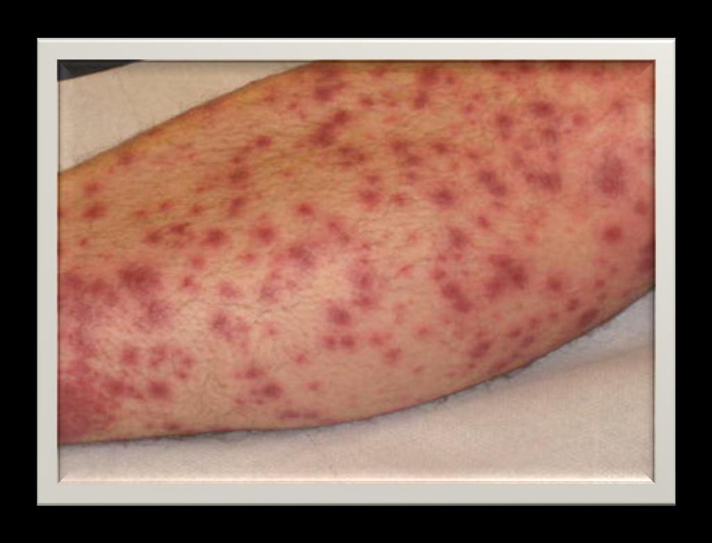
3 A 60-year-old female patient presented with 3 weeks history of coughing and itchy generalized skin rash mostly in lower limbs. She is a known diabetic and hypertensive on oral medications. The rash was not associated with fever, night sweat, diarrhea, joint pain or weight loss.
4 Physical examination revealed nonblanching petechial rash and vesiculobullous lesions containing clear or hemorrhagic fluid. Some lesions are crusted, ulcerated with central necrosis. The rash was mainly on lower limbs, also on back, abdomen, axillae and upper limbs.
5


6
7 On admission Lab. investigations : CBC, LFT s, renal function tests, electrolytes and coagulation profile were normal ANA, ds-dna, ENA, ANCA, C4, C3, IgA, RF and cryoglobulin were negative HIV and hepatitis profile were negative LDH, CK, CRP and blood glucose levels were high
8 Investigation CT Thorax with contrast Negative CT Abdomen/Pelvis with contrast Negative US LE Venous Doppler Bilateral No evidence of DVT Skin biopsy : Skin punch taken from early erythematous lesion from chest
9 Histopathogy Acanthosis, hyperkeratosis, parakeratosis and focal parakeratotic crust infiltrated with neutrophils Heavy perivascular chronic inflammatory cell infiltration predominantly lymphocytic infiltrate penetrating the dermal blood vessel walls with mild extravasation of erythrocytes DIAGNOSIS : LYMPHOCYTIC VASCULITIS
10 Based on the diagnosis of vasculitis Oral Prednisolone tab. 100 mg daily was administered, patient showed good response but following the tapering of prednisolone tab. the rash relapsed On re-admission, skin biopsy was repeated The specimen was taken from well developed lesion including the central adherent keratotic plug on right leg

11
12 Histopathology Epidermal ulcerative lesion with dermal invagination. The ulcerative lesion shows full thickness epidermal necrosis with neutrophilic infiltrate The underlying dermis demonstrates foci with altered collagen. Vertical collagen fibers are noted within necrotic epidermal lesion Masson Trichrome special stain highlights the extruded epidermal collagen Elastic stain fails to demonstrate elastic fibers Gomori-Methamine silver stain is negative for fungal organisms Dermal blood vessels demonstrate reactive changes associated with perivascular neutrophils There is absence of vasculitis

13 Hyperkeratosis overlying a cup shaped depression or invagination in the epidermis. Prominent epidermal hyperplasia, which encompasses a central basophilic plug of keratin, collagen and inflammatory debris.

14 Central plug of basophilic debris and collagen with surrounding epidermal hyperplasia


15 The central crusted keratotic plug contains keratin, cellular debris and collagen fibres Perforating collagen bundles Elastica van Gieson stain

16 Transepidermal elimination of red collagen fibers through the spinous layer and into the stratum corneum (Verhoeff van Gieson stain) Verhoeff's stain Elastic fibers and cell nuclei are stained black, collagen fibers are stained red other tissue elements including cytoplasm are stained yellow

17 Vertically oriented collagen fibers, perforating the epidermis from below (Masson-Trichrome stain, 400) Masson Trichome Collagen fibers stain green or blue Muscle and keratin will be red. Cytoplasm will be pink to red. Nuclei will be black..
18 DIAGNOSIS Reactive Perforating Collagenosis
19 Doxycycline capsule 100 mg daily started while oral steroids gradually tapered over 2 weeks till complete withdrawal of steroid Well controlled DM This resulted in almost complete clearance of skin lesions Successful treatment of acquired reactive perforating collagenosis with doxycycline. Acta Derm. Venereol. 2002; 82 (5); 393-5

20 2 weeks after doxycycline therapy
21 2 weeks later

22


23
24 Perforating Dermatosis It is a group of disorders with trans-epidermal elimination of collagen, elastic tissue or necrotic connective tissue caused by unrelated pathologic abnormalities These are papulo-squamous disorders characterized by keratotic plug or crust, in which dermal connective tissue is eliminated through epidermis
25 Transepithelial Elimination (TEE) It is a phenomenon in which material from the dermis is extruded through the epidermis to the exterior with little or no disruption of the surrounding structures The extruded material may include inflammatory cells, red cells, micro-organisms and extracellular substances, such as mucin or altered connective tissue components

26
27 Molecular Mechanism AGE: advanced glycation end product KC : keratinocytes TEE : transepidermal elimination Journal of Investigative Dermatology (2010) 130,
28 TEE in RPC TEE in EPS TEE in Perforating Folliculitis TEE in Kyrle s disease collagen elastic fibers hair follicle keratinocyte Journal of Investigative Dermatology (2010) 130,


29 Perforating folliculitis papules typically are concentrated on hairbearing portions of the extremities (arms, thighs) and buttocks Dilated follicular infundibulum filled with mixture of keratin, basophilic debris, inflammatory cells and degenerated collagen fibers

30 Kyrle s disease large keratotic papules distributed widely throughout the body. The papules contain a central keratotic plug, which histologically correlates with keratin and necrotic debris


31 Elastosis perforating serpiginosa small papules erupt and grouped in a confined area, eventually becoming serpiginous. The central core of each papule contains a compressed aggregate of fibrous material and cellular debris
32 Reactive Perforating Collagenosis The major abnormality in RPC is focal damage to collagen and the elimination of the disrupted collagen through the epidermis The underlying cause of reactive perforating collagenosis is unknown Abnormal response to superficial trauma: A frequent association with pruritus, the tendency to the Koebner phenomenon and the distribution of lesions on trauma-prone areas provides evidence that superficial trauma (e.g. scratching) may play a part in its etiology
33 Reactive perforating collagenosis 2 forms Inherited Acquired
34 Reactive Perforating Collagenosis The inherited form Starts in early childhood as small papules on the extensor surface of the hands, the elbows and the knees following superficial trauma Each skin colored papule increases to a size of about 6 mm over 3 5 weeks and then becomes umbilicated, with a keratinous plug The lesions regress spontaneously in 6 8 weeks to leave a hypopigmented area or slight scar, but new lesions may appear J Eur Acad Dermatol Venereol. Jul 2007;21(6):766-70
35 Reactive Perforating Collagenosis Acquired form The bulk of the coarse granular basophilic material that is extruded by TEE appears to derive from the nuclei of PMNL Lysosomal enzymes derived from leukocytes might be responsible for the altered staining of collagen fibres and the impairment of keratinocyte adhesion, which allows TEE of dermal components
36 The diagnostic criteria for the adult (acquired) form of RPC, as follows : Onset of lesions after age 18 years Umbilicated papules or nodules with a central, adherent keratotic plug Elimination of necrotic basophilic collagen bundles into a cup-shaped epidermal depression as seen in biopsy specimens J Am Acad Dermatol 1994;30: Faver IR, Daoud MS
37 Associations Chronic renal failure, often with underlying diabetes * Other nephropathies without diabetes Hypothyroidism, hyperparathyroidism Liver dysfunction Malignancies ** Indinavir*** * Brenner and Rector's the Kidney, Ninth Edition 2012 **Br. J. Dermatol. - ; 142 (2); ***Eur J Dermatol. Jan-Feb 2008;18(1):84-5
38 Specific investigations Skin biopsy with Masson trichrome stains and Verhoeff van Gieson stains Serum blood urea nitrogen, creatinine ALT, AST, alkaline phosphatase, bilirubin and uric acid Serum glucose, oral glucose tolerance test or hemoglobin A1C Hepatitis C virus antibodies Thyroid function tests
39 Management When the underlying cause is not apparent, serum chemistry for renal and liver function tests and oral glucose tolerance test or hemoglobin A1C may be helpful Most often, conditions such as diabetes mellitus and renal failure will be known to the patient who presents with perforating skin lesions The lesion can disappear with stabilization of renal failure and/or correction of hyperglycaemia
40 Management Minimizing pruritus is important because many of the perforating disorders typically exhibit a Koebner phenomenon, meaning that lesions develop in traumatized or scratched skin. So it is important to instruct the patient to avoid scratching the lesions.
41 Therapeutic Lines 1 st line 2 nd line 3 rd line Tretinoin 0.1% Tazarotene gel 0.1% Narrowband UVB Allopurinol Isotretinoin PUVA Acitretin Doxycycline Oral metronidazole Oral clindamycin Oral hydroxychloroquine Imiquimod Surgical debridement Cryotherapy Cantharidin Ultrapulse laser CO 2 laser Photodynamic therapy Treatment of Skin Disease: Comprehensive therapeutic strategies. Mark G. Lebwohl, Warren R. Heymann, John Berth-Jones.
42 Summary Consider Perforating disorders when ulcer with keratotic plugs is found A good interdisciplinary cooperation is crucial for the early recognition by histopathology Definitive diagnosis of the perforating disorders depends on the demonstration of trans-epidermal elimination on skin biopsy Differentiation between the different forms of perforating disorders can be accomplished by Masson trichrome stains for collagen (RPC), Verhoeff van Gieson stains for elastic tissue (EPS)
43 Conclusions Management of the perforating diseases involves determination of underlying etiologies Once the diagnosis of underlying diseases is ascertained, treatment is directed at associated symptoms
44 References 1. Jean L Bolognia. Joseph L Jorizzo. Ronald p rapini dermatology 2 nd edition 2. Lever s histology of the skin ninth edition 3. Nih center for macromolecular modeling & Bioformatics. University of Illinois at Urbana- Champaign 4-Indian Journal Dermatology,Venereology and Leprology vol.77, Num.1,2011,pp Journal of Investigative Dermatology (2010) 130, ;doi: /jid Indian Journal Pathology & Microbiology Year 2013/Volume 56/ Issue 4/ Page Saray Y, Seçkin D, Bilezikçi B. J Eur Acad Dermatol Venereol 2006; 20: Ramesh V, Sood N, Kubba A, Singh B. J Eur Acad Dermatol Venereol 2007; 21: Outland JD, Brown TS, Callen JP. Arch Dermatol 2002; 138: Ohe S, Danno K, Sasaki H, Isei T, Okamoto H. J Am Acad Dermatol 2004; 50: Hoque SR, Ameen M, Holden CA. Br J Dermatol 2006; 154: Satchell AC, Crotty K, Lee S. Australas J Dermatol 2001; 42: Kelly SC, Purcell SM. Arch Dermatol 2006; 142: Brinkmeier T, Schaller J, Herbst RA, Frosch PJ. Acta Derm Venereol 2002; 82: Humphrey S, Hemmati I, Randhawa R, J Cutan Med Surg 2010; 14: Wong J, Phelps R, Levitt J. Arch Dermatol 2012; 148: Sezer E, Erkek E. Photodermatol Photoimmunol Photomed 2012; 28: 50 2.


45 THANK YOU
Acquired Perforating Dermatosis in Patients with Chronic Renal Failure and Diabetes Mellitus
 J Korean Med Sci 2004; 19: 283-8 ISSN 1011-8934 Copyright The Korean Academy of Medical Sciences Acquired Perforating Dermatosis in Patients with Chronic Renal Failure and Diabetes Mellitus Acquired perforating
Histopathology: skin pathology
 Histopathology: skin pathology These presentations are to help you identify, and to test yourself on identifying, basic histopathological features. They do not contain the additional factual information
MECHANISMS OF HUMAN DISEASE: LABORATORY SESSION PATHOLOGY OF THE SKIN LAB. Friday, February 12, :30 am 11:00 am
 MECHANISMS OF HUMAN DISEASE: LABORATORY SESSION PATHOLOGY OF THE SKIN LAB Friday, February 12, 2012 9:30 am 11:00 am FACULTY COPY GOALS: Describe the basic clinical and morphologic features of various
CPC. Chutika Srisuttiyakorn, M.D. Kobkul Aunhachoke, M.D. Phramongkutklao Hospital Bangkok, Thailand
 CPC Chutika Srisuttiyakorn, M.D. Kobkul Aunhachoke, M.D. Phramongkutklao Hospital Bangkok, Thailand A 53 year-old woman with fever, facial swelling and rashes on face, trunk and upper extremities for 3
Conflicts. Objectives. University of Texas Health Science Center at San Antonio. Pediatrics Grand Rounds 24 August Pediatric Dermatology 101
 Pediatric Dermatology 101 John C. Browning, MD, FAAD, FAAP Conflicts Investigator: ViroXis Advisor: ViroXis Advisory Board: TopMD Speaker: Galderma Objectives Understand the meaning and importance of cutaneous
Case Rep Dermatol 2009;1:66 70 DOI: / Key Words Coma Blister Barbiturate Overdose Meningoencephalitis
 66 Coma Blisters Joana Rocha a Teresa Pereira a Filipa Ventura a Fernando Pardal b Celeste Brito a Departments of a Dermatology and b Pathology, Hospital de São Marcos, Braga, Portugal Key Words Coma Blister
Pimples and Boils!! Dr Nathan Harvey Anatomical Pathology, PathWest
 Pimples and Boils!! Dr Nathan Harvey Anatomical Pathology, PathWest Overview & Learning Objectives Review the cardinal signs/symptoms of acute inflammation Review the histological features of acute inflammation
MECHANISMS OF HUMAN DISEASE: LABORATORY SESSION PATHOLOGY OF THE SKIN LAB. Friday, February 13, :30 am 11:00 am
 MECHANISMS OF HUMAN DISEASE: LABORATORY SESSION PATHOLOGY OF THE SKIN LAB Friday, February 13, 2009 9:30 am 11:00 am FACULTY COPY GOALS: Describe the basic clinical and morphologic features of various
HEMORRHAGIC BULLOUS HENOCH- SCHONLEIN PURPURA: A CASE REPORT
 HEMORRHAGIC BULLOUS HENOCH- SCHONLEIN PURPURA: A CASE REPORT Nirmala Ponnuthurai, Sabeera Begum, Lee Bang Rom Paediatric Dermatology Unit, Institute of Paediatric, Hospital Kuala Lumpur, Malaysia Abstract
Citation The Journal of Dermatology, 37(8), available at
, available at") NAOSITE: Nagasaki University's Ac Title Two cases of blaschkitis with promi Author(s) Utani, Atsushi Citation The Journal of Dermatology, 37(8), Issue Date 2010-08 URL Right http://hdl.handle.net/10069/25634
NAOSITE: Nagasaki University's Ac Title Two cases of blaschkitis with promi Author(s) Utani, Atsushi Citation The Journal of Dermatology, 37(8), Issue Date 2010-08 URL Right http://hdl.handle.net/10069/25634
Retrospective 10 years review of 100 patients with psoriasis in the Kingdom of Saudi Arabia (KSA)
") Retrospective 10 years review of 100 patients with psoriasis in the Kingdom of Saudi Arabia (KSA) Ahmed Abdullah Alhumidi King saud university, Riyadh, kingdom of Saudi Arabia Abstract Background: This
Retrospective 10 years review of 100 patients with psoriasis in the Kingdom of Saudi Arabia (KSA) Ahmed Abdullah Alhumidi King saud university, Riyadh, kingdom of Saudi Arabia Abstract Background: This
S003 CPC Self-Assessment
 S003 CPC Self-Assessment Alina G. Bridges, D.O. Associate Professor Program Director, Dermatopathology Fellowship Department of Dermatology, Division of Dermatopathology and Cutaneous Immunopathology Mayo
Observations on the Pathology of Lesions Associated with Stephanofilaria dinniki Round, 1964 from the Black Rhinoceros (Diceros bicornis)
") Journal of Helminthology, ~ol. XXXVIII, Nos. 1/2, 1964, pp. 171-174. Observations on the Pathology of Lesions Associated with Stephanofilaria dinniki Round, 1964 from the Black Rhinoceros (Diceros bicornis)
Journal of Helminthology, ~ol. XXXVIII, Nos. 1/2, 1964, pp. 171-174. Observations on the Pathology of Lesions Associated with Stephanofilaria dinniki Round, 1964 from the Black Rhinoceros (Diceros bicornis)
BSD Self Assessment Workshop 7 th July 2013 CASE 27 RAC6123
 BSD Self Assessment Workshop 7 th July 2013 CASE 27 RAC6123 M55. 4/7 tender lesions on knee, legs and arms. Also iritis/ weight loss/headache, synovitis.?vasculitis. Sarcoidosis. Biopsy from left elbow
Case 16.1 A 67-year-old Thai man from Nontaburi Chief complaint: Asymptomatic erythematous papules and plaques for 2 months Present illness: Two
 Case 16.1 A 67-year-old Thai man from Nontaburi Chief complaint: Asymptomatic erythematous papules and plaques for 2 months Present illness: Two months, he developed asymptomatic multiple erythematous
THE TIP OF THE ICEBERG SAMER BOLIS, DO PGY-3 LEHIGH VALLEY HEALTH NETWORK, ALLENTOWN PA
 THE TIP OF THE ICEBERG SAMER BOLIS, DO PGY-3 LEHIGH VALLEY HEALTH NETWORK, ALLENTOWN PA Case The patient is a 48 year-old female, who recently returned from a trip to Puerto Rico. She presents to the ED
EXPERIMENTAL THERMAL BURNS I. A study of the immediate and delayed histopathological changes of the skin.
 EXPERIMENTAL THERMAL BURNS I A study of the immediate and delayed histopathological changes of the skin. RJ Brennan, M.D. and B. Rovatti M.D. The purpose of this study was to determine the progressive
Actinic keratosis (AK): Dr Sarma s simple guide
: Dr Sarma s simple guide") Actinic keratosis (AK): Dr Sarma s simple guide Actinic keratosis is a very common lesion that you will see in your day-to-day practice. First, let me explain the name Actinic keratosis. It means keratosis
Actinic keratosis (AK): Dr Sarma s simple guide Actinic keratosis is a very common lesion that you will see in your day-to-day practice. First, let me explain the name Actinic keratosis. It means keratosis
Basal cell carcinoma 5/28/2011
 Goal of this Presentation A practical approach to the diagnosis of cutaneous carcinomas and their mimics Thaddeus Mully, MD University of California San Francisco To review common non-melanoma skin cancers
Degos Disease: A Case Report and Review of Literature
 Degos Disease: A Case Report and Review of Literature Monira waked Egyptian Dermatology Online Journal 4 (1): 5, June 2008 Al Houd Al Marsod Hospital Submitted for publication: May 25 th, 2008 Accepted
This section covers the basic knowledge of normal skin structure and function required to help understand how skin diseases occur.
 Background Knowledge Functions of normal skin Background Knowledge This section covers the basic knowledge of normal skin structure and function required to help understand how skin diseases occur. Learning
SESSION 1: GENERAL (BASIC) PATHOLOGY CONCEPTS Thursday, October 16, :30am - 11:30am FACULTY COPY
 PATHOLOGY CONCEPTS Thursday, October 16, :30am - 11:30am FACULTY COPY") SESSION 1: GENERAL (BASIC) PATHOLOGY CONCEPTS Thursday, October 16, 2008 9:30am - 11:30am FACULTY COPY GOAL: Describe the basic morphologic (structural) changes which occur in various pathologic conditions.
SESSION 1: GENERAL (BASIC) PATHOLOGY CONCEPTS Thursday, October 16, 2008 9:30am - 11:30am FACULTY COPY GOAL: Describe the basic morphologic (structural) changes which occur in various pathologic conditions.
Grover s disease: A case report.
 320 Case report Thai J Dermatol, October-December 2011 ABSTRACT: Grover s disease: A case report. Supicha Chavanich MD, Praneet Sajjachareonpong MD. CHAVANICH C, SAJJACHAREONPONG P. GROVER S DISEASE: A
Lymphomatoid Papulosis 3 Case Reports
 IOSR Journal of Dental and Medical Sciences (IOSR-JDMS) e-issn: 2279-0853, p-issn: 2279-0861.Volume 14, Issue 7 Ver. III (July. 2015), PP 31-35 www.iosrjournals.org Lymphomatoid Papulosis 3 Case Reports
Skin lesions The Good and the Bad. Dr Virginia Hubbard Ipswich Hospital NHS Trust Barts and the London School of Medicine and Dentistry
 Skin lesions The Good and the Bad Dr Virginia Hubbard Ipswich Hospital NHS Trust Barts and the London School of Medicine and Dentistry Case 1 32 year old woman Australian Lesion on back New hair growing
CELL AND TISSUE INJURY COURSE-II PATHOLOGY LABORATORY
 CELL AND TISSUE INJURY COURSE-II PATHOLOGY LABORATORY PATHOLOGY of INFECTIOUS DISEASES MICROSCOPY Rengin Ahıskalı Macroscopy samples are shown in the macroscopy presentations of the first two courses.
Name the condition: Canine sterile neutrophilic dermatosis (Sweet s syndrome)
") 5-year-old male miniature Schnauzer dog with acute onset of severe macular erythema and multiple tender violaceus plaques all over the body. Which of the following is the most likely diagnosis? 1. Canine
5-year-old male miniature Schnauzer dog with acute onset of severe macular erythema and multiple tender violaceus plaques all over the body. Which of the following is the most likely diagnosis? 1. Canine
Challenging Cases in Dermatopathology. Rosalie Elenitsas, M.D. Professor of Dermatology Director, Dermatopathology University of Pennsylvania
 Challenging Cases in Dermatopathology Rosalie Elenitsas, M.D. Professor of Dermatology Director, Dermatopathology University of Pennsylvania DISCLOSURE OF RELATIONSHIPS WITH INDUSTRY Rosalie Elenitsas
A dinical and histopathologic entity associated with an increased risk of nonmelanoma skin cancer
 PUVA keratosis A dinical and histopathologic entity associated with an increased risk of nonmelanoma skin cancer M. C. G. van Praag, MD, a J. N. Bouwes Bavinck, MD, a W. Bergman, MD, PhD, a F. R. Rosendaal,
Rameshwar Gutte and Uday Khopkar
 Extragenital unilateral lichen sclerosus et atrophicus in a child: a case report Rameshwar Gutte and Uday Khopkar Department of Dermatolgy, Seth GSMC and KEM Hospital, Parel, Mumbai-400012, India Egyptian
Pathology of the skin. Dr Fónyad László, 1sz. Patológiai és Kísérleti Rákkutató Intézet, SE
 Pathology of the skin Dr Fónyad László, 1sz. Patológiai és Kísérleti Rákkutató Intézet, SE The skin Biggest organ Kb. 1.8 nm Kb. 10 kg Most frequent site for tumor development (BCC) Pathology of the skin
Cutanous Manifestation of Lupus Erythematosus. Presented By: Dr. Naif S. Al Shahrani Salman Bin Abdaziz university
 Cutanous Manifestation of Lupus Erythematosus Presented By: Dr. Naif S. Al Shahrani Salman Bin Abdaziz university A 50-year old lady, who is otherwise healthy, presented to the dermatology clinic with
Case No. 5; Slide No. B13/8956/2
 Interface diseases Case No. 5; Slide No. B13/8956/2 Histological findings Severe hydropic vacuolation of epidermal and follicular basal cells/ interface dermatitis Multifocally apoptotic keratinocytes
Dermoscopy: Recognizing Top Five Common In- Office Diagnoses
 Dermoscopy: Recognizing Top Five Common In- Office Diagnoses Vu A. Ngo, DO Department of Family Medicine and Dermatology Choctaw Nation Health Services Authority Learning Objectives Introduction to dermoscopy
Melanoma Case Scenario 1
 Melanoma Case Scenario 1 History and physical 11/5/16 Patient is a single, 48-year-old male in good health who presented to his primary physician for a yearly physical exam during which a 3.4 x 2.8 x 1.5
Extreme dermatoheliosis: How to approach the severely sun damaged patient
 Extreme dermatoheliosis: How to approach the severely sun damaged patient Anokhi Jambusaria MD, MSCE Staff Dermatologist Baylor Scott and White Health Round Rock, TX I have no relevant conflicts of interest
Melanoma Case Scenario 1
 Melanoma Case Scenario 1 History and physical 11/5/16 Patient is a single, 48-year-old male in good health who presented to his primary physician for a yearly physical exam during which a 3.4 x 2.8 x 1.5
Infections and nonmicrobial inflammatory stimuli can cause leukocytosis (as seen in Lab 1) as well as lymph node enlargement (lymphadenopathy).
 as well as lymph node enlargement (lymphadenopathy).") LAB 5: LYMPHOID TISSUE AND SKIN The focus of this week s lab will be pathology of the lymphoid tissue and skin. The lymphoid organs include the thymus, spleen, and lymph nodes. Abnormalities in the lymph
LAB 5: LYMPHOID TISSUE AND SKIN The focus of this week s lab will be pathology of the lymphoid tissue and skin. The lymphoid organs include the thymus, spleen, and lymph nodes. Abnormalities in the lymph
Acantholytic Anaplastic Extramammary Paget s Disease: A Case Report and Review of the Literature
 Ann Dermatol Vol. 23, Suppl. 2, 2011 http://dx.doi.org/10.5021/ad.2011.23.s2.s226 CASE REPORT Acantholytic Anaplastic Extramammary Paget s Disease: A Case Report and Review of the Literature Yu-Jin Oh,
Benign and malignant epithelial lesions: Seborrheic keratosis: A common benign pigmented epidermal tumor occur in middle-aged or older persons more
 Benign and malignant epithelial lesions: Seborrheic keratosis: A common benign pigmented epidermal tumor occur in middle-aged or older persons more common on the trunk; but extremities, head and neck are
4 Skin and Body Membranes Study Guide
 Name: SKIN AND BODY MEMBRANES: 4 Skin and Body Membranes Study Guide Period: Body membranes, which cover body surfaces, line its cavities, and form protective sheets around organs, fall into two major
Interesting Case Series. Skin Grafting in Pyoderma Gangrenosum
 Interesting Case Series Skin Grafting in Pyoderma Gangrenosum Marco Romanelli, MD, PhD, Agata Janowska, MD, Teresa Oranges, MD, and Valentina Dini, MD, PhD Department of Dermatology, University of Pisa,
Introduction. Results. Discussion. Histopathologic and immunohistochemical findings. Results. conclusions,
 1/5 2/5 Carcinoma distinctive carcinoma. form erysipeloides (CE), metastasis. which clinically Itfrom has resembles been termed erysipelas, is an uncommon, but may extend It164 toclassically back, presents
INTEGUMENTARY 1-Epidermis, 2-Dermis, Structure of thick and thin skin I- Epidermis . Stratum basale
 INTEGUMENTARY The skin (integument, cutis ) and its derivatives constitute the integumentary system. It form the external covering of the body and is the largest organ of the body. The skin consists of
Dual Wavelength Phototherapy System
 Dual Wavelength Phototherapy System The AKLARUS Blue and Red Combination System is an effective, drugfree alternative for treating acne & photodamaged skin. The non-invasive Aklarus treatment has been
Dermatopathology: The tumor is composed of keratinocytes which show atypia, increase mitoses and abnormal mitoses.
 Squamous cell carcinoma (SCC): A common malignant tumor of keratinocytes arising in the epidermis, usually from a precancerous condition: 1- UV induced actinic keratosis, usually of low grade malignancy.
Pseudoxanthoma Elasticum-like Syndrome: Report of a Case and Discussion of Pathogenesis
 ISPUB.COM The Internet Journal of Dermatology Volume 5 Number 2 Pseudoxanthoma Elasticum-like Syndrome: Report of a Case and Discussion of Pathogenesis C Meade, J Shehan Citation C Meade, J Shehan.. The
Darier's Disease: Report Of A New Case With A Rare Clinical Appearance
 ISPUB.COM The Internet Journal of Dermatology Volume 1 Number 2 Darier's Disease: Report Of A New Case With A Rare Clinical Appearance A Darjani, A Ramezanpour Citation A Darjani, A Ramezanpour.. The Internet
Annular elastolytic giant cell granuloma presented with annular erythematous patches over the face and cheek in a Chinese lady
 Hong Kong J. Dermatol. Venereol. (2009) 17, 151-155 Case Report Annular elastolytic giant cell granuloma presented with annular erythematous patches over the face and cheek in a Chinese lady SKF Loo, LY
THE INTEGUMENTARY SYSTEM. Body Membranes & Skin
 THE INTEGUMENTARY SYSTEM Body Membranes & Skin TYPES OF MEMBRANES Epithelial Membranes includes layer of epithelial cells and connective tissue Serous Cutaneous Mucous Connective Tissue Membranes solely
Progressive symmetrical Erythrokeratoderma: A case report and literature review.
 214 Case report Thai J Dermatol, October-December 2010 Progressive symmetrical Erythrokeratoderma: A case report and literature review. Pasu Piamphongsant MD, Kowit Kampirapap MD. ABSTRACT: PIAMPHONGSANT
DERMATITIS CHRONICA HELICIS
 J. clin. Path. (1957), 10, 46. THE HISTOLOGICAL APPEARANCES OF CHONDRO- DERMATITIS CHRONICA HELICIS BY E. M. McCONNELL From the Department of Pathology, Liverpool Radium Institute, Liverpool (RECEIVED
A case of rosacea fulminans in a pregnant woman
 Hong Kong J. Dermatol. Venereol. (2018) 26, 122-126 Views and Practice A case of rosacea fulminans in a pregnant woman JE Seol, SH Park, JU Kim, GJ Cho, SH Moon, H Kim Introduction Rosacea fulminans (RF)
Anatomy Ch 6: Integumentary System
 Anatomy Ch 6: Integumentary System Introduction: A. Organs are body structures composed of two or more different tissues. B. The skin and its accessory organs make up the integumentary system. Types of
Disseminated epidermolytic acanthoma probably related to trauma
 Disseminated epidermolytic acanthoma probably related to trauma I. Sánchez-Carpintero, A. España and M.A. Idoate* Departments of Dermatology and *Pathology, University Clinic of Navarra, School of Medicine,
Fever of unknown origin
 Fever of unknown origin Case B History of the present illness 75 years old women presented at our hospital with since months daily fevers between 38 to 39.5 Celsius (100.4-103.1 F) with night sweats. Her
Mucinoses Diverse group of disorders which have in common deposition of basophilic, finely granular and stringy material in the connective tissues of
 Cutaneous Mucinoses Nathan C. Walk, M.D. Mucinoses Diverse group of disorders which have in common deposition of basophilic, finely granular and stringy material in the connective tissues of the dermis.
Skin and Body Membranes Body Membranes Function of body membranes Cover body surfaces Line body cavities Form protective sheets around organs
 Skin and Body Membranes Body Membranes Function of body membranes Cover body surfaces Line body cavities Form protective sheets around organs Classification of Body Membranes Epithelial membranes Cutaneous
Introduction. Skin and Body Membranes. Cutaneous Membranes Skin 9/14/2017. Classification of Body Membranes. Classification of Body Membranes
 Introduction Skin and Body Membranes Body membranes Cover surfaces Line body cavities Form protective and lubricating sheets around organs Classified in 5 categories Epithelial membranes 3 types- cutaneous,
Psoriasis Case presentation 2 Ahmad is 50 years old male came to the on call dermatologist with a 3 day history of feeling generally unwell and
 Psoriasis Case presentation 2 Ahmad is 50 years old male came to the on call dermatologist with a 3 day history of feeling generally unwell and redness of all skin associated with de-sequamation scaling.
The Utility Of Congo Red Stain And Cytokeratin Immunostain In The Detection Of Primary Cutaneous Amyloidosis
 ISPUB.COM The Internet Journal of Pathology Volume 17 Number 1 The Utility Of Congo Red Stain And Cytokeratin Immunostain In The Detection Of Primary Cutaneous A,A Citation A, A.. The Internet Journal
Glistening, Skin-Colored Nodule
 To Print: Click your browser's PRINT button. NOTE: To view the article with Web enhancements, go to: http://www.medscape.com/viewarticle/436334 Medscape Dermatology Clinic Glistening, Skin-Colored Nodule
Histopathology: Glomerulonephritis and other renal pathology
 Histopathology: Glomerulonephritis and other renal pathology These presentations are to help you identify basic histopathological features. They do not contain the additional factual information that you
Pathology of the skin. 2nd Department of Pathology, Semmelweis University
 Pathology of the skin 2nd Department of Pathology, Semmelweis University Histology of the skin Epidermis: Stratum corneum Stratum granulosum Stratum spinosum Stratum basale Dermis: papillary and reticular
المركب النموذج--- سبيتز وحمة = Type Spitz's Nevus, Compound SPITZ NEVUS 1 / 7
 SPITZ NEVUS 1 / 7 Epidemiology An annual incidence rate of 1.4 cases of Spitz nevus per 100,000 individuals has been estimated in Australia, compared with 25.4 per 100,000 individuals for cutaneous melanoma
Prelab #4 BLOOD; BONE MARROW; RESPIRATORY; INTEGUEMENT Page 1
 Prelab #4 BLOOD; BONE MARROW; RESPIRATORY; INTEGUEMENT Page 1 Blood Slide 101 This a classic slide of blood cells using a Wright stain. Inspect red blood cells and their appearance. Note the approximate
Skin (Integumentary System) Wheater, Chap. 9
 Wheater, Chap. 9") Skin (Integumentary System) Wheater, Chap. 9 Skin (Integument) Consists of skin and associated derivatives Largest organ of body (21 ft 2 ; 9 lbs.; has 11 miles of blood vessels) Functions: Protection
Skin (Integumentary System) Wheater, Chap. 9 Skin (Integument) Consists of skin and associated derivatives Largest organ of body (21 ft 2 ; 9 lbs.; has 11 miles of blood vessels) Functions: Protection
Antonella Tosti Fredric Brandt Endowed Professor of Dermatology & Cutaneous Surgery
 Dermoscopy in the evaluation and treatment of hair loss Antonella Tosti Fredric Brandt Endowed Professor of Dermatology & Cutaneous Surgery DISCLOSURE OF RELATIONSHIPS WITH INDUSTRY Antonella Tosti, MD
Some skin conditions
 Some skin conditions Some skin conditions Acute Inflammatory Dermatoses Chronic Inflammatory Dermatoses Blistering (Bullous) Diseases Panniculitis Disorders of Epidermal Appendages -Urticaria -Acute eczematous
Elsevier B.V.; この論文は出版社版でありま Right 引用の際には出版社版をご確認ご利用ください This is
 Title Refractory cutaneous lichenoid sarc tranilast. Author(s) Nakahigashi, Kyoko; Kabashima, Kenj Utani, Atsushi; Miyachi, Yoshiki Citation Journal of the American Academy of 63(1): 171-172 Issue Date
Case Report. A Surgical Case of Venous Aneurysm of the Cephalic Vein. with Unique Clinicopathological Findings for Venous Dissection
 Case Report A Surgical Case of Venous Aneurysm of the Cephalic Vein with Unique Clinicopathological Findings for Venous Dissection Takashi Kobata, 1 Sohsuke Yamada, 2,3* Ken-ichi Mizutani, 2 Nozomu Kurose,
Chapter 6 Squamous Cell Carcinoma: Variants and Challenges
 Chapter 6 Squamous Cell Carcinoma: Variants and Challenges Michael B. Morgan EPIDEMIOLOGY: Second most common skin cancer, rare in the dark-skinned races. ETIOLOGY: Ultraviolet light, HPV infection. PATHOGENESIS:
PowerPoint Lecture Slide Presentation by Patty Bostwick-Taylor, Florence-Darlington Technical College Skin and Body Membranes
 PowerPoint Lecture Slide Presentation by Patty Bostwick-Taylor, Florence-Darlington Technical College Skin and Body Membranes 4 Body Membranes Function of body membranes Cover body surfaces Line body cavities
Unit 4 - The Skin and Body Membranes 1
 Unit 4 - The Skin and Body Membranes 1 I. Unit 4: Skin and Body Membranes A. Body Membranes 1. Function of body membranes a) Cover body surfaces b) Line body cavities c) Form protective sheets around organs
Presented by: Dr. Giuseppe Molinaro Dr. Davide De Biase
 Presented by: Dr. Giuseppe Molinaro Dr. Davide De Biase Dog Spayed Female LABRADOR RETRIEVER 3 Years old VACCINATIONS ANTIPARASITIC COMMERCIAL DIET VOMITING FOR A MONTH DULLNESS WEIGHT LOSS INAPPETANCE
أملس عضلي غرن = Leiomyosarcoma. Leiomyosarcoma 1 / 5
 Leiomyosarcoma 1 / 5 EPIDEMIOLOGY Exact incidence is unknown, but older studies suggest that leiomyosarcomas comprise approximately 3 percent of soft-tissue sarcomas. Superficial leiomyosarcoma occurs
THERE IS A GROUP OF PAtients. Defining Urticarial Dermatitis. A Subset of Dermal Hypersensitivity Reaction Pattern
 STUDY Defining Urticarial Dermatitis A Subset of Dermal Hypersensitivity Reaction Pattern Steven Kossard, FACD; Ian Hamann, FACD; Barbara Wilkinson, BSc Background: Urticarial dermatitis may represent
Papular Elastorrhexis: A Case Report and Review of the Literature
 CLINICAL VIGNETTE Proceedings of UCLA Healthcare Papular Elastorrhexis: A Case Report and Review of the Literature. Aparche Yang, M.D., Jamie Zussman, M.D., Chandra Smart, M.D., Joseph Diehl, M.D., Dina
Corresponding author: Alan Irvine, Department of Dermatology, Our Lady s
 Congenital Reticular Ichthyosiform Erythroderma V. Dvorakova, 1 RM Watson, 1 A. Terron Kwiatkowski, 2 N. Andrew 2 and AD. Irvine 1,3,4 1 Department of Dermatology, Our Lady s Children s Hospital, Crumlin,
Due next week in lab - Scientific America Article Select one article to read and complete article summary
 Due in Lab 1. Skeletal System 33-34 2. Skeletal System 26 3. PreLab 6 Due next week in lab - Scientific America Article Select one article to read and complete article summary Cell Defenses and the Sunshine
Skin and Body Membranes
 4 Skin and Body Membranes PowerPoint Lecture Slide Presentation by Jerry L. Cook, Sam Houston University ESSENTIALS OF HUMAN ANATOMY & PHYSIOLOGY EIGHTH EDITION ELAINE N. MARIEB Skin and Body Membranes
Describe the functions of the vertebrate integumentary system. Discuss the structure of the skin and how it relates to function.
 Chapter 5 Describe the functions of the vertebrate integumentary system. Discuss the structure of the skin and how it relates to function. Explain the basis for different skin colors. Describe the structure
Ch. 4: Skin and Body Membranes
 Ch. 4: Skin and Body Membranes I. Body Membranes A. Function of body membranes 1. Cover body surfaces 2. Line body cavities 3. Form protective sheets around organs II. Classification of Body Membranes
Interstitial Granulomatous Dermatitis -A Case Report Associated with Rheumatoid Arthritis
 Interstitial Granulomatous Dermatitis -A Case Report Associated with Rheumatoid Arthritis Wen-Yu Chang Gwo-Shing Chen Interstitial granulomatous dermatitis is a rare entity first described by Ackerman
Progressive Symmetric Erythrokeratodermia
 * * Progressive Symmetric Erythrokeratodermia A Case Report Shu-Feng Kan Chung-Hong Hu Woan-Ruoh Lee* Progressive symmetric erythrokeratodermia (PSEK) is a rare disorder of cornification characterized
CASE REPORT GRANULOMA ANNULARE MIMICKING SARCOIDOSIS AND TREATED WITH ACITRETIN: A CASE REPORT
 GRANULOMA ANNULARE MIMICKING SARCOIDOSIS AND TREATED WITH ACITRETIN: A CASE REPORT M.G. Gopal 1, Divya Gupta 2, Sharath Kumar B.C 3, Ramesh M 4, Nandini 5 HOW TO CITE THIS ARTICLE: MG Gopal, Divya Gupta,
ACNE VULGARIS: DIAGNOSIS AND TREATMENT
 ACNE VULGARIS: DIAGNOSIS AND TREATMENT Federal Bureau of Prisons Clinical Guidance DECEMBER 2017 Clinical guidance is made available to the public for informational purposes only. The Federal Bureau of
PDF of Trial CTRI Website URL -
 Clinical Trial Details (PDF Generation Date :- Wed, 25 Jul 2018 13:50:16 GMT) CTRI Number Last Modified On 10/05/2013 Post Graduate Thesis Type of Trial Type of Study Study Design Public Title of Study
Histopathology: granulomatous inflammation, including tuberculosis
 Histopathology: granulomatous inflammation, including tuberculosis These presentations are to help you identify basic histopathological features. They do not contain the additional factual information
SWISS SOCIETY OF NEONATOLOGY. A newborn with a papulonodular rash at birth
 SWISS SOCIETY OF NEONATOLOGY A newborn with a papulonodular rash at birth June 2001 2 Glanzmann R, Neonatal Intensive Care Unit, UKKB Basel, Switzerland Swiss Society of Neonatology, Thomas M Berger, Webmaster
2/5/2019. Organ System: Skin or Integumentary System. Hypodermis (or superficial fascia) Integumentary System - Learn and Understand
 Integumentary System - Learn and Understand") Integumentary System - Learn and Understand Skin is an organ comprised of all four tissues Each layer of the skin contributes to one or more of its numerous functions Skin is both strong and flexible Keratinization
Integumentary System - Learn and Understand Skin is an organ comprised of all four tissues Each layer of the skin contributes to one or more of its numerous functions Skin is both strong and flexible Keratinization
Coagulative Necrosis of Myocardium. Dr Rodney Itaki Division of Pathology
 Coagulative Necrosis of Myocardium Dr Rodney Itaki Division of Pathology Coagulative Necrosis Gross pathology: 3 day old infarct: Yellow necrosis surrounded by hyperemic borders. Arrow points to a transmural
A. Erythema multiforme and related diseases
 Go Back to the Top To Order, Visit the Purchasing Page for Details Chapter Erythema, Erythroderma (Exfoliative Dermatitis) Erythema is caused by telangiectasia or hyperemia in the papillary and reticular
Discoid Lupus Erythematosus
 S023 Hair and Scalp Dermoscopy Discoid Lupus Erythematosus Bruna Duque Estrada, M.D. Instituto de Dermatologia Prof. Rubem David Azulay Rio de Janeiro, Brazil. Disclosure of Relationship with Industry
Prurigo nodularis is an intense pruritic eruption
 Case Report Diffuse Prurigo Nodularis Masquerading as Dermatitis Herpetiformis Treated With Cyclosporine Susun Bellew, DO; James Q. Del Rosso, DO; Narciss Mobini, MD; Saira B. Momin, DO Prurigo is a group
Histopathology: healing
 Histopathology: healing These presentations are to help you identify, and to test yourself on identifying, basic histopathological features. They do not contain the additional factual information that
manifestations are uncommon. Initial descriptions of the disease (Rosai and Dorfman, 1969) specifically
 specifically") Postgraduate Medical Journal (July 1980) 56, 521-525 Diffuse cutaneous involvement and sinus histiocytosis with massive lymphadenopathy A. A. WOODCOCK B.Sc., M.B., Ch.B., M.R.C.P. Summary Severe skin involvement
Postgraduate Medical Journal (July 1980) 56, 521-525 Diffuse cutaneous involvement and sinus histiocytosis with massive lymphadenopathy A. A. WOODCOCK B.Sc., M.B., Ch.B., M.R.C.P. Summary Severe skin involvement
Title: Erythema annulare centrifugum associated with chronic lymphocytic leukaemia. Authors: Helbling I, Walewska R, Dyer MJS, Bamford M, Harman KE
 Title: Erythema annulare centrifugum associated with chronic lymphocytic leukaemia Authors: Helbling I, Walewska R, Dyer MJS, Bamford M, Harman KE Sir, A wide range of conditions have been described as
Egyptian Dermatology Online Journal Vol. 6 No 1: 14, June 2010
 Wells Syndrome H. Gammaz, H. Amer, A. Adly and S. Mahmoud Egyptian Dermatology Online Journal 6 (1): 14 Al-Haud Al-Marsoud Hospital, Cairo, Egypt e-mail: hananderma@hotmail.com Submitted: April 15, 2010
Psoriasiform pemphigus foliaceus: a report of two cases
 J Cutan Pathol 2012: 39: 549 553 doi: 10.1111/j.1600-0560.2012.01866.x John Wiley & Sons. Printed in Singapore Copyright 2012 John Wiley & Sons A/S Journal of Cutaneous Pathology Psoriasiform pemphigus