Skin Pathology. SCBM342-Systemic Pathology. Somphong Narkpinit, M.D. Department of Pathobiology Faculty of Science, Mahidol university
|
|
|
- Christopher Scott
- 5 years ago
- Views:
Transcription

1 Skin Pathology SCBM342-Systemic Pathology Somphong Narkpinit, M.D. Department of Pathobiology Faculty of Science, Mahidol university
2 Dermatologic history: details of onset evolution of symptoms previous diagnosis and treatment PMH of skin disease PMH of allergies PMH of chronic disease environmental/occupational history family history of skin disease (distinguish between inherited and acquired disease in the family) medications other medical problems sexual history
3 Manifestration of skin disorders Skin lesion Location of lesions Configurations Distribution of lesions
4 Topics Normal Skin Structure Pathological Skin Lesion Macroscopic Microscopic Common Skin Disease Infection inflammation Neoplasm of the skin Pigmentary Disoders Disorders of Epidermal Maturation Disorders of Epidermal Appendages

5 Structure of Skin 1. Epidermis stratified squamous epithelium epidermal ridges 2. Dermis a. Papillary layer small blood vessels, lymph & nerves fine collagen & elastic fibers b. Reticular layer vascular plexus, lymph, nerves & appendages compact collagen fibers & thick elastic fibers 3. Hypodermis : Subcutaneous

6
7 Pathological Finding Macroscopic finding Primary lesion Secondary lesion Microscopic finding Epidermis Dermis Hypodermis
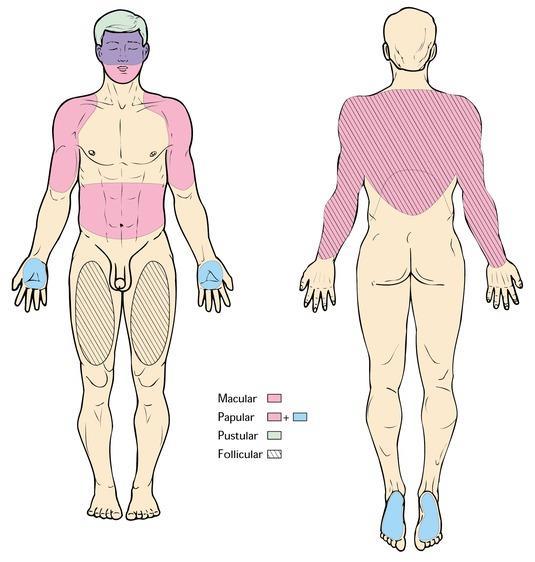
8 Distributions of lesions
9 Macroscopic Lesion Primary lesion basic lesion Secondary lesion lesions develop during procession of disease

10 Classification of skin lesions Primary lesions Secondary lesions
11 Macroscopic Lesion Primary lesion Macule Patch Plaque Papule Nodule Vesicle Bullae Pustule Wheal Cyst Secondary lesion Crust Scale Erosion Ulcer Scar Atrophy

12 Macule Circumscribed color change No elevation or depress Macule : diameter < 0.5 cm Patch : > 0.5 cm
 in the dermis as in Mongolian spots C.")
13 The colors of macules A. Brown : melanin pigmentation in the epidermis B. Blue : melanin or particulates (tattoo) in the dermis as in Mongolian spots C. Red : vasodilatation in the dermis = erythema extravasated red blood cell.= purpura D. Red : inflammatory cells infiltration

14 Macule A macule is a circumscribed color change, flat lesion. Macules may have any size or shape. Some macular lesions are associated with fine scaling Maculosquamous

15 Patch A small well-defined area of the skin distinct in color or appearance. such as large macule, thin and scaling plaque.

16 Papule A small, solid, elevated lesion Flat, pointed or round <0.5 cm in diameter Coalesce into Plaque

17 mucin cell A. The accumulated material may be a metabolic deposit, amyloid or mucin. B. A cellular infiltrate of inflammatory or neoplastic cells C. A proliferation of cells in the epidermis papule tightly packed,the lession is verrucous or warty Dermal tend to have indistinct margins Epidermal very well-demarcated margins

18 Papule Multiple, well-defined papules of varying sizes are seen Flat tops and glistening surface are characteristic of Lichen planus

19 Plaque Circumscribed Solid elevation Usually flat-topped It may be a confluence of papules over a surface

20 Plaque Well-demarcated pink plaques with a silvery scale representing psoriasis vulgaris

21 Nodule Palpable, variably shaped lesion Epidermal, Dermal, SC. Usually elevated,any size T umor is term for any mass

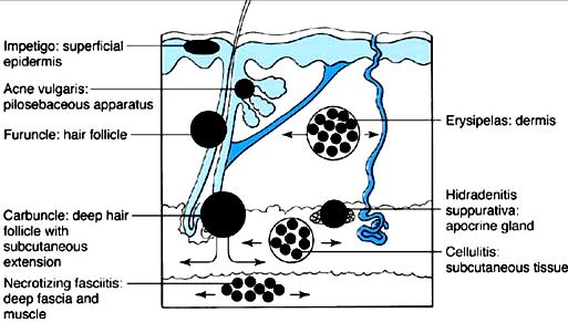
22 Nodule A nodular basal cell carcinoma Well - defined, firm nodule with a smooth and glistening surface through which telangiectasia can be seen


23 Wheals Firm edematous plaque. Infilrating fluid may cause blanching. Usually transient.

24 Vesicles and Bullae Circumscribed fluid filled lesion Vesicle < 0.5 cm or less. Bulla >0.5 cm.


25 Vesicles and Bullae A vesicle is a circumscribed, elevated lesion that contains fluid that they are translucent and the serum, lymph, blood, or extracellular fluid. A vesicle with a diameter greater than 0.5 cm is a bulla.


26 Erosion Focal loss of epidermis. Loss does not penetrate into dermis Heal without scarring Habif: Clinical Dermatology, 5th ed.copyright 2009 Mosby, An Imprint of Elsevier

27 Ulcer Focal loss of epidermis and dermis Loss does penetrate into at least upper dermis Heals with scarring


28 Scar - An abnormal formation of connective tissue implying dermal damage; after injury or surgery - scars are initially thick and pink but with time become white and atrophic Habif: Clinical Dermatology, 5th ed.copyright 2009 Mosby, An Imprint of Elsevier


29 Pustule A vesicle /bulla with pus exudate May/may not be sterile Vary in size & shape
30 Pustules may present different levels: Epidermis : pustule Dermis : abcess Follicle : Folliculitis Deep follicle : Furuncle Several Furuncle : Carbuncle
31

32 Folliculitis An inflamed follicular papule Habif: Clinical Dermatology, 5th ed. Copyright 2009 Mosby, An Imprint of Elsevier

33 Furuncle An inflamed deep follicular-based nodule with a central necrotic plug crusting over the surface

34 Abscess A tender red erythematous fluctuant abscess on the leg dermis

35 Cysts A circumscribed lesion With a wall and a lumen The lumen may contain fluid or solid matter

36 Atrophy Depression of the skin Results from thinning of the epidermis +/- dermis Potent topical steroid can cause

37 Scale Abnormal shedding or accumulation of stratum corneum Habif: Clinical Dermatology, 5th ed. Copyright 2009 Mosby, An Imprint of Elsevier

38 Erythema craquele (crack dense scale) Habif: Clinical Dermatology, 5th ed. Copyright 2009 Mosby, An Imprint of Elsevier

39 Pityriasis rosea (collarette) Habif: Clinical Dermatology, 5th ed. Copyright 2009 Mosby, An Imprint of Elsevier

40 Psoriasis (silvery) Habif: Clinical Dermatology, 5th ed. Copyright 2009 Mosby, An Imprint of Elsevier

41 Tinea versicolor (fine) Habif: Clinical Dermatology, 5th ed. Copyright 2009 Mosby, An Imprint of Elsevier

42 Ichthyosis Habif: Clinical Dermatology, 5th ed. Copyright 2009 Mosby, An Imprint of Elsevier

43 Crust A collection of dried serum and cellular debris a scab
44 Impetigo Crusts yellow - dried serum green - purulent exudate brown or dark red - blood. Acute eczematous dermatitis and impetigo honey-colored, glistening crusts

45 Excoriations Superficial excavations of epidermis result from scratching Linear loss of epidermis and punctate excoriations

46 Fissure A linear loss of epidermis and dermis with sharply defined, nearly vertical walls


47 Fissures linear cleavages or cracks in the skin in palmar/plantar psoriasis in chronic eczematous dermatitis of the hands and feet

48 Poikiloderma Refers to the combination of Atrophy Telangiectasia Pigmentary changes (hyper- and hypo-). Poikilodermatous lesions can be seen in Radiodermatitis Dermatomyositis mycosis fungoides lupus erythematosus
49 Microscopic Lesion
50 Microscopic finding Concise Pathology > Chapter 61. Diseases of the Skin >
51 Microscopic finding layer Concise Pathology > Chapter 61. Diseases of the Skin >
52 Microscopic finding Anaplasia Dedifferentiation Hyperplasia Physiological proli. Neoplasia Abn. proliferation Dysplasia Abn. maturation Metaplasia cell type conversion Dyskeratosis-Abn. keratinization Concise Pathology > Chapter 61. Diseases of the Skin >

53 Epidermal atrophy (Epidermal Hypoplasia_ผ วบาง) Reduction of keratinocytes leads to thinning of the epidermis the papillary processes are diminished or lost It is often found in senile skin, actinic keratosis

54 Acanthosis (Epidermal Hyperplasia_ผ วหนา) Acanthosis is diffuse epidermal hyperplasia implies increased thickness of stratum spinosum It is classified into flat : the entire site thickens moderately chronic eczema psoriasiform: epidermal protrusions/elongated rete ridge psoriasis papillomatous : the epidermis projects upwards e.g warts or seborrheic keratosis pseudocarcinomatous : irregularly downward project e.g chronic ulcer margin, deep mycoses Concise Pathology > Chapter 61. Diseases of the Skin >

55 Acanthosis Atrophy

56 Hyperkeratosis Thickening of the stratum corneum associated with a qualitative abnormality of the keratin Parakeratosis: retained nuclei Orthohyperkeratosis without retained nuclei

57 Parakeratosis Caused by incomplete keratinization nuclei remain in the cells of the horny cell layer such as psoriasis vulgaris Column parakeratosis, cornoid lamellae. porokeratosis

58 Hypergranulosis A thickening of the granular cell layers to > 4 layers (normal 1-3) It is often found in lichen planus warts congenital ichthyosis
 http://www.derm-hokudai.")
59 Granular degeneration In the granular cell layer vacuolated cells containing large keratohyaline granules appear It is characteristic of Vörner palmoplantar keratosis ichthyosiform erythroderma (bullous congenital)

60 Spongiosis Separation of spinous layer because of increased fluid in epidermis secondary to inflammation Excessive spongiosis can lead to intraepidermal vesicles It is found in Atopic dermatitis Acute eczema

61 Intracellular edema Ballooning Degeneration Ballooning degeneration Intracellular swelling The cells become spherical such as herpes simplex

62 Acantholysis Loss of intercellular bridges of keratinocyte (desmosomes) resulting in The dispersion of keratinocytes call Acantholytic cell Form Intercellular spaces and blisters Acantholytic cells have a tendency to become dyskeratotic The phenomenon is found in Pemphigus Hailey-Hailey disease Darier s disease

63 Dyskeratosis Abnormal keratinization occurring prematurely within individual cells below the stratum corneum Dyskeratosis, acantholysis and the intraepidermal cleft formation Darier's disease

64 Blister Intraepidermal and subepidermal seperation resulting in blister formation Blister contents are cytoplasm and infiltrating cells

65 Pemphigus vulgaris. Habif: Clinical Dermatology, 5th ed.copyright 2009 Mosby, An Imprint of Elsevier The epidermal separation occurs low in the epidermis (Suprabasal blister)

66 Bullous pemphigoid. Habif: Clinical Dermatology, 5th ed.copyright 2009 Mosby, An Imprint of Elsevier A subepidermal blister contains numerous eosinophils
67 Exocytosis The infiltration of inflammatory cells and erythrocytes into the dermis It is mostly found in spongiotic space

68 Pautrier s microabscess Infiltration of tumorous lymphocytes Cutaneous Tcell lymphoma (CTCL)

69 Munro s microabcess A blister containing purulent (mainly neutrophils) A small pustule below the horny cell layer
70 Dermal Infiltration There are several infiltration patterns Perivascular infiltration : Inflammatory cells infiltrate around the blood vessels Lichenoid infiltration : the cells infiltrate in a band resembling that in lichen planus Vasculitis : the cells cause fibrinoid degeneration, blood clots, or bleeding in the blood vessels Nodular infiltration
71 Subcutaneous fat tissue Panniculitis is an inflammation of the subcutaneous fat tissue erythema nodosum. erythema induratum
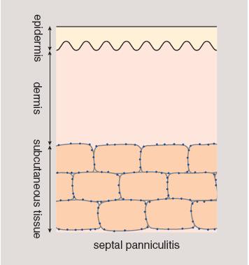
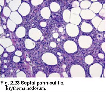
72 Septal Panniculitis
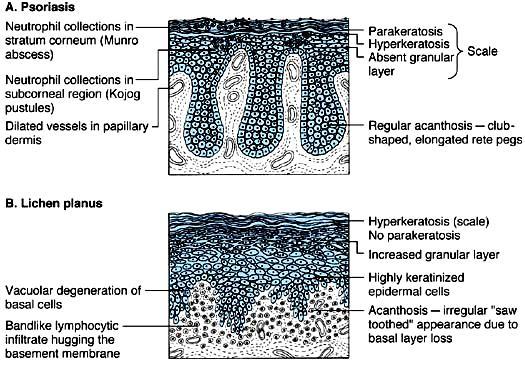
73 Common Skin Diseases
74 Common Skin Diseases Infection diseases of the skin Bacterial Viral Fungal Inflammatory diseases of the skin Atopic dermatitis Psoriasis Lichen planus Bullous Disease Neoplasm of the skin Benign Lesion : Actinic keratosis, Bowen Malignant Lesion : SCC BCC Pigmentary Disoders NEVUS MELANOMA Disorders of Epidermal Maturation ICHTHYOSIS Disorders of Epidermal Appendages ACNE VULGARIS
75 Infection Verrucae (Warts) Molluscum Contagiosum Impetigo Superficial Fungal Infections

76 Verrucae (Warts) Multiple papules with rough pebble-like surfaces Papillomatous epidermal hyperplasia Vacuolar degeneration_perinuclear halo (koilocyte) Enlarge/clump keratohyaline granules In situ hybridization demonstrating viral DNA within epidermal cells Kumar: Robbins and Cotran Pathologic Basis of Disease, Professional Edition, 8th ed. Copyright 2009 Saunders, An Imprint of Elsevier


77 Molluscum contagiosum Kumar: Robbins and Cotran Pathologic Basis of Disease, Professional Edition, 8th ed. Copyright 2009 Saunders, An Imprint of Elsevier A rounded, pink to flesh skin color A central umbilication Molluscum Body Verrucous epidermal hyperplasia KC eosinophilic intracytoplasmic inclusions (molluscum bodies) Viral particle contain
78 Impetigo contagiosum Honey-colored, glistening crusts


79 Impetigo Bullosa Source: Weedon: Skin Pathology 2nd edition Intraepidermal Vesicle Subcorneal pustule with Neutrophil infiltration Special stains : bacteria foci
80 Superficial Fungal Infections Tinea Dermatophyte 3 organism Trichophyton Epidermpphyton Microsporum Various forms : location Scalp :Tinea Capitis Face Body Inguinal Hand Foot Nail :Tinea Faciei :Tinea Corporis :TineaCruis :Tinea Manuum :Tinea Pedis :Tinea Ungium Tinea versicolor (Pityriasis versicolor) Malassezia furfur, a yeast Condition : Humidity area Seborrheic area Location : upper chest Upper back Lesion : hypo-hyperpigment macule With fine scale
 : hyphae within the S.corneum. Kumar: Robbins and Cotran Pathologic Basis of Disease, Professional Edition, 8th ed.")
81 Tinea Corporis Tinea A : well circumscribed erythematous macule, papule to plaque with active border and central regression B : mild spongiosis and focal neutrophilic abscesses with fungal hyphae C : Periodic Acid Schiff stain (PAS) : hyphae within the S.corneum. Kumar: Robbins and Cotran Pathologic Basis of Disease, Professional Edition, 8th ed. Copyright 2009 Saunders, An Imprint of Elsevier

82 Psoriasis Psoriasis Kumar: Robbins and Cotran Pathologic Basis of Disease, Professional Edition, 8th ed. Copyright 2009 Saunders, An Imprint of Elsevier A: erythema plaque with silvery-white scale B: Histologically : hyperkeratosis with parakeratosis regular acanthosis with clubbing an absent granular layer neutrophils infiltrationin the stratum corneum (Munro microabscesses) subcorneal layer (Kojog spongiform pustules)
 * Mostly idiopathic but the")
83 Lichen planus A : This flat-topped pink-purple, polygonal papule a white lacelike pattern that is referred to as Wickham stria. B : A bandlike infiltrate of lymphocytes at the dermoepidermal junction hyperkeratosis hypergranulosis pointed rete ridges (saw toothing) * Mostly idiopathic but the possibility of a cell mediated immunologic mechanism
84
 C : Subepidermal blister the entire epidermis separates from the dermis (as in bullous")
85 Histologic levels of blister formation A : Subcorneal blister the stratum corneum forms the roof of the bulla (as in pemphigus foliaceus) B : Suprabasal blister a portion of the epidermis, including the stratum corneum, forms the roof (as in pemphigus vulgaris) C : Subepidermal blister the entire epidermis separates from the dermis (as in bullous pemphigoid)

86 Pemphigus foliaceus. A The delicate, superficial (subcorneal) blisters are much less erosive than seen in pemphigus vulgaris. B Subcorneal separation of the epithelium is seen. Kumar: Robbins and Cotran Pathologic Basis of Disease, Professional Edition, 8th ed. Copyright 2009 Saunders, An Imprint of Elsevier

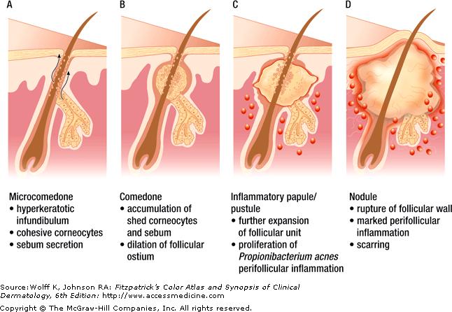
87 Pemphigus Pemphigus vulgaris A : flaccid bullae and thin-roofed =>Eroded plaques B : Suprabasal acantholysis => intraepidermal blister C : Ulcerated blisters in the oral mucosa are also common Kumar: Robbins and Cotran Pathologic Basis of Disease, Professional Edition, 8th ed. Copyright 2009 Saunders, An Imprint of Elsevier

88 Direct immunofluorescence of IgG Pemphigus vulgaris the intercellular membranes in a reticular pattern Pemphigus foliaceus the deposits are more superficial

89 Bullous Pemphigoid Bullous pemphigoid. A : Tense bullae, filled with clear fluid, on normal or erythematous skin B : Histopathology shows Basal cell layer vacuolization, producing tense, intact subepidermal blisters With eosinophils, as well as lymphocytes or neutrophils

90 Linear deposition of complement along the dermoepidermal junction in bullous pemphigoid; the ribbon candy pattern
91 Neoplasm of the skin

92 Seborrheic Keratosis Common benign tumor usually in elderly persons This lesion occurring on the face, trunk and extremities The lesions are flat, raised, soft, sharply demarcated, and brown

93 Seborrheic keratosis A well-demarcated coin like pigmented lesion with warty surface stuck on appearance Histologically of benign basaloid cells proliferation interspersed with keratin filled horny cysts

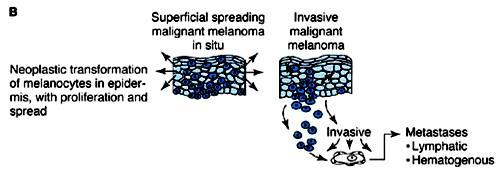
94 Actinic Keratosis Hypertrophic Actinic keratosis A : A cutaneous horn horn like projection keratin B : Basal cell layer atypia (dysplasia) is associated with marked hyperkeratosis and parakeratosis C : Progression to full-thickness nuclear atypia, with or without the presence of superficial epidermal maturation, heralds the development of squamous cell carcinoma in situ : Actinic keratosis is a premalignant lesion

95 Squamous Cell Carcinoma Invasive squamous cell carcinoma A: Lesions are often nodular and ulcerated as seen in this scalp tumor. B: Atypical squamous epithelium invading BM into the dermis C: A magnified image : invasive tumor cells showing enlarged nuclei with angulated contours and prominent nucleoli.

96 Basal Cell Carcinoma Basal cell carcinoma A : Pearly, telangiectatic nodules B : Nests of uniformly atypical basaloid cells C : Often separated from the adjacent stroma by clefts
97 Disorders of Epidermal Maturation Ichthyosis

98 Ichthyosis prominent fishlike scales compacted, thickened stratum corneum - Group of genetically inherited disorders - Ichthyosis exists in several forms with different inheritance patterns (dominant, recessive, X-linked)


99 Acne Divided into 2 types Non inflammatory types Open comedones : black keratin plug Closed comedones the keratin plug is trapped beneath the epidermal surface Inflammatory types erythematous papules, nodules, and pustules Severe : acne conglobata => sinus tract formation & physical scarring
100
101 Pigmentary Disorder NEVUS MELANOMA
102
103 Melanoma

104 Melanocytic nevus : junctional type A small, relatively flat, symmetric, and uniform. B On histologic examination, junctional nevi are characterized by rounded nests of nevus cells originating at the tips of rete ridges along DEJ

105 Melanocytic nevus, compound type (A) the compound nevus is more raised and dome-shaped. The symmetry and uniform pigment distribution suggest a benign process. (B) Histologically : combine the features of junctional nevi (intraepidermal nevus cell nests) with nests cords of nevus cells in the underlying dermis

106 A : Typically lesions : irregular in contour and pigmentation Macule area : radial growth phase Raise area : verticle growth phase B : Radial growth phase irregular nested and melanoma cells within the epidermis an inflammatory response within the dermis C : Vertical growth phase nodular aggregates of infiltrating cells
107 Reference 1. Kumar: Robbins and Cotran Pathologic Basis of Disease, Professional Edition, 8th ed. 2. Fitzpatrick's Dermatology in General Medicine, 7e Klaus Wolff, Lowell A. Goldsmith, Stephen I. Katz, Barbara A. Gilchrest, Amy S. Paller, David J. Leffell 3. Concise Pathology, 3th ed. Chapter 61, Diseases of the Skin, Copyright The McGraw-Hill Companies. All rights reserved. 4. Weedon: Skin Pathology 2nd edition 5. Histology and Cell Biology: An Introduction to Pathology (2002). Kierszenbaum.Mosby. 6. Wolff K, Johnson RA, Section 1. Disorders of Sebaceous and Apocrine Glands (Chapter). Wolff K, Johnson RA: Fitzpatrick s Color Atlas & Synopsis of Clinical Dermatology, 6e:
108 Thank you for Attention
Benign and malignant epithelial lesions: Seborrheic keratosis: A common benign pigmented epidermal tumor occur in middle-aged or older persons more
 Benign and malignant epithelial lesions: Seborrheic keratosis: A common benign pigmented epidermal tumor occur in middle-aged or older persons more common on the trunk; but extremities, head and neck are
SKIN PATHOLOGY. Topics. Manifestration of skin disorders. Dermatologic history: Skin lesion Location of lesions Configurations Distribution of lesions
 SKIN PATHOLOGY SOMPHONG NARKPINIT, M.D. PATHOBIOLOGY DEPARTMENT FACULTY OF SCIENCE Dermatologic history: details of onset evolution of symptoms previous diagnosis and treatment PMH of skin disease PMH
Pathology of the skin. 2nd Department of Pathology, Semmelweis University
 Pathology of the skin 2nd Department of Pathology, Semmelweis University Histology of the skin Epidermis: Stratum corneum Stratum granulosum Stratum spinosum Stratum basale Dermis: papillary and reticular
Histopathology: skin pathology
 Histopathology: skin pathology These presentations are to help you identify, and to test yourself on identifying, basic histopathological features. They do not contain the additional factual information
Actinic keratosis (AK): Dr Sarma s simple guide
: Dr Sarma s simple guide") Actinic keratosis (AK): Dr Sarma s simple guide Actinic keratosis is a very common lesion that you will see in your day-to-day practice. First, let me explain the name Actinic keratosis. It means keratosis
Actinic keratosis (AK): Dr Sarma s simple guide Actinic keratosis is a very common lesion that you will see in your day-to-day practice. First, let me explain the name Actinic keratosis. It means keratosis
Conflicts. Objectives. University of Texas Health Science Center at San Antonio. Pediatrics Grand Rounds 24 August Pediatric Dermatology 101
 Pediatric Dermatology 101 John C. Browning, MD, FAAD, FAAP Conflicts Investigator: ViroXis Advisor: ViroXis Advisory Board: TopMD Speaker: Galderma Objectives Understand the meaning and importance of cutaneous
Pathology of the skin. Dr Fónyad László, 1sz. Patológiai és Kísérleti Rákkutató Intézet, SE
 Pathology of the skin Dr Fónyad László, 1sz. Patológiai és Kísérleti Rákkutató Intézet, SE The skin Biggest organ Kb. 1.8 nm Kb. 10 kg Most frequent site for tumor development (BCC) Pathology of the skin
Dermatopathology: The tumor is composed of keratinocytes which show atypia, increase mitoses and abnormal mitoses.
 Squamous cell carcinoma (SCC): A common malignant tumor of keratinocytes arising in the epidermis, usually from a precancerous condition: 1- UV induced actinic keratosis, usually of low grade malignancy.
Benign versus Cancerous Lesions How to tell the difference FMF 2014 Christie Freeman MD, CCFP, DipPDerm, MSc
 1 Benign versus Cancerous Lesions How to tell the difference FMF 2014 Christie Freeman MD, CCFP, DipPDerm, MSc Benign lesions Seborrheic Keratoses: Warty, stuck-on Genetics and birthdays Can start in late
This section covers the basic knowledge of normal skin structure and function required to help understand how skin diseases occur.
 Background Knowledge Functions of normal skin Background Knowledge This section covers the basic knowledge of normal skin structure and function required to help understand how skin diseases occur. Learning
Some skin conditions
 Some skin conditions Some skin conditions Acute Inflammatory Dermatoses Chronic Inflammatory Dermatoses Blistering (Bullous) Diseases Panniculitis Disorders of Epidermal Appendages -Urticaria -Acute eczematous
Pimples and Boils!! Dr Nathan Harvey Anatomical Pathology, PathWest
 Pimples and Boils!! Dr Nathan Harvey Anatomical Pathology, PathWest Overview & Learning Objectives Review the cardinal signs/symptoms of acute inflammation Review the histological features of acute inflammation
4. Pityriasis lichenoides
 Go Back to the Top To Order, Visit the Purchasing Page for Details usually more than 5 cm in diameter and accompanied by poikiloderma. Some but not all patients may develop mycosis fungoides (Fig. 22.35).
المركب النموذج--- سبيتز وحمة = Type Spitz's Nevus, Compound SPITZ NEVUS 1 / 7
 SPITZ NEVUS 1 / 7 Epidemiology An annual incidence rate of 1.4 cases of Spitz nevus per 100,000 individuals has been estimated in Australia, compared with 25.4 per 100,000 individuals for cutaneous melanoma
Dermoscopy: Recognizing Top Five Common In- Office Diagnoses
 Dermoscopy: Recognizing Top Five Common In- Office Diagnoses Vu A. Ngo, DO Department of Family Medicine and Dermatology Choctaw Nation Health Services Authority Learning Objectives Introduction to dermoscopy
COPYRIGHTED MATERIAL. Introduction CHAPTER 1. Introduction
 CHAPTER 1 Introduction OVERVIEW The clinical features of skin lesions are related to the underlying pathological processes. Broadly skin conditions fall into three clinical groups: (a) those with a well-defined
Gross Appearance & Histology of Skin Cancer. Kyle Mannion M.D. January 21, 2005
 Gross Appearance & Histology of Skin Cancer Kyle Mannion M.D. January 21, 2005 Actinic Keratosis 5-20% will develop squamous/basal cell ca Almost solely from solar damage Usually develop during 4 th decade
Rash Decisions Approach to the patient with a skin condition
 National Conference for Nurse Practitioners April 25, 2014 Rash Decisions Approach to the patient with a skin condition Margaret A. Bobonich, DNP, FNP C, DCNP, FAANP Assistant Professor, Case Western Reserve
MECHANISMS OF HUMAN DISEASE: LABORATORY SESSION PATHOLOGY OF THE SKIN LAB. Friday, February 12, :30 am 11:00 am
 MECHANISMS OF HUMAN DISEASE: LABORATORY SESSION PATHOLOGY OF THE SKIN LAB Friday, February 12, 2012 9:30 am 11:00 am FACULTY COPY GOALS: Describe the basic clinical and morphologic features of various
Inflammatory skin disease I Jade Wititsuwannakul, MD Chulalongkorn University, Thailand
 Inflammatory skin disease I Jade Wititsuwannakul, MD Chulalongkorn University, Thailand Superficial Perivascular Dermatitis Interface Dermatitis Vacuolar Dermatitis Lichenoid Dermatitis Barnhill Textbook
MECHANISMS OF HUMAN DISEASE: LABORATORY SESSION PATHOLOGY OF THE SKIN LAB. Friday, February 13, :30 am 11:00 am
 MECHANISMS OF HUMAN DISEASE: LABORATORY SESSION PATHOLOGY OF THE SKIN LAB Friday, February 13, 2009 9:30 am 11:00 am FACULTY COPY GOALS: Describe the basic clinical and morphologic features of various
Basal cell carcinoma 5/28/2011
 Goal of this Presentation A practical approach to the diagnosis of cutaneous carcinomas and their mimics Thaddeus Mully, MD University of California San Francisco To review common non-melanoma skin cancers
Mucinoses Diverse group of disorders which have in common deposition of basophilic, finely granular and stringy material in the connective tissues of
 Cutaneous Mucinoses Nathan C. Walk, M.D. Mucinoses Diverse group of disorders which have in common deposition of basophilic, finely granular and stringy material in the connective tissues of the dermis.
ISPUB.COM. Seborrheic Keratosis: A Pictorial Review of the Histopathologic Variations. D Sarma, S Repertinger
 ISPUB.COM The Internet Journal of Dermatology Volume 7 Number 2 Seborrheic Keratosis: A Pictorial Review of the Histopathologic Variations D Sarma, S Repertinger Citation D Sarma, S Repertinger.. The Internet
04/09/2018. Squamous Cell Neoplasia and Precursor Lesions. Agenda. Squamous Dysplasia. Squamo-proliferative lesions. Architectural features
 Squamous Cell Neoplasia and Precursor Lesions Jennifer L. Hunt, MD, MEd Aubrey J. Hough Jr, MD, Endowed Professor of Pathology Chair of Pathology and Laboratory Medicine University of Arkansas for Medical
Medical History. Oral Medicine and General Medicine
 Medical History Oral Medicine and General Medicine Gingivitis herpetica acuta NECROTIZÁLÓ SIALOMETAPLASIA SOOR Medical History The life expectancy has recently increased and increasing By dental prevention
Skin and Body Membranes Body Membranes Function of body membranes Cover body surfaces Line body cavities Form protective sheets around organs
 Skin and Body Membranes Body Membranes Function of body membranes Cover body surfaces Line body cavities Form protective sheets around organs Classification of Body Membranes Epithelial membranes Cutaneous
THE INTEGUMENTARY SYSTEM. Body Membranes & Skin
 THE INTEGUMENTARY SYSTEM Body Membranes & Skin TYPES OF MEMBRANES Epithelial Membranes includes layer of epithelial cells and connective tissue Serous Cutaneous Mucous Connective Tissue Membranes solely
Integumentary System (Skin) Unit 6.3 (6 th Edition) Chapter 7.3 (7 th Edition)
 Unit 6.3 (6 th Edition) Chapter 7.3 (7 th Edition)") Integumentary System (Skin) Unit 6.3 (6 th Edition) Chapter 7.3 (7 th Edition) 1 Learning Objectives Identify the major components (anatomy) of skin Differentiate between the two types of skin glands Explain
Integumentary System (Skin) Unit 6.3 (6 th Edition) Chapter 7.3 (7 th Edition) 1 Learning Objectives Identify the major components (anatomy) of skin Differentiate between the two types of skin glands Explain
Basics in Dermoscopy
 Basics in Dermoscopy Manal Bosseila Professor of Dermatology, Cairo University Member of European Academy Dermatology & Venereology EADV Member of International Dermoscopy Society IDS Member of Aesthetic
General Dermatology Objectives Learn to recognize some common dermatologic disorders d and some associated with systemic diseases Learn the causative
 General Dermatology Julia R. Nunley, MD, FAAD, FACP Professor Program Director Department of Dermatology General Dermatology Objectives Learn to recognize some common dermatologic disorders d and some
Chapter 6 Squamous Cell Carcinoma: Variants and Challenges
 Chapter 6 Squamous Cell Carcinoma: Variants and Challenges Michael B. Morgan EPIDEMIOLOGY: Second most common skin cancer, rare in the dark-skinned races. ETIOLOGY: Ultraviolet light, HPV infection. PATHOGENESIS:
Squamous Cell Neoplasia and Precursor Lesions
 Squamous Cell Neoplasia and Precursor Lesions Jennifer L. Hunt, MD, MEd Aubrey J. Hough Jr, MD, Endowed Professor of Pathology Chair of Pathology and Laboratory Medicine University of Arkansas for Medical
Unit 4 - The Skin and Body Membranes 1
 Unit 4 - The Skin and Body Membranes 1 I. Unit 4: Skin and Body Membranes A. Body Membranes 1. Function of body membranes a) Cover body surfaces b) Line body cavities c) Form protective sheets around organs
Lid Lesions: Relax or Refer
 Lid Lesions: Relax or Refer Blair Lonsberry, MS, OD, MEd., FAAO Professor of Optometry Pacific University College of Optometry blonsberry@pacificu.edu Agenda Benign vs. Malignant lesions Benign Eyelid
PowerPoint Lecture Slide Presentation by Patty Bostwick-Taylor, Florence-Darlington Technical College Skin and Body Membranes
 PowerPoint Lecture Slide Presentation by Patty Bostwick-Taylor, Florence-Darlington Technical College Skin and Body Membranes 4 Body Membranes Function of body membranes Cover body surfaces Line body cavities
Ch. 4: Skin and Body Membranes
 Ch. 4: Skin and Body Membranes I. Body Membranes A. Function of body membranes 1. Cover body surfaces 2. Line body cavities 3. Form protective sheets around organs II. Classification of Body Membranes
Chapter 8 Skin Disorders and Diseases
 Chapter 8 Skin Disorders and Diseases Attitude is more important than the past, than education, than money, than circumstances, than what people do or say. It is more important than appearance, giftedness,
Skin lesions The Good and the Bad. Dr Virginia Hubbard Ipswich Hospital NHS Trust Barts and the London School of Medicine and Dentistry
 Skin lesions The Good and the Bad Dr Virginia Hubbard Ipswich Hospital NHS Trust Barts and the London School of Medicine and Dentistry Case 1 32 year old woman Australian Lesion on back New hair growing
Appendix : Dermoscopy
 Go Back to the Top To Order, Visit the Purchasing Page for Details APP Appendix : Dermoscopy Dermoscopy, also known as dermatoscopy, epiluminoscopy and epiluminescent microscopy, is an effective non-invasive
A. Erythema multiforme and related diseases
 Go Back to the Top To Order, Visit the Purchasing Page for Details Chapter Erythema, Erythroderma (Exfoliative Dermatitis) Erythema is caused by telangiectasia or hyperemia in the papillary and reticular
CH 05 THE INTEGUMENTARY SYSTEM
 CH 05 THE INTEGUMENTARY SYSTEM This system consists of skin and its derivatives. The skin is one of the largest organs of the body in terms of surface area. The functions of the integumentary system include:
DERMATOLOGY SKIN DISEASE: APPROACH TO DIAGNOSIS
 DERMATOLOGY SKIN DISEASE: APPROACH TO DIAGNOSIS History Clinical Examination List and Prioritise Differentials Diagnostic Testing/Trials (eg Treatment Trial) Correlate All Findings History Signalment age,
Spongiotic Dermatitis
 Prepared by Kurt Schaberg Introduction to Inflammatory Dermpath Spongiotic Dermatitis intraepidermal intercellular edema (spongiosis) - presence of widened intercellular spaces between keratinocytes, with
DESCRIPTIONS FOR MED 3 ROTATIONS Dermatology A3S
 Regardless of your future field of practice, you will be exposed to a considerable amount of dermatology and this rotation provides you the chance to see a range of skin diseases. You will have the opportunity
Hole s Human Anatomy and Physiology. Eleventh Edition. Chapter 6
 Hole s Human Anatomy and Physiology Eleventh Edition Shier Butler Lewis Chapter 6 Copyright The McGraw-Hill Companies, Inc. Permission required for reproduction or display. 1 Referred to as Cutaneous Membrane
Premalignant lesions may expose to a promoting. factor & may be induced to undergo malignant. Carcinoma in situ displays the cytologic features of
 بسم رلاهللا Def. Premalignant lesions may expose to a promoting factor & may be induced to undergo malignant transformation. Carcinoma in situ displays the cytologic features of malignancy without invasion
Introduction. Skin and Body Membranes. Cutaneous Membranes Skin 9/14/2017. Classification of Body Membranes. Classification of Body Membranes
 Introduction Skin and Body Membranes Body membranes Cover surfaces Line body cavities Form protective and lubricating sheets around organs Classified in 5 categories Epithelial membranes 3 types- cutaneous,
My Algorithm. Questions to ask. Do you or your family have a history of?... Allergic rhinitis, Sensitive skin, Asthma Skin Cancer
 Tracey C. Vlahovic, DPM Associate Professor, Temple University School of Podiatric Medicine My Algorithm Inflammatory Skin Disorder on Feet Family hx, clinical exam, look at hands! Defined plaques: Psoriasis
NEOPLASMS OF THE SURFACE EPITHELIUM (KERATINOCYTES)
") NEOPLASMS OF THE SURFACE EPITHELIUM (KERATINOCYTES) Papillary Lesions Precancerous Lesions Keratinocyte Proliferations Carcinomas Melanotic Lesions Melanomas Normal Mucosa Keratin layer Spinous layer Basal
NEOPLASMS OF THE SURFACE EPITHELIUM (KERATINOCYTES) Papillary Lesions Precancerous Lesions Keratinocyte Proliferations Carcinomas Melanotic Lesions Melanomas Normal Mucosa Keratin layer Spinous layer Basal
Disclosure. Objectives. PAFP CME Conference Lou Mancano MD, FAAFP Reading Health System November 18, 2016
 PAFP CME Conference Lou Mancano MD, FAAFP Reading Health System November 18, 2016 1 Disclosure The speaker has no conflict of interest, financial agreement, or working affiliation with any group or organization.
B. Autoimmune blistering diseases
 Go Back to the Top To Order, Visit the Purchasing Page for Details formation immediately above the basal layer. The dermal papillae, which are covered by basal cells in the single layer that is left in
Integumentary System. Packet #12
 Integumentary System Packet #12 Introduction Skin/Integument Skin, considered an organ, is the major component of the integumentary system. The integumentary system is also composed of other accessory
What are the functions of the integumentary system? What are some disorders of the integumentary system?
 Essential Questions: What are the functions of the integumentary system? What are some disorders of the integumentary system? How are integumentary system disorders treated? How do you relate the integumentary
Table of Contents: Part 1 Medical Dermatology. Chapter 1 Acneiform Disorders. Acne. Acne Vulgaris. Pomade Acne. Steroid Acne
 Table of Contents: Part 1 Medical Dermatology Chapter 1 Acneiform Disorders Acne Acne Vulgaris Pomade Acne Steroid Acne Infantile Acne Pediatric Perspectives Neonatal Acne (Acne Neonatorum) Pediatric Perspectives
Lumps and Bumps: The Dermatology of Lid Lesions
 Lumps and Bumps: The Dermatology of Lid Lesions Thomas J. Joly, MD, PhD Assistant Professor of Ophthalmology Eastern Virginia Medical School Ophthalmic Plastic Surgery Service Virginia Eye Consultants
Chapter 6 Skin and the Integumentary System. Skin Cells. Layers of Skin. Epidermis Dermis Subcutaneous layer beneath dermis not part of skin
 Chapter 6 Skin and the Integumentary System Composed of several tissues Maintains homeostasis Protective covering Retards water loss Regulates body temperature Houses sensory receptors Contains immune
Supplementary Online Content
 Supplementary Online Content Ross NA, Chung H-J, Li Q, Andrews JP, Keller MS, Uitto J. Pityriasis rubra pilaris: a case series of patients. Published online March 9, 26. JAMA Dermatol. doi:./jamadermatol.26.9.
An Approach to Common and not so Common Rashes in the Office FMF 2014 Christie Freeman MD, CCFP, DipPDerm, MSc
 An Approach to Common and not so Common Rashes in the Office FMF 2014 Christie Freeman MD, CCFP, DipPDerm, MSc 1 Common Rashes Tinea Corporis: Annular- this is not the only criteria Advancing erythematous
WR SKIN. DERMATOLOGY
 WR SKIN. DERMATOLOGY 1 Societies 11 History 13 Dictionaries. Encyclopaedias. Bibliographies Use for general works only. Classify with specific aspect 15 Classification. Nomenclature 16 Tables. Statistics
Skin lesions & Abrasions
 Skin lesions & Abrasions What Are Skin Lesions? A skin lesion is a part of the skin that has an abnormal growth or appearance compared to the skin around it Types of Skin Lesions Two types of skin lesions
Principles of Anatomy and Physiology
 Principles of Anatomy and Physiology 14 th Edition CHAPTER 5 The Integumentary System Introduction The organs of the integumentary system include the skin and its accessory structures including hair, nails,
Diseases of the breast (1 of 2)
") Diseases of the breast (1 of 2) Introduction A histology introduction Normal ducts and lobules of the breast are lined by two layers of cells a layer of luminal cells overlying a second layer of myoepithelial
Diseases of the breast (1 of 2) Introduction A histology introduction Normal ducts and lobules of the breast are lined by two layers of cells a layer of luminal cells overlying a second layer of myoepithelial
Skin and Body Membranes
 4 Skin and Body Membranes PowerPoint Lecture Slide Presentation by Jerry L. Cook, Sam Houston University ESSENTIALS OF HUMAN ANATOMY & PHYSIOLOGY EIGHTH EDITION ELAINE N. MARIEB Skin and Body Membranes
The Integumentary System
 The Integumentary System The Integumentary System Integument is skin Skin and its appendages make up the integumentary system A fatty layer (hypodermis) lies deep to it Two distinct regions Epidermis Dermis
Cornell Notes Name: Date: Topic: CH 4
 *We are revisiting Ch 3B on body tissues (Connective) prior to our study of Ch 4 Integumentary. Start on p.90 I. Connective Tissue A. Functions of Connective 1. Protection 2. Support 3. Binding Together
The Integumentary System
 The Integumentary System The Integumentary System Integument is skin Skin and its appendages make up the integumentary system (See if you can name some appendages) A fatty layer (hypodermis) lies deep
Classification: 1. Infective: 2. Traumatic: 3. Idiopathic: Recurrent Aphthous Stomatitis (RAS) 4. Associated with systemic disease:
 4. Associated with systemic disease:") Classification: 1. Infective: 2. Traumatic: 3. Idiopathic: Recurrent Aphthous Stomatitis (RAS) 4. Associated with systemic disease: Hematological GIT Behcet s HIV 5. Associated with dermatological diseases:
Classification: 1. Infective: 2. Traumatic: 3. Idiopathic: Recurrent Aphthous Stomatitis (RAS) 4. Associated with systemic disease: Hematological GIT Behcet s HIV 5. Associated with dermatological diseases:
22/04/2015. Dermoscopy of Melanoma. Ilsphi Browne. Overview
 Dermoscopy of Melanoma Ilsphi Browne Overview The device Dermoscopic criteria (terminology) Colour Patterns Global features Local features Approach to diagnosing pigmented lesions Other uses in general
LESSON ASSIGNMENT. Primary and Secondary Skin Lesions. After completing this lesson, you should be able to:
 LESSON ASSIGNMENT LESSON 3 Primary and Secondary Skin Lesions. LESSON ASSIGNMENT Paragraphs 3-1 through 3-5. LESSON OBJECTIVES After completing this lesson, you should be able to: 3-1. Identify different
Ch 5: Integumentary System
 Ch 5: Integumentary System You gotta have skin; All you really need is skin. Skin's the thing, that if you've got it outside, It helps keep your insides in. Alan Sherman (1924-1973) Developed by John Gallagher,
1 Assessment Techniques General Survey Skin, Hair, and Nails. 2 Cultivating Your Senses
 1 Assessment Techniques General Survey Skin, Hair, and Nails 2 Cultivating Your Senses Inspection Always performed first Palpation Purpose Use different parts of the hands Light vs. deep palpation 3 Cultivating
Diagnosis and Management of Common and Infective Skin Diseases in Children at primary care level
 Diagnosis and Management of Common and Infective Skin Diseases in Children at primary care level Dr Ng Su Yuen Paediatrician and Paediatric Dermatologist Hospital Pulau Pinang Outline Common inflammatory
Integumentary System
 Integumentary System Overview Functions 1. Protection 2. Excretion of wastes 3. Maintenance of T b 4. Synthesis of vitamin D 3 5. Storage of lipids 6. Detection of sensory stimuli Epidermis Tissue types
Chapter 6: Integumentary System
 Chapter 6: Integumentary System 6.1 Introduction Why is skin considered to be an organ? What makes up the integumentary system? Integumentary System Skin (cutaneous membrane) Skin derivatives Sweat glands
Observations on the Pathology of Lesions Associated with Stephanofilaria dinniki Round, 1964 from the Black Rhinoceros (Diceros bicornis)
") Journal of Helminthology, ~ol. XXXVIII, Nos. 1/2, 1964, pp. 171-174. Observations on the Pathology of Lesions Associated with Stephanofilaria dinniki Round, 1964 from the Black Rhinoceros (Diceros bicornis)
Journal of Helminthology, ~ol. XXXVIII, Nos. 1/2, 1964, pp. 171-174. Observations on the Pathology of Lesions Associated with Stephanofilaria dinniki Round, 1964 from the Black Rhinoceros (Diceros bicornis)
Doctors of Optometry Course Notes
 Doctors of Optometry Course Notes OD19 1CE COPE: 43871-AS Eyelid Lumps and Bumps Sunday, February 26, 2017 2:40 pm 3:30 pm Regency C 3 rd Floor Presenter: Blair Lonsberry, OD, FAAO Dr. Lonsberry is a Full
CHAPTER 7:3 INTEGUMENTARY SYSTEM
 CHAPTER 7:3 INTEGUMENTARY SYSTEM I. OBJECTIVES A. Label a diagram of a cross section of the skin B. Differentiate between the two types of skin glands C. Identify six functions of the skin D. Provide the
Contents. QAaptm-2. CAaptei-3. CAaptm-4. Cftapte%-5. Qfiaptvt-6. QhapteK-7. Qkaptefc-8 Clinical Immunology and Allergy 71
 Contents Ckaptm-1 Aaatomy, Physiology, Embryology, Bacteriology and Pathology ~ 1 Anatomy 1 Physiology 10 Embryology 14 Pathology 19 Bacteriology 22 Laboratory and other aids in dermatological pratice
EXPERIMENTAL THERMAL BURNS I. A study of the immediate and delayed histopathological changes of the skin.
 EXPERIMENTAL THERMAL BURNS I A study of the immediate and delayed histopathological changes of the skin. RJ Brennan, M.D. and B. Rovatti M.D. The purpose of this study was to determine the progressive
Histopathology of Melanoma
 THE YALE JOURNAL OF BIOLOGY AND MEDICINE 48, 409-416 (1975) Histopathology of Melanoma G. J. WALKER SMITH Department ofpathology, Yale University School ofmedicine, 333 Cedar Street, New Haven, Connecticut
Pathology. Skin Tumor. Bayan N. Mohammad 15/10/2015. Mohammad al-orjani. Page 0 of 23
 #7 35 Pathology Skin Tumor Bayan N. Mohammad 15/10/2015 Mohammad al-orjani Page 0 of 23 بسم هللا الرحمن الرحيم GREETINGS This lecture is about skin tumors, all the slides are included and every slide will
The Integumentary System. Disorders, Conditions, and Diseases
 The Integumentary System Disorders, Conditions, and Diseases Definitions Disease- an abnormal condition of the body or the mind that causes dysfunction or discomfort. Disorder- a functional abnormality,
PowerPoint Lecture Slide Presentation by Patty Bostwick-Taylor, Florence-Darlington Technical College Skin and Body Membranes
 PowerPoint Lecture Slide Presentation by Patty Bostwick-Taylor, Florence-Darlington Technical College Skin and Body Membranes 4 Body Membranes Function of body membranes Cover body surfaces Line body cavities
Learning Objectives. Tanning. The Skin. Classic Features. Sun Reactive Skin Type Classification. Skin Cancers: Preventing, Screening and Treating
 Learning Objectives Skin Cancers: Preventing, Screening and Treating Robert A. Baldor, MD, FAAFP Professor, Family Medicine & Community Health University of Massachusetts Medical School Distinguish the
Chapter 5 The Integumentary System. Copyright 2009, John Wiley & Sons, Inc. 1
 Chapter 5 The Integumentary System Copyright 2009, John Wiley & Sons, Inc. 1 Introduction The organs of the integumentary system include the skin and its accessory structures including hair, nails, and
Non-melanocytic Patterns
 Non-melanocytic Lesions Non-melanocytic Patterns Michelle Tarbox, MD Assistant Professor of Dermatology and Dermatopathology Texas Tech University Health Sciences Center 2018 Seborrheic keratoses Acanthotic
LUMPS AND BUMPS: AN ORGANIZED APPROACH TO DIAGNOSIS AND MANAGEMENT
 LUMPS AND BUMPS: AN ORGANIZED APPROACH TO DIAGNOSIS AND MANAGEMENT Tammy P. Than, M.S., O.D., F.A.A.O. The University of Alabama at Birmingham / School of Optometry 1716 University Blvd. Birmingham, AL
Evaluation of Epidermal Reaction Pattern and Assessment of Histopathological Findings of Various Skin Disorders
 ORIGINAL RESEARCH www.ijcmr.com Evaluation of Epidermal Reaction Pattern and Assessment of Histopathological Findings of Various Skin Disorders Shweta Sharma 1, Dhara P Trivedi 2, Ronak Vyas 3 ABSTRACT
2/5/2019. Organ System: Skin or Integumentary System. Hypodermis (or superficial fascia) Integumentary System - Learn and Understand
 Integumentary System - Learn and Understand") Integumentary System - Learn and Understand Skin is an organ comprised of all four tissues Each layer of the skin contributes to one or more of its numerous functions Skin is both strong and flexible Keratinization
Integumentary System - Learn and Understand Skin is an organ comprised of all four tissues Each layer of the skin contributes to one or more of its numerous functions Skin is both strong and flexible Keratinization
Integumentary System
 Integumentary System Physiology of Touch Skin: our most sensitive organ Touch: first sense to develop in embryos Most important but most neglected sense How many sensory receptors do we have? (We have
IN THE NAME OF GOD. Dr.kheirandish DDS,MSC Oral and maxillofacial pathology
 IN THE NAME OF GOD Dr.kheirandish DDS,MSC Oral and maxillofacial pathology Dermatologic Diseases Chapter 16 ECTODERMAL DYSPLASIA o Two or more ectodermally derived anatomic structures fail to develop o
1. Introduction (Open your text to the image of a cross section of skin) i. Organ of the Integument. Connective Tissues. Epithelial Tissues
 i. Organ of the Integument. Connective Tissues. Epithelial Tissues") Integumentary System 1. Introduction (Open your text to the image of a cross section of skin) A. Integumentary System i. Organ of the Integument a. Tissues Connective Tissues * Tissue / Location Relationships
Integumentary System 1. Introduction (Open your text to the image of a cross section of skin) A. Integumentary System i. Organ of the Integument a. Tissues Connective Tissues * Tissue / Location Relationships
Integumentary System
 Integumentary System The integumentary system is commonly known as the Skin Largest organ of human body 10% total body weight and would cover over 20 square feet Functions of Skin 1. Protection Barrier
Chapter 6 The Integumentary System
 Chapter 6 The Integumentary System Copyright 2015 Wolters Kluwer Health Lippincott Williams & Wilkins Overview Key Terms apocrine epidermis melanin alopecia erythema melanocyte arrector pili exfoliation
Describe the functions of the vertebrate integumentary system. Discuss the structure of the skin and how it relates to function.
 Chapter 5 Describe the functions of the vertebrate integumentary system. Discuss the structure of the skin and how it relates to function. Explain the basis for different skin colors. Describe the structure
Diseases of the vulva
 Diseases of the vulva 1. Bartholin Cyst - Infection of the Bartholin gland produces an acute inflammation within the gland (adenitis) and may result in an abscess. Bartholin duct cysts - Are relatively
Due next week in lab - Scientific America Article Select one article to read and complete article summary
 Due in Lab 1. Skeletal System 33-34 2. Skeletal System 26 3. PreLab 6 Due next week in lab - Scientific America Article Select one article to read and complete article summary Cell Defenses and the Sunshine
Lesions & Lifestyles
 Lesions & Lifestyles attended a 3 hour Continuing Education Seminar on Oral Pathology presented by Nancy Dewhirst, RDH,BS on (date) at (location):. Course material is directly related patient care. Notes: