MUSCLE DISEASE ANTIBODIES NOVOCASTRA ADVANCING MUSCLE DISEASE DIAGNOSIS, MANAGEMENT AND RESEARCH RESULTS YOU CAN RELY ON
|
|
|
- Rafe Cannon
- 5 years ago
- Views:
Transcription


1 MUSCLE DISEASE ANTIBODIES ADVANCING MUSCLE DISEASE DIAGNOSIS, MANAGEMENT AND RESEARCH NOVOCASTRA RESULTS YOU CAN RELY ON
2 Novocastra Muscle Disease Antibodies The Novocastra muscle disease portfolio comprises 23 key clones for scientific research or in vitro diagnostic use, for use in IHC or western blotting. The majority of hybridomas are exclusive to Leica Biosystems, indicated by the EXCLUSIVE symbol in this catalog. Proteins Involved in Muscular Dystrophy Different types of muscular dystrophy can arise from mutations in numerous genes. Immunohistochemical stains may demonstrate alteration in expression or localization of the mutant protein, thus facilitating the diagnostic process. Direct or functional association between several of these proteins is reflected by the presence of specific secondary abnormalities. In particular, the expression of proteins of the dystrophin glycoprotein complex (DGC) appears to be strictly connected. Extracellular Matrix N Muscle Membrane Sarcoplasm F-Actin Limb Girdle MD Type 2B and Miyoshi Myopathy Chr. 2p13 Dysferlin Dystrophin Sarcospan Duchenne and Becker MD Chr. Xp21.2 Limb Girdle MD Type 1A and Myofibrillar Myopathy Chr. 5q31 Congenital MD Chr. 6q22-23 α 2 Laminin 2 α Dystroglycans β α β γ δ Sarcoglycans Dystrobrevin α1 nnos β1 Syntrophins C Myotilin Emerin Emery-Dreifuss MD1 Chr. Xq28 Limb Girdle MD Type 2D (SCARMD) Chr. 17q21 Limb Girdle MD Type 2E Chr. 4q12 Limb Girdle MD Type 2C (SCARMD) Chr. 13q12 Limb Girdle MD Type 2F Chr. 5q33-34 Caveolin 3 Limb Girdle MD Type 2A Chr. 15q15.1-q21.1 Dystrophin is a rod-like cytoskeletal protein which lies close to the muscle membrane and forms part of a system which links actin on the inside of muscle fibers, through a complex of trans-membrane proteins to extracellular matrix proteins which surround the muscle fibers. The dystrophin glycoprotein complex contains two groups of membrane proteins: alpha and beta-dystroglycans and alpha, beta, gamma and delta-sarcoglycans. The dystroglycans form a trans-membrane link between the cytoskeletal protein, dystrophin and the extracellular matrix proteins, such as laminin and agrin. The sarcoglycans form a group of trans-membrane proteins that are closely associated with the dystroglycans. Mutations which lead to altered dystrophin expression cause Duchenne and Becker muscular dystrophy. Mutations in the gene for the muscle laminin alpha 2 (merosin) chain cause a form of congenital muscular dystrophy. In addition, four forms of limb girdle muscular dystrophy: LGMD2D, LGMD2E, LGMD2C and LGMD2F, are caused by mutations in the genes for alpha, beta, gamma and delta-sarcoglycans, respectively; therefore, many forms of muscular dystrophy are caused by defective expression of one or more of the component of the DGC and a differential diagnosis may be achieved by labeling muscle biopsies with antibodies to these proteins. Proteins affected in muscular dystrophies may have diverse localization. Dysferlin is usually expressed at the sarcolemma and mutations in the DYSF gene cause LGMD2B and Miyoshi Myopathy. Mutations in genes encoding the intracellular proteins myotilin and calpain 3 cause LGMD1A (and myofibrillar myopathy) and LGMD2A, respectively. Emerin is a component of the nuclear envelope and mutations in the EMD gene are responsible for Emery-Dreifuss Muscular Dystrophy. Calpain 3 A schematic diagram illustrating the arrangement of proteins linked to muscular dystrophies. Labeling with an antibody to beta-spectrin, to monitor the quality of the sample (e.g. membrane integrity, protein degradation), is an essential immunohistochemical control. 2
3 Product Details and Utility Marker Material Code Clone Regulatory Status * VoIume Utility Page Alpha-Sarcoglycan (Adhalin) A-SARC-L-CE Ad1/20A6 IVD 1 ml IHC/ Frozen Tissue 9 Beta-Dystroglycan B-DG-CE 43DAG1/8D5 IVD 1 ml IHC/ Frozen Tissue 4 Beta-Sarcoglycan B-SARC-L-CE βsarc1/5b1 IVD 1 ml IHC/ Frozen Tissue 9 Calpain CALP-12A2 Calp3c/12A2 RUO 2.5 ml Western Blotting 4 Calpain CALP-2C4 Calp3d/2C4 RUO 2.5 ml Western Blotting 4 Delta-Sarcoglycan D-SARC-CE δsarc3/12c1 IVD 1 ml IHC/ Frozen Tissue 10 Dysferlin HAMLET-CE Ham1/7B6 IVD 1 ml IHC / Frozen & Paraffin Tissue 5 Dysferlin HAMLET-2-CE Ham3/17B2 IVD 1 ml IHC / Frozen & Paraffin Tissue 5 Dystrophin (Rod Domain) DYS1-CE Dy4/6D3 IVD 2.5 ml IHC/ Frozen Tissue 5 Dystrophin (C-terminus) DYS2-CE Dy8/6C5 IVD 2.5 ml IHC/ Frozen Tissue 6 Dystrophin (N-terminus) DYS3-CE Dy10/12B2 IVD 2.5 ml IHC/ Frozen Tissue 6 Dystrophin DYSA 13H6 RUO 1 ml IHC/ Paraffin Tissue 6 Dystrophin DYSB 34C5 RUO 1 ml IHC/ Paraffin Tissue 6 Emerin EMERIN-CE 4G5 IVD 1 ml IHC / Frozen & Paraffin Tissue 7 Gamma-Sarcoglycan G-SARC-CE 35DAG/21B5 IVD 1 ml IHC/ Frozen Tissue 10 Merosin Laminin Alpha 2 Chain MEROSIN-CE Mer3/22B2 IVD 1 ml IHC/ Frozen Tissue 7 Myosin Heavy Chain (developmental) MHCD RNMy2/9D2 RUO 1 ml IHC/ Frozen Tissue 8 Myosin Heavy Chain (fast) MHCF WB-MHCf RUO 1 ml IHC/ Frozen Tissue 8 Myosin Heavy Chain (neonatal) MHCN WB-MHCn RUO 1 ml IHC/ Frozen Tissue 8 Myosin Heavy Chain (slow) MHCS WB-MHCs RUO 1 ml IHC/ Frozen Tissue 8 Myotilin MYOTILIN RSO34 RUO 1 ml IHC / Frozen & Paraffin Tissue 9 Spectrin SPEC1-CE RBC2/3D5 IVD 1 ml IHC/ Frozen Tissue 11 Utrophin (N-terminus) DRP2-CE DRP3/20C5 IVD 2.5 ml IHC/ Frozen Tissue 11 *Regulatory status in USA and EU. Regulatory status varies geographically. Please consult your Leica Biosystems representative for more information about regulatory classification in your country. 3
4 Beta-Dystroglycan EXCLUSIVE Dystrophin associated glycoproteins (DAGs) are involved in the attachment of dystrophin to muscle membranes. The biological significance of this dystrophin/glycoprotein complex is not fully understood, but it appears to form an essential linkage between actin on the inside of the muscle fiber and muscle laminin in the basal lamina which surrounds the fiber. Betadystroglycan spans the muscle membrane and it has been suggested that it is the member of the complex which binds directly to dystrophin. 1 ml Lyophilized Monoclonal (NCL-b-DG) 43DAG1/8D5. Qualitative identification by light microscopy of beta-dystroglycan by immunohistochemistry. Synthetic peptide containing 15 of the last 16 amino acids at the extreme C-terminus of the human betadystroglycan sequence (PKNMTPYRSPPPYVP-PCOOH). Human beta-dystroglycan (43 kd). Also crossreacts strongly with beta-dystroglycan in sections of mouse, rat, rabbit, dog and chicken, hamster and toad muscle. Other animal species not tested. Calpain Antibodies EXCLUSIVE The gene responsible for LGMD2A has been identified as the chromosome 15q15-encoded muscle-specific calcium-activated neutral protease, calpain 3. Calpain 3 enzyme is only stable in human muscle when homogenized in treatment buffer immediately after harvest (Anderson LVB et al. Am. J. of Pathol. 153(4), (1998)), and in homogenates containing SDS and is therefore well suited for analysis by Western blot. NCL-CALP-2C4 reacts with the full-size calpain 3 (94kD) and an additional fragment (30kD) in human skeletal muscle. NCL-CALP-12A2 reacts with fullsize protein plus apparent degradation products at approximately 60kD. Western blot: analysis of human skeletal muscle showing detection of calpain proteins. Lane A, calpain 3 bands at 94 and 30 kd detected with NCL-CALP- 2C4. Lane B, calpain 3 bands at 94 and approximately 60 kd detected with NCL-CALP-12A2. Photograph supplied courtesy of Dr Louise V B Anderson. 2.5 ml Lyophilized Monoclonal (NCL-CALP-2C4) Calp3d/2C4. Western blotting. Synthetic peptide containing amino acids 1 19 of the human calpain 3 sequence. This antibody reacts with full-size calpain 3 (94 kd) plus an additional fragment at 30 kd in human skeletal muscle. A band of 94 kd is seen with rabbit and dog muscle while extracts of hamster muscle show reactivity with the 94 kd band and a larger species of approximately 110 kd. This 110 kd band is the principal immunoreactive species seen in rat muscle extracts. This antibody produces no bands with mouse, pig or chicken muscle. 2.5 ml Lyophilized Monoclonal (NCL-CALP-12A2) Calp3c/12A2. Western blotting. Synthetic peptide containing amino acids of the human calpain 3 sequence. This antibody reacts with full-size calpain 3 (94 kd) plus an additional breakdown product at 60 kd in human skeletal muscle. The 94 kd band can be seen in muscle extracts from rabbit, mouse, dog, chicken, hamster, pig and rat. Degraded calpain 3 bands starting at approximately 60 kd are also usually present. Additional bands corresponding in size to calpains 1 and/or 2 can be detected in skeletal muscle from mouse, rat, chicken and hamster. 4
5 Dysferlin Antibodies EXCLUSIVE Dysferlin is the protein product of the 2p13 gene that is defective in patients with Limb-Girdle Muscular Dystrophy type 2B (LGMD2B) and Miyoshi Myopathy (MM). Dysferlin is normally localized to the muscle plasma membrane. In patients with LGMD2B and MM, immunoreactivity to dysferlin is severely reduced or lost. Patients with other neuromuscular conditions demonstrate normal labeling patterns. NCL-Hamlet may require heatinduced epitope retrieval in some cases. 1 ml Lyophilized Monoclonal (NCL-Hamlet) Ham1 /7B6. Qualitative identification by light microscopy of dysferlin by immunohistochemistry. Recommended for use on paraffin or frozen sections. Synthetic peptide containing amino acids of the human dysferlin molecule. Reactive with the dysferlin molecule in human skeletal muscle. Also present in many non-muscle tissues. 1 ml Lyophilized Monoclonal (NCL-Hamlet-2) Ham3/17B2. Qualitative identification by light microscopy of dysferlin by immunohistochemistry. Recommended for use on paraffin or frozen sections. Synthetic peptide containing amino acids , spanning exons 11 and 12, of the human dysferlin molecule. Reactive with the dysferlin molecule in human skeletal muscle. Also present in many non-muscle tissues. Dystrophin Antibodies EXCLUSIVE Duchenne Muscular dystrophy (DMD) is the most common of the muscular dystrophies resulting in progressive muscular wasting and death. Dystrophin is the 427kD protein product of the DMD gene located on the X chromosome at position Xp21. Abnormalities in protein expression occur in patients with DMD/BMD and dystrophin analysis may be used to distinguish these conditions from other neuromuscular diseases. Severe Duchenne muscular dystrophy is associated with a marked dystrophin deficiency, whereas patients with the milder form of Becker muscular dystrophy show less pronounced abnormalities of protein expression. The immunolabeling patterns for NCL-DYS1, NCL-DYS2 and NCL-DYS3 are similar; however, the use of all three antibodies is recommended to avoid the possibility of occasional false negative results. Dystrophin (Rod Domain) 2.5 ml Lyophilized Monoclonal (NCL-DYS1) Dy4/6D3. Qualitative identification by light microscopy of dystrophin (rod domain) by immunohistochemistry. Bacterial fusion protein (Hoffman EP et al., 1987). Reacts strongly with the rod domain (between amino acids 1181 and 1388) of human dystrophin. Also reacts with skeletal, cardiac and smooth muscle dystrophin from normal mouse, rat, rabbit, dog, hamster and pig. No reactivity with mdx mouse tissue or DMD/BMD patients who have a gene modification which removes the antibody binding site. No reaction is seen with chicken dystrophin. 5


6 Dystrophin (C-terminus) 2.5 ml Lyophilized Monoclonal (NCL-DYS2) Dy8/6C5. Qualitative identification by light microscopy of dystrophin (C-terminus) by immunohistochemistry. Synthetic polypeptide consisting of the last 17 amino acids at the carboxy terminus of the human dystrophin sequence. Reacts strongly with the carboxy terminus (between amino acids 3669 and 3685) of human dystrophin. Also cross-reacts strongly with skeletal, cardiac and smooth muscle dystrophin from normal mouse, rat, rabbit, dog, chicken and hamster. No cross-reactivity with mdx mouse tissue. Cross-reacts very weakly with pig dystrophin. Dystrophin (N-terminus) 2.5 ml Lyophilized Monoclonal (NCL-DYS3) Dy10/12B2. Qualitative identification by light microscopy of dystrophin (N-terminus) by immunohistochemistry. Fusion protein containing amino acids 67 to 713. Reacts strongly with the amino terminal domain (between amino acids 321 and 494) of human dystrophin. Patient immunoreactivity indicates epitope is near exons 10 to 12. Epitope mapping suggests that sequences from amino acids 308 to 351 are involved in antibody binding. This region spans the junction of exons 9 and 10 and the epitope recognized may be part of a hinge region joining the amino domain to the central rod domain. No reactivity with DMD/BMD patients deleted for exons 10 to 12. No cross-reaction is observed with mouse (high background only), rat, rabbit, dog, chicken, hamster and pig dystrophin. Dystrophin NCL-DYSA is raised to an area of the dystrophin molecule, upstream from the C-terminal region and NCL-DYSB is raised to an area of the N-terminus of the dystrophin molecule. These two antibodies will be of particular interest in the investigation of archived formalin-fixed, paraffin-embedded material. Human skeletal muscle: immunohistochemical staining for dystrophin using NCL-DYSA. Note membrane staining of normal muscle fibers (A) and reduced and variable staining of revertant muscle fibers in an individual with Duchenne muscular dystrophy (B). Paraffin section. 1 ml Lyophilized Monoclonal (NCL-DYSA) 13H6. Immunohistochemistry. Recommended for use on paraffin sections. Prokaryotic recombinant protein corresponding to a region of the rod domain of the human dystrophin molecule. Human dystrophin molecule. 1 ml Lyophilized Monoclonal (NCL-DYSB) 34C5. Immunohistochemistry. Recommended for use on paraffin sections. Prokaryotic recombinant protein corresponding to amino acids 321 to 494 of the dystrophin molecule. Human dystrophin molecule. 6

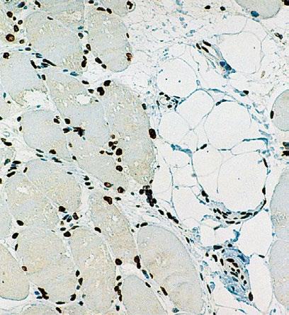
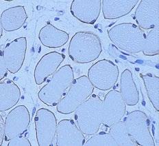
7 Emerin EXCLUSIVE Emery-Dreifuss muscular dystrophy (EDMD) is a late onset, X-linked, recessive disorder characterized by slowly progressing contractures, wasting of skeletal muscle and cardiomyopathy usually presented as heart block. Contractures are seen in the elbows, Achilles tendons and postcervical muscles with humero-peroneal distribution early in the course of the disease. The STA gene, at Xq28 locus, encodes a serine-rich 34kD protein, emerin, which is ubiquitous in tissues and is found in highest concentration in skeletal and cardiac muscle. Emerin is localized in the nuclear membrane of normal muscle cells and its deficiency plays a crucial part in the pathology of EDMD. Merosin Laminin Alpha 2 Chain EXCLUSIVE The muscle-specific form of laminin, merosin, is composed of three chains: alpha 2, beta 1 and gamma 1. Mutations in the chromosome 6 encoded gene for the laminin alpha 2 chain of merosin are responsible for a form of congenital muscular dystrophy (CMD). Merosin-negative CMD is characterized by a severe clinical phenotype and is associated with white matter changes on brain imaging. Human skeletal muscle: immunohistochemical staining for emerin using NCL-EMERIN. Note perinuclear staining of all cell nuclei. Paraffin section. 1 ml Lyophilized Monoclonal (NCL-EMERIN) 4G5. For the qualitative identification by light microscopy of emerin by immunohistochemistry. Recommended for use on paraffin or frozen sections. Prokaryotic recombinant protein corresponding to a 222 amino acid region near the N-terminus of the emerin protein. Human emerin protein. Human skeletal muscle: immunohistochemical staining for merosin using NCL-MEROSIN. Note membrane staining of normal muscle fibers (A) and absence of staining of muscle fibers in an individual with chromosome 6-linked congenital muscular dystrophy (B). Frozen sections. Photographs supplied courtesy of Dr Louise V B Anderson. 1 ml Lyophilized Monoclonal (NCL-MEROSIN) Mer3/22B2. Qualitative identification by light microscopy of merosin laminin alpha 2 chain by immunohistochemistry. Purified protein from placenta. Reacts strongly with laminin alpha 2 chain of merosin in human and rabbit skeletal muscle. No reaction is observed in muscle sections from mouse, rat, dog, chicken, hamster or pig. 7

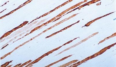
8 Myosin Heavy Chain Antibodies Myosin is a contractile muscle specific protein composed of two heavy and four light chains. The myosin heavy chain has many isoforms which are specific for different muscles or fiber types, some of which are developmentally regulated. The range of myosin heavy chain antibodies may prove useful for investigating development of intrafusal and extrafusal muscle fibers and the course of muscle fiber regeneration. At the ultrastructural level, antibodies can reveal architectural details of the myofilament as well as the cytoplasmic and membrane sites of new myosin integration. Myosin Heavy Chain (developmental) EXCLUSIVE 1 ml Lyophilized Monoclonal (NCL-MHCd) RNMy2/9D2. Immunohistochemistry. Recommended for use on frozen sections. Native myosin extracted from the hind limb muscle of 7 day old rats. Human myosin developmental type heavy chain. Note that this antibody recognizes a myosin heavy chain (MHC) present during the embryonic and neonatal period in the development of skeletal muscle. The same MHC occurs during regeneration of muscle fibers. Also reacts with rat myosin developmental type heavy chain. Myosin Heavy Chain (fast) 1 ml Lyophilized Monoclonal (NCL-MHCf) WB-MHCf. Immunohistochemistry. Recommended for use on frozen sections. Native myosin extracted from rabbit psoas muscle. Rabbit myosin fast type heavy chain. Cross-reacts with human myosin fast type heavy chain. The antibody also reacts with type II myosin heavy chain (both IIa and IIb) in rat, mouse, dog, sheep, pig and goat muscle. Myosin Heavy Chain (neonatal) 1 ml Lyophilized Monoclonal (NCL-MHCn) Human skeletal muscle: immunohistochemical staining for myosin heavy chain using NCL-MHCn. Note intense staining of muscle fibers. Frozen section. WB-MHCn. Immunohistochemistry. Recommended for use on frozen sections. Myosin extracted from the hind limb muscle of a 3 day old rabbit, denatured with sodium dodecyl sulphate. Rabbit myosin neonatal type heavy chain. Cross-reacts with human myosin neonatal type heavy chain. Note that this antibody recognizes a myosin heavy chain present during the neonatal period in rabbit limb muscle. The temporal appearance of an equivalent epitope may differ in different species and consequently it may not be correct to label the epitope as neonatal in some circumstances. Myosin Heavy Chain (slow) 1 ml Lyophilized Monoclonal (NCL-MHCs) WB-MHCs. Immunohistochemistry. Recommended for use on frozen sections. Native myosin extracted from rabbit soleus muscle. Rabbit myosin slow type heavy chain. Cross-reacts with human myosin slow type heavy chain. The antibody also reacts with type I myosin heavy chain in rat, mouse, dog, sheep, pig and goat muscle. 8

 and one form of Myofibrillar Myopathy.")
9 Myotilin EXCLUSIVE The myotilin gene on chromosome 5q31 encodes a 498 amino acid polypeptide with a molecular weight of 57kD. Myotilin is a structural protein of sarcomeric Z discs and sarcolemma in human skeletal and cardiac muscle. It is homologous to palladin and titin in the two C-terminal lg-domains and also to palladin in its unique serine-rich N-terminal region. Myotilin interacts with alpha-actinin, actin and gamma-filamin. Mutations in the myotilin gene are associated with limb-girdle muscular dystrophy 1 A (LGMD1A) and one form of Myofibrillar Myopathy. It is highly conserved between human and mouse with its expression being more widespread in the embryo than in the adult. Expression of myotilin has been reported in adult skeletal and cardiac muscle with variable expression reported in the peripheral nervous system, lung, liver and kidney. NCL-MYOTILIN will be of use in studies to determine the expression of myotilin in normal and pathological tissues. 1 ml Lyophilized Monoclonal (NCL-MYOTILIN) A RSO34. Immunohistochemistry. Recommended for use on paraffin and frozen sections. Prokaryotic recombinant protein corresponding to a C-terminal region of 266 amino acids of the human myotilin molecule. Human myotilin. B Sarcoglycan Antibodies EXCLUSIVE In normal skeletal muscle, dystrophin, the protein product of the gene which is defective in Duchenne and Becker muscular dystrophy, is attached to the muscle membrane via a complex of proteins (dystrophinassociated glycoproteins, DAG s). Dystrophin-deficient muscle shows a generalized reduction in DAG labeling. The expression of different members of the dystrophin glycoprotein complex is altered in several types of muscular dystrophy. For example, patients with LGMD2D have mutations in the gene for alpha-sarcoglycan, those with LGM2E have mutations in the beta-sarcoglycan gene, those with LGM2C have mutations in the gamma-sarcoglycan gene and those with LGM2F have mutations in the delta-sarcoglycan gene. As the sarcoglycans function together as a subcomplex, mutations in any one of the sarcoglycan genes usually results in variable expression for the whole group. 1 ml Alpha-Sarcoglycan (Adhalin) Liquid Monoclonal (NCL-L-a-SARC) Ad1/20A6. NCL-L-a-SARC is intended for the qualitative identification by light microscopy of alphasarcoglycan (adhalin) by immunohistochemistry. Fusion protein containing amino acids 217 to 289 of the rabbit adhalin sequence (Roberds SL et al., 1993). Human alpha-sarcoglycan, also known as adhalin. Also cross-reacts strongly with alpha-sarcoglycan in sections of muscle from mouse, rat, rabbit, hamster and pig. Does not react with chicken muscle. 1 ml Beta-Sarcoglycan Liquid Monoclonal (NCL-L-b-SARC) βsarc1/5b1. NCL-L-b-SARC is intended for the qualitative identification by light microscopy of betasarcoglycan by immunohistochemistry. Fusion protein RBSG-NT of the human beta-sarcoglycan sequence. Human beta-sarcoglycan (43 kd). Human skeletal muscle: immunohistochemical staining for myotilin using NCL-MYOTILIN antibody. Note sarcoplasmic staining of normal muscle fibers (A) and presence of protein aggregates in an individual with myofibrillar myopathy (B). Frozen sections. Photographs supplied courtesy of Dr Rita Barresi. 9
10 Sarcoglycan Antibodies EXCLUSIVE 1 ml Delta-Sarcoglycan Lyophilized Monoclonal (NCL-d-SARC) δsarc3/12c1. NCL-d-SARC is intended for the qualitative identification by light microscopy of Delta- Sarcoglycan by immunohistochemistry. Synthetic peptide containing amino acids 1-19 at the N-terminus of the human deltasarcoglycan sequence (Jung D. et al.,1966). Human delta-sarcoglycan (35 kd). Does not react with delta-sarcoglycan in sections of mouse, rat, rabbit, dog, chicken, hamster or pig muscle. Other animal species not tested. 1 ml Gamma-Sarcoglycan Lyophilized Monoclonal (NCL-g-SARC) 35DAG/21B5. NCL-g-SARC is intended for the qualitative identification by light microscopy of gammasarcoglycan by immunohistochemistry. Synthetic peptide containing amino acids of the rabbit gamma-sarcoglycan sequence (Noguchi et al., 1995). Human gamma-sarcoglycan (35 kd). 10


11 Spectrin EXCLUSIVE Spectrin is a cytoskeletal protein which has some structural homology with dystrophin, the protein that is defective in Duchenne and Becker muscular dystrophy. Subtle membrane damage frequently occurs during the excision and freezing of muscle biopsies. Labeling for spectrin must be used to monitor membrane integrity. NCL-SPEC1 recognizes the beta chain of spectrin in erythrocytes and muscle. NCL-SPEC1 reacts with human beta-spectrin. 1 ml Lyophilized Monoclonal (NCL-SPEC1) RBC2/3D5. NCL-SPEC1 is intended for the qualitative identification by light microscopy of spectrin by immunohistochemistry. Human red blood cell membrane ghosts. Beta chain of spectrin in human red blood cells and muscle. Utrophin EXCLUSIVE The utrophin gene is located on chromosome 6. The protein is a homologue of dystrophin and is known as dystrophin-related protein. In normal muscle, utrophin is restricted to neuromuscular junctions; however, in dystrophin-deficient muscle, utrophin expression may be upregulated and labeling appears around the periphery of most fibers. lmmunohistochemical staining with NCL-DRP2 labels vessels and neuromuscular junctions and the upregulated form of utrophin, located around fiber membranes. 2.5 ml Lyophilized Monoclonal (NCL-DRP2) DRP3/20C5. NCL-DRP2 is intended for the qualitative identification by light microscopy of utrophin (N-terminus) by immunohistochemistry. Fusion protein containing the first 261 amino acids of the published UTRN gene sequence. Amino terminal domain of the human homolog of human dystrophin, utrophin (also known as dystrophinrelated protein or DRP ). Also cross-reacts with utrophin in sections of muscle from rat and dog. Other animal species have not been tested. A B Human skeletal muscle: immunohistochemical staining for utrophin using NCL-DRP2 antibody. In control muscle the antibody labels blood vessels and neuromuscular junctions (A). Utrophin is expressed at the sarcolemma in individuals with mutations in the DMD gene (B). Frozen sections. Photographs supplied courtesy of Dr Rita Barresi. 11


12 LeicaBiosystems.com CUSTOM-MADE KREATECH FISH4U PROBES Leica Biosystems FISH4U service provides custom-made DNA FISH probes designed to your specifications. Our flexible, custom probe service gives you access to your probe of choice. We will develop completely new probe designs to your specifications or you can request a probe from our existing portfolio to be labeled with an alternative from our range of colors. FISH4U probes are developed at Leica Biosystems in collaboration with you. The probes will be optimized on your specimens with the probe color of your choice. Custom FISH4U probes: Leading-edge design Designed to meet your specific needs Flexible format and volume Repeat-free:»» A clearer background»» Greater signal intensity Manual or automated For more information contact: fish4u@leicabiosystems.com LEICA BIOSYSTEMS Leica Biosystems is a global leader in workflow solutions and automation. As the only company to own the workflow from biopsy to diagnosis, we are uniquely positioned to break down the barriers between each of these steps. Our mission of Advancing Cancer Diagnostics, Improving Lives is at the heart of our corporate culture. Our easy-to-use and consistently reliable offerings help improve workflow efficiency and diagnostic confidence. The company is represented in over 100 countries. It has manufacturing facilities in 9 countries, sales and service organizations in 19 countries, and an international network of dealers. The company is headquartered in Nussloch, Germany. Visit LeicaBiosystems.com for more information. Advancing cancer diagnostics to improve lives Leica Biosystems an international company with a strong network of worldwide customer services. For detailed contact information on your nearest sales office or distributor please visit our website: LeicaBiosystems.com This catalog was developed with assistance from Dr. Rita Barresi, Consultant Clinical Scientist and Head of the Muscle Immunoanalysis Unit, Newcastle NHS Foundation Trust, UK. * Regulatory status can vary geographically. Please consult your Leica Biosystems representative for information about regulatory classification in your country. Copyright 2018 Leica Biosystems Newcastle Ltd. All rights reserved. LEICA and the Leica Logo are registered trademarks of Leica Microsystems IR GmbH. Novocastra, REPEAT-FREE and BOND are trademarks of the Leica Biosystems group of companies in the USA and optionally in other countries. Other logos, product and/or company names might be trademarks of their respective owners Rev C 08/2018
Immunohistochemical Study of Dystrophin Associated Glycoproteins in Limb-girdle Muscular Dystrophies
 Dystrophin Immunohistochemical Study of Dystrophin Associated Glycoproteins in Limb-girdle Muscular Dystrophies NSC 89-2314-B-002-111 88 8 1 89 7 31 ( Peroxidase -AntiPeroxidase Immnofluorescence) Abstract
Muscular Dystrophy. Biol 405 Molecular Medicine
 Muscular Dystrophy Biol 405 Molecular Medicine Duchenne muscular dystrophy Duchenne muscular dystrophy is a neuromuscular disease that occurs in ~ 1/3,500 male births. The disease causes developmental
Three Muscular Dystrophies: Loss of Cytoskeleton-Extracellular Matrix Linkage
 Cell, Vol. 80, 675-679, March 10, 1995, Copyright 1995 by Cell Press Three Muscular Dystrophies: Loss of Cytoskeleton-Extracellular Matrix Linkage Review Kevin P. Campbell Howard Hughes Medical Institute
READ ORPHA.NET WEBSITE ABOUT BETA-SARCOGLYOCANOPATHY LIMB-GIRDLE MUSCULAR DYSTROPHIES
 READ ORPHA.NET WEBSITE ABOUT BETA-SARCOGLYOCANOPATHY LIMB-GIRDLE MUSCULAR DYSTROPHIES (LGMD) Limb-girdle muscular dystrophies (LGMD) are a heterogeneous group of genetically determined disorders with a
A rare case of muscular dystrophy with POMT2 and FKRP gene mutation. Present by : Ghasem Khazaei Supervisor :Dr Mina Mohammadi Sarband
 A rare case of muscular dystrophy with POMT2 and FKRP gene mutation Present by : Ghasem Khazaei Supervisor :Dr Mina Mohammadi Sarband Index : Congenital muscular dystrophy (CMD) Dystroglycanopathies Walker-Warburg
Molecular mechanisms of muscular dystrophies: old and new players
 MECHANISMS OF DISEASE Molecular mechanisms of muscular dystrophies: old and new players Kay E Davies* and Kristen J Nowak Abstract The study of the muscle cell in the muscular dystrophies (MDs) has shown
Pathology of Neuromuscular Disease Part 1: muscle
 Pathology of Neuromuscular Disease Part 1: muscle Zarife Sahenk, MD. PhD. Research Institute at Nationwide Children s Hospital Center for gene therapy, Neuromuscular Program Experimental & Clinical Neuromuscular
Muscle Diseases: The Muscular Dystrophies
 Annu. Rev. Pathol. Mech. Dis. 2007. 2:87 109 The Annual Review of Pathology: Mechanisms of Disease is online at pathmechdis.annualreviews.org This article s doi: 10.1146/annurev.pathol.2.010506.091936
Electron microscopy in the investigation and diagnosis of muscle disease
 Electron microscopy in the investigation and diagnosis of muscle disease Roy Weller Clinical Neurosciences University of Southampton School of Medicine Normal Muscle Normal Muscle The Sarcomere The names
The functional role of dystrophin in the heart: implications for inherited and non-inherited heart disease. Matthew Scott Barnabei
 The functional role of dystrophin in the heart: implications for inherited and non-inherited heart disease by Matthew Scott Barnabei A dissertation submitted in partial fulfillment of the requirements
Skeletal Muscle and the Molecular Basis of Contraction. Lanny Shulman, O.D., Ph.D. University of Houston College of Optometry
 Skeletal Muscle and the Molecular Basis of Contraction Lanny Shulman, O.D., Ph.D. University of Houston College of Optometry Like neurons, all muscle cells can be excited chemically, electrically, and
Chapter Skeletal Muscle Structure and Function
 Chapter 10.2 Skeletal Muscle Structure and Function Introduction to Muscle Physiology Movement is a fundamental characteristic of all living things All muscle cells (skeletal, cardiac, and smooth) are
18 (2), DOI: /bjmg
, DOI: /bjmg") 18 (2), 2015 71-76 DOI: 10.1515/bjmg-2015-0088 CASE REPORT SARCOLEMMAL DEFICIENCY OF SARCOGLYCAN COMPLEX IN AN 18-MONTH-OLD TURKISH BOY WITH A LARGE DELETION IN THE BETA SARCOGLYCAN GENE Diniz G 1,*, Tekgul
18 (2), 2015 71-76 DOI: 10.1515/bjmg-2015-0088 CASE REPORT SARCOLEMMAL DEFICIENCY OF SARCOGLYCAN COMPLEX IN AN 18-MONTH-OLD TURKISH BOY WITH A LARGE DELETION IN THE BETA SARCOGLYCAN GENE Diniz G 1,*, Tekgul
Single section Western blot
 Single section Western blot Improving the molecular diagnosis of the muscular dystrophies Sandra T. Cooper, PhD; Harriet P. Lo, BSc; and Kathryn N. North, MD Brief Communications Abstract Single section
III./10.4. Diagnosis. Introduction. A.) Laboratory tests. Laboratory tests, electrophysiology, muscle biopsy, genetic testing, imaging techniques
 Laboratory tests. Laboratory tests, electrophysiology, muscle biopsy, genetic testing, imaging techniques") III./10.4. Diagnosis Laboratory tests, electrophysiology, muscle biopsy, genetic testing, imaging techniques After studying this chapter, you will become familiar with the most commonly used diagnostic
III./10.4. Diagnosis Laboratory tests, electrophysiology, muscle biopsy, genetic testing, imaging techniques After studying this chapter, you will become familiar with the most commonly used diagnostic
DMD Genetics: complicated, complex and critical to understand
 DMD Genetics: complicated, complex and critical to understand Stanley Nelson, MD Professor of Human Genetics, Pathology and Laboratory Medicine, and Psychiatry Co Director, Center for Duchenne Muscular
Muscle Tissue. Dr. Heba Kalbouneh Associate Professor of Anatomy and Histology
 Muscle Tissue Dr. Heba Kalbouneh Associate Professor of Anatomy and Histology Functions of muscle tissue Movement Maintenance of posture Joint stabilization Heat generation Tendon Belly Tendon Types of
Medical Biology. Dr. Khalida Ibrahim
 Dr. Khalida Ibrahim Medical Biology MUSCLE TISSUE 1. Muscle tissue is characterized by its well-developed properties of contraction. 2. Muscle is responsible for the movements of the body and the various
Muscle Histology. Dr. Heba Kalbouneh Assistant Professor of Anatomy and Histology
 Muscle Histology Dr. Heba Kalbouneh Assistant Professor of Anatomy and Histology Functions of muscle tissue Movement Maintenance of posture Joint stabilization Heat generation Types of Muscle Tissue Skeletal
The Pathogenesis and Therapy of Muscular Dystrophies
 I GG16CH13-Davies ARI 11 May 2015 13:45 R E V I E W S Review in Advance first posted online on June 4, 2015. (Changes may still occur before final publication online and in print.) E N C A D V A N The
Dysferlinopathies. LGMD2B, Miyoshi & Others. 2B Empowered Conference
 Dysferlinopathies LGMD2B, Miyoshi & Others 2B Empowered Conference Matthew P. Wicklund, MD, FAAN Professor of Neurology and Pediatrics Penn State Health May 24, 2015 Outline A. Empower you with knowledge
Genetic diagnosis of limb girdle muscular dystrophy type 2A, A Case Report
 Genetic diagnosis of limb girdle muscular dystrophy type 2A, A Case Report Roshanak Jazayeri, MD, PhD Assistant Professor of Medical Genetics Faculty of Medicine, Alborz University of Medical Sciences
About This Chapter. Skeletal muscle Mechanics of body movement Smooth muscle Cardiac muscle Pearson Education, Inc.
 About This Chapter Skeletal muscle Mechanics of body movement Smooth muscle Cardiac muscle Skeletal Muscle Usually attached to bones by tendons Origin: closest to the trunk or to more stationary bone Insertion:
Iowa Wellstone Center Muscle Tissue and Cell Culture Repository
 Iowa Wellstone Center Muscle Tissue and Cell Culture Repository Steven A. Moore, M.D., Ph.D. The University of Iowa Department of Pathology and Iowa Wellstone Muscular Dystrophy Cooperative Research Center
Muscle and Neuromuscular Junction. Peter Takizawa Department of Cell Biology
 Muscle and Neuromuscular Junction Peter Takizawa Department of Cell Biology Types and structure of muscle cells Structural basis of contraction Triggering muscle contraction Skeletal muscle consists of
ANSC/FSTC 607 Biochemistry and Physiology of Muscle as a Food PRIMARY, SECONDARY, AND TERTIARY MYOTUBES
 ANSC/FSTC 607 Biochemistry and Physiology of Muscle as a Food PRIMARY, SECONDARY, AND TERTIARY MYOTUBES I. Satellite Cells A. Proliferative, myoblastic cells that lie in invaginations in the sarcolemma
A novel mutation in the DYSF gene in a patient with a presumed inflammatory myopathy
 2018; 38, 433 437 doi:10.1111/neup.12474 Case Report A novel mutation in the DYSF gene in a patient with a presumed inflammatory myopathy Jin Tang, Xueqin Song, Guang Ji, Hongran Wu, Shuyan Sun, Shan Lu,
Muscle Tissue. General concepts. Classification of muscle. I. Functional classification is based on the type of neural control.
 Muscle Tissue LEARNING OBJECTIVES 1. Identify the three types of muscle tissue at the light microscopic level. 2. List and compare the structural and functional features of each of the three muscle fiber
Skeletal Muscle : Structure
 1 Skeletal Muscle : Structure Dr.Viral I. Champaneri, MD Assistant Professor Department of Physiology 2 Learning objectives 1. Gross anatomy of the skeletal muscle 2. Myofilaments & their molecular structure
Familial DilatedCardiomyopathy Georgios K Efthimiadis, MD
 Familial DilatedCardiomyopathy Georgios K Efthimiadis, MD Dilated Cardiomyopathy Dilated LV/RV, reduced EF, in the absence of CAD valvulopathy pericardial disease Prevalence:40/100.000 persons Natural
The limb girdle muscular dystrophies (LGMDs)
") The limb gird muscular dystrophies (LGMDs) This factsheet is for all peop for whom a diagnosis of limb gird muscular dystrophy (LGMD) has been suggested. This is a complicated subject since there are many
The limb gird muscular dystrophies (LGMDs) This factsheet is for all peop for whom a diagnosis of limb gird muscular dystrophy (LGMD) has been suggested. This is a complicated subject since there are many
Muscular Tissue. Functions of Muscular Tissue. Types of Muscular Tissue. Skeletal Muscular Tissue. Properties of Muscular Tissue
 Muscular Tissue Functions of Muscular Tissue Muscle makes up a large percentage of the body s weight (40-50%) Their main functions are to: Create motion muscles work with nerves, bones, and joints to produce
muscle biopsy How to do it
 How to do it muscle biopsy Gillian Hall Department of Clinical Neurosciences, Western General Hospital, Edinburgh EH4 2XU. Email: ghall@skull.dcn.ed.ac.uk INTRODUCTION Good clinical evaluation remains
Anti-Lamin B1/LMNB1 Picoband Antibody
 Anti-Lamin B1/LMNB1 Picoband Antibody Catalog Number:PB9611 About LMNB1 Lamin-B1 is a protein that in humans is encoded by the LMNB1 gene. The nuclear lamina consists of a two-dimensional matrix of proteins
Connective tissue MUSCLE TISSUE
 Connective tissue MUSCLE TISSUE Part 1 General features of MT Develop from mesoderm Many cells, less intercellular matrix Function contraction (shortening) Skeletal (striated, voluntary) Types of MT Cardiac
Leucine and exercise improve skeletal muscle function in the mdx mouse. Kevin Andrew Voelker
 Leucine and exercise improve skeletal muscle function in the mdx mouse Kevin Andrew Voelker Dissertation submitted to the faculty of the Virginia Polytechnic Institute and State University in partial fulfillment
Muscle Tissue- 3 Types
 AN INTRODUCTION TO MUSCLE TISSUE Muscle Tissue- 3 Types Skeletal muscle (focus on these) Cardiac muscle Smooth muscle FUNCTIONS OF SKELETAL MUSCLES Produce movement of the skeleton Maintain posture and
Advances and Perspectives in Muscular Dystrophies Abstract Key words X-Linked Muscular Dystrophies
 Advances and Perspectives in Muscular Dystrophies Corrado Angelini Department of Neurological and Psychiatric Sciences, University of Padova, Padova, Italy Abstract The muscular dystrophies are inherited
BIOLOGY - CLUTCH CH.49 - MUSCLE SYSTEMS.
 !! www.clutchprep.com BIOLOGY - CLUTCH Muscle system organ system that includes skeletal, cardiac, and smooth muscle Muscle tissue capable of contracting through the interaction of actin and myosin proteins
Seminar. Muscular dystrophies
 Muscular dystrophies Eugenio Mercuri, Francesco Muntoni Muscular dystrophies are a heterogeneous group of inherited disorders that share similar clinical features and dystrophic changes on muscle biopsy.
CHAPTER 6 2/9/2016. Learning Objectives List the four traits that all muscle types have in common.
 Learning Objectives List the four traits that all muscle types have in common. CHAPTER 6 The Muscular System Demonstrate and explain the use of antagonistic muscle pairs. Describe the attachment of muscle
T cell Receptor. Chapter 9. Comparison of TCR αβ T cells
 Chapter 9 The αβ TCR is similar in size and structure to an antibody Fab fragment T cell Receptor Kuby Figure 9-3 The αβ T cell receptor - Two chains - α and β - Two domains per chain - constant (C) domain
MRI and proteomic studies in normal and
 MRI and proteomic studies in normal and pathological muscles. Isabel ILLA. Catedrática Medicina de la Universitat Autònoma de Barcelona. Chief Neuromuscular Diseases Unit. Hospital Santa Creu i Sant Pau.
The New England Journal of Medicine MUTATIONS IN THE SARCOGLYCAN GENES IN PATIENTS WITH MYOPATHY
 MUTATIONS IN THE SARCOGLYCAN GENES IN PATIENTS WITH MYOPATHY DAVID J. DUGGAN, B.S., J. RAFAEL GOROSPE, M.D., PH.D., MARINA FANIN, M.S., ERIC P. HOFFMAN, PH.D., A CORRADO ANGELINI, M.D. ABSTRACT Background
Topics Covered. General muscle structure non-muscle components, macro-structure, contractile elements, membrane components.
 1 Topics Covered General muscle structure non-muscle components, macro-structure, contractile elements, membrane components. Contractile function Contractile and regulatory proteins, force production.
Histology Exam 2: Smith Lecture 5 (Muscle)
") Histology Exam 2: Smith Lecture 5 (Muscle) What are the three types of muscle? Skeletal: morphology and functionality? Cardiac: morphology and functionality? Smooth: morphology and functionality? Skeletal
Histology Exam 2: Smith Lecture 5 (Muscle) What are the three types of muscle? Skeletal: morphology and functionality? Cardiac: morphology and functionality? Smooth: morphology and functionality? Skeletal
DOWNLOAD PDF STRUCTURE AND REGULATION OF CARDIAC AND SKELETAL MUSCLE THIN FILAMENTS
 Chapter 1 : ACTC1 - Wikipedia LARRY S. TOBACMAN STRUCTURE AND REGULATION OF CARDIAC AND The biological production of force and movement can be understood only when it is. Structure[ edit ] There are three
Smooth Cardiac Skeletal Location Around tubes Heart tissue attached to skeleton Moves stuff thru Heart beat pumps Moves body parts
 Biology 067 - Muscular system A. Type of muscles: Smooth Cardiac Skeletal Location Around tubes Heart tissue attached to skeleton Function Moves stuff thru Heart beat pumps Moves body parts tubes blood
Muscular System Module 3: Contraction and Relaxation *
 OpenStax-CNX module: m48498 1 Muscular System Module 3: Contraction and Relaxation * Donna Browne Based on Muscle Fiber Contraction and Relaxation by OpenStax This work is produced by OpenStax-CNX and
Functional significance of dystrophin positive fibres
 632 Muscular Dystrophy Group Research Laboratories, Regional Neurosciences Centre, Newcastle General Hospital, Newcastle upon Tyne L V B Nicholson M A Johnson K M D Bushby D Gardner-Medwin Correspondence
Cardiac Considerations and Care in Children with Neuromuscular Disorders
 Cardiac Considerations and Care in Children with Neuromuscular Disorders - importance of early and ongoing treatment, management and available able medications. Dr Bo Remenyi Department of Cardiology The
Limb Girdle Muscular Dystrophy
 Limb Girdle Muscular Dystrophy Reza Shervin Badv MD, Pediatric Neurologist Children s Medical Center Pediatrics Center of Excellence Tehran University of Medical Sciences Limb-girdle muscular dystrophies(lgmd)
9/16/2009. Fast and slow twitch fibres. Properties of Muscle Fiber Types Fast fibers Slow fibers
 Muscles, muscle fibres and myofibrils Fast and slow twitch fibres Rat hindlimb muscle ATPase staining at different ph and NADH Muscle fibre shortening velocity lengths/second Properties of Muscle Fiber
Dystrophy Patients Lacking COOH-terminal Domains of Dystrophin
 Deficiency of Dystrophin-associated Proteins in Duchenne Muscular Dystrophy Patients Lacking COOH-terminal Domains of Dystrophin Kiichiro Matsumura, * Fernando M. S. Tome,t Victor lonasescu, James M. Ervasti,
Thyroidal Trophic Influence Skeletal Muscle. Myosin >>>CLICK HERE<<<
 Thyroidal Trophic Influence Skeletal Muscle Myosin Even though skeletal muscle by sheer volume alone is the ample, antibody to myosin heavy chains from red Thyroidal trophic influence on skeletal muscle.
Genetic modifiers of muscular dystrophy: Implications for therapy
 Biochimica et Biophysica Acta 1772 (2007) 216 228 www.elsevier.com/locate/bbadis Review Genetic modifiers of muscular dystrophy: Implications for therapy Ahlke Heydemann a, Katherine R. Doherty b, Elizabeth
Muscle Tissue. Muscle Development and Repair. Development: fusion of myoblasts. Repair: Satellite cells (S) 3 Types of Muscle
 3 Types of Muscle") ANNOUNCEMENTS Review Session Every Friday at 12:20 Muscle Tissue 3 Types of Muscle Function: Force generation Lab Practical Coming up! October 26 th, 27 th Muscle Tissue Striated Nonstriated Skeletal Smooth
ANNOUNCEMENTS Review Session Every Friday at 12:20 Muscle Tissue 3 Types of Muscle Function: Force generation Lab Practical Coming up! October 26 th, 27 th Muscle Tissue Striated Nonstriated Skeletal Smooth
Skeletal muscle. General features :
 Muscular tissues In the first embryonic life the muscular tissues arise from mesoderm, The function of movement in multicellular organisms is usually assumed by specialized cells called muscle fibers which
How many skeletal muscles are present in our body? Muscles are excitable & contractile, extensible and elastic to some extent.
 Muscles How many skeletal muscles are present in our body? -646 muscles The functions of the muscles are: Movement Maintenance of posture Generation of heat Stabilization of joints : amount of muscle surrounding
Lecture 9A. Muscle structure. Outline
 Lecture 9A Muscle structure Outline Smooth, skeletal, and cardiac muscle tissues Structure and function of skeletal muscle cells. Sarcomeres structure and contraction Actin-myosin interaction and sliding
MP Genetic Testing for Limb-Girdle Muscular Dystrophies
 BCBSA Ref. Policy: 2.04.132 Last Review: 04/30/2018 Effective Date: 04/30/2018 Section: Medicine Related Policies 2.04.86 Genetic Testing for Duchenne and Becker Muscular Dystrophy 2.04.105 Genetic Testing
근이양증의분자적병리학적진단 Molecular and Pathological Diagnosis of Muscular Dystrophies
 HANYANG MEDICAL REVIEWS Vol. 26, No. 1, 2006 근이양증의분자적병리학적진단 Molecular and Pathological Diagnosis of Muscular Dystrophies 최영철연세대학교의과대학신경과교실 Young-Chul Choi, M.D., Ph.D. Department of Neurology, Yonsei University
Uncovering the complexity of muscular dystrophy pathology through disease signaling
 Uncovering the complexity of muscular dystrophy pathology through disease signaling A dissertation submitted to the Graduate School of the University of Cincinnati in partial fulfillment of the requirements
Myopathies of Unknown Etiology (Western Blot)
") Myopathies of Unknown Etiology (Western Blot) April 2013 DISCLAIMER: This document was originally drafted in French by the Institut national d'excellence en santé et en services sociaux (INESSS), and that
Myopathies of Unknown Etiology (Western Blot) April 2013 DISCLAIMER: This document was originally drafted in French by the Institut national d'excellence en santé et en services sociaux (INESSS), and that
Corporate Medical Policy
 Corporate Medical Policy File Name: Origination: Last CAP Review: Next CAP Review: Last Review: mutation_testing_for_limb_girdle_muscular_dystrophies 01/01/2019 N/A 01/01/2020 01/01/2019 Description of
Current Research Strategies and Therapeutic Approaches in Duchenne Muscular Dystrophy
 Current Research Strategies and Therapeutic Approaches in Duchenne Muscular Dystrophy H. Lee Sweeney, Ph.D. Department of Physiology University of Pennsylvania Perelman School of Medicine Current Research
Microanatomy of Muscles. Anatomy & Physiology Class
 Microanatomy of Muscles Anatomy & Physiology Class Three Main Muscle Types Objectives: By the end of this presentation you will have the information to: 1. 2. 3. 4. 5. 6. Describe the 3 main types of muscles.
Skeletal muscle in the light of its structure
 Mechanism of contraction of Skeletal muscle in the light of its structure By Dr. Mudassar Ali Roomi (MBBS, M. Phil) Muscle Tissue Skeletal Muscle Cardiac Muscle Smooth Muscle Skeletal Muscle Long cylindrical
MUSCLE TISSUE (MUSCLE PHYSIOLOGY) PART I: MUSCLE STRUCTURE
 PART I: MUSCLE STRUCTURE") PART I: MUSCLE STRUCTURE Muscle Tissue A primary tissue type, divided into: skeletal muscle cardiac muscle smooth muscle Functions of Skeletal Muscles Produce skeletal movement Maintain body position Support
PART I: MUSCLE STRUCTURE Muscle Tissue A primary tissue type, divided into: skeletal muscle cardiac muscle smooth muscle Functions of Skeletal Muscles Produce skeletal movement Maintain body position Support
Muscles, muscle fibres and myofibrils
 Muscles, muscle fibres and myofibrils Properties of Muscle Fiber Types Fast fibers Slow fibers Characteristic IIb IIx IIa Type I V max (speed of shortening) Highest Intermediate Low Resistance to fatigue
Outline. Bio 105: Muscular System. Muscular System. Types of Muscles. Smooth Muscle. Cardiac Muscle 4/6/2016
 Outline Bio 105: Muscular System Lecture 11 Chapter 6 Characteristics of muscles 3 types of muscles Functions of muscles Structure of skeletal muscles Mechanics of muscle contraction Energy sources for
BIOH111. o Cell Module o Tissue Module o Integumentary system o Skeletal system o Muscle system o Nervous system o Endocrine system
 BIOH111 o Cell Module o Tissue Module o Integumentary system o Skeletal system o Muscle system o Nervous system o Endocrine system Endeavour College of Natural Health endeavour.edu.au 1 TEXTBOOK AND REQUIRED/RECOMMENDED
Quality in Control. ROS1 Analyte Control. Product Codes: HCL022, HCL023 and HCL024
 Quality in Control ROS1 Analyte Control Product Codes: HCL022, HCL023 and HCL024 Contents What is ROS1? 2 The Role of ROS1 in Cancer 3 ROS1 Assessment 3 ROS1 Analyte Control Product Details 4 ROS1 Analyte
Skeletal Muscle Contraction 4/11/2018 Dr. Hiwa Shafiq
 Skeletal Muscle Contraction 4/11/2018 Dr. Hiwa Shafiq Skeletal Muscle Fiber About 40 per cent of the body is skeletal muscle, and 10 per cent is smooth and cardiac muscle. Skeletal muscles are composed
CLASS SET Unit 4: The Muscular System STUDY GUIDE
 NPHS Anatomy & Physiology Questions to answer: 1) List three functions of the muscular system. 1) movement 2) thermogenesis (generates heat) 3) posture & body/joint support CLASS SET Unit 4: The Muscular
Genetic Testing for Limb-Girdle Muscular Dystrophies
 Applies to all products administered or underwritten by Blue Cross and Blue Shield of Louisiana and its subsidiary, HMO Louisiana, Inc.(collectively referred to as the Company ), unless otherwise provided
Ch.10 Muscle Tissue. Copyright 2009, John Wiley & Sons, Inc.
 Ch.10 Muscle Tissue Preview Chapter 10 In groups we will define the following terms 1. Skeletal muscle 2. Smooth muscle 3. Cardiac muscle 4. Sarcomere 5. Myofibril 6. Myofilament 7. Sarcoplasmic reticulum
Muscle fatigue, nnos and muscle fiber atrophy in limb girdle muscular dystrophy
 Acta Myologica 2014; XXXIII: p. 119-126 invited review Muscle fatigue, nnos and muscle fiber atrophy in limb girdle muscular dystrophy Corrado Angelini 1, Elisabetta Tasca 1, Anna Chiara Nascimbeni 2 and
Skeletal Muscle Contraction 5/11/2017 Dr. Hiwa Shafiq
 Skeletal Muscle Contraction 5/11/2017 Dr. Hiwa Shafiq Skeletal Muscle Fiber About 40 per cent of the body is skeletal muscle, and 10 per cent is smooth and cardiac muscle. Skeletal muscles are composed
CARDIAC muscle is commonly affected in muscular
 362 THE NEW ENGLAND JOURNAL OF MEDICINE Feb. 8, 1996 BRIEF REPORT: DEFICIENCY OF A DYSTROPHIN-ASSOCIATED GLYCOPROTEIN (ADHALIN) IN A PATIENT WITH MUSCULAR DYSTROPHY AND CARDIOMYOPATHY RICARDO FADIC, M.D.,
Anti-PD-L1 antibody [28-8] ab205921
![Anti-PD-L1 antibody [28-8] ab205921](/thumbs/84/91196262.jpg "Anti-PD-L1 antibody [28-8] ab205921") Anti-PD-L1 antibody [28-8] ab205921 2 Abreviews 16 References 15 Images Overview Product name Anti-PD-L1 antibody [28-8] Description Tested applications Species reactivity Immunogen Rabbit monoclonal [28-8]
Anti-PD-L1 antibody [28-8] ab205921 2 Abreviews 16 References 15 Images Overview Product name Anti-PD-L1 antibody [28-8] Description Tested applications Species reactivity Immunogen Rabbit monoclonal [28-8]
RECESSIVELY INHERITED LIMBgirdle
 ORIGINAL CONTRIBUTION Cardiac Involvement in Patients With Limb-Girdle Muscular Dystrophy Type 2 and Becker Muscular Dystrophy Marie-Louise Sveen, MD; Jens Jakob Thune, MD; Lars Køber, MD, DMSci; John
Sarcoglycanopathies: A Multiplex Molecular Analysis for the Most Common Mutations
 ORIGINAL ARTICLE Sarcoglycanopathies: A Multiplex Molecular Analysis for the Most Common Mutations Telma L.F. Gouveia, MS,* Julia F.O. Paim, MD, PhD,w Rita C. Pavanello, MD,* Mayana Zatz, PhD,* and Mariz
Mutations. A2 Biology For WJEC
 12. Mutation is a change in the amount, arrangement or structure in the DNA of an organism. 13. There are two types of mutations, chromosome mutations and gene mutations. Mutations A2 Biology For WJEC
Antigen Receptor Structures October 14, Ram Savan
 Antigen Receptor Structures October 14, 2016 Ram Savan savanram@uw.edu 441 Lecture #8 Slide 1 of 28 Three lectures on antigen receptors Part 1 (Today): Structural features of the BCR and TCR Janeway Chapter
Dystrophin-glycoprotein complex: molecular organization and critical roles in skeletal muscle
 Dystrophin-glycoprotein complex: molecular organization and critical roles in skeletal muscle Yoshihide Sunada and Kevin P. Campbell Howard Hughes Medical Institute, Department of Physiology and Biophysics,
The dystrophin glycoprotein complex (DGC)1 consists
1 consists") Molecular Organization of Sarcoglycan Complex in Mouse Myotubes in Culture Yiu-mo Chan,* Carsten G. Bönnemann,* Hart G.W. Lidov, and Louis M. Kunkel* *Howard Hughes Medical Institute, Division of Genetics,
Molecular Organization of Sarcoglycan Complex in Mouse Myotubes in Culture Yiu-mo Chan,* Carsten G. Bönnemann,* Hart G.W. Lidov, and Louis M. Kunkel* *Howard Hughes Medical Institute, Division of Genetics,
Page 1. Introduction Skeletal muscle cells have unique characteristics which allow for body movement.
 Anatomy Review: Skeletal Muscle Tissue Graphics are used with permission of: adam.com (http://www.adam.com/) Benjamin Cummings Publishing Co (http://www.awl.com/bc) Page 1. Introduction Skeletal muscle
Class XI Chapter 20 Locomotion and Movement Biology
 Question 1: Draw the diagram of a sarcomere of skeletal muscle showing different regions. The diagrammatic representation of a sarcomere is as follows: Question 2: Define sliding filament theory of muscle
Muscular System- Part 1. Unit 5 Miss Wheeler
 Muscular System- Part 1 Unit 5 Miss Wheeler Fun Facts! The tongue is the strongest muscle in your body The smallest muscles in the body are in the middle ear The largest muscle in the body is the gluteus
The Muscular System PART A
 6 The Muscular System PART A PowerPoint Lecture Slide Presentation by Jerry L. Cook, Sam Houston University ESSENTIALS OF HUMAN ANATOMY & PHYSIOLOGY EIGHTH EDITION ELAINE N. MARIEB The Muscular System
CONTRIBUTION OF DYSFERLIN-CONTAINING MEMBRANES TO MEMBRANE REPAIR IN SKELETAL MUSCLE. Joel Ryan McDade
 CONTRIBUTION OF DYSFERLIN-CONTAINING MEMBRANES TO MEMBRANE REPAIR IN SKELETAL MUSCLE by Joel Ryan McDade A dissertation submitted in partial fulfillment of the requirements for the degree of Doctor of
Abnormalities of dystrophin, the sarcoglycans, and laminin a2 in the muscular dystrophies
 JMed Genet 1998;35:379-386 Department of Clinical Genetics, Royal Alexandra Hospital for Children, Westmead, Sydney, Australia K J Jones Neurogenetics Research Unit, Clinical Sciences Building, Royal Alexandra
MUSCULAR TISSUE. Dr. Gary Mumaugh
 MUSCULAR TISSUE Dr. Gary Mumaugh MUSCLE OVERVIEW The three types of muscle tissue are skeletal, cardiac, and smooth These types differ in structure, location, function, and means of activation FUNCTIONAL
Product Datasheet. SERCA2 ATPase Antibody (IID8) NB Unit Size: 100uL. Store at -20C. Avoid freeze-thaw cycles.
 NB Unit Size: 100uL. Store at -20C. Avoid freeze-thaw cycles.") Product Datasheet SERCA2 ATPase Antibody (IID8) NB300-529 Unit Size: 100uL Store at -20C. Avoid freeze-thaw cycles. Publications: 5 Protocols, Publications, Related Products, Reviews, Research Tools and
Product Datasheet SERCA2 ATPase Antibody (IID8) NB300-529 Unit Size: 100uL Store at -20C. Avoid freeze-thaw cycles. Publications: 5 Protocols, Publications, Related Products, Reviews, Research Tools and
Muscle Physiology. Dr. Ebneshahidi Ebneshahidi
 Muscle Physiology Dr. Ebneshahidi Skeletal Muscle Figure 9.2 (a) Functions of the muscular system 1. Locomotion body movements are due to skeletal muscle contraction. 2. Vasoconstriction and vasodilatation
Muscle Tissue. PowerPoint Lecture Presentations prepared by Jason LaPres. Lone Star College North Harris Pearson Education, Inc.
 10 Muscle Tissue PowerPoint Lecture Presentations prepared by Jason LaPres Lone Star College North Harris An Introduction to Muscle Tissue Muscle Tissue A primary tissue type, divided into: Skeletal muscle
Ch 12: Muscles sarcolemma, t-tubules, sarcoplasmic reticulum, myofibrils, myofilaments, sarcomere...
 Ch 12: Muscles Review micro-anatomy of muscle tissue Terminology examples: sarcolemma, t-tubules, sarcoplasmic reticulum, myofibrils, myofilaments, sarcomere... SLOs Differentiate levels of muscle structure:
Product Datasheet. EMMPRIN/CD147 Antibody (MEM-M6/1) NB Unit Size: 0.1 mg. Store at 4C. Do not freeze. Publications: 2
 NB Unit Size: 0.1 mg. Store at 4C. Do not freeze. Publications: 2") Product Datasheet EMMPRIN/CD147 Antibody (MEM-M6/1) NB500-430 Unit Size: 0.1 mg Store at 4C. Do not freeze. Publications: 2 Protocols, Publications, Related Products, Reviews, Research Tools and Images
Product Datasheet EMMPRIN/CD147 Antibody (MEM-M6/1) NB500-430 Unit Size: 0.1 mg Store at 4C. Do not freeze. Publications: 2 Protocols, Publications, Related Products, Reviews, Research Tools and Images
Muscle Dr. Ted Milner (KIN 416)
") Muscle Dr. Ted Milner (KIN 416) Muscles are biological motors which actively generate force and produce movement through the process of contraction. The molecular mechanism responsible for muscle contraction
Muscle Dr. Ted Milner (KIN 416) Muscles are biological motors which actively generate force and produce movement through the process of contraction. The molecular mechanism responsible for muscle contraction