EasyScan: Smart Retinal Imaging
|
|
|
- Angelica Pitts
- 5 years ago
- Views:
Transcription
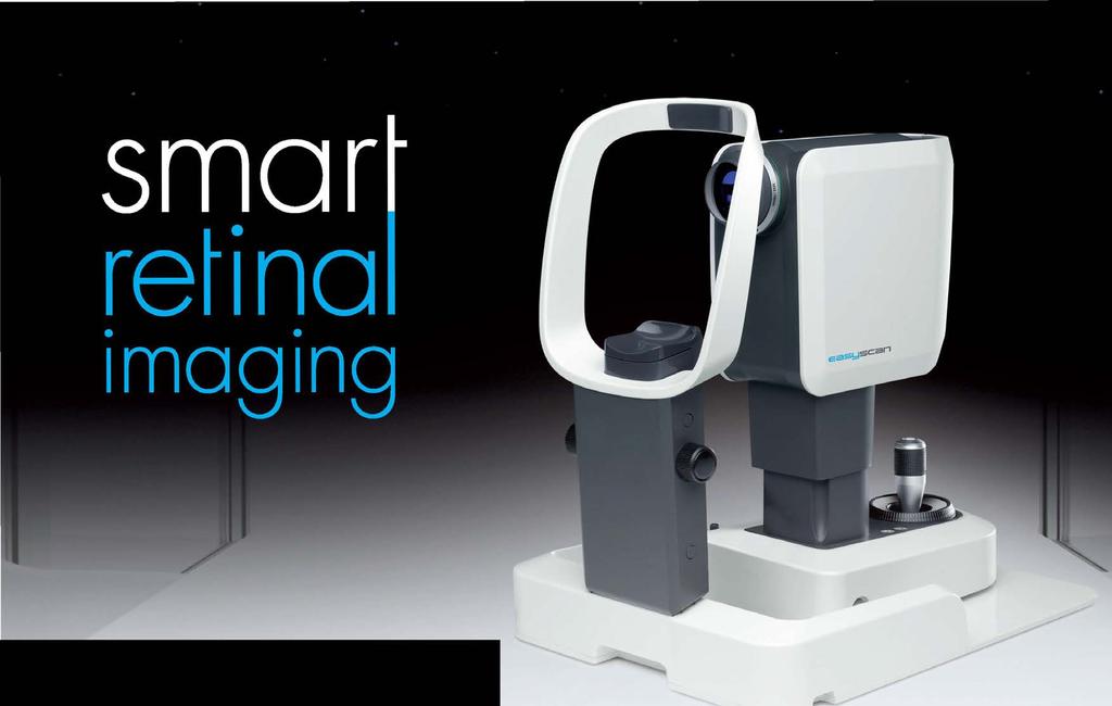

1 easyscan
2 EasyScan: Smart Retinal Imaging Superior Imaging Enjoy the benefits of SLO technology and capture high-quality images easily for accurate diagnosis. Never Dilate Reduce examination time, capture images through small pupils and increase patient satisfaction with the fact that no dilation is ever needed. Mobility EasyScan is a compact, light-weight device that will fit every office space and enable it to be easily transported between locations. Connectivity EasyScan can be used in single practices, mobile workstations and with multiple review stations.


3 Superior Imaging No pathology EasyScan employs Scanning Laser Ophthalmoscope (SLO) technology for superior imaging Retinopathies become more easily detectable with a clear and sharper image for accurate diagnosis. EasyScan, OS, merged central EasyScan, OS, infrared central EasyScan, OS, green central Benefits of SLO technology include: AMD.Earlier diagnosis of retinal conditions Greater contrast than a fundus camera Better penetration of media opacities such as cataract EasyScan, glaucoma suspect, color EasyScan, early AMD, infrared Conventional fundus, early AMD, color Diabetic retinopathy EasyScan, DR, color EasyScan, DR, green Conventional fundus, DR, color

 Infra red laser (785nm) Undilated EasyScan image")


4 Never dilate There is never any need for dilation when capturing images with EasyScan. This saves time and increases patient satisfaction. EasyScan also has the ability to scan through mild to moderate cataract. Green laser (532nm) Infra red laser (785nm) Undilated EasyScan image through cataract Dilated fundus camera image through same cataract Minimum pupil size with a fundus camera: 3.3 mm Unique with EasyScan: Minimum pupil size 1.5 mm Pigment epithelium Retina 1.5 mm Choroid Sclera EasyScan can always image through small pupils; there is no need to dilate. EasyScan s green laser reveals any problems in the upper layers of the retina; its infrared laser enables you to spot issues in the pigment epithelium, choroid, and sclera.


5 Mobility EasyScan s compact design allows it to fit in every office space and to be stored and moved between locations with ease. The simple USB connection to a computer gives you the flexibility to run your clinics anywhere. With a weight of less than 11kg/24lb and easy plug n play installation it opens up high-end eye care to patients all over the world in the blink of an eye. User-friendly and convenient technology EasyScan is user-friendly; anyone can capture high-quality images. It can be operated almost anywhere without having to darken a room. Internal fixation lights and the self-guiding software do the trick for you.


6 Connectivity EasyScan can be used in single practices, mobile workstations and with multiple review stations. Images can be reviewed straight away if necessary. Network integration is easy and connection, updates and service can all be performed remotely. Use for single practice Multiple review stations Mobile workstation Plug & Play Large network, scalable, remote updating
7 EasyScan Uses A breakthrough, zero-dilation retinal imaging system that uses SLO technology to diagnose retinal diseases including diabetic retinopathy, age-related macular degeneration (AMD) and glaucoma. Accurate diagnosis for Eye Care Professionals EasyScan gives us a great view of the internal retina which allows for early detection of maculopathies not typically seen by a dilated fundus exam. Dr. Steven Squillace, O.D., Johnson Memorial Medical Center, Stafford Spring, Connecticut, USA A full service program for opticians to differentiate from their competition. The program drives increased traffic, enhanced customer loyalty, increased consumer spending, improved business results and positioning as an eye expertise center. Boost your revenue simply by providing a three minute eye health service for your patients We experienced a turnover growth of 12% and a 70% higher average price paid for glasses. Dr. Engelke, Optik Engelke GmbH, Neukirchen-Vluyn, Germany An effective and cost-efficient retinal disease screening service for diabetic and other patients to help prevent vision loss. Increase patient throughput with high volume screening With EasyScan, we were able to increase our patient throughput from 8 to 12 patients per hour. Thijs Veerman, CEO Star MDC, The Netherlands
 Field angle: 45 0 Minimum pupil size: 1.")

8 ..... Specifications Optical engine: Confocal SLO Capture mode: green (532 nm), near infrared (785 nm) and combined (pseudo color) Field angle: 45 0 Minimum pupil size: 1.5 mm Autofocus and auto capture: Yes Alignment help: See what you get with IR live imaging Emmetropia compensation: +/- 10D Networking capabilities including telediagnosis Image formats: TIFF, JPEG, BMP, PNG, DICOM, PDF Compact and portable Weight < 11kg/24lb 445 mm/ mm/ mm/12.7 ES.BR.EN.1.0
Five Things You re Missing with Your Fundus Camera
 ebook Five Things You re Missing with Your Fundus Camera By Donald J. Siegel, OD, Sun City West Eye Care Sponsored by: Before I began incorporating EIDON true-color imaging into my practice, my retinal
The ideal tool for early detection and monitoring of AMD.
 The ideal tool for early detection and monitoring of AMD. presenting maia 1 MAIA, the new frontier of Fundus Perimetry (microperimetry) assesses the function of the macula representing an effective clinical
The Evolution of Fundus Perimetry
 The Evolution of Fundus Perimetry Company Profile CenterVue designs and manufactures highly automated medical devices for the diagnosis and management of ocular pathologies, including those that represent
The New Frontier of Microperimetry
 Macular Integrity Assessment The New Frontier of Microperimetry Microperimetry is attracting our attention more and more as a method that is superior to standard automated perimetry for visual function
The New Frontier of Microperimetry
 Macular Integrity Assessment The New Frontier of Microperimetry Index 4 Company Profile Microperimetry is attracting our attention more and more as a method that is superior to standard automated perimetry
Patient AB. Born in 1961 PED
 Clinical Atlas Patient AB Born in 1961 PED Autofluorescence Dilated 45 EasyScan Zero-dilation IR 45 Fundus Dilated 45 In the fundus photos (Canon CX1) the PED is not able to be seen. However, the extent
Experience Spectacular Retinal Imaging with the new NIDEK F-10 Digital Ophthalmoscope
 Experience Spectacular Retinal Imaging with the new NIDEK F-10 Digital Ophthalmoscope The F-10 was developed to give Ophthalmologists a high definition (HD) diagnostic imaging system. Designed to provide
Widefield Retinal Imaging with Auto Fluorescence Technology in the Optometric Practice
 Widefield Retinal Imaging with Auto Fluorescence Technology in the Optometric Practice This course will define ultra-widefield retinal imaging and autofluorescence for the attendee. Will show how it is
SOCT Copernicus REVO. * - Currently import and overlay are avaibale in manual mode only
 SOCT Copernicus REVO Easy Operation (Full auto & Auto mode) Auto alignment (Z-position, C-gate, Focus, Tomogram) Voice guide (support patient through examination) Powerful analysis tools Enhanced tomograms
PRIMUS 200 from ZEISS The essential OCT
 PRIMUS 200 from ZEISS The essential OCT Seeing beyond the surface. ZEISS PRIMUS 200 // INNOVATION MADE BY ZEISS Clear Visualization. Advanced Technology. Reliability. Essential elements of your first OCT.
Cirrus TM HD-OCT. Details define your decisions
 Cirrus TM HD-OCT Details define your decisions 2 With high-definition OCT Carl Zeiss Meditec takes you beyond standard spectral domain Built on 10 years experience at the vanguard of innovation, Carl Zeiss
Cirrus TM HD-OCT. Details defi ne your decisions
 Cirrus TM HD-OCT Details defi ne your decisions 2 With high-defi nition OCT Carl Zeiss Meditec takes you beyond standard spectral domain Built on 10 years experience at the vanguard of innovation, Carl
PRIMUS 200 from ZEISS The essential OCT
 EN 00_00I The contents of the brochure may differ from the current status of approval of the product in your country. Please contact your regional representative for more information. Subject to change
Moving forward with a different perspective
 Moving forward with a different perspective The Leader In Vision Diagnostics Offers A New Perspective Marco has served the eyecare community by offering exceptional lane products and automated high tech
DRI OCT Triton Series A Multimodal Swept Source OCT
 DRI OCT Triton Series A Multimodal Swept Source OCT Color Red-Free FA FAF Posterior Anterior See what others can t see. A Multimodal Swept Source OCT DEEP RANGE IMAGING Swept Source OCT imaging massively
HOCT-1I 1F All-in-One Optical Coherence Tomography with Fundus
 HOCT-1I 1F All-in-One Optical Coherence Tomography with Fundus Specification Type Resolution(in Tissue) A scan Rate Scan Range SD-OCT / Fundus Z :6~7um, XY:20um 68,000 A-scan/sec. [Fundus] X:6-12mm, Y:6-9mm,
Delivering Top Performance. Outstanding Value. Exceptional Reliability. ACUSON X150 Ultrasound System. Answers for life.
 Delivering Top Performance. Outstanding Value. Exceptional Reliability. ACUSON X150 Ultrasound System Answers for life. Gynecology Imaging Ovarian Follicles Abdominal Imaging Renal Vasculature, Color Doppler
Fundus Autofluorescence. Jonathan A. Micieli, MD Valérie Biousse, MD
 Fundus Autofluorescence Jonathan A. Micieli, MD Valérie Biousse, MD The retinal pigment epithelium (RPE) has many important functions including phagocytosis of the photoreceptor outer segments Cone Rod
OCT Image Analysis System for Grading and Diagnosis of Retinal Diseases and its Integration in i-hospital
 Progress Report for1 st Quarter, May-July 2017 OCT Image Analysis System for Grading and Diagnosis of Retinal Diseases and its Integration in i-hospital Milestone 1: Designing Annotation tool extraction
CS Introducing the CS 1600 Intraoral Camera Revolutionary caries detection aid. FIRE technology. Superior patient care.
 CS 1600 Introducing the CS 1600 Intraoral Camera Revolutionary caries detection aid. FIRE technology. Superior patient care. An aid in the process of early caries detection Caries discovered in early stages
Detached and Torn. Se Habla Español
 Detached and Torn Retina www.fleyedocs.com Se Habla Español Retinal Detachments Occur in 1 Out of 10,000 Americans Each Year A retinal detachment is not as common as other eye conditions such as glaucoma
DIABETIC RETINOPATHY
 THE UK GUIDE DIABETIC RETINOPATHY Everything you need to know about diabetic retinopathy Jaheed Khan BSc (Hons) MBBS MD FRCOphth Fellow of the Royal College of Ophthalmologists Association for Research
C a t a r a c t G l a u c o m a R e t i n a R e f r a c t i v e. The GDxVCC Early answers and ongoing assessment for glaucoma
 C a t a r a c t G l a u c o m a R e t i n a R e f r a c t i v e The GDxVCC Early answers and ongoing assessment for glaucoma The quantifiable approach to quality care Only Humphrey GPA software Early insight
The Measure of Confidence
 Heidelberg_936357.qxd:Layout 1 5/9/08 12:01 PM 12:02 Page 1 (Cyan (Magenta (Yellow (Black (UV Five Powerful Solutions to Fit Your Practice PowerCheck Glaucoma FastCheck+ GPS Software and Retina Edema Index
The World s fastest OCT. As simple as pressing. the start button
 The World s fastest OCT As simple as pressing the start button lution continues Optopol engineering team, designers of the first commercially available Spectral Domain OCT in the world, are proud to present
Lasers and Imaging PHOTOCOAGULATION PHOTODISRUPTION SLT PHOTOREGENERATION DIAGNOSTIC ULTRASOUND
 Lasers and Imaging PHOTOCOAGULATION PHOTODISRUPTION SLT PHOTOREGENERATION DIAGNOSTIC ULTRASOUND One Powerful Vision Photodisruption The tough demands of today s new-generation intraocular lenses (IOLs)
Screening for Diabetic Retinopathy in Europe Impact of New Technologies
 Screening for Diabetic Retinopathy in Europe Impact of New Technologies 23 rd June 2016 Hardware developments Dr Dag Fosmark Oslo University Hospital Norway 1 Discussion - Latest UWF-imaging sufficient
GE Healthcare. Total-access ultrasound LOGIQ e compact ultrasound system for general imaging and shared services.
 GE Healthcare Total-access ultrasound LOGIQ e compact ultrasound system for general imaging and shared services. Opening possibilities. Where should an ultrasound exam be performed? And how big does a
IMAGE-GUIDED RADIATION THERAPY
 IMAGE-GUIDED RADIATION THERAPY Your Single Source Oncology Solutions Provider Plan. Target. Treat. At Best NOMOS, we design products and solutions that help medical professionals treat a variety of cancers.
Retinal Tears and Detachments
 Retinal Tears and Detachments Understanding Retinal Problems When your eyes are working well, it s easy to take them for granted. But a tear or detachment of your eye s retina (the light-sensing lining
The MP-1 Microperimeter Clinical Applications in Retinal Pathologies
 The MP-1 Microperimeter Clinical Applications in Retinal Pathologies Nelson R. Sabates, MD Director, Retina/Vitreous Service Vice-Chairman Department of Ophthalmology University of Missouri Kansas City
D-EYE Smartphone-Based Healthcare System
 D-EYE Smartphone-Based Healthcare System Alberto Scarpa CEO alberto.scarpa@d-eyecare.com Our Vision We see a world where healthcare monitoring and prevetion is available for evryone and evrywhere. Our
Lasers and Imaging PHOTOCOAGULATION PHOTODISRUPTION SLT PHOTOREGENERATION DIAGNOSTIC ULTRASOUND
 Lasers and Imaging PHOTOCOAGULATION PHOTODISRUPTION SLT PHOTOREGENERATION DIAGNOSTIC ULTRASOUND Diagnostic Ultrasound The Eye Cubed delivers highest-quality image resolution and unparalleled sensitivity
THE OPHTHALMOLOGIST S NEEDS FOR THE ANALYSIS OF THE RETINA
 biophotonics end-users needs THE OPHTHALMOLOGIST S NEEDS FOR THE ANALYSIS OF THE RETINA Dr Matonti Frédéric CHU Nord / INT AMU Marseille ANATOMY OF THE RETINA ANATOMY OF THE RETINA ANATOMY OF THE RETINA
CS Introducing the CS 1600 Intraoral Camera Revolutionary technology. Superior workflow.
 CS 1600 Introducing the CS 1600 Intraoral Camera Revolutionary technology. Superior workflow. Early caries identfication for healthier patients Earliest caries identification Early detection is the most
NEPTUNE RED BANK BRICK
 NEPTUNE RED BANK BRICK Diabetes & The Eye Diabetics are more likely to develop Cataracts at a younger age. Diabetics are twice as likely to develop Glaucoma when compared to non-diabetics. The primary
GE Healthcare 9900 Innovation Drive Wauwatosa, WI U.S.A.
 2011 General Electric Company All rights reserved. General Electric Company reserves the right to make changes in specifications and features shown herein, or discontinue the product described at any time
DermoGenius ultra The advanced video dermoscopy system High-Tech, Lightweight, Handy Outstanding Image Quality Modular Expansion
 DermoGenius ultra The advanced video dermoscopy system High-Tech, Lightweight, Handy Outstanding Image Quality Modular Expansion www.mydermoscan.com DermoGenius ultra at a glance: DermoGenius ultra is
Diabetic retinopathy damage to the blood vessels in the retina. Cataract clouding of the eye s lens. Cataracts develop at an earlier age in people
 Diabetic Retinopathy What is diabetic eye disease? Diabetic eye disease refers to a group of eye problems that people with diabetes may face as a complication of diabetes. All can cause severe vision loss
3/23/2016. Diagnostic Services Taylor Pannell CRA, OCT-C. Services Available. Important info for the Tech to know. Visual Fields
 Services Available Diagnostic Services Taylor Pannell CRA, OCT-C Static and Kinetic Visual Fields Pachymetry Anterior and Posterior Segment OCT Fundus Photos FAF,FA,ICG Slit Lamp Photography Confocal HRT
4/19/2018 FUNDUS AUTOFLUORESCENCE. Fluorescence Imaging. Fundus Autofluorescence (FAF) Fluorescence. Fluorescence
 Fluorescence. Fluorescence") I have no financial or proprietary interest in the subject matter of this presentation. FUNDUS AUTOFLUORESCENCE Timothy J. Bennett, CRA, OCT-C, FOPS Penn State Eye Center Hershey, PA Fluorescence Imaging
I have no financial or proprietary interest in the subject matter of this presentation. FUNDUS AUTOFLUORESCENCE Timothy J. Bennett, CRA, OCT-C, FOPS Penn State Eye Center Hershey, PA Fluorescence Imaging
You can see clearly now. Heidelberg Retina Angiograph 2
 You can see clearly now Heidelberg Retina Angiograph 2 Wishes come true The way ahead is clear Highest image contrast and detail Lowest light exposure Simultaneous FA and ICGA Infra-red and Blue Reflectance
Scrub In. What is the function of vitreous humor? What does the pupil do when exposed to bright light? a. Maintain eye shape and provide color vision
 Scrub In What is the function of vitreous humor? a. Maintain eye shape and provide color vision b. Maintain eye shape and refract light rays c. Provide night vision and color vision d. Provide night vision
As with most body systems, there are vision changes that will occur for most of use these are typical or expected changes.
 1 As with most body systems, there are vision changes that will occur for most of use these are typical or expected changes. 2 Few parts to mention Pupil Lens 3 Question How do your eyes change with age?
Year 2 MBChB Clinical Skills Session Ophthalmoscopy. Reviewed & ratified by: Mr M Batterbury Consultant Ophthalmologist
 Year 2 MBChB Clinical Skills Session Ophthalmoscopy Reviewed & ratified by: o Mr M Batterbury Consultant Ophthalmologist Learning objectives o To understand the anatomy and physiology of the external and
Tips and Tactics for Retinal Imaging
 Tips and Tactics for Retinal Imaging Retinal Imaging Fundus camera Scanning Laser Ophthalmoscope (cslo) SD-Optical Coherence Tomography Timothy J. Bennett, CRA, OCT-C, FOPS Penn State Eye Center Hershey,
On Different Wavelengths: The Spectrum of Retinal Imaging. On Different Wavelengths: The Spectrum of Retinal Imaging. Wavelength Specific Imaging
 On Different Wavelengths: The Spectrum of Retinal Imaging Timothy J. Bennett, CRA, FOPS, OCT-C Penn State Hershey Eye Center Hershey, PA On Different Wavelengths: The Spectrum of Retinal Imaging Wavelengths
Fundus Autofluorescence
 Brittany Bateman, BS Fundus autofluorescence imaging is used to record fluorescence that may occur naturally in ocular structures or as a byproduct of a disease process. This technique allows the topographic
ABSTRACT 1. INTRODUCTION. Proceedings of Photonics West BIOS, San Francisco, CA,2014
 Portable, Low-Priced Retinal Imager for Eye Disease Screening Peter Soliz* 1, Sheila Nemeth 1, Richard VanNess 1, E Simon Barriga 1, and Gilberto Zamora 1 1 VisionQuest Biomedical LLC, Albuquerque, NM
Your eyes are. a window to your health. Take a closer look with optomap.
 Your eyes are a window to your health. Take a closer look with optomap. Protect your vision. Bringing the most advanced technology to our patients, we recommend optomap ultra-wide digital retinal imaging
OPHTHALMOLOGY DEPARTMENT Primary care referral guidelines
 OPHTHALMOLOGY DEPARTMENT Primary care referral guidelines Contents REFERRAL CATEGIES... 2 Emergency... 2 Urgent... 2 Semi urgent/routine... 2 Not accepted... 2 OPHTHALMOLOGY CONDITIONS NOT ACCEPTED...
Plug-and-play goes online
 The next-generation Accu-Chek T Smart Pix software Plug-and-play goes online Proven tools for efficient and effective diabetes management 1 even for patients who are not in your office How many of these
Navigated Laser Therapy. A New Era in Retinal Disease Management
 Navigated Laser Therapy A New Era in Retinal Disease Management Bringing Navigation to Retina Treatment Navilas Laser System To unleash the full potential of Retina Navigation, the Navilas Laser System
LEE EYE CENTRE. YOUR VISION, OUR PASSION LEC EyeNews
 LEE EYE CENTRE YOUR VISION, OUR PASSION LEC EyeNews FOR INTERNAL CIRCULATION ONLY www.lec.com.my ISSUE 51/003 SEPT OCT 2017 The American Society of Cataract and Refractive Surgery is one of the leading
PLEX Elite 9000 from ZEISS Swept-Source OCT
 PLEX Elite 9000 from ZEISS Swept-Source OCT Uncovering the undiscovered. ZEISS PLEX Elite 9000 // INNOVATION MADE BY ZEISS 2 Ultra-wide angiography En face montage Image courtesy of Prof. G. Querques,
DP-50 Mindray DP-50 Digital Ultrasonic Diagnostic Imaging System Outstanding image performance in a compact design
 DP-50 Mindray DP-50 Digital Ultrasonic Diagnostic Imaging System Outstanding image performance in a compact design DP-50 Digital Ultrasonic Diagnostic Imaging System The DP-50 is engineered to help you
Somers Eye Center. Full Service Practice: We offer everything from routine eye exams to Cataract and Lasik
 Somers Eye Center Full Service Practice: We offer everything from routine eye exams to Cataract and Lasik Surgeries. Dr. Somers has been in practice for 27 years and is as passionate about the care of
Premium performance compact ultrasound. Philips CX50 CompactXtreme ultrasound system
 Premium performance compact ultrasound Philips CX50 CompactXtreme ultrasound system Philips CX50 CompactXtreme system Now you can be confident in the data from your exams, including your most technically
Examining the Eyes. Mr. Nicholas Lee Lead Clinician Consultant Ophthalmologist at Hillingdon and The Western Eye Hospitals
 Examining the Eyes Mr. Nicholas Lee Lead Clinician Consultant Ophthalmologist at Hillingdon and The Western Eye Hospitals Ophthalmology - Eyes Ophthalmology Medical - Busiest outpatient department Glaucoma
2018 OPTIONS FOR INDIVIDUAL MEASURES: REGISTRY ONLY. MEASURE TYPE: Process
 Quality ID #14 (NQF 0087): Age-Related Macular Degeneration (AMD): Dilated Macular Examination National Quality Strategy Domain: Effective Clinical Care 2018 OPTIONS FOR INDIVIDUAL MEASURES: REGISTRY ONLY
f.a.q s Q. Why should I have an iwellness exam?
 f.a.q s The iwellness exam is a quick and non-invasive scan of your eye that lets your doctor see the layers of your retina to aid in the diagnosis of sight-threatening eye disease. Q. Why should I have
2016 PQRS Recommended Measures for: Ophthalmology
 Measures Groups Choose 1 Measures Group Report on a minimum of 20 eligible patients (at least 11 must be Medicare Part B FFS patients) #130: Documentation of Current Medications in the Medical Record #226:
Mark Dunbar: Disclosure
 Important Things to Understand About OCT Mark T. Dunbar, O.D., F.A.A.O. Bascom Palmer Eye Institute University of Miami, School of Medicine Mark Dunbar: Disclosure Optometry Advisory Board for: Allergan
Envisu TM C-Class SD-OCT System
 Bioptigen SDOIS is intended to acquire, process, display and save depth-resolved images of Ocular tissue microstructure using Spectral Domain Optical Coherence Tomography (SDOCT). The SDOIS is indicated
Lasers and Imaging PHOTOCOAGULATION PHOTODISRUPTION SLT PHOTOREGENERATION DIAGNOSTIC ULTRASOUND
 Lasers and Imaging PHOTOCOAGULATION PHOTODISRUPTION SLT PHOTOREGENERATION DIAGNOSTIC ULTRASOUND Diagnostic Ultrasound Innovative Imaging has long been considered the premier name in diagnostic ophthalmic
Glaucoma. Cornea. Iris
 Glaucoma Introduction Glaucoma is a group of eye diseases that can lead to blindness if not treated. Openangle glaucoma, the most common form of glaucoma, affects about 3 million Americans. Half of those
Retinal Tear and Detachment
 Retinal Tear and Detachment Introduction The retina is the layer of tissue in the back of the eye that is responsible for vision. It is attached to the choroid tissue, which supplies the retina with blood.
RETINAL CONDITIONS RETINAL CONDITIONS
 GENERAL INFORMATION RETINAL CONDITIONS RETINAL CONDITIONS WHAT ARE RETINAL CONDITIONS? Retinal conditions affect the light-sensitive tissue at the back of eye known as the retina. They include diseases
Advanced Examination of the Retina: Scleral Indentation & Retinal 3-Mirror
 Advanced Examination of the Retina: Scleral Indentation & Retinal 3-Mirror Meredith Whiteside, OD, FAAO Nimesh Patel, OD, FAAO John Shan, OD, FAAO Please silence all mobile devices. Unauthorized recording
ACUSON P500. Ultrasound Anytime, Anywhere. ACUSON P500. siemens.com/acusonp500. siemens.com/acusonp500 1
 Ultrasound Anytime, Anywhere. ACUSON P500 siemens.com/acusonp500 siemens.com/acusonp500 1 Enabling ultrasound imaging anytime, anywhere. 2 siemens.com/acusonp500 Ultrasound Anytime, Anywhere Siemens Healthineers
Flashers and Floaters
 Flashers and Floaters Introduction Sometimes people see small, moving spots or specks in their field of vision. These sensations are called floaters. About 7 out of 10 people experience floaters at some
A new class of compact ultrasound. Philips CX50 CompactXtreme ultrasound system
 A new class of compact ultrasound Philips CX50 CompactXtreme ultrasound system Introducing the Philips CX50 CompactXtreme system Now you can be confident in the data from your exams, including your most
Assisting in Ophthalmology. Copyright 2011, 2007, 2003, 1999 by Saunders, an imprint of Elsevier Inc. All rights reserved.
 Assisting in Ophthalmology Learning Objectives Define, spell, and pronounce the terms listed in the vocabulary. Apply critical thinking skills in performing patient assessment and care. Explain the differences
Diagnosis in AMD. Managing your AMD Patients
 Managing your AMD Patients Robert W. Dunphy, O.D., F.A.A.O. Diagnosis in AMD Have suspicion Identify relative risk Conduct surveillance Biometry Utilize technology to facilitate detection of change / stability
ZEISS presents innovations that support ophthalmologists in their work
 ZEISS presents innovations that support ophthalmologists in their work At the 2015 Annual Meeting of the American Academy of Ophthalmology (AAO) in Las Vegas, ZEISS announces innovations for clinical excellence
Premium performance compact ultrasound. Philips CX50 CompactXtreme ultrasound system
 Premium performance compact ultrasound Philips CX50 CompactXtreme ultrasound system Philips CX50 CompactXtreme system Now you can be confident in the data from your portable exams, including your most
True Low Dose. Exact time to display image on screen may vary upon computer and network configuration.
 RAYSCAN ALPHA PLUS True Low Dose Cone Beam CT Industry Leading Resolution High resolution images provide all the clinical information needed while keeping radiation exposure low. Endodontics - Smallest
Frequently Asked Questions about General Ophthalmology:
 1. Normal Eye Structure The eye is a slightly asymmetrical globe, about an inch in diameter. The parts of the eye include: Cornea (a clear dome over the iris), Iris (the pigmented part); Pupil (the black
Facts About Diabetic Eye Disease
 Facts About Diabetic Eye Disease Points to Remember 1. Diabetic eye disease comprises a group of eye conditions that affect people with diabetes. These conditions include diabetic retinopathy, diabetic
OCT Interpretation in Retinal Disease
 OCT Interpretation in Retinal Disease Jay M. Haynie, OD, FAAO Financial Disclosure I have received honoraria or am on the advisory board for the following companies: Carl Zeiss Meditec Advanced Ocular
The Human Eye. Cornea Iris. Pupil. Lens. Retina
 The Retina Thin layer of light-sensitive tissue at the back of the eye (the film of the camera). Light rays are focused on the retina then transmitted to the brain. The macula is the very small area in
UVEITIS IN GENERAL. Information for patients UVEITIS CLINIC WHAT IS UVEITIS? MAIN CATEGORIES OF UVEITIS
 Information for patients UVEITIS CLINIC UVEITIS IN GENERAL WHAT IS UVEITIS? The uvea is a name given to the pigmented layer of tissue inside the eye. When all or part of the uvea becomes inflamed, the
Optos: The design challenges and business tribulations
 Optos: The design challenges and business tribulations Lecture at the Royal Society of Edinburgh (RSE) Mr Douglas Anderson, Founder, Optos plc Monday 10 March 2008 2 The Royal Academy of Engineering and
powerful versatile mobile
 M9 Premium Compact Ultrasound System powerful versatile mobile M9 Premium Compact Ultrasound System powerful versatile mobile Powerful Platform With the next generation of technologies, the Mindray M9
Eye screening for patients taking hydroxychloroquine (Plaquenil )
") Eye screening for patients taking hydroxychloroquine (Plaquenil ) This leaflet contains important information for people taking hydroxychloroquine. This leaflet is available on audio CD. No one need face
Eye screening for patients taking hydroxychloroquine (Plaquenil ) This leaflet contains important information for people taking hydroxychloroquine. This leaflet is available on audio CD. No one need face
Technologies and Methods for Visualizing the Retina
 Transcript Details This is a transcript of an educational program accessible on the ReachMD network. Details about the program and additional media formats for the program are accessible by visiting: https://reachmd.com/programs/revealing-retina/technologies-and-methods-for-visualizing-theretina/3663/
Eye screening for patients taking hydroxychloroquine (Plaquenil )
") Eye screening for patients taking hydroxychloroquine (Plaquenil ) Support throughout central vision loss This leaflet contains important information about eye health checks for people taking hydroxychloroquine.
Eye screening for patients taking hydroxychloroquine (Plaquenil ) Support throughout central vision loss This leaflet contains important information about eye health checks for people taking hydroxychloroquine.
How can I provide her with the most advanced care?
 How can I provide her with the most advanced care? A Premium-Performance Ultrasound Solution that Supports Women s Health and Your Practice. ACUSON S2000 Ultrasound System Women s Imaging Answers for life.
X X X. GXS-700 Direct USB Digital Intraoral Sensors. Buy a Sensor Combo and a Digital Pan Unit, Receive $600 Off! GO.BENCO benco.
 8 0 0. G O. B E N C O b e n c o. c o m GS-700 Direct USB Digital Intraoral Sensors Designed to make migrating from film, or upgrading an existing digital system, easier than ever High quality image capture
Digital mammography imaging from Carestream Health solutions for great workflow, productivity, and patient care.
 Digital Mammography Imaging on KODAK CR Systems Digital mammography imaging from Carestream Health solutions for great workflow, productivity, and patient care. Commercial distribution of the CR Mammography
Direct Ophthalmoscopes Introduction
 14 Direct Ophthalmoscopes Introduction Direct Ophthalmoscopes Introduction Direct Ophthalmoscopes A combination of optical perfection, superb ergonomics and versatile features make Keeler direct ophthalmoscopes
Premium performance compact ultrasound. Philips CX50 POC CompactXtreme ultrasound system
 Premium performance compact ultrasound Philips CX50 POC CompactXtreme ultrasound system Philips CX50 POC CompactXtreme system for regional anesthesia and pain medicine I need to clearly and quickly see
Introducing ANGIOVUE ESSENTIAL. Built on the Avanti Widefield OCT Platform. OCT Angiography for Primary Eye Care
 Introducing ANGIOVUE ESSENTIAL Built on the Avanti Widefield OCT Platform OCT Angiography for Primary Eye Care Transform Your View of the Retina OCT Angiography (OCTA) is a quick non-invasive test that
and at the same patient encounter. Code has been deleted. For scanning computerized ophthalmic diagnostic imaging of optic nerve and retin
 92227: Remote imaging for detection of retinal disease (eg, retinopathy in a patient with diabetes) with analysis and report under physician supervision, unilateral or bilateral. For Medicare, bill only
Original Policy Date
 MP 9.03.08 Photocoagulation of Macular Drusen Medical Policy Section Miscellaneous Policies Issue 12/2013 Original Policy Date 12/2013 Last Review Status/Date Reviewed with literature search/12/2013 Return
2/6/2018 RAPID FIRE PANEL: CO-MANAGEMENT OF UNUSUAL SITUATIONS IN CATARACT SURGERY. Andrew Siedlecki, M.D. Richard Orlando, M.D.
 POLL QUESTION: HOW DID YOU DEVELOP THE CLINICAL SKILLS TO CO MANAGE RAPID FIRE PANEL: CO-MANAGEMENT OF UNUSUAL SITUATIONS IN CATARACT SURGERY Andrew Siedlecki, M.D. Richard Orlando, M.D. A) Working in
BRAINLAB ELEMENTS RADIOTHERAPY PLANNING
 BRAINLAB ELEMENTS RADIOTHERAPY PLANNING BUILD REACH AND RICHNESS BRAINLAB ELEMENTS Medical technology just got a whole lot easier and innovation is within reach with pre-planning software apps by Brainlab.
Preparing for laser treatment for diabetic retinopathy and maculopathy
 Preparing for laser treatment for diabetic retinopathy and maculopathy Information for patients Preparing for laser treatment for diabetic retinopathy and maculopathy. This leaflet sets out to answer the
NEW AUTOMATED PERIMETERS NEW. Fast and precise perimetry at your fingertips. ZETA strategy EyeSee recording DPA analysis
 NEW AUTOMATED PERIMETERS Fast and precise perimetry at your fingertips NEW ZETA strategy EyeSee recording DPA analysis PTS 920 PTS 925W I PTS 2000 PTS AUTOMATED PERIMETER SERIES THRESHOLD IN 3 MINUTES**
2018 OPTIONS FOR INDIVIDUAL MEASURES: CLAIMS ONLY. MEASURE TYPE: Process
 Quality ID #14 (NQF 0087): Age-Related Macular Degeneration (AMD): Dilated Macular Examination National Quality Strategy Domain: Effective Clinical Care 2018 OPTIONS FOR INDIVIDUAL MEASURES: CLAIMS ONLY