The World s fastest OCT. As simple as pressing. the start button
|
|
|
- Edwin Hudson
- 5 years ago
- Views:
Transcription

1 The World s fastest OCT As simple as pressing the start button


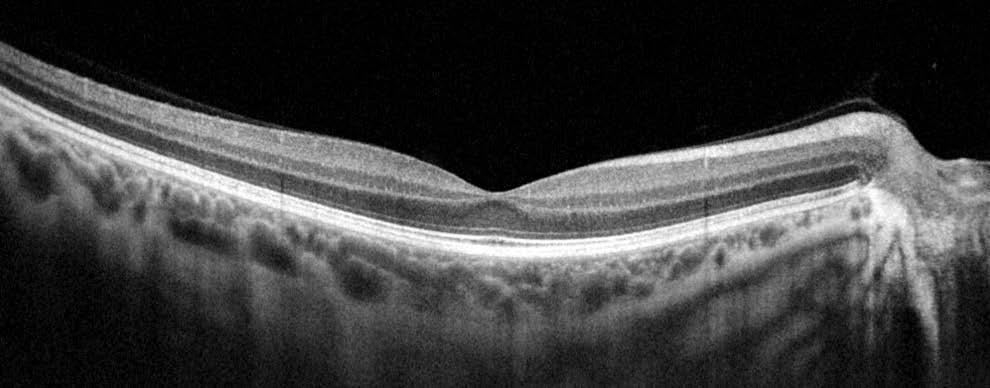
2 lution continues Optopol engineering team, designers of the first commercially available Spectral Domain OCT in the world, are proud to present the World s fastest OCT. Our supreme experience in Spectral Domain OCT allows us to provide the market with the state of the art instrument, offering advanced technologies and remarkable simplicity of operation. The new REVO NX software meets all demands of a daily routine in a modern ophthalmic practice. The new angiography module expands the precision of your diagnosis with minimum patient fatigue. Need for speed The world s fastest available scanning speed allows for more achievable and more detailed exams with reduction of the scanning time. It brings benefits for both clinicians and patients by reducing errors often caused by involuntary eye movements. OCT made simple as never before Position the patient and press the START button to acquire examinations of both eyes. The Revo NX, using vocal messages, guides the patient through the process, increasing comfort and reducing patient chair time. Short scanning time ensures less fatigue for the patient. Creating customized scanning protocols of different diagnostic scenarios speed s up the workflow. A perfect fit for every practice. With a small system footprint and access for both operator and patient only necessary from one side, space saving is further enhanced. In addition, connection by a single cable allows the installation of REVO NX into the smallest of examination room spaces. Revo s variety of examination and analysis tools enables it to effortlessly function as a screening or advanced diagnostic device. High quality of OCT image The noise reduction technology provides the finest details proven to be important for early disease detection.


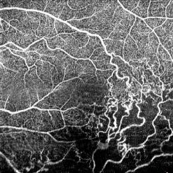
3 offers the newest standards available in OCT technology ANGIOGRAPHY SOCT* This non-invasive dye free technique allows the visualization of the microvasculature of the retina. Both blood flow and structural visualization will give additional information in the diagnosis of many retinal diseases. Angiography scan allows assessment of the structural vasculature of the macula, periphery or the optic disc. Extremely short scanning time 1.6 second in standard resolution or in high resolution within ~3 seconds. Superficial plexus Deep Plexus Optic Nerve Head Peripheral area Now Angiography OCT can become a routine diagnosis in your practice. RETINA A single 3D macula scan performs both Retina and Glaucoma analysis.the software automatically recognizes 8 retinal layers which assists with a precise diagnosis and the mapping of any changes in the patient s condition. A variety of result analysis and presentation methods allows for the best selection suitable to increase efficiency of work. Single Comparison Both Progression WIDEFIELD SCAN 12x12 mm Widefield Central scan is perfect for fast and precise screening of the patient s retina. Dense scanning in high resolution tomograms guarantee the discovery of most of the early changes. Peripheral scanning reveals diseases in the far periphery. * SOCT Angio is an optional software module to purchase. Combined view of two examinations of peripheral scan 12 mm + 12 mm. Done in external software.


4 GLAUCOMA Comprehensive glaucoma analysis tools for quantification of the Nerve Fiber Layer, Ganglion layer and Optic Head with DDLS allows for precise diagnosis and the monitoring of glaucoma over time. Asymmetry Analysis of Ganglion layers between hemispheres and between eyes allows easier identification and detection of glaucoma in early stages and in non-typical patients. ONH Both GCL+IPL Both ONH Progression GCL+IPL Progression ANTERIOR For a standard anterior examination, no additional lens is required. This allows the examiner to quickly complete the scanning procedure. Presentation of results for both eyes allows quick and precise evaluation of the condition of the anterior segment. Cornea Both Angle Both Additional adapter provided with the device increases range of clinical application in Anterior chamber observation. FOLLOW UP Revo s standard high density scanning capability and blood vessel structure recognition enable a precise alignment of past and current scans The Operator can analyze changes is morphology, quantified progression maps and evaluate the progression trends. Progression Morphology Progression Quantification DICOM, EMR, NETWORK INTEGRATION A proficient networking solution increases productivity and enhances the patient experience. It allows you to view and manage multiple examinations from review stations in your practice. Effortlessly helping to facilitate patient education by allowing you to interactively show examination results to patients. Every practice will have different requirements which we can provide by tailoring a bespoke service. DICOM connectivity allows the connection of the REVO into large hospital medical systems. Receive the Worklist (MWL) and send report (C-storage) or whole exam into view stations. CMDL interface allows for the integration of the REVO in to practice management systems. There is no additional charge for the networking and DICOM functionality.


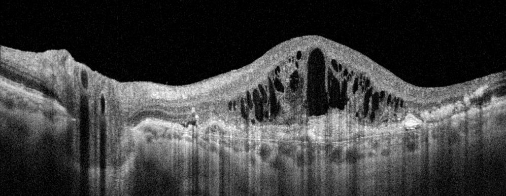
5 Clinical images CNV BRVO GA Choroidal observation Central 12 mm scan Angle to Angle scan

6 t e c h n i c a l S p e c i f i c at i o n Technology Spectral Domain OCT Light Source SLED, Wavelength 830 nm Bandwidth 50 nm half bandwidth Scanning speed measurements per second Axial resolution 5 μm in tissue 2,6 μm digital Transverse Resolution 12 μm, typical 18 μm Overall scan depth 2.4 mm Min. pupil size 3 mm Focus adjustment range -25 D to +25 D Scan range Posterior 5-12 mm, Angio 3-9 mm, Anterior 3-16 mm Scan types 3D, Angio*, Radial (HD), B-scan (HD), Raster (HD), Cross (HD) Fundus image Live Fundus Reconstruction Alignment method Fully automatic, Automatic Retina analysis Retina thickness, Inner retinal thickness, Outer retinal thickness RNFL+GCL+IPL thickness, GCL+IPL thickness, RNFL thickness, RPE deformation, IS/OS thickness Angiography OCT* Superficial plexus, Deep Plexus, Outer Retina, Choriocapilaries, Depth Coded, Custom, Enface, Thickness map Glaucoma analysis RNFL, ONH morphology, DDLS, OU and Hemisphere asymmetry, Ganglion analysis as RNFL+GCL+IP and GCL+IPL Anterior Pachymetry, LASIK flap, Angle Assessment, AIOP, AOD 500/750, TISA 500/750 Anterior Wide Scan Angle to Angle view (Adapter required) Connectivity DICOM Storage SCU, DICOM MWL SCU, CMDL, Networking Dimensions (WxDxH) 382 x mm Weight 23 kg Fixation target OLED display (The target shape and position can be changed), External fixation arm Power supply V, 50/60 Hz Power consumption VA OPTOPOL Technology Sp. z o. o. ul. Żabia 42, Zawiercie, Poland Tel/Fax: info@optopol.com.pl ver. REVO NX
7 Biometry OCT in SOCT software 8.0 version
8 Biometry OCT Biometry OCT Innovative method of using the posterior OCT device to measure ocular structure along eye axis. OCT-B based on measurements of the mutual position of individual eye elements. The Eye elements are measured individually. System captures separate exams and measures distances between structures of eye elements.
9 Biometry OCT OCT Biometry provides: AL Axial Length SD +/ mm ACD Anterior chamber depth SD +/ mm LT Lens thickness SD +/ mm CCTCornea thickness SD +/- 6 um Axial Length ACD LT CCT


10 Biometry OCT 2 Biometry programs: AL AL.,CCT, ACD, LT parameter ACD CCT, ACD parameter Acquisition method: Full Auto Auto Number of repeats 5, 10, 15 Acquisition time 5 AL. full measurements ~ 4 sec


11 Analysis Single view Biometry OCT Both eyes view Result review window Single view and Both View Results table Result verification Excluding deviated results AVG and SD for measurements series

12 Biometry OCT Analysis Verification and correction Full screen view Precise review of each measurement Manual correction of boundaries Echogram or precise manual alignment
13 Biometry OCT Biometrical measurement sample cases Healthy Cataract Dense cataract still manual measurement is possible
14 Biometry OCT Clinical use Axial distances measurement UBM verification AL in monitoring Hi myopic eyes ACD management of glaucoma Benefits Extends the use of Standard OCT OCT Biometry provides exact measured boundaries of ocular structures Manual correction of boundaries which is not available in Gold standard Verification and precise correction for non typical cases Precise IOL detection for pseudo phakic eyes Limits Slightly lower penetration of dense cataract patient than newest SS optical biometers with 1050 nm wavelength


15 Biometry OCT Male, 37 y/old Healthy Healthy subject with visible peak in the vitrous behind the crystalline lens Berger space and vitrous


16 Biometry OCT Male, 24 y/old Healthy Patient with contact lens on the cornea Only REVO is able provide correct measurement. All other devices cannot detect the lens.


17 Biometry OCT Female, 67 y. Cataract


18 Biometry OCT Female, 40 y/old Cataract over posterior lens capsule


19 B-OCT Verification Female, 68 y. Patient with Macular hole after PPV treatment IOL 700 LT measurement error
![B-OCT Verification IOL 700 REVO NX AL [mm] 27,16 27,16 ACD [mm] 4,13 4,16](/docs-images/88/116116812/images/20-1.jpg "LT [mm] 3,83 3,80 CCT [mm] 0,674 0,666 Patient: Male, 40 y.")

20 B-OCT Verification IOL 700 REVO NX AL [mm] 27,16 27,16 ACD [mm] 4,13 4,16 LT [mm] 3,83 3,80 CCT [mm] 0,674 0,666 Patient: Male, 40 y. Contact lens on the cornea, myopic patient. In REVO NX it is possible to modified detected boundary position In IOL it is not possible to modify detected boundaries.
![B-OCT Verification IOL 700 REVO NX AL [mm] 23,27 23,27 ACD [mm]](/docs-images/88/116116812/images/21-1.jpg "3,58 3,57 LT [mm] 4,22 4,22 CCT [mm] 0,583 0,587 Female, 50 y.")
21 B-OCT Verification IOL 700 REVO NX AL [mm] 23,27 23,27 ACD [mm] 3,58 3,57 LT [mm] 4,22 4,22 CCT [mm] 0,583 0,587 Female, 50 y. Healthy

![3,66 LT [mm] 4,86 4,93 CCT [mm]](/docs-images/88/116116812/images/22-2.jpg "0,519 0,515 Patient: Male, 72 y.")
22 B-OCT Verification IOL 700 REVO NX AL [mm] 23,50 23,50 ACD [mm] 3,67 3,66 LT [mm] 4,86 4,93 CCT [mm] 0,519 0,515 Patient: Male, 72 y. Cataract
![B-OCT Verification IOL 700 REVO NX AL [mm] 21,94 22,03 ACD [mm]](/docs-images/88/116116812/images/23-2.jpg "2,81 2,98 LT [mm] 3,99 3,92 CCT [mm] 0,559 0,599 Female, 31 y.")
23 B-OCT Verification IOL 700 REVO NX AL [mm] 21,94 22,03 ACD [mm] 2,81 2,98 LT [mm] 3,99 3,92 CCT [mm] 0,559 0,599 Female, 31 y. Healthy

![4,15 4,00 CCT [mm] 0,567 0,586 Patient: Female, 55 y.](/docs-images/88/116116812/images/24-2.jpg "Cataract The biggest difference in comaparison study IOL does")
24 B-OCT Verification? IOL 700 REVO NX AL [mm] 28,94 29,17 ACD [mm] 3,49 3,54 LT [mm] 4,15 4,00 CCT [mm] 0,567 0,586 Patient: Female, 55 y. Cataract The biggest difference in comaparison study IOL does not show detected boundaries Long eye ball AL~29mm

25
SOCT Copernicus REVO. * - Currently import and overlay are avaibale in manual mode only
 SOCT Copernicus REVO Easy Operation (Full auto & Auto mode) Auto alignment (Z-position, C-gate, Focus, Tomogram) Voice guide (support patient through examination) Powerful analysis tools Enhanced tomograms
Visualize. Analyze. Personalize. OCT + OCTA
 Visualize. Analyze. Personalize. OCT + OCTA A New Approach to Protecting Vision AngioVue OCT Angiography brings valuable new information to clinical practice. Non-invasive visualization of retinal vasculature.
Visualize. Analyze. Personalize. OCT + OCTA. with
 Visualize. Analyze. Personalize. OCT + OCTA with Avanti Widefield OCT with AngioVue OCTA Imaging Comprehensive Structural and Functional Imaging in a Single Imaging Platform Comprehensive OCT Imaging The
Cirrus TM HD-OCT. Details define your decisions
 Cirrus TM HD-OCT Details define your decisions 2 With high-definition OCT Carl Zeiss Meditec takes you beyond standard spectral domain Built on 10 years experience at the vanguard of innovation, Carl Zeiss
PRIMUS 200 from ZEISS The essential OCT
 PRIMUS 200 from ZEISS The essential OCT Seeing beyond the surface. ZEISS PRIMUS 200 // INNOVATION MADE BY ZEISS Clear Visualization. Advanced Technology. Reliability. Essential elements of your first OCT.
NEW AUTOMATED PERIMETERS NEW. Fast and precise perimetry at your fingertips. ZETA strategy EyeSee recording DPA analysis
 NEW AUTOMATED PERIMETERS Fast and precise perimetry at your fingertips NEW ZETA strategy EyeSee recording DPA analysis PTS 920 PTS 925W I PTS 2000 PTS AUTOMATED PERIMETER SERIES THRESHOLD IN 3 MINUTES**
DRI OCT Triton Series A Multimodal Swept Source OCT
 DRI OCT Triton Series A Multimodal Swept Source OCT Color Red-Free FA FAF Posterior Anterior See what others can t see. A Multimodal Swept Source OCT DEEP RANGE IMAGING Swept Source OCT imaging massively
Swept-Source OCT Angiography: SS OCT Angio TM
 Swept-Source OCT Angiography: SS OCT Angio TM Not available in all countries, please check with your distributor. 2015.09 Swept-Source OCT Angiography: SS OCT Angio TM Introduction Optical coherence tomography
Introducing ANGIOVUE ESSENTIAL. Built on the Avanti Widefield OCT Platform. OCT Angiography for Primary Eye Care
 Introducing ANGIOVUE ESSENTIAL Built on the Avanti Widefield OCT Platform OCT Angiography for Primary Eye Care Transform Your View of the Retina OCT Angiography (OCTA) is a quick non-invasive test that
3/23/2016. Diagnostic Services Taylor Pannell CRA, OCT-C. Services Available. Important info for the Tech to know. Visual Fields
 Services Available Diagnostic Services Taylor Pannell CRA, OCT-C Static and Kinetic Visual Fields Pachymetry Anterior and Posterior Segment OCT Fundus Photos FAF,FA,ICG Slit Lamp Photography Confocal HRT
HOCT-1I 1F All-in-One Optical Coherence Tomography with Fundus
 HOCT-1I 1F All-in-One Optical Coherence Tomography with Fundus Specification Type Resolution(in Tissue) A scan Rate Scan Range SD-OCT / Fundus Z :6~7um, XY:20um 68,000 A-scan/sec. [Fundus] X:6-12mm, Y:6-9mm,
Advances in OCT Murray Fingeret, OD
 Disclosures Advances in OCT Murray Fingeret, OD Consultant Alcon, Allergan, Bausch & Lomb, Carl Zeiss Meditec, Diopsys, Heidelberg Engineering, Reichert, Topcon Currently Approved OCT Devices OCT Devices
Cirrus TM HD-OCT. Details defi ne your decisions
 Cirrus TM HD-OCT Details defi ne your decisions 2 With high-defi nition OCT Carl Zeiss Meditec takes you beyond standard spectral domain Built on 10 years experience at the vanguard of innovation, Carl
PRIMUS 200 from ZEISS The essential OCT
 EN 00_00I The contents of the brochure may differ from the current status of approval of the product in your country. Please contact your regional representative for more information. Subject to change
What is the Value of Swept Source oct Technology in Biometry? Experts discussed the IOLMaster 700 at the ESCRS ebook. Content provided by:
 ebook Content provided by: What is the Value of Swept Source oct Technology in Biometry? Experts discussed the IOLMaster 700 at the ESCRS 2016 Participating experts: G. Barrett, MD, Australia; D. Chang,
Experience Spectacular Retinal Imaging with the new NIDEK F-10 Digital Ophthalmoscope
 Experience Spectacular Retinal Imaging with the new NIDEK F-10 Digital Ophthalmoscope The F-10 was developed to give Ophthalmologists a high definition (HD) diagnostic imaging system. Designed to provide
The Measure of Confidence
 Heidelberg_936357.qxd:Layout 1 5/9/08 12:01 PM 12:02 Page 1 (Cyan (Magenta (Yellow (Black (UV Five Powerful Solutions to Fit Your Practice PowerCheck Glaucoma FastCheck+ GPS Software and Retina Edema Index
OCT Image Analysis System for Grading and Diagnosis of Retinal Diseases and its Integration in i-hospital
 Progress Report for1 st Quarter, May-July 2017 OCT Image Analysis System for Grading and Diagnosis of Retinal Diseases and its Integration in i-hospital Milestone 1: Designing Annotation tool extraction
FA vs. OCTA? The status of OCTA, today. Fukuoka, JSOS 2016 Gerd Klose. Korobelnik J Fr Ophthalmol (2015)
") FA vs. OCTA? The status of OCTA, today Korobelnik J Fr Ophthalmol (2015) Fukuoka, JSOS 2016 Gerd Klose 1 2 FA / ICGA a well-founded Gold standard! Benefits Useful for many pathologies High contrast, detailed
FA vs. OCTA? The status of OCTA, today Korobelnik J Fr Ophthalmol (2015) Fukuoka, JSOS 2016 Gerd Klose 1 2 FA / ICGA a well-founded Gold standard! Benefits Useful for many pathologies High contrast, detailed
PLEX Elite 9000 from ZEISS Swept-Source OCT
 PLEX Elite 9000 from ZEISS Swept-Source OCT Uncovering the undiscovered. ZEISS PLEX Elite 9000 // INNOVATION MADE BY ZEISS 2 Ultra-wide angiography En face montage Image courtesy of Prof. G. Querques,
Incorporating OCT Angiography Into Patient Care
 Incorporating OCT Angiography Into Patient Care Beth A. Steele, OD, FAAO OCT A: Introduction Isolates microvascular circulation from OCT image data Axial resolution = 5 microns (i.e. fine capillaries visible)
The ideal tool for early detection and monitoring of AMD.
 The ideal tool for early detection and monitoring of AMD. presenting maia 1 MAIA, the new frontier of Fundus Perimetry (microperimetry) assesses the function of the macula representing an effective clinical
LEE EYE CENTRE. YOUR VISION, OUR PASSION LEC EyeNews
 LEE EYE CENTRE YOUR VISION, OUR PASSION LEC EyeNews FOR INTERNAL CIRCULATION ONLY www.lec.com.my ISSUE 51/003 SEPT OCT 2017 The American Society of Cataract and Refractive Surgery is one of the leading
3/16/2018. Ultrasound Biomicroscopy in Glaucoma By Ahmed Salah Abdel Rehim. Prof. of Ophthalmology Al-Azhar University
 Ultrasound Biomicroscopy in Glaucoma By Ahmed Salah Abdel Rehim Prof. of Ophthalmology Al-Azhar University 1 Ultrasound biomicroscopy (UBM) is a recent technique to visualize anterior segment with the
SOUTH-EAST EUROPEAN JOURNAL of OPHTHALMOLOGY 2015; 1 (1) 34 40
 34 40") Review article SOUTH-EAST EUROPEAN JOURNAL of OPHTHALMOLOGY 2015; 1 (1) 34 40 Retinal nerve fiber layer versus peripapillary capillary density assessment A powerful tool for detecting optic nerve head
Review article SOUTH-EAST EUROPEAN JOURNAL of OPHTHALMOLOGY 2015; 1 (1) 34 40 Retinal nerve fiber layer versus peripapillary capillary density assessment A powerful tool for detecting optic nerve head
ZEISS AngioPlex OCT Angiography Making the revolutionary, routine.
 ZEISS AngioPlex OCT Angiography Making the revolutionary, routine. The moment that revolutionary insight becomes routine. // OCT ANGIOGRAPHY MADE BY ZEISS CIRRUS with AngioPlex creates a new era in both
Cornea/Anterior Segment OCT. User Experience
 Cornea/Anterior Segment OCT User Experience User Experience Case#1 Post Penetrating Keratoplasty Tokyo Medical University / Kohsei Chuo General Hospital Hideki Mori MD, PhD Almost eight years have passed
Simply the best OCT & OCTA image quality.
 Avanti Widefield OCT with AngioVue OCT Angiography Simply the best OCT & OCTA image quality. Dear Friends of Optovue, Since introducing Spectral Domain OCT to the ophthalmology market in 2006, Optovue
Mark Dunbar: Disclosure
 Important Things to Understand About OCT Mark T. Dunbar, O.D., F.A.A.O. Bascom Palmer Eye Institute University of Miami, School of Medicine Mark Dunbar: Disclosure Optometry Advisory Board for: Allergan
Lasers and Imaging PHOTOCOAGULATION PHOTODISRUPTION SLT PHOTOREGENERATION DIAGNOSTIC ULTRASOUND
 Lasers and Imaging PHOTOCOAGULATION PHOTODISRUPTION SLT PHOTOREGENERATION DIAGNOSTIC ULTRASOUND Diagnostic Ultrasound The Eye Cubed delivers highest-quality image resolution and unparalleled sensitivity
Evolving glaucoma management True diagnostic integration for the preservation of vision
 Evolving glaucoma management True diagnostic integration for the preservation of vision // GLAUCOMA MANAGEMENT MADE BY ZEISS The moment you are certain it is glaucoma. This is the moment we work for. There
Structural examina.on: Imaging
 ManaMa: Glaucoma Structural examina.on: Imaging Luís Abegão Pinto, MD, PhD Department of Ophthalmology CHLC Lisbon Faculty of Medicine, Lisbon University 1 11-10- 2013 Structural changes Qualitative changes
OCT Interpretation in Retinal Disease
 OCT Interpretation in Retinal Disease Jay M. Haynie, OD, FAAO Financial Disclosure I have received honoraria or am on the advisory board for the following companies: Carl Zeiss Meditec Advanced Ocular
Ultrahigh Speed Imaging of the Rat Retina Using Ultrahigh Resolution Spectral/Fourier Domain OCT
 Ultrahigh Speed Imaging of the Rat Retina Using Ultrahigh Resolution Spectral/Fourier Domain OCT The MIT Faculty has made this article openly available. Please share how this access benefits you. Your
THE CHRONIC GLAUCOMAS
 THE CHRONIC GLAUCOMAS WHAT IS GLAUCOMA? People with glaucoma have lost some of their field of all round vision. It is often the edge or periphery that is lost. That is why the condition can be missed until
Moving forward with a different perspective
 Moving forward with a different perspective The Leader In Vision Diagnostics Offers A New Perspective Marco has served the eyecare community by offering exceptional lane products and automated high tech
Lasers and Imaging PHOTOCOAGULATION PHOTODISRUPTION SLT PHOTOREGENERATION DIAGNOSTIC ULTRASOUND
 Lasers and Imaging PHOTOCOAGULATION PHOTODISRUPTION SLT PHOTOREGENERATION DIAGNOSTIC ULTRASOUND One Powerful Vision Photodisruption The tough demands of today s new-generation intraocular lenses (IOLs)
R&M Solutions
 Mohamed Hosny El-Bradey, MD., Assistant Professor of Ophthalmology, Tanta University. Wael El Haig, MD., Professor of Ophthalmology. Zagazeeg University. 1 Myopic CNV is considered the most common vision
ZEISS AngioPlex OCT Angiography Overview ZEISS OCT Angiography
 ZEISS AngioPlex OCT Angiography Overview ZEISS OCT Angiography California, ZEISS AngioPlex Ultra-clear visualization of microvascular blood flow using non-invasive OCT angiography 2 AngioPlex OCT Angiography
Corporate Medical Policy
 Corporate Medical Policy Optical Coherence Tomography (OCT) Anterior Segment of the Eye File Name: Origination: Last CAP Review: Next CAP Review: Last Review: optical_coherence_tomography_(oct)_anterior_segment_of_the_eye
Widefield Retinal Imaging with Auto Fluorescence Technology in the Optometric Practice
 Widefield Retinal Imaging with Auto Fluorescence Technology in the Optometric Practice This course will define ultra-widefield retinal imaging and autofluorescence for the attendee. Will show how it is
Exceptional versatility without compromise
 Introducing the VICTUS femtosecond laser platform Exceptional versatility without compromise FEMTOSECOND TECHNOLOGY that empowers Introducing VICTUS the first femtosecond laser capable of exceptional performance
The Evolution of Fundus Perimetry
 The Evolution of Fundus Perimetry Company Profile CenterVue designs and manufactures highly automated medical devices for the diagnosis and management of ocular pathologies, including those that represent
Will OCT-Angiography replace FA?
 ASL Roma A PRESIDIO TERRITORIALE NUOVO REGINA MARGHERITA AMBULATORIO PATOLOGIE RETINICHE Resp. Dott.ssa SUSANNA CATALANO CENTRO ITALIANO MACULA Will OCT-Angiography replace FA? Marco Rispoli, Luca di Antonio,
OCT in the Diagnosis and Follow-up of Glaucoma
 OCT in the Diagnosis and Follow-up of Glaucoma Karim A Raafat MD. Professor Of Ophthalmology Cairo University Hmmmm! Do I have Glaucoma or not?! 1 Visual Function 100% - N Gl Structure : - 5000 axon /
Deeper visualizations for intervening with confidence.
 CIRRUS OCT with AngioPlex from ZEISS Making the revolutionary routine New vascular quantification Deeper visualizations for intervening with confidence. CIRRUS OCT with AngioPlex from ZEISS can be a much
Technologies and Methods for Visualizing the Retina
 Transcript Details This is a transcript of an educational program accessible on the ReachMD network. Details about the program and additional media formats for the program are accessible by visiting: https://reachmd.com/programs/revealing-retina/technologies-and-methods-for-visualizing-theretina/3663/
Lasers and Imaging PHOTOCOAGULATION PHOTODISRUPTION SLT PHOTOREGENERATION DIAGNOSTIC ULTRASOUND
 Lasers and Imaging PHOTOCOAGULATION PHOTODISRUPTION SLT PHOTOREGENERATION DIAGNOSTIC ULTRASOUND Diagnostic Ultrasound Innovative Imaging has long been considered the premier name in diagnostic ophthalmic
HOW TO MAKE THE MOST OF A NEW OCT. with Kelly Kerksick, OD
 HOW TO MAKE THE MOST OF A NEW OCT with Kelly Kerksick, OD 3 How to Make the Most of a New OCT Kelly Kerksick, OD, graduated from Southern College of Optometry and immediately started her own private practice
On Different Wavelengths: The Spectrum of Retinal Imaging. On Different Wavelengths: The Spectrum of Retinal Imaging. Wavelength Specific Imaging
 On Different Wavelengths: The Spectrum of Retinal Imaging Timothy J. Bennett, CRA, FOPS, OCT-C Penn State Hershey Eye Center Hershey, PA On Different Wavelengths: The Spectrum of Retinal Imaging Wavelengths
New Technologies in Glaucoma Management: From ERG to OCT
 What s New and What s Next in Glaucoma New Technologies in Glaucoma Management: From ERG to OCT Ben Gaddie, OD FAAO Murray Fingeret, OD FAAO IOP 24- Hour IOP Role of hysteresis in glaucoma risk Cerebrospinal
Interpretation of corneal tomography
 Interpretation of corneal tomography Presented by Chameen Samarawickrama - Westmead Hospital - Liverpool Hospital - University of Sydney - University of New South Wales The University of Sydney Page 1
Patient Referral. Date: Doctor s Name: Phone: Contact Fax: Preferred Method of Communication (circle one): Fax Letter
: Fax Letter") Patient Referral Date: Doctor s Name: Phone: Contact Email: Fax: Preferred Method of Communication (circle one): Email Fax Letter Patient s Name: Phone: DOB: Please check your doctor preference: Savannah
Patient Referral Date: Doctor s Name: Phone: Contact Email: Fax: Preferred Method of Communication (circle one): Email Fax Letter Patient s Name: Phone: DOB: Please check your doctor preference: Savannah
Il contributo dell'angio-oct: valutazione integrata della componente nervosa e vascolare della malattia glaucomatosa
 SIMPOSIO G.O.A.L. - LE NUOVE FRONTIERE DIAGNOSTICHE E LE LINEE DI INDIRIZZO AMBULATORIALI DEL GLAUCOMA Coordinatore e moderatore: D. Mazzacane Presidente: L. Rossetti Il contributo dell'angio-oct: valutazione
C a t a r a c t G l a u c o m a R e t i n a R e f r a c t i v e. The GDxVCC Early answers and ongoing assessment for glaucoma
 C a t a r a c t G l a u c o m a R e t i n a R e f r a c t i v e The GDxVCC Early answers and ongoing assessment for glaucoma The quantifiable approach to quality care Only Humphrey GPA software Early insight
Optical Coherence Tomography (OCT)
") Understanding and Interpreting OCT Mark Dunbar: Disclosure The Swiss Army Pocket Knife of Eye Care Mark T. Dunbar, O.D., F.A.A.O. Bascom Palmer Eye Institute University of Miami, School of Medicine Consultant
Understanding and Interpreting OCT Mark Dunbar: Disclosure The Swiss Army Pocket Knife of Eye Care Mark T. Dunbar, O.D., F.A.A.O. Bascom Palmer Eye Institute University of Miami, School of Medicine Consultant
EasyScan: Smart Retinal Imaging
 easyscan EasyScan: Smart Retinal Imaging Superior Imaging Enjoy the benefits of SLO technology and capture high-quality images easily for accurate diagnosis. Never Dilate Reduce examination time, capture
Yasser R. Serag, MD Tamer Wasfi, MD El- Saied El-Dessoukey, MD Magdi S. Moussa, MD Anselm Kampik, MD
 Microperimetric Evaluation of Brilliant Blue G- assisted Internal Limiting Membrane Peeling By Yasser R. Serag, MD Tamer Wasfi, MD El- Saied El-Dessoukey, MD Magdi S. Moussa, MD Anselm Kampik, MD The internal
IN NICU OCT UTILIZES A CONCEPT KNOWN AS INTERFEROMETRY APPLICATIONS FOR OCT THE PRIMARY USE IN THE EYE - RETINA
 2016 25 YEARS OF OPTICAL COHERENCE TOMOGRAPHY OPTICAL COHERENCE TOMOGRAPHY IN NICU Marcin Stopa, MD, PhD, FEBO Department of Ophthalmology, Chair of Ophthalmology and Optometry. Poznan University of Medical
Is OCT-A Needed As An Investigative Tool During The Management Of Diabetic Macular Edema
 Is OCT-A Needed As An Investigative Tool During The Management Of Diabetic Macular Edema Ayman M Khattab MD, FRCS Professor of Ophthalmology Cairo University Diabetic Macular Edema (DME) Diabetic macular
ZEISS presents innovations that support ophthalmologists in their work
 ZEISS presents innovations that support ophthalmologists in their work At the 2015 Annual Meeting of the American Academy of Ophthalmology (AAO) in Las Vegas, ZEISS announces innovations for clinical excellence
OCT Angiography in Primary Eye Care
 OCT Angiography in Primary Eye Care An Image Interpretation Primer Julie Rodman, OD, MS, FAAO and Nadia Waheed, MD, MPH Table of Contents Diabetic Retinopathy 3-6 Choroidal Neovascularization 7-9 Central
Diagnosis in AMD. Managing your AMD Patients
 Managing your AMD Patients Robert W. Dunphy, O.D., F.A.A.O. Diagnosis in AMD Have suspicion Identify relative risk Conduct surveillance Biometry Utilize technology to facilitate detection of change / stability
10/17/2017. FDA Approved. Zeiss AngioPlex TM Optovue AngioVue TM
 Images retinal microvasculature without dye injection Displays structure and function from a single imaging system Standard of Care-2011 DFE, Fundus Photos, VF 10-2, SD-OCT, FAF, or mferg 2016-AAO Baseline
Learn Connect Succeed. JCAHPO Regional Meetings 2017
 Learn Connect Succeed JCAHPO Regional Meetings 2017 Faculty Biometry and IOL Calculations ASCRS and ASOA Symposium and Congress Los Angeles, CA Daniel H. Chang, M.D. - Empire Eye and Laser Center Bakersfield,
QUANTIFICATION OF PROGRESSION OF RETINAL NERVE FIBER LAYER ATROPHY IN FUNDUS PHOTOGRAPH
 QUANTIFICATION OF PROGRESSION OF RETINAL NERVE FIBER LAYER ATROPHY IN FUNDUS PHOTOGRAPH Hyoun-Joong Kong *, Jong-Mo Seo **, Seung-Yeop Lee *, Hum Chung **, Dong Myung Kim **, Jeong Min Hwang **, Kwang
WORKSHOP B Ophthalmic Imaging: All Hands on Tech! COPE Course PS
 WORKSHOP B Ophthalmic Imaging: All Hands on Tech! COPE Course 44334-PS Ophthalmic Imaging: All Hands on Tech! Southern College of Optometry April 17, 2015 COPE #44334-PS Faculty Dr. Michael Gerstner Dr.
4/19/2018 FUNDUS AUTOFLUORESCENCE. Fluorescence Imaging. Fundus Autofluorescence (FAF) Fluorescence. Fluorescence
 Fluorescence. Fluorescence") I have no financial or proprietary interest in the subject matter of this presentation. FUNDUS AUTOFLUORESCENCE Timothy J. Bennett, CRA, OCT-C, FOPS Penn State Eye Center Hershey, PA Fluorescence Imaging
I have no financial or proprietary interest in the subject matter of this presentation. FUNDUS AUTOFLUORESCENCE Timothy J. Bennett, CRA, OCT-C, FOPS Penn State Eye Center Hershey, PA Fluorescence Imaging
Learn Connect Succeed. JCAHPO Regional Meetings 2016
 Learn Connect Succeed JCAHPO Regional Meetings 2016 pearls and pitfalls of ophthalmic imaging JCHAPO 2016 Conference Vikas Chopra, M.D. Medical Director, UCLA Doheny Eye Centers Pasadena Principal Investigator,
and at the same patient encounter. Code has been deleted. For scanning computerized ophthalmic diagnostic imaging of optic nerve and retin
 92227: Remote imaging for detection of retinal disease (eg, retinopathy in a patient with diabetes) with analysis and report under physician supervision, unilateral or bilateral. For Medicare, bill only
OCT Angiography: The Next Step in Retinal Imaging Jonathan Zelenak D.O.
 OCT Angiography: The Next Step in Retinal Imaging Jonathan Zelenak D.O. Hillsdale Hospital Michigan State University Overview Evolution of OCT How does OCT angiography work? Clinical examples Potential
Choroidal Mapping; a Novel Approach for Evaluating Choroidal Thickness and Volume
 Imaging Technique Choroidal Mapping; a Novel Approach for Evaluating Choroidal Thickness and Volume Jila Noori 1, MD; Mohammad Riazi Esfahani 1,2, MD Fedra Hajizadeh 2, MD; Mohammad-Mehdi Zaferani 1, MD
Continuing Femtosecond Laser Innovation
 VICTUS Femtosecond Laser Platform Continuing Femtosecond Laser Innovation See better. Live better. VICTUS Femtosecond Laser Platform The VICTUS Femtosecond Laser combines sophisticated high-speed OCT technology
How to Be Efficient and Effective. Disclosure. Topics CASE CM. Case JF 2007 OHTN / POAG? How to Be Efficient and Effective with. with New Technology
 How to Be Efficient and Effective with Disclosure COPE Course ID: 40750 GL Michael Chaglasian has the following disclosures: 1. Advisory Board: Allergan, Inc., Alcon Labs, B+L Carl Zeiss Meditec 2. Research:
Is this glaucoma? Leo Semes, OD Michael Chaglasian, OD Danica Marrelli, OD. Optometry s Meeting 2015 Seattle, WA
 Is this glaucoma? Leo Semes, OD Michael Chaglasian, OD Danica Marrelli, OD Optometry s Meeting 2015 Seattle, WA Case 1. 54 WM Engineer is referred to UAB Eye Care as a glaucoma suspect. Mild myopic refractive
Measuring of the fovea and foveola using line scans and 3D Macular scans obtained with spectral domain optical coherent tomography.
 Measuring of the fovea and foveola using line scans and 3D Macular scans obtained with spectral domain optical coherent tomography. Vakhrameeva O.A. 1, Moiseenko G.A. 1, Maltsev D.S. 2, Sukhinin M.V. 2,
Glaucoma. Cornea. Iris
 Glaucoma Introduction Glaucoma is a group of eye diseases that can lead to blindness if not treated. Openangle glaucoma, the most common form of glaucoma, affects about 3 million Americans. Half of those
Overview. Macular OCT Artifact Study
 Imaging Artifacts Sarah Moyer, CRA, OCT-C Director, Ophthalmic Imaging Kittner Eye Center University of North Carolina Chapel Hill, NC Disclose financial interest now Overview Sarah s Thoughts on Artifacts
History/principles of the OCT What does the normal retinal OCT look like Vitreal disorders Retinal/RPE disorders Choroidal disorders
 Nathan Lighthizer, O.D., F.A.A.O. Assistant Professor Assistant Dean for Clinical Care Director of Continuing Education Chief of Specialty Care Clinics Chief of Electrodiagnostics Clinic Oklahoma College
THE CHRONIC GLAUCOMAS
 THE CHRONIC GLAUCOMAS WHAT IS GLAUCOMA People with glaucoma have lost some of their field of all round vision. It is often the edge or periphery that is lost. That is why the condition can be missed until
ZONARE Ultrasound for the Emergency Department. Living Technology
 ZONARE Ultrasound for the Emergency Department Living Technology We ve talked about the ultrasound challenges you face every day: I need consistent image performance across all my imaging applications.
Optical Coherence Tomography: Pearls for the Anterior Segment Surgeon Basic Science Michael Stewart, M.D.
 Optical Coherence Tomography: Pearls for the Anterior Segment Surgeon Basic Science Michael Stewart, M.D. Disclosure OCT Optical Coherence Tomography No relevant financial relationships I will refer to
Navigated Laser Therapy. A New Era in Retinal Disease Management
 Navigated Laser Therapy A New Era in Retinal Disease Management Bringing Navigation to Retina Treatment Navilas Laser System To unleash the full potential of Retina Navigation, the Navilas Laser System
Macular Ganglion Cell Complex Measurement Using Spectral Domain Optical Coherence Tomography in Glaucoma
 Med. J. Cairo Univ., Vol. 83, No. 2, September: 67-72, 2015 www.medicaljournalofcairouniversity.net Macular Ganglion Cell Complex Measurement Using Spectral Domain Optical Coherence Tomography in Glaucoma
Ganglion cell analysis by optical coherence tomography (OCT) Jonathan A. Micieli, MD Valérie Biousse, MD
 Jonathan A. Micieli, MD Valérie Biousse, MD") Ganglion cell analysis by optical coherence tomography (OCT) Jonathan A. Micieli, MD Valérie Biousse, MD Figure 1. Normal OCT of the macula (cross section through the line indicated on the fundus photo)
Ganglion cell analysis by optical coherence tomography (OCT) Jonathan A. Micieli, MD Valérie Biousse, MD Figure 1. Normal OCT of the macula (cross section through the line indicated on the fundus photo)
OCT Angiography The Next Frontier
 Choroid Retina avascular 5/13/2017 OCT Angiography The Next Frontier Pierce Kenworthy OD, FAAO June 9, 2017 OCT Angiography (OCTA) 2016 Non-invasive, motion contrast imaging Represents erythrocyte movement
University Hospital Basel. Optical Coherence Tomography Emerging Role in the Assessment of MS PD Dr. Konstantin Gugleta
 University Hospital Basel Optical Coherence Tomography Emerging Role in the Assessment of MS PD Dr. Konstantin Gugleta 15th State of the Art SMSS, Lucerne January 2013 Retinal Nerve Fiber Layer 1.200.000
Course # Getting to Know Your OCT
 Course # 140 Getting to Know Your OCT Course Title: Lecturer: Getting to Know Your OCT Brad Sutton, OD, FAAO IU School of Optometry Financial Disclosures No financial disclosures Optical Coherence Tomography-OCT
Optical Coherence Tomography (OCT) in Uveitis Piergiorgio Neri, BMedSc, MD, PhD Head Ocular Immunology Unit
 in Uveitis Piergiorgio Neri, BMedSc, MD, PhD Head Ocular Immunology Unit") The Eye Clinic Polytechnic University of Marche Head: Prof Alfonso Giovannini November, 1991 Optical Coherence Tomography (OCT) in Uveitis Piergiorgio Neri, BMedSc, MD, PhD Head Ocular Immunology Unit
The Eye Clinic Polytechnic University of Marche Head: Prof Alfonso Giovannini November, 1991 Optical Coherence Tomography (OCT) in Uveitis Piergiorgio Neri, BMedSc, MD, PhD Head Ocular Immunology Unit
Method for comparing visual field defects to local RNFL and RGC damage seen on frequency domain OCT in patients with glaucoma.
 Method for comparing visual field defects to local RNFL and RGC damage seen on frequency domain OCT in patients with glaucoma. Donald C. Hood 1,2,* and Ali S. Raza 1 1 Department of Psychology, Columbia
Focus on your needs. Ultrasound system HS60 SAMSUNG MEDISON CO., LTD. CT-HS60 V1.0-GI-FT EN
 Samsung Medison, an affiliate of Samsung Electronics, is a global medical company founded in 1985. With a mission to bring health and well-being to people's lives, the company manufactures diagnostic ultrasound
tracking progression we can better manage our patients. Like any tool, any instrument you ve got to
 EIYESS ALBEINUTI, MD 1 As we know OCT has become very instrumental in taking care of glaucoma patients whether we have the ability to objectively image the RNFL and therefore pickup earlier signs of damage
Science & Technologies
 STANDARD COMPUTERIZED PERIMETRY IN FUNCTION OF DIAGNOSTIC GLAUCOMA Iljaz Ismaili, 1 Gazepov Strahil, 2, Goshevska Dashtevska Emilija 1 1 University Eye Clinic,Skopje 2 Clinical Hospital, Shtip Abstract
8/6/17. Disclosures Aerie Pharmaceuticals Alcon BioTissue Diopsys Optovue Shire
 Nathan Lighthizer, O.D., F.A.A.O. Associate Professor Assistant Dean for Clinical Care Director of Continuing Education Chief of Specialty Care Clinics Oklahoma College of Optometry Tahlequah, OK lighthiz@nsuok.edu
Choosing Mindray SpO2
 Choosing Mindray SpO2 Aug. 2014 Choosing Mindray SpO2 The culmination of over 20 years of in-house SpO2 technology research. 4 advanced technologies make Mindray a leading SpO2 provider 5 dimensions of
Reliable versatility. Philips HD5 ultrasound system
 Reliable versatility Philips HD5 ultrasound system Affordability and features in one Designed to perform as you need it Every day, your patients come to you for high-quality care. Now, there s an ultrasound
THE PENTACAM AXL. Improving Cataract Surgery Outcomes. Optical biometry and anterior segment tomography in one device
 Insert to November/December 2016 Sponsored by OCULUS THE PENTACAM AXL Improving Cataract Surgery Outcomes Optical biometry and anterior segment tomography in one device A New Way to Calculate IOL Power
Distinction layer by layer. HRT II Rostock Cornea Module
 Distinction layer by layer HRT II Rostock Cornea Module Homogenously illuminated, undistorted images Movie capture Manual Pachymetry Epithelial and intra-corneal pachymetry Full corneal thickness Post-LASIK
When optical coherence tomography (OCT)
") Macular Imaging: SD-OCT in nterior Segment Surgical Practice Many pathologic processes of the macula can be visualized or quantified only with this modality. y Steven G. Safran, MD When optical coherence
Macular Imaging: SD-OCT in nterior Segment Surgical Practice Many pathologic processes of the macula can be visualized or quantified only with this modality. y Steven G. Safran, MD When optical coherence
Optical Coherence Tomography in Diabetic Retinopathy. Mrs Samantha Mann Consultant Ophthalmologist Clinical Lead of SEL-DESP
 Optical Coherence Tomography in Diabetic Retinopathy Mrs Samantha Mann Consultant Ophthalmologist Clinical Lead of SEL-DESP Content OCT imaging Retinal layers OCT features in Diabetes Some NON DR features