Clinical Study A Study on Clinical and Pathologic Features in Lupus Nephritis with Mainly IgA Deposits and a Literature Review
|
|
|
- Kelley Hensley
- 5 years ago
- Views:
Transcription
1 Clinical and Developmental Immunology Volume 2013, Article ID , 5 pages Clinical Study A Study on Clinical and Pathologic Features in Lupus Nephritis with Mainly IgA Deposits and a Literature Review Liu Hongyan, 1 Zheng Yi, 1 Dong Bao, 2 Lu Yuewu, 1 and Meng Juan 1 1 Department of Rheumatology, Beijing Chao-Yang Hospital, Capital Medical University, Beijing , China 2 Department of Nephrology, Peking University People s Hospital, Beijing , China Correspondence should be addressed to Zheng Yi; fsmyzy2012@163.com Received 2 May 2013; Accepted 19 August 2013 Academic Editor: Xuan Zhang Copyright 2013 Liu Hongyan et al. This is an open access article distributed under the Creative Commons Attribution License, which permits unrestricted use, distribution, and reproduction in any medium, provided the original work is properly cited. Objective. To study the clinical and pathologic features of systemic lupus erythematosus (SLE) that has atypical lupus nephritis (LN) with mainly IgA deposits.methods. We searched the SLE patients who had nephritis with mainly IgA deposits in our hospital and selected the information including clinical manifestations, laboratory tests, treatments, and prognosis. Results. From January 2009 to June 2012, 5 patients were definitely diagnosed as SLE according to both 1982 and 2009 ACR classification criteria. But renal biopsy showed that all cases had mainly IgA deposits and were free of IgG, C1q, and fibrinogen-related antigen deposits under immunofluorescent microscopy, which did not match with typical LN. There were 2 males and 3 females, aging from 31 to 64 years and with an average of(42.20 ± 13.59) years. The 5 cases had multiple-system involvements, mainly the renal system. Compared to primary IgAN, the atypical LN showed some differences: older than primary IgAN, more women than men, no previous infection history, lower incidence of serum IgA elevation, and ACL positive rate as high as 100%. Conclusion. Nephritis with mainly IgAN deposits, as an atypical LN, may be a special subtype of SLE. 1. Introduction In clinic, LN is divided into six types, which are minimal mesangial LN (class I), mesangial proliferative LN (class II), focal proliferative LN (class III), diffuse proliferative LN (class IV), membranous LN (class V), and advanced sclerosing LN (class VI) [1]. The previous typical LN are characterized by the so-called Full House stain under immunofluorescent microscopy, staining positively for IgG, IgA, IgM, C3, and C1q [2]. Besides the typical changes, some SLE patients were reported to have IgAN established by renal biopsy [3 8], staining positively for mainly IgA. Here, we report 5 cases of SLE patients who had nephritis with mainly IgA deposits. 2. Information and Methods 2.1. Patients and Information Collecting. Select the SLE patients who had nephritis with IgA-predominant deposits in our hospital from January 2009 to June The information collected included sex, age, duration, clinical and pathological manifestations, laboratory tests, treatments, and prognosis. The detailed tests were as follows. (1) Blood routine: blood leukocyte, platelet, and hemoglobin. (2) Urine routine: urine leukocyte, erythrocyte, cast. (3) 24-hour urine protein and bacteria culture of clean midstream urine. (4) Auto-antibodies: antinuclear antibody (ANA), anti-sm antibody, anti-dsdna antibody, anticardiolipin antibody (ACL), and anti-β2-glycoprotein-1 antibody (anti-β2-gp-1). (5) Renal biopsies examinations: HE staining, MASSON staining, PAS staining, PASM staining, and immunofluorescent staining. (6) Others: serum IgG, IgA, IgM, C3, and C4 levels and SLEDAI scores for disease activity assessment Main Reagents and Detection Methods. ANA and antidsdna were both detected by indirect immunofluorescence assay, and the reagents were from German EU. Both anti- β2-gp-1 and ACL were tested by way of ELISA, and the ELISA kits were from German Human and German Orgenpec, respectively. Anti-Sm antibody was detected by linear immunoassay, and the reagent was from Germany Human.
2 2 Clinical and Developmental Immunology Serums IgA, IgG, and IgM, complements C3 and C4 were all tested by rate nephelometry, and the reagents were from the American Beckman Diagnostic Criteria. SLE was diagnosed according to both 1982 and 2009 ACR classification criteria [9, 10] Renal Biopsy Examination. The histological changes of renal biopsies were observed by HE staining, MASSON staining, PAS staining, and PASM staining. Use the method of direct immunofluorescence to detect accumulation of IgA, IgG, IgM, complement C3, C1q, and fibrinogen-related antigen (FRA) in renal tissues, and determine the fluorescence intensity under a fluorescence microscope: indicated no or weak fluorescence; + indicated only clearly visible fluorescence; ++ indicated bright fluorescence; +++ indicated dazzling fluorescent. 3. Results 3.1. General Information. From January 2009 to June 2012, 5 SLE patients were established to have mainly IgA deposits by renal biopsy. In the 5 cases, 2 cases were males, and 3 were females (M/F ratio 2 : 3). The age ranged from 31 to 64 years, with an average of (42.20 ± 13.59) years. The duration lasted from 1.0 to 108 months, with an average of (29.40 ± 44.91) months Clinical Manifestations and Laboratory Tests. In clinic manifestations, the 5 cases had multiple-system involvements, mainly renal system (5/5), manifesting in hematuria (gross hematuria 2 cases, microhematuria 3 cases), proteinuria (5/5), pyuria (2/5), cylindruria (4/5), renal dysfunction (2/5), and edema (2/5). Besides, the patients also presented hematologic involvement (3/5), serositis (2/5), and joint synovitis (1/5). In laboratory tests, anti-ana was positive in all cases (5/5), anti-dsdna in 3 cases (3/5), anti-sm in 2 cases (2/5), anti-β2-gp-1 in 1 case (1/5), and ACL in all cases (5/5). Besides, complement C3 or C4 decreased in 4 cases (4/5), immunoglobin increased in 2 cases (2/5), and IgA increased in only 1 case (1/5). In diagnosis, all cases fulfilled both 1982 and 2009 ACR classification criteria for SLE. According to 1982 criteria for SLE,cases1and5satisfiedfiveofthecriteria,andtherest cases satisfied four. According to 2009 criteria for SLE, case 1, 2, and 4 satisfied six of the criteria, case 3 satisfied five, and case 5 satisfied eight of the criteria, including at least 2 clinical criteria and 3 immunologic criteria. SLEDAI score ranged from 12 to 25, with an average of (19.2 ± 5.12). The details were as follows. Case 1 showed gross hematuria, edema of lower limbs, pericardial effusion, and pleural effusion under ultrasound examination.thepatienthadnofever,cutaneouslupus, photosensitivity, oral/nasal ulcers, alopecia, inflammatory synovitis, or neurologic symptoms. Electroencephalogram was normal. Blood routine showed anemia (hemoglobin 96 g/l, normal range is g/l) and thrombocytopenia ( /L, normal /L). Urine test showed hematuria, pyuria, cylindruria, and proteinuria (24-hour urine protein 2393 mg, normal mg). Renal function was normal. ANA was positive, with the titer S1:320. ACL IgM was positive, while anti-sm, anti-dsdna, anti-β2-gp-1, ACLIgA,andIgGwerenegative.ComplementC3decreased (67.70 mg/dl, normal range is mg/dl), while C4 was normal. SLEDAI score was 23. Case 2 had no fever, cutaneous lupus, photosensitivity, oral/nasal ulcers, alopecia, serous cavity effusion, inflammatory synovitis, or neurologic symptoms. Blood routine showed anemia (hemoglobin 98 g/l) and thrombocytopenia ( /L). Urine test showed hematuria, cylindruria, and proteinuria (24-hour urine protein 2497 mg), without pyuria. Renal function was abnormal (serum creatinine was umol/l, normal range umol/l; serum urea nitrogen mmol/l, normal range mmol/l). ANA titer was S1:320, and anti-dsdna titer was 1 : 10. ACL IgM, and IgG were positive, while anti-sm, anti-β2gp-1, and ACL IgA were negative. Both C3 and C4 decreased (resp., mg/dl and mg/dl; normal range of C4 is mg/dl). SLEDAI score was 17. Case 3 had repeated oral ulcers but no fever, cutaneous lupus, photosensitivity, alopecia, serous cavity effusion, inflammatory synovitis, or neurologic symptoms. Urine test showed hematuria and proteinuria (24-hour urine protein 651 mg), without pyuria or cylindruria. Blood routine, renal function, C3, and C4 were all normal. ANA titer was S1:320, and anti-dsdna titer was 1 : 10. Anti-β2GP-1, ACL IgM, and IgAwerepositive,whileanti-SmandACLIgGwerenegative. SLEDAI score was 12. Case 4 had hands Raynaud s phenomenon and edema of lower limbs, without fever, cutaneous lupus, photosensitivity, oral/nasal ulcers, alopecia, serous cavity effusion, inflammatory synovitis, or neurologic symptoms. Blood routineshowedleukopenia(whitebloodcell /L, normal /L). Urine test showed hematuria, pyuria, cylindruria, and proteinuria (24-hour urine protein 3619 mg). Renal function was abnormal (serum creatinine umol/l; serum urea nitrogen mmol/l). ANA was positive, with a titer of S1:3200. Anti-Sm and ACL IgM were positive,while anti-dsdna, anti-β2gp-1, ACL IgA, and IgG were negative. C3 decreased (53.10 mg/dl), while C4 was normal. SLEDAI score was 19. Case 5 presented fever, arthritis, pericardial effusion, and pleural effusion under lung HRCT. He had no infection, cutaneous lupus, photosensitivity, oral/nasal ulcers, alopecia, or neurologic symptoms. Blood routine and renal function were normal. Urine test showed hematuria, cylindruria, and proteinuria (24-hour urine protein 4692 mg), without pyuria. ANA titer was S1:3200, and anti-dsdna titer was 1 : Anti-Sm and ACL IgG were positive, while anti-β2gp-1, ACL IgA, and IgM were negative. Both C3 and C4 decreased (resp., mg/dl and 4.50 mg/dl). SLEDAI score was 25. Besides, all the cases had no allergic purpura, gastrointestinal, or urinary tract irritation symptoms. Bacteria cultures of clean midstream urine in all patients were negative. 3 of the 5 patients had hypertension (3/5), and 1 case had slightly abnormal coagulation (1/5; prothrombin time was 9.1 seconds, while normal value is 9.6 to 13.0 seconds; activated partial prothrombin time was 16.2 seconds, while normal
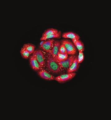
3 Clinical and Developmental Immunology 3 Case HGB (g/l) Table 1: Laboratory test results of SLE that has nephritis with mainly IgA deposits. WBC ( 10 9 /L) PLT ( 10 9 /L) ANA dsdna Sm C3 (mg/dl) C4 (mg/dl) Anti-β2 GP : IgM(+) :320 1: IgM, G(+) : : IgM, A(+) : IgM(+) : : IgG(+) 25 ACL SLEDAI score Indicates lower than normal; + indicates positive; indicates negative. Normal range: (hemoglobin) HGB g/l; (white blood cells) WBC /L; (platelet) PLT /L; C mg/dl; C mg/dl. Case Sex age Table 2: Renal involvement of SLE that has nephritis with mainly IgA deposits. Red cells (per HPF) White cells (per HPF) Casts (per HPF) Protein (g/24 h) Serum creatinine (umol/l) Serum urea nitrogen (mmol/l) 1 F/64 All view All view 8 10 granular M/45 All view 1-2 granular F/ F/ granular M/ granular Indicates higher than normal range. Red cells, white cells, cast, and protein were all tested in urine. Normal range: urine protein (g/24 h): mg; serum creatinine umol/l; serum urea nitrogen mmol/l. value is 21 to 34 seconds; thrombin time was normal). Serum immunoglobulin was abnormal in 2 cases (2/5), and IgA increased in only 1 case (1/5). In case 1, IgG was 2340 mg/dl (normal range was mg/dl), IgA 1050 mg/dl (normal range was mg/dl), and IgM 349 mg/dl (normal range was 46 to 304 mg/dl). In case 4, only IgG slightly increased (1690 mg/dl), while IgA and IgM were normal. Most of the previous laboratory results were listed in Tables 1 and Renal Biopsy Findings. All cases performed light and immunofluorescent microscopy. The result showed that all cases had mainly IgA deposits and did not match with LN. Under light microscope (Figure 1), all cases showed mild diffuse hyperplasia of glomerular mesangium and matrix, with focal and segmental aggravation. The renal tubular epithelial cell showed vacuolar degeneration, granular degeneration, andspottyorflakeatrophy,whiletherenalinterstitialshowed fibrosis and infiltration of lymphocytes and monocytes. In case 2, glomerular sclerosis can be clearly seen. Immune complex deposits were seen in glomerular mesangium under immunofluorescent microscope. All cases had IgA deposits (Figure 1) and were free of IgG, C1q, and FRA deposits. In addition, as well as IgA deposit, 1 case had C3 deposit, and the other 4 cases had IgM and C3 deposits Treatment and Prognosis. All cases were given prednisone at a dose of 1 mg/(kg d) after percutaneous renopuncture, and cases 1, 2, and 5 also received intravenous cyclophosphamide treatment. All the cases achieved remission after therapy, for example, clinical symptoms got relief (such as arthritis, edema, orrhomeningitis, Raynaud s phenomenon, and oral ulcers), blood routine, urine tests, and immunological tests improved, including reduction of protein,redbloodcells,whitebloodcells,andcastsinurine, decrease of SLEDAI score, as well as increase of white blood cells, platelet, C3, and C4. 4. Discussion 4.1. Diagnosis of SLE. In 1982 ACR classification criteria for SLE,ifthepatientsatisfiesfourormorethanfourofthe criteria, we can classify the patient as having SLE. According to that, cases 1 and 5 satisfied five of the criteria, and the rest cases satisfied four. So they can be definitely diagnosed as SLE. In 2009 ACR classification criteria for SLE, if (1) the patient has biopsy-proven LN with ANA or anti-dsdna or (2) the patient satisfied four of the criteria, including at least one clinical and one immunologic criterion, we classify the patient as having SLE. The 5 patients in our study were in the second case. They satisfied 5 to 8 criteria, including at least 2 clinical criteria and 3 immunologic criteria. Even if we exclude renal injury, the patients still satisfied 4 to 7 criteria and can be diagnosed as SLE. So, whichever criteria we choose or whether we include renal injury, the five cases can be diagnosed as SLE. TypicalLNarecharacterizedby FullHouse stainunder immunofluorescent microscopy, staining positively for IgG, IgA, IgM, C3, and C1q. However, the five SLE patients showed mainly IgA deposits and free of IgG and C1q deposits, which didnotmatchwithtypicalln.itisunusualinaclinicfor SLE patients to have nephritis with mainly IgA deposits, so we made a review to get a further understanding of the problem Relationship between SLE and Nephritis with Mainly IgA Deposits. In the recent 3.5 years, as many as 5 SLE

, (b2), (b3), (b4), and (b5), respectively, indicate MASSON staining of cases 1 5 under light microscope ( 400).")
, (c2), (c3), (c4), and (c5), respectively, indicate IgA deposits of cases 1 5 under immunofluorescence microscope and all cases are")
 since 1995, 8 pieces of literature reported a total of 10")
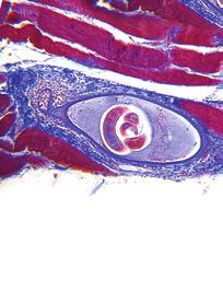
![SLE patients who have nephritis with mainly IgA deposits, all of whom were regarded as IgAN [3 8].](/docs-images/89/99596820/images/4-4.jpg "In these pieces of literature, the relationship between SLE and IgAN was discussed, and the result is still controversial.")


![[6] reported a case of a male SLE patient whose renal biopsy was established as class II LN.](/docs-images/89/99596820/images/4-9.jpg "He was given the second renal biopsy because of repeated proteinuria, and the result suggested IgAN.")


4 4 Clinical and Developmental Immunology (a1) (a2) (a3) (a4) (a5) (b1) (b2) (b3) (b4) (b5) (c1) (c2) (c3) (c4) (c5) Figure 1: Light and immunofluorescent microscope findings of SLE that has nephritis with mainly IgA deposits. (a1), (a2), (a3), (a4), and (a5), respectively, indicate PASM staining of cases 1 5 under light microscope ( 400). (b1), (b2), (b3), (b4), and (b5), respectively, indicate MASSON staining of cases 1 5 under light microscope ( 400). Hyperplasia of glomerular mesangium and matrix can be seen in all cases. In addition, case 2 has clear glomerular sclerosis. (c1), (c2), (c3), (c4), and (c5), respectively, indicate IgA deposits of cases 1 5 under immunofluorescence microscope and all cases are positive (++ +++). patients in our study were found to have mainly IgA deposits by renal biopsy. Retrieved from the Pubmed and Chinese National Knowledge Infrastructure (CKNI) since 1995, 8 pieces of literature reported a total of 10 SLE patients who have nephritis with mainly IgA deposits, all of whom were regarded as IgAN [3 8]. In these pieces of literature, the relationship between SLE and IgAN was discussed, and the result is still controversial. Most scholars believe that the typical LN includes the previous 6 types but not IgAN. They regard IgAN as a complication of SLE when the two diseases coexist [3, 4, 7], which means that the patient is affected by SLE and IgAN simultaneously. In typical LN, the complements C1q, C3, and C4 and the IgG-predominant accumulation of polyclonal immune complexes can be seen in the capillary basal membrane. In IgAN, however, IgA is the major deposit. At the same time, 22 60% of IgAN patients combine with IgM deposit, and 80% combine with C3 deposit. The previous features of IgAN do not match with the typical LN. Some other scholars believe that IgAN may be a special clinical subtype of SLE. In 2010, the Japanese scholar Horino et al. [6] reported a case of a male SLE patient whose renal biopsy was established as class II LN. He was given the second renal biopsy because of repeated proteinuria, and the result suggested IgAN. The authors proposed that IgAN may be a special clinical subtype of SLE. As there is a mutual transition among the types of typical LN, typical LN may also convert into IgAN. In our study, we found some differences in clinical characteristics between atypical LN with mainly IgA deposits and primary IgAN. The primary IgAN mainly occurred in young men, mostly 20 to 30 years old. Before the onset of IgAN, the patients can affect upper respiratory or gastrointestinal infection. Primary IgAN is characterized by gross hematuria or asymptomatic microscopic. They may also have edema, hypertension, renal dysfunction, and other clinical manifestations. Besides, serum IgA level is higher than normal in about 50% patients. The five cases in our report, similar to primary IgAN, showed gross hematuria or microscopic hematuria, edema, hypertension, renal dysfunction, and so on. However, compared with primary IgAN, the five patients have some different characteristics: older than primary IgAN, more women than men, no previous infection history before disease onset, and low incidence of serum IgA elevation. Some SLE patients are ACL positive, and the rates reported are different but not more than 60% [11]. However, the ACL positive rate in this report is as high as 100%. ACL can interfere with blood clotting mechanism, resulting in generation and aggravation of hypercoagulable state. It can also cause coagulation in the glomerular capillary and aggravate kidney damage. This may be one of the reasons why the five cases of SLE patients have much more serious kidney damage. However, it is still unclear why the ACL positive rate increases in SLE that had nephritis with mainly IgA deposits. We speculate that nephritis with mainly IgA deposits may be a special clinical subtype of SLE. In other words, nephritis with mainly IgA deposits, as an atypical LN, may be another nephropathy of SLE in addition to the typical LN. The supporting points are as follows. (1) The cases of nephritis
5 Clinical and Developmental Immunology 5 with mainly IgA deposits are not rare. As many as 10 cases have already been reported before, and other 5 cases are found here in only 3.5 years. Also, it is possible that some similar cases were not reported because of neglect. (2) There was no significant difference between SLE with typical LN and SLE with atypical LN in clinical manifestations or laboratory tests. (3) Compared with primary IgAN, nephritis with mainly IgA deposits observed in SLE has its own clinical characteristics as mentioned previously: older than primary IgAN, more women than men, low incidence of serum IgA elevation, and ACLpositiverateashighas100%.Toconclude,nephritis with mainly IgA deposits, as an atypical LN, may be a special clinicalsubtypeofsle,althoughitstillneedsalotoffurther research Pathogenesis. SLE is also an immune-complex-mediated disease. Although C1q, C3, C4, and IgG depositions [2] are more common in typical LN, IgA deposition can also be seen. We speculated that, when some uncertain mechanisms lead to a majority of IgA deposition, the renal biopsy finding may present atypical LN with mainly IgA deposits Prognosis. In this research, five patients got relief after treatment of glucocorticoids and immunosuppressive drugs. The relief indicates good effect of glucocorticoids and immunosuppressive drugs on SLE that has atypical LN with maily IgA deposits. But there are also some complex and ineffective cases reported. Lai et al. [8] reported one patient who died of systemic infection. This alerts clinicians that they should give early diagnosis and treatment to such patients and be aware of the appearance of complications, especially infection. In summary, nephritis with maily IgAN deposits is not rare in SLE and has its own clinical characteristics which are different from those of primary IgAN. We speculated that nephritiswithmainlyigandeposits,asanatypicalln,may beaspecialsubtypeofsle.itmaybeanothernephropathy of SLE in addition to the typical LN and has a relative good prognosis. However, the speculation still needs further clinical observation and research. [4] E.Fujikura,T.Kimura,A.Otakaetal., IgAnephropathyinthe patient with systemic lupus erythematosus in remission, Nihon Naika Gakkai Zasshi, vol. 91, no. 11, pp , [5] A. Corrado, L. Quarta, A. M. Di Palma, L. Gesualdo, and F. P. Cantatore, IgA nephropathy in systemic lupus erythematosus, Clinical and Experimental Rheumatology,vol.25,no.3,pp , [6] T. Horino, T. Takao, and Y. Terada, IgA nephropathy in a patient with systemic lupus erythematosus, Lupus, vol. 19, no. 5,pp ,2010. [7] S.Kobak,O.HudaverdI,G.Keser,andF.Oksel, Coexistenceof systemic lupus erythematosus, Hashimoto s thyroiditis and IgA nephropathy in the same patient, Modern Rheumatology, vol. 21, no. 1, pp , [8]F.M.M.Lai,E.K.M.Li,N.L.S.Tang,P.K.T.Li,S.F.Lui, and K. N. Lai, IgA nephropathy: a rare lesion in systemic lupus erythematosus, Modern Pathology, vol. 8, no. 1, pp. 5 10, [9]E.M.Tan,A.S.Cohen,J.F.Friesetal., The1982revised criteria for the classification of systemic lupus erythrematosus, Arthritis and Rheumatism,vol.25,no.11,pp ,1982. [10] M. Petri, Systemic Lupus International Collaborating Clinic (SLICC). SLICC revision of the ACR classification criteria for SLE, Arthritis and Rheumatism,vol.60,supplement10,p.895, [11] N. Bizzaro, E. Tonutti, and D. Villalta, Prevalence and clinical correlation of anti-phospholipid-binding protein antibodies in anticardiolipin-negative patients with systemic lupus erythematosus and women with unexplained recurrent miscarriages, Archives of Pathology and Laboratory Medicine, vol.129,no.1, pp.61 68,2005. Conflict of Interests The authors declare that they have no conflict of interests. References [1] J. J. Weening, V. D. D Agati, and M. M. Schwartz, The classification of glomerulonephritis in systemic lupus erythematosus revisited, Kidney International,vol.65, pp ,2004. [2]R.N.Verma,A.K.Banerjee,andK.Bhattacharya, Lupus nephritis-light, electron and immunofluorescent microscopy (a clinicopathological study), Indian Pathology and Microbiology,vol.32,no.4,pp ,1989. [3] C. Basile, A. Semeraro, A. Montanaro et al., IgA nephropathy in a patient with systemic lupus erythematosus, Nephrology Dialysis Transplantation,vol.13,no.7,pp ,1998.


6 MEDIATORS of INFLAMMATION The Scientific World Journal Gastroenterology Research and Practice Diabetes Research International Endocrinology Immunology Research Disease Markers Submit your manuscripts at BioMed Research International PPAR Research Obesity Ophthalmology Evidence-Based Complementary and Alternative Medicine Stem Cells International Oncology Parkinson s Disease Computational and Mathematical Methods in Medicine AIDS Behavioural Neurology Research and Treatment Oxidative Medicine and Cellular Longevity
A clinical syndrome, composed mainly of:
 Nephritic syndrome We will discuss: 1)Nephritic syndrome: -Acute postinfectious (poststreptococcal) GN -IgA nephropathy -Hereditary nephritis 2)Rapidly progressive GN (RPGN) A clinical syndrome, composed
Clinical Study Glomerulonephritis with Crescents in Children: Etiology and Predictors of Renal Outcome
 International Scholarly Research Network ISRN Pediatrics Volume 2011, Article ID 507298, 5 pages doi:10.5402/2011/507298 Clinical Study Glomerulonephritis with Crescents in Children: Etiology and Predictors
J Renal Inj Prev. 2018; 7(1): Journal of Renal Injury Prevention
: Journal of Renal Injury Prevention") J Renal Inj Prev. 2018; 7(1): 22-26. Journal of Renal Injury Prevention DOI: 10.15171/jrip.2018.05 Comparison of clinical and histopathological findings in patients with lupus nephritis having IgG deposits
J Renal Inj Prev. 2018; 7(1): 22-26. Journal of Renal Injury Prevention DOI: 10.15171/jrip.2018.05 Comparison of clinical and histopathological findings in patients with lupus nephritis having IgG deposits
Glomerular pathology in systemic disease
 Glomerular pathology in systemic disease Lecture outline Lupus nephritis Diabetic nephropathy Glomerulonephritis Associated with Bacterial Endocarditis and Other Systemic Infections Henoch-Schonlein Purpura
Significance of Anti-C1q Antibodies in Patients with Systemic Lupus Erythematosus as A Marker of Disease Activity and Lupus Nephritis
 THE EGYPTIAN JOURNAL OF IMMUNOLOGY Vol. 23 (1), 2016 Page: 00-00 Significance of Anti-C1q Antibodies in Patients with Systemic Lupus Erythematosus as A Marker of Disease Activity and Lupus Nephritis 1
RENAL HISTOPATHOLOGY
 RENAL HISTOPATHOLOGY Peter McCue, M.D. Department of Pathology, Anatomy & Cell Biology Sidney Kimmel Medical College There are no conflicts of interest. 1 Goals and Objectives! Goals Provide introduction
C1q nephropathy the Diverse Disease
 C1q nephropathy the Diverse Disease Danica Galešić Ljubanović School of Medicine, University of Zagreb Dubrava University Hospital Zagreb, Croatia Definition Dominant or codominant ( 2+), mesangial staining
Surgical Pathology Report
 Louisiana State University Health Sciences Center Department of Pathology Shreveport, Louisiana Accession #: Collected: Received: Reported: 6/1/2012 09:18 6/2/2012 09:02 6/2/2012 Patient Name: Med. Rec.
Overview of glomerular diseases
 Overview of glomerular diseases *Endothelial cells are fenestrated each fenestra: 70-100nm in diameter Contractile, capable of proliferation, makes ECM & releases mediators *Glomerular basement membrane
An unusual association between focal segmental sclerosis and lupus nephritis: a distinct concept from lupus podocytopathy?
 CEN Case Rep (2015) 4:70 75 DOI 10.1007/s13730-014-0142-1 CASE REPORT An unusual association between focal segmental sclerosis and lupus nephritis: a distinct concept from lupus podocytopathy? Hironari
Case Report A Case of Proliferative Glomerulonephritis with Monoclonal IgG Deposits That Showed Predominantly Membranous Features
 Hindawi Case Reports in Nephrology Volume 2017, Article ID 1027376, 5 pages https://doi.org/10.1155/2017/1027376 Case Report A Case of Proliferative Glomerulonephritis with Monoclonal IgG Deposits That
Glomerular diseases mostly presenting with Nephritic syndrome
 Glomerular diseases mostly presenting with Nephritic syndrome 1 The Nephritic Syndrome Pathogenesis: proliferation of the cells in glomeruli & leukocytic infiltrate Injured capillary walls escape of RBCs
High Impact Rheumatology
 High Impact Rheumatology Systemic Lupus Erythematosus Bernard Rubin, DO MPH Case 1: History A 45-year-old woman presents with severe dyspnea and cough. She was in excellent health until 4 weeks ago when
Approach to Glomerular Diseases: Clinical Presentation Nephrotic Syndrome Nephritis
 GLOMERULONEPHRITIDES Vivette D Agati Jai Radhakrishnan Approach to Glomerular Diseases: Clinical Presentation Nephrotic Syndrome Nephritis Heavy Proteinuria Renal failure Low serum Albumin Hypertension
Autoimmune diseases. SLIDE 3: Introduction to autoimmune diseases Chronic
 SLIDE 3: Introduction to autoimmune diseases Chronic Autoimmune diseases Sometimes relapsing : and remitting. which means that they present as attacks Progressive damage Epitope spreading more and more
Year 2004 Paper one: Questions supplied by Megan
 QUESTION 53 Endothelial cell pathology on renal biopsy is most characteristic of which one of the following diagnoses? A. Pre-eclampsia B. Haemolytic uraemic syndrome C. Lupus nephritis D. Immunoglobulin
Journal of Nephropathology
 www.nephropathol.com DOI: 10.12860/jnp.2014.22 J Nephropathol. 2014; 3(3): 115-120 Journal of Nephropathology Clinicopathological correlations in lupus nephritis; a single center experience Hamid Nasri
Xi Yang, Ri-Bao Wei, Ping Li, Yue Yang, Ting-Yu Su, Yu-Wei Gao, Qing-Ping Li, Xue-Guang Zhang, Xiang-Mei Chen
 Int J Clin Exp Pathol 2016;9(9):9401-9407 www.ijcep.com /ISSN:1936-2625/IJCEP0028098 Original Article Correlation between serum hepatitis B virus DNA replication level and clinicopathology in 235 patients
Lupus Related Kidney Diseases. Jason Cobb MD Assistant Professor Renal Division Emory University School of Medicine October 14, 2017
 Lupus Related Kidney Diseases Jason Cobb MD Assistant Professor Renal Division Emory University School of Medicine October 14, 2017 Financial Disclosures MedImmune Lupus Nephritis Kidney Biopsy Biomarkers
Glomerular pathology-2 Nephritic syndrome. Dr. Nisreen Abu Shahin
 Glomerular pathology-2 Nephritic syndrome Dr. Nisreen Abu Shahin 1 The Nephritic Syndrome Pathogenesis: inflammation proliferation of the cells in glomeruli & leukocytic infiltrate Injured capillary walls
Systemic Lupus Erythematosus
 Systemic Lupus Erythematosus Marc C. Hochberg, MD, MPH Professor of Medicine and Head, Division of Rheumatology University of Maryland School of Medicine CASE: HISTORY A 26-year-old woman is seen for migratory
Histopathology: Glomerulonephritis and other renal pathology
 Histopathology: Glomerulonephritis and other renal pathology These presentations are to help you identify basic histopathological features. They do not contain the additional factual information that you
Case Report Pseudothrombocytopenia due to Platelet Clumping: A Case Report and Brief Review of the Literature
 Case Reports in Hematology Volume 2016, Article ID 3036476, 4 pages http://dx.doi.org/10.1155/2016/3036476 Case Report Pseudothrombocytopenia due to Platelet Clumping: A Case Report and Brief Review of
CHAPTER 2. Primary Glomerulonephritis
 2nd Report of the PRIMARY GLOMERULONEPHRITIS CHAPTER 2 Primary Glomerulonephritis Sunita Bavanandan Lee Han Wei Lim Soo Kun 21 PRIMARY GLOMERULONEPHRITIS 2nd Report of the 2.1 Introduction This chapter
Definition Chronic autoimmune disease The body s immune system starts attacking itself Can affect most organs and tissues in the body Brain, lungs, he
 LIVING WITH SYSTEMIC LUPUS ERYTHEMATOSUS Stacy Kennedy, M.D.,M.B.A. Rowan Diagnostic Clinic Salisbury, N.C. May 11, 2013 Agenda What is lupus Who is affected Causes of lupus Symptoms and organ involvement
GOODPASTURE'S SYNDROME WITH CONCOMITANT IMMUNE COMPLEX MIXED MEMBRANOUS AND PROLIFERATIVE GLOMERULONEFRITIS
 GOODPASTURE'S SYNDROME WITH CONCOMITANT IMMUNE COMPLEX MIXED MEMBRANOUS AND PROLIFERATIVE GLOMERULONEFRITIS VESNA JURČIĆ 1, ANDREJA ALEŠ RIGLER 2, INSTITUTE OF PATHOLOGY, FACULTY OF MEDICINE, UNIVERSITY
Renal Pathology 1: Glomerulus. With many thanks to Elizabeth Angus PhD for EM photographs
 Renal Pathology 1: Glomerulus With many thanks to Elizabeth Angus PhD for EM photographs Anatomy of the Kidney http://www.yalemedicalgroup.org/stw/page.asp?pageid=stw028980 The Nephron http://www.beltina.org/health-dictionary/nephron-function-kidney-definition.html
THE URINARY SYSTEM. The cases we will cover are:
 THE URINARY SYSTEM The focus of this week s lab will be pathology of the urinary system. Diseases of the kidney can be broken down into diseases that affect the glomeruli, tubules, interstitium, and blood
THE URINARY SYSTEM. The cases we will cover are:
 THE URINARY SYSTEM The focus of this week s lab will be pathology of the urinary system. Diseases of the kidney can be broken down into diseases that affect the glomeruli, tubules, interstitium, and blood
Case Presentation Turki Al-Hussain, MD
 Case Presentation Turki Al-Hussain, MD Director, Renal Pathology Chapter Saudi Society of Nephrology & Transplantation Consultant Nephropathologist & Urological Pathologist Department of Pathology & Laboratory
Dr Ian Roberts Oxford. Oxford Pathology Course 2010 for FRCPath Illustration-Cellular Pathology. Oxford Radcliffe NHS Trust
 Dr Ian Roberts Oxford Oxford Pathology Course 2010 for FRCPath Present the basic diagnostic features of the commonest conditions causing proteinuria & haematuria Highlight diagnostic pitfalls Nephrotic
9/25/2013 SYSTEMIC LUPUS ERYTHEMATOSUS (SLE)
") SYSTEMIC LUPUS ERYTHEMATOSUS (SLE) 1 Other Types of Lupus Discoid Lupus Erythematosus Lupus Pernio --- Sarcoidosis Lupus Vulgaris --- Tuberculosis of the face Manifestations of SLE Fever Rashes Arthritis
SYSTEMIC LUPUS ERYTHEMATOSUS (SLE) 1 Other Types of Lupus Discoid Lupus Erythematosus Lupus Pernio --- Sarcoidosis Lupus Vulgaris --- Tuberculosis of the face Manifestations of SLE Fever Rashes Arthritis
Case Presentation Turki Al-Hussain, MD
 Case Presentation Turki Al-Hussain, MD Director, Renal Pathology Chapter Saudi Society of Nephrology & Transplantation Consultant Nephropathologist & Urological Pathologist Department of Pathology & Laboratory
Research Article Clinicopathological Correlation in Asian Patients with Biopsy-Proven Lupus Nephritis
 International Nephrology Volume 2015, Article ID 857316, 6 pages http://dx.doi.org/10.1155/2015/857316 Research Article Clinicopathological Correlation in Asian Patients with Biopsy-Proven Lupus Nephritis
Lupus Nephritis: Pathophysiology, Diagnosis, And Collaborative Management.: An Article From: Nephrology Nursing Journal [HTML] [Digital] By D.
![Lupus Nephritis: Pathophysiology, Diagnosis, And Collaborative Management.: An Article From: Nephrology Nursing Journal [HTML] [Digital] By D.](/thumbs/74/71077666.jpg "Lupus Nephritis: Pathophysiology, Diagnosis, And Collaborative Management.: An Article From: Nephrology Nursing Journal [HTML] [Digital] By D.") Lupus Nephritis: Pathophysiology, Diagnosis, And Collaborative Management.: An Article From: Nephrology Nursing Journal [HTML] [Digital] By D. Michelle Smith;E. Michelle Fortune- Faulkner;Brenda L. Spurbeck
Lupus Nephritis: Pathophysiology, Diagnosis, And Collaborative Management.: An Article From: Nephrology Nursing Journal [HTML] [Digital] By D. Michelle Smith;E. Michelle Fortune- Faulkner;Brenda L. Spurbeck
CIBMTR Center Number: CIBMTR Recipient ID: RETIRED. Today s Date: Date of HSCT for which this form is being completed: &
 Systemic Lupus Erythematosus Pre-HSCT Data Sequence Number: Date Received: Registry Use Only Today s Date: Date of HSCT for which this form is being completed: & HSCT type: o autologous o allogeneic, o
Dr. Rai Muhammad Asghar Head of Paediatric Department BBH Rawalpindi
 Dr. Rai Muhammad Asghar Head of Paediatric Department BBH Rawalpindi Acute Post streptococcal Glomerulonephritis Sudden onset of Gross hematuria Edema Hypertension Renal insufficiency Cause of AGN Post
.,Dr Ali Alkazzaz Babylon collage of medicine 2016
 .,Dr Ali Alkazzaz Babylon collage of medicine 2016 Lupus history Lupus is the Latin word for wolf 1 st used medically in the 10 th century Described clinically in the 19 th century Butterfly rash in 1845
Dense deposit disease with steroid pulse therapy
 Case Report Dense deposit disease with steroid pulse therapy Jun Odaka, Takahiro Kanai, Takane Ito, Takashi Saito, Jun Aoyagi, and Mariko Y Momoi Abstract Treatment of dense deposit disease DDD has not
CHAPTER 2 PRIMARY GLOMERULONEPHRITIS
 CHAPTER 2 Sunita Bavanandan Lim Soo Kun 19 5th Report of the 2.1: Introduction This chapter covers the main primary glomerulonephritis that were reported to the MRRB from the years 2005-2012. Minimal change
Journal of Nephropathology
 www.nephropathol.com DOI: 10.15171/jnp.2018.24 J Nephropathol. 2018;7(3):101-105 Journal of Nephropathology Relationship of CD147 kidney expression with various pathologic lesions, biochemical and demographic
Nephrotic syndrome minimal change disease vs. IgA nephropathy. Hadar Meringer Internal medicine B Sheba
 Nephrotic syndrome minimal change disease vs. IgA nephropathy Hadar Meringer Internal medicine B Sheba The Case 29 year old man diagnosed with nephrotic syndrome 2 weeks ago and complaining now about Lt.flank
Disorders of the kidney. Urine analysis. Nephrotic and nephritic syndrome.
 Disorders of the kidney. Urine analysis. Nephrotic and nephritic syndrome. Azotemia and Urinary Abnormalities Disturbances in urine volume oliguria, anuria, polyuria Abnormalities of urine sediment red
Glomerular diseases with organized deposits
 Glomerular diseases with organized deposits Banu Sis, MD, FRCPC University of Alberta, Edmonton, AB, Canada Ulusal Patoloji Kongresi, Manavgat, Antalya 8/11/2012 What is an organized deposit? A number
Membranous nephropathy. By Mohammed Kamal Nassar, MD Lecturer of Nephrology Mansoura University
 Membranous nephropathy By Mohammed Kamal Nassar, MD Lecturer of Nephrology Mansoura University Membranous nephropathy Definition: Immune complex glomerular disease in which immune deposits of IgG and complement
UNDERSTANDING SYSTEMIC LUPUS ERYTHEMATOSUS
 UNDERSTANDING SYSTEMIC LUPUS ERYTHEMATOSUS Stacy Kennedy, M.D.,M.B.A. October 20, 2012 Agenda What is lupus Who is affected Causes of lupus Symptoms and organ involvement Diagnosis Treatment Pregnancy
Case 3. ACCME/Disclosure. Laboratory results. Clinical history 4/13/2016
 Case 3 Lynn D. Cornell, M.D. Mayo Clinic, Rochester, MN Cornell.Lynn@mayo.edu USCAP Renal Case Conference March 13, 2016 ACCME/Disclosure Dr. Cornell has nothing to disclose Clinical history 57-year-old
ACUTE GLOMERULONEPHRITIS. IAP UG Teaching slides
 ACUTE GLOMERULONEPHRITIS 1 Definition Etiology Pathology/pathogenesis Risk factors Clinical Presentation Investigation Differential Diagnosis Management Outcome/Prognosis Indication for Renal Biopsy Summary
THE KIDNEY AND SLE LUPUS NEPHRITIS
 THE KIDNEY AND SLE LUPUS NEPHRITIS JACK WATERMAN DO FACOI 2013 NEPHROLOGY SIR RICHARD BRIGHT TERMINOLOGY RENAL INSUFFICIENCY CKD (CHRONIC KIDNEY DISEASE) ESRD (ENDSTAGE RENAL DISEASE) GLOMERULONEPHRITIS
Ordering Physician. Collected REVISED REPORT. Performed. IgG IF, Renal MCR. Lambda IF, Renal MCR. C1q IF, Renal. MCR Albumin IF, Renal MCR
 RenalPath Level IV Wet Ts IgA I Renal IgM I Renal Kappa I Renal Renal Bx Electron Microscopy IgG I Renal Lambda I Renal C1q I Renal C3 I Renal Albumin I Renal ibrinogen I Renal Mayo Clinic Dept. of Lab
Interesting case seminar: Native kidneys Case Report:
 Interesting case seminar: Native kidneys Case Report: Proximal tubulopathy and light chain deposition disease presented as severe pulmonary hypertension with right-sided cardiac dysfunction and nephrotic
Study of Interleukin-12 Cytokine and Anti-C1q Antibodies in Lupus Nephritis Patients
 International Journal of Internal Medicine 214, 3(1): 13-26 DOI: 1.5923/j.ijim.21431.3 Study of Interleukin-12 Cytokine and Anti-C1q Antibodies in Lupus Nephritis Patients Mohamed N. AL Alfy 1,*, Mohamed
Policy. Section: Medicine Effective Date: January 15, 2015 Subsection: Pathology/Laboratory Original Policy Date: December 5, 2014 Subject:
 Last Review Status/Date: December 2014 Page: 1 of 10 Summary Systemic lupus erythematosus (SLE) is an autoimmune connective tissue disease that can be difficult to diagnose because patients often present
Glomerular Pathology- 1 Nephrotic Syndrome. Dr. Nisreen Abu Shahin
 Glomerular Pathology- 1 Nephrotic Syndrome Dr. Nisreen Abu Shahin The Nephrotic Syndrome a clinical complex resulting from glomerular disease & includes the following: (1) massive proteinuria (3.5 gm /day
Dr Ian Roberts Oxford. Oxford Pathology Course 2010 for FRCPath Illustration-Cellular Pathology. Oxford Radcliffe NHS Trust
 Dr Ian Roberts Oxford Oxford Pathology Course 2010 for FRCPath Plan of attack: Diagnostic approach to the renal biopsy Differential diagnosis of the clinical syndromes of renal disease Microscopy Step
CASE 4 A RARE CASE OF INTRALUMINAL GLOMERULAR CAPILLARY DEPOSITS
 CASE 4 A RARE CASE OF INTRALUMINAL GLOMERULAR CAPILLARY DEPOSITS DR ANNIE JOJO, Dr Seethalekshmy N V, Dr Nanda Kachare DEPARTMENT OF PATHOLOGY, AMRITA INSTITUTE OF MEDICAL SCIENCES, KOCHI. 54 yrs female,
Committee Approval Date: May 9, 2014 Next Review Date: May 2015
 Medication Policy Manual Policy No: dru248 Topic: Benlysta, belimumab Date of Origin: May 13, 2011 Committee Approval Date: May 9, 2014 Next Review Date: May 2015 Effective Date: June 1, 2014 IMPORTANT
Secondary IgA Nephropathy & HSP
 Secondary IgA Nephropathy & HSP Anjali Gupta, MD 1/11/11 AKI sec to Hematuria? 65 cases of ARF after an episode of macroscopic hematuria have been reported in the literature in patients with GN. The main
substance staining with IgG, C3 and IgA (trace) Linear deposition of IgG(+), IgA.M(trace) and C3(+++) at the DEJ
 Linear deposition of IgG(+), IgA.M(trace) and C3(+++) at the DEJ") Direct Immunofluorescence: Skin Diagnosis Findings Picture Pemphigus Vulgaris and it s Intracellular cement variants substance staining with IgG, C3 and IgA (trace) Bullous Pemphigoid and it s variants
Direct Immunofluorescence: Skin Diagnosis Findings Picture Pemphigus Vulgaris and it s Intracellular cement variants substance staining with IgG, C3 and IgA (trace) Bullous Pemphigoid and it s variants
Mayo Clinic/ RPS Consensus Report on Classification, Diagnosis, and Reporting of Glomerulonephritis
 Mayo Clinic/ RPS Consensus Report on Classification, Diagnosis, and Reporting of Glomerulonephritis Sanjeev Sethi, MD, PhD Department of Laboratory Medicine and Pathology Disclosure Relevant Financial
Monoclonal Gammopathies and the Kidney. Tibor Nádasdy, MD The Ohio State University, Columbus, OH
 Monoclonal Gammopathies and the Kidney Tibor Nádasdy, MD The Ohio State University, Columbus, OH Monoclonal gammopathy of renal significance (MGRS) Biopsies at OSU (n=475) between 2007 and 2016 AL or AH
Familial DDD associated with a gain-of-function mutation in complement C3.
 Familial DDD associated with a gain-of-function mutation in complement C3. Santiago Rodríguez de Córdoba, Centro de investigaciones Biológicas, Madrid Valdés Cañedo F. and Vázquez- Martul E., Complejo
Nephritic vs. Nephrotic Syndrome
 Page 1 of 18 Nephritic vs. Nephrotic Syndrome Terminology: Glomerulus: A network of blood capillaries contained within the cuplike end (Bowman s capsule) of a nephron. Glomerular filtration rate: The rate
RECURRENT AND DE NOVO RENAL DISEASES IN THE ALLOGRAFT. J. H. Helderman,MD,FACP,FAST
 RECURRENT AND DE NOVO RENAL DISEASES IN THE ALLOGRAFT J. H. Helderman,MD,FACP,FAST Vanderbilt University Medical Center Professor of Medicine, Pathology and Immunology Medical Director, Vanderbilt Transplant
Original. IgAN. Key words : IgA nephropathy, IgM deposition, proteinuria, tonsillectomy, steroid pulse therapy. Introduction
 Showa Univ J Med Sci 27 3, 167 174, September 2015 Original Prominent IgM Deposition in Glomerulus Is Associated with Severe Proteinuria and Reduced after Combined Treatment of Tonsillectomy with Steroid
Lab 3, case 1. Is this an example of nephrotic or nephritic syndrome? Why? Which portion of the nephron would you expect to be abnormal?
 Lab 3, case 1 12-year-old Costa Rican boy is brought into clinic by his parents because of dark brownish-red urine over the last 24 hours. The family has been visiting friends in Indianapolis for two weeks.
Xiao-wei Yang 1,2,3,4, Ying Tan 1,2,3,4, Feng Yu 1,2,3,4 and Ming-hui Zhao 1,2,3,4. Introduction
 Nephrol Dial Transplant (2012) 27: 3552 3559 doi: 10.1093/ndt/gfs179 Advance Access publication 13 June 2012 Combination of anti-c1q and anti-dsdna antibodies is associated with higher renal disease activity
Insights into the DX of Pediatric SLE
 Insights into the DX of Pediatric SLE Dr. John H. Yost Pediatric Rheumatology Children s Hospital at Dartmouth Assistant Professor of Medicine Geisel School of Medicine at Dartmouth john.h.yost@hitchcock.org
Clinical Study IVIG Effects on Erythrocyte Sedimentation Rate in Children
 International Pediatrics, Article ID 981465, 4 pages http://dx.doi.org/10.1155/2014/981465 Clinical Study IVIG Effects on Erythrocyte Sedimentation Rate in Children Farhad Salehzadeh, Ahmadvand Noshin,
Cutaneous manifestations and systemic correlation in patients with lupus erythematosus and its subsets: a study of 40 cases
 International Journal of Research in Dermatology Mahajan R et al. Int J Res Dermatol. 2018 Nov;4(4):479-483 http://www.ijord.com Original Research Article DOI: http://dx.doi.org/10.18203/issn.2455-4529.intjresdermatol20183407
Clinicopathologic Characteristics of IgA Nephropathy with Steroid-responsive Nephrotic Syndrome
 J Korean Med Sci 2009; 24 (Suppl 1): S44-9 ISSN 1011-8934 DOI: 10.3346/jkms.2009.24.S1.S44 Copyright The Korean Academy of Medical Sciences Clinicopathologic Characteristics of IgA Nephropathy with Steroid-responsive
Pathogenesis of IgA Nephropathy. Shokoufeh Savaj MD Associate Professor of Medicine Firoozgar hospital- IUMS
 Pathogenesis of IgA Nephropathy Shokoufeh Savaj MD Associate Professor of Medicine Firoozgar hospital- IUMS History Immunoglobin A nephropathy was first described by Berger and Hinglais in 1968 in Paris
What will we discuss today?
 Autoimmune diseases What will we discuss today? Introduction to autoimmune diseases Some examples Introduction to autoimmune diseases Chronic Sometimes relapsing Progressive damage Epitope spreading more
Policy. Background
 Last Review Status/Date: December 2016 Page: 1 of 11 Summary Systemic lupus erythematosus (SLE) is an autoimmune connective tissue disease that can be difficult to diagnose because patients often present
Classification of Glomerular Diseases and Defining Individual Glomerular Lesions: Developing International Consensus
 Classification of Glomerular Diseases and Defining Individual Glomerular Lesions: Developing International Consensus Mark Haas MD, PhD Department of Pathology & Laboratory Medicine Cedars-Sinai Medical
Case Report Pauci-Immune Crescentic Glomerulonephritis in Connective Tissue Disease
 Case Reports in Rheumatology Volume 2016, Article ID 9070487, 6 pages http://dx.doi.org/10.1155/2016/9070487 Case Report Pauci-Immune Crescentic Glomerulonephritis in Connective Tissue Disease Supraja
Nephrotic Syndrome. Department of pediatrics The first affiliated hospital Sun Yat Sen University. Yue Zhihui ( 岳智慧 )
") Nephrotic Syndrome Department of pediatrics The first affiliated hospital Sun Yat Sen University Yue Zhihui ( 岳智慧 ) yuezhihui810@yahoo.com.cn Contents Definition Pathophysiology Clinical manifestation
Nephrotic Syndrome Department of pediatrics The first affiliated hospital Sun Yat Sen University Yue Zhihui ( 岳智慧 ) yuezhihui810@yahoo.com.cn Contents Definition Pathophysiology Clinical manifestation
Case Report Diagnostic Challenges of Tuberculous Lymphadenitis Using Polymerase Chain Reaction Analysis: A Case Study
 Case Reports in Infectious Diseases Volume 2015, Article ID 723726, 4 pages http://dx.doi.org/10.1155/2015/723726 Case Report Diagnostic Challenges of Tuberculous Lymphadenitis Using Polymerase Chain Reaction
Rejection or Not? Interhospital Renal Meeting 10 Oct Desmond Yap & Sydney Tang Queen Mary Hospital
 Rejection or Not? Interhospital Renal Meeting 10 Oct 2007 Desmond Yap & Sydney Tang Queen Mary Hospital Case Presentation F/61 End stage renal failure due to unknown cause Received HD in private hospital
Immunoadsorption in Lupus
 Immunoadsorption in Lupus Myocarditis Griveas I. 1, Visvardis G. 1, Zarifis I. 2, Papadopoulou D. 1, Manou E. 1, Kyriklidou P. 1, Nouskas I. 2, Mitsopoulos E. 1, Sourgounis A. 2, Meimaridou D. 1, Ginikopoulou
Long-term follow-up of juvenile acute nonproliferative glomerulitis (JANG)
") Pediatr Nephrol (2007) 22:1957 1961 DOI 10.1007/s00467-007-0555-6 BRIEF REPORT Long-term follow-up of juvenile acute nonproliferative glomerulitis (JANG) Teruo Fujita & Kandai Nozu & Kazumoto Iijima &
Pediatr Nephrol (2007) 22:1957 1961 DOI 10.1007/s00467-007-0555-6 BRIEF REPORT Long-term follow-up of juvenile acute nonproliferative glomerulitis (JANG) Teruo Fujita & Kandai Nozu & Kazumoto Iijima &
UNUSUAL PRESENTATIONS OF SYSTEMIC LUPUS ERYTHEMATOSUS
 UNUSUAL PRESENTATIONS OF SYSTEMIC LUPUS ERYTHEMATOSUS Presenter- Dr. Meghana B S Prof Dr. NAGARAJA B S Prof Dr. NIRMALA A C Dr. SIVARANJANI H Dr. B C PRAKASH Dr. MUMTAZ ALI KHAN A 60 year old lady, k/c/o
Nephrology Grand Rounds. Mansi Mehta November 24, 2015
 Nephrology Grand Rounds Mansi Mehta November 24, 2015 Case 51yo F with PMH significant for Hypertension referred to renal clinic for evaluation of elevated Cr. no known history of CKD; baseline creatinine
Research Article Urinary TWEAK Level as a Marker of Lupus Nephritis Activity in 46 Cases
 Biomedicine and Biotechnology Volume 212, Article ID 359647, 7 pages doi:1.1155/212/359647 Research Article Urinary TWEAK Level as a Marker of Lupus Nephritis Activity in 46 Cases Zhu Xuejing, 1 Tan Jiazhen,
Budsakorn Darawankul, MD. Maharat Nakhon Ratchasima Hospital
 Budsakorn Darawankul, MD. Maharat Nakhon Ratchasima Hospital Outline What is ANA? How to detect ANA? Clinical application Common autoantibody in ANA diseases Outline What is ANA? How to detect ANA? Clinical
Dr P Sigwadi 30 May 2012
 Dr P Sigwadi 30 May 2012 Introduction Haematuria Positive blood on urine dipstick 5 red blood cells/ microliter of urine Prevalence Gross haematuria ( macroscopic) 0.13 % Microscopic- 1.5% Haematuria +
Title: A novel differential diagnostic model based on multiple biological parameters for immunoglobulin A nephropathy
 Author's response to reviews Title: A novel differential diagnostic model based on multiple biological parameters for immunoglobulin A nephropathy Authors: Nan Zhen Dong (dongzn@301hospital.com.cn) Yong
Benlysta (belimumab) Prior Authorization Criteria Program Summary
 Prior Authorization Criteria Program Summary") Benlysta (belimumab) Prior Authorization Criteria Program Summary This prior authorization applies to Commercial, NetResults A series, NetResults F series and Health Insurance Marketplace formularies.
Benlysta (belimumab) Prior Authorization Criteria Program Summary This prior authorization applies to Commercial, NetResults A series, NetResults F series and Health Insurance Marketplace formularies.
FIBRILLARY GLOMERULONEPHRITIS DIAGNOSTIC CRITERIA, PITFALLS, AND DIFFERENTIAL DIAGNOSIS
 FIBRILLARY GLOMERULONEPHRITIS DIAGNOSTIC CRITERIA, PITFALLS, AND DIFFERENTIAL DIAGNOSIS Guillermo A. Herrera MD Louisiana State University, Shreveport Fibrils in bundles 10-20 nm d Diabetic fibrillosis
Section: Medicine Effective Date: January 15, 2016 Subsection: Pathology/Laboratory Original Policy Date: December 5, 2014 Subject:
 Last Review Status/Date: December 2015 Page: 1 of 11 Summary Systemic lupus erythematosus (SLE) is an autoimmune connective tissue disease that can be difficult to diagnose because patients often present
Development of SLE among Possible SLE Patients Seen in Consultation: Long-Term Follow-Up. Disclosures. Background. Evidence-Based Medicine.
 Development of SLE among Patients Seen in Consultation: Long-Term Follow-Up Abstract # 1699 May Al Daabil, MD Bonnie L. Bermas, MD Alexander Fine Hsun Tsao Patricia Ho Joseph F. Merola, MD Peter H. Schur,
Additional file 2: Details of cohort studies and randomised trials
 Reference Randomised trials Ye et al. 2001 Abstract 274 R=1 WD=0 Design, numbers, treatments, duration Randomised open comparison of: (45 patients) 1.5 g for 3, 1 g for 3, then 0.5 to 0.75 g IV cyclophosphamide
Case Report Features of the Atrophic Corpus Mucosa in Three Cases of Autoimmune Gastritis Revealed by Magnifying Endoscopy
 Volume 2012, Article ID 368160, 4 pages doi:10.1155/2012/368160 Case Report Features of the Atrophic Corpus Mucosa in Three Cases of Autoimmune Gastritis Revealed by Magnifying Endoscopy Kazuyoshi Yagi,
Pathology of Complement Mediated Renal Disease
 Pathology of Complement Mediated Renal Disease Mariam Priya Alexander, MD Associate Professor of Pathology GN Symposium Hong Kong Society of Nephrology July 8 th, 2017 2017 MFMER slide-1 The complement
Clinical Features and Outcome of Systemic Lupus Erythematosus
 SHORT COMMUNICATION Clinical Features and Outcome of Systemic Lupus Erythematosus INDIRA AGARWAL, T SATHISH KUMAR, KALA RANJINI, CHELLAM KIRUBAKARAN AND *DEBASHISH DANDA From the Departments of Child Health
RENAL EVENING SPECIALTY CONFERENCE
 RENAL EVENING SPECIALTY CONFERENCE Harsharan K. Singh, MD The University of North Carolina at Chapel Hill Disclosure of Relevant Financial Relationships No conflicts of interest to disclose. CLINICAL HISTORY
Willcocks et al.,
 ONLINE SUPPLEMENTAL MATERIAL Willcocks et al., http://www.jem.org/cgi/content/full/jem.20072413/dc1 Supplemental materials and methods SLE and AASV cohorts The UK SLE cohort (n = 171) was obtained from
LUPUS CAN DO EVERYTHING, BUT NOT EVERYTHING IS LUPUS LUPUS 101 SLE SUBSETS AUTOIMMUNE DISEASE 11/4/2013 HOWARD HAUPTMAN, MD IDIOPATHIC DISCOID LUPUS
 LUPUS 101 LUPUS CAN DO EVERYTHING, BUT NOT EVERYTHING IS LUPUS HOWARD HAUPTMAN, MD IDIOPATHIC DISCOID LUPUS SLE SUBSETS SUBACUTE CUTANEOUS LUPUS DRUG INDUCED LUPUS NEONATAL LUPUS LATE ONSET LUPUS ANTI-PHOSPHOLIPID
Systemic examination
 PROLONGED FEVER IN AN ADOLESCENT BOY Dr.Praveena Lionel, DNB PG, Dr.Kannan (HOD) Railway Hospital, Perambur History 11 yrs old adolescent boy was admitted with c/o Fever -1 wk Myalgia -1 wk Arthralgia
Efficacy and Safety of Belimumab in the treatment of Systemic Lupus Erythematosus: a Prospective Multicenter Study.
 1. Title Efficacy and Safety of Belimumab in the treatment of Systemic Lupus Erythematosus: a Prospective Multicenter Study. 2. Background Systemic Lupus Erythematosus (SLE) is a chronic, autoimmune and