Case Report A Case of Proliferative Glomerulonephritis with Monoclonal IgG Deposits That Showed Predominantly Membranous Features
|
|
|
- Brian Greene
- 5 years ago
- Views:
Transcription
1 Hindawi Case Reports in Nephrology Volume 2017, Article ID , 5 pages Case Report A Case of Proliferative Glomerulonephritis with Monoclonal IgG Deposits That Showed Predominantly Membranous Features Homare Shimohata, Kentaro Ohgi, Hiroshi Maruyama, Yasunori Miyamoto, Mamiko Takayashu, Kouichi Hirayama, and Masaki Kobayashi Department of Nephrology, Tokyo Medical University Ibaraki Medical Center, Ibaraki, Japan Correspondence should be addressed to Homare Shimohata; h-shimo@tokyo-med.ac.jp Received 19 July 2017; Revised 23 August 2017; Accepted 4 October 2017; Published 25 October 2017 Academic Editor: Ichiei Narita Copyright 2017 Homare Shimohata et al. This is an open access article distributed under the Creative Commons Attribution License, which permits unrestricted use, distribution, and reproduction in any medium, provided the original work is properly cited. In 2004, the novel category of monoclonal IgG deposition disease has been proposed and termed proliferative glomerulonephritis with monoclonal IgG deposits (PGNMID). This disease is characterized by membranoproliferative glomerulonephritis and staining for a single light-chain isotype and gamma heavy-chain subclass. A 76-year-old male who had monoclonal gammopathy was referred to our hospital because of proteinuria. The renal biopsy showed diffuse thickening of the glomerular capillary walls with focal mesangial proliferation. On immunofluorescence study, only IgG1 among the four subclasses and lambda light chains were detected mainly in the glomerular capillary walls. From these results, we diagnosed our case as PGNMID showing predominantly membranous features. Almost all pathological findings on light microscopy of PGNMID are membranoproliferative GN or endocapillary proliferative GN, while membranous GN cases are rare. Here, we present the case of PGNMID that showed predominantly membranous features on light microscopy. 1. Introduction Although AL amyloidosis and monoclonal immunoglobulin deposition disease (light-chain deposition disease and heavy-chain deposition disease, resp.) are both characterized by monoclonal immunoglobulin deposits in tissues, these diseases are distinguishable strictly by Congo red staining and characteristic appearance on electron microscopy. Furthermore, almost all fragments of light-chain deposition in AL amyloidosis are of the lambda type, whereas LCDD depositionsareofthekappatype.in2004,nasretal.reported the novel category of monoclonal IgG deposition disease characterized by membranoproliferative glomerulonephritis or endocapillary glomerulonephritis on light microscopic findings, staining for a single light-chain isotype and a single gamma heavy-chain subclass on immunofluorescence findings, and granular electron-dense deposits on electron microscopic findings [1]. Thereafter, they gathered 37 similar cases and termed this novel category of glomerular involvement proliferative glomerulonephritis with monoclonal IgG deposits (PGNMID) [2]. After their disease concept proposal, further cases were reported by other groups [3, 4]. According to Nasr et al. s report, almost all pathological findings on light microscopy of PGNMID are membranoproliferative GN or endocapillary proliferative GN, while membranous GN cases are rare. Here, we present the case of PGNMID that showed predominantly membranous features on light microscopy. 2. Case Presentation A 76-year-old Japanese male was referred to our hospital because of pretibial edema and proteinuria. On examination, hisbloodpressurewas124/90mmhgandthepulserate was 74 beats/min (regular sinus rhythm). Laboratory data showed serum creatinine and blood urea nitrogen of 3.6 and 46.7 mg/dl, respectively. Hemoglobin and serum albumin were 9.7 and 2.7 g/dl, respectively. C-reactive protein, alanine aminotransferase, aspartate aminotransferase, lactate dehydrogenase, blood glucose, hemoglobin A1c, and electrolytes
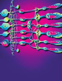
 Periodic acid-methenamine silver (PAM) staining showed thickening of glomerular capillary walls (original magnification 400).")


![Because the bone marrow examination showed 3.3% plasma cells and the patient had no features of hematologic malignancy, he was diagnosed with monoclonal gammopathy of renal significance [5].](/docs-images/90/103467723/images/2-3.jpg "Renal biopsy was performed to investigate the reason for the proteinuria and elevated serum creatinine levels.")
). Severe tubular atrophy, interstitial fibrosis, and monocyte infiltration were observed in the tubulointerstitium.")
 and only lambda light chains were detected in the glomerular capillary walls (Figures 2 and 3).")

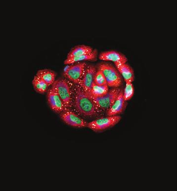
2 2 Case Reports in Nephrology (a) (b) (c) Figure 1: Light microscopy findings. (a) Periodic acid-schiff stain (PAS) staining showed focal mesangial proliferation (original magnification 400). (b) Periodic acid-methenamine silver (PAM) staining showed thickening of glomerular capillary walls (original magnification 400). (c) Masson trichrome (MT) staining showed red staining in the subepithelial area (original magnification 400). were all normal. Serum cryoglobulin, hepatitis B virus surface antigen, and hepatitis C virus antibodies were negative. Urinalysis showed proteinuria (4.9 g/day) and hematuria (5 9 erythrocytes per high-power field) with granular casts (5 9 per high-power field) and lipid casts (5 9 per whole field). Proteinuria was in the nephrotic range. Immunoglobulin and complement levels were all within normal limits. We detected monoclonal IgG-kappa protein, not Bence-Jones protein, by serum and urine immunoelectrophoretic study. Because the bone marrow examination showed 3.3% plasma cells and the patient had no features of hematologic malignancy, he was diagnosed with monoclonal gammopathy of renal significance [5]. Renal biopsy was performed to investigate the reason for the proteinuria and elevated serum creatinine levels. Light microscopy showed diffuse thickening of the glomerular capillary walls with focal mesangial proliferation (Figures 1(a) and 1(b)), and red staining was observed in the subepithelial area by Masson s trichrome stain (Figure 1(c)). Severe tubular atrophy, interstitial fibrosis, and monocyte infiltration were observed in the tubulointerstitium. On immunofluorescence study, IgG and C3 granular deposits were detected in the peripheral capillary walls. Moreover, only IgG1 among the four subclasses (BindingSite, Birmingham, UK) and only lambda light chains were detected in the glomerular capillary walls (Figures 2 and 3). Granular electron-dense deposits were observed in subepithelial, intramembranous, and mesangial area by electron microscopy (Figure 4). From the above pathological findings, we considered that our case was consistent with the conception of PGNMID. Thereafter, in spite of conservative therapy such as angiotensin-converting enzyme inhibitors, calcium blocker, and erythropoietin-stimulating agents, hemodialysis therapy was started because of the progression to end-stage renal disease two years after the renal biopsy. 3. Discussion Glomerular disease with monoclonal immunoglobulin deposition is divided into two categories by electron microscopy, those with organized deposits and those with disorganized deposits [6]. Amyloidosis, type 1 cryoglobulinemic glomerulonephritis, and immunotactoid glomerulonephritis are included in the first group and monoclonal immunoglobulin deposition disease is included in the second group. In 2004, Nasr et al. reported a novel form of glomerular injury related to monoclonal IgG deposition and termed the disease proliferative glomerulonephritis with monoclonal IgG deposits [4]. Although they described typical features of light microscopy as membranoproliferative glomerulonephritis or endocapillary proliferative glomerulonephritis,

. various histologic patterns have been reported.")


![deposits without detection of monoclonal proteins in serum or LCDD without the deposition of light chains in the glomerulus [7, 8].](/docs-images/90/103467723/images/3-6.jpg "Furthermore, Nasr et al.")


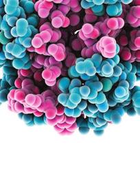
3 Case Reports in Nephrology 3 IgG C3 Figure 2: Immunofluorescence findings. IgG was strongly positive mainly in the peripheral capillary walls. C3 was also positive in peripheral pattern. In light-chain staining, kappa chain was entirely negative, but lambda chain was positive in the peripheral capillaries (original magnification 400). various histologic patterns have been reported. However, according to their report, membranous glomerulonephritis is a rare pathological finding in PGNMID, like mesangial proliferative glomerulonephritis. In the present report, we report a case of membranous glomerulonephritis associated with focal mesangial proliferation with monoclonal IgG deposits. Komatsuda et al. reported a case of immunoglobulin deposition disease with a membranous pattern. Their case showed IgG lambda bands in serum electrophoresis and IgG1-lambda immunofluorescence staining in glomeruli. Although the pathological features and underling disease were similar to our case, their case had no mesangial hypercellularity whereas our case showed a serum light-chain isotype that differed from the glomerular immune deposits. The reason for this discrepancy is not obvious, but there have been some previous reports of glomerulonephritis with monoclonal IgG deposits without detection of monoclonal proteins in serum or LCDD without the deposition of light chains in the glomerulus [7, 8]. Furthermore, Nasr et al. demonstrated that thirty percent of PGNMID patients had a detectable circulating monoclonal protein with the same light-chain isotype as the glomerular deposits [9]. Therefore, discordance between the light-chain isotypes of serum and glomerular deposits is not a rare situation. Komatsuda et al. also reported monoclonal immunoglobulin deposition disease associated with membranous features [10]. They described the light microscopic findings of their cases as thickening of the glomerular capillary walls and spike formation without proliferative lesions and immunofluorescence staining of glomerular deposits that were all of IgG-kappa type. Furthermore, steroid therapy was very effective in their cases and renal function was preserved in all patients. On the other hand, Nasr et al. summarized clinical outcomes of 32 patients who had PGNMID, of whom 21.9% progressed to end-stage renal disease [4]. At the time of renal biopsy, our patient showed the elevation of serum creatinine, and he progressed to end-stage renal disease within two years after renal biopsy. This poor clinical outcome was similar to those of Nasr et al. s report. Light microscopy of our case revealed membranous glomerulonephritis with focal mesangial proliferation and lambdatype light-chain deposits. These findings are more similar to the pathological features of PGNMID. From these above considerations, we diagnosed our case as PGNMID with a predominantly membranous glomerulonephritis character. It
.")


![for staining IgG subclasses. References [1] S.H.Nasr,G.S.Markowitz,M.B.Stokesetal.](/docs-images/90/103467723/images/4-7.jpg ", Proliferative glomerulonephritis with monoclonal IgG deposits: a distinct entity mimicking immune-complex glomerulonephritis, Kidney International,vol.65,no.")
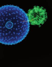
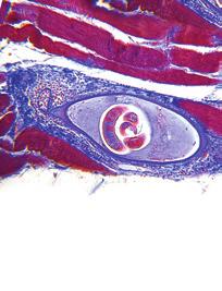
4 4 Case Reports in Nephrology Figure 3: Findings of IgG subclass staining. IgG1 was positive in peripheral granular pattern. On the other hand, IgG2, IgG3, and IgG4 were all negative (original magnification 400). Conflicts of Interest The authors declare that there are no conflicts of interest regarding the publication of this paper. Acknowledgments Figure 4: Electron microscopic finding. Electron microscopy showed huge electron-dense deposits in subepithelial, intramembranous, and mesangial lesions (original magnification 3,000). is unclear whether monoclonal immunoglobulin deposition disease associated with membranous features reported by de Seigneux et al. and Komatsuda et al. is a distinct disease concept from PGNMID. Further accumulation of cases of monoclonal immunoglobulin deposition disease associated with membranous features is needed to confirm whether these pathological features belong to PGNMID. Here, we present the case of PGNMID that showed predominantly membranous features on light microscopy. Whenever membranous glomerulonephritis with monoclonal gammopathy is diagnosed, immunoglobulin staining for IgG subclass and immunoglobulin light chains should be conducted to clarify the immunoglobulin deposit disease. The authors would like to thank Professor Yoshihiko Ueda of Department of Diagnostic Pathology, Dokkyo Medical University Koshigaya Hospital, Saitama, Japan, for staining IgG subclasses. References [1] S.H.Nasr,G.S.Markowitz,M.B.Stokesetal., Proliferative glomerulonephritis with monoclonal IgG deposits: a distinct entity mimicking immune-complex glomerulonephritis, Kidney International,vol.65,no.1,pp.85 96,2004. [2]S.H.Nasr,A.Satoskar,G.S.Markowitzetal., Proliferative glomerulonephritis with monoclonal IgG deposits, the American Society of Nephrology, vol.20,no.9,pp , [3] R. Masai, H. Wakui, A. Komatsuda et al., Characteristics of proliferative glomerulonephritis with monoclonal IgG deposits associated with membranoproliferative features, Clinical Nephrology,vol.72,no.1,pp.46 54,2009. [4] S. de Seigneux, P. Bindi, H. Debiec et al., Immunoglobulin Deposition Disease With a Membranous Pattern and a Circulating Monoclonal Immunoglobulin G With Charge-Dependent
5 Case Reports in Nephrology 5 Aggregation Properties, American Kidney Diseases, vol.56,no.1,pp ,2010. [5]N.Leung,F.Bridoux,C.A.Hutchisonetal., Monoclonal gammopathy of renal significance: when MGUS is no longer undetermined or insignificant, Blood,vol.120,no.22,pp , [6]P.M.Ronco,M.-A.Alyanakian,B.Mougenot,andP.Aucouturier, Light chain deposition disease: A model of glomerulosclerosis defined at the molecular level, the American Society of Nephrology,vol.12,no.7,pp ,2001. [7]A.Komatsuda,H.Ohtani,K.Sawada,K.Joh,andH.Wakui, Proliferative glomerulonephritis with discrete deposition of monoclonal immunoglobulin γ1 CH2 heavy chain and κ light chain: A new variant of monoclonal immunoglobulin deposition disease, Pathology International, vol. 63, no. 1, pp , [8] S. Darouich, R. Goucha, M. H. Jaafoura, S. Zekri, A. Kheder, and H. B. Maiz, Light-chain deposition disease of the kidney: Acasereport, Ultrastructural Pathology,vol.36,no.2,pp , [9] S. H. Nasr, S. Sethi, L. D. Cornell et al., Proliferative glomerulonephritis with monoclonal IgG deposits recurs in the allograft, Clinical the American Society of Nephrology,vol. 6, no. 1, pp , [10] A. Komatsuda, R. Masai, H. Ohtani et al., Monoclonal immunoglobulin deposition disease associated with membranous features, Nephrology Dialysis Transplantation,vol.23,no. 12, pp , 2008.


6 MEDIATORS of INFLAMMATION The Scientific World Journal Gastroenterology Research and Practice Diabetes Research International Endocrinology Immunology Research Disease Markers Submit your manuscripts at BioMed Research International PPAR Research Obesity Ophthalmology Evidence-Based Complementary and Alternative Medicine Stem Cells International Oncology Parkinson s Disease Computational and Mathematical Methods in Medicine AIDS Behavioural Neurology Research and Treatment Oxidative Medicine and Cellular Longevity
Monoclonal Gammopathies and the Kidney. Tibor Nádasdy, MD The Ohio State University, Columbus, OH
 Monoclonal Gammopathies and the Kidney Tibor Nádasdy, MD The Ohio State University, Columbus, OH Monoclonal gammopathy of renal significance (MGRS) Biopsies at OSU (n=475) between 2007 and 2016 AL or AH
Case Report Nephrotic Syndrome Secondary to Proliferative Glomerulonephritis with Monoclonal Immunoglobulin Deposits of Lambda Light Chain
 Hindawi Publishing Corporation Case Reports in Nephrology Volume 214, Article ID 164694, 6 pages http://dx.doi.org/1.1155/214/164694 Case Report Nephrotic Syndrome Secondary to Proliferative Glomerulonephritis
Interesting case seminar: Native kidneys Case Report:
 Interesting case seminar: Native kidneys Case Report: Proximal tubulopathy and light chain deposition disease presented as severe pulmonary hypertension with right-sided cardiac dysfunction and nephrotic
Mayo Clinic/ RPS Consensus Report on Classification, Diagnosis, and Reporting of Glomerulonephritis
 Mayo Clinic/ RPS Consensus Report on Classification, Diagnosis, and Reporting of Glomerulonephritis Sanjeev Sethi, MD, PhD Department of Laboratory Medicine and Pathology Disclosure Relevant Financial
Ordering Physician. Collected REVISED REPORT. Performed. IgG IF, Renal MCR. Lambda IF, Renal MCR. C1q IF, Renal. MCR Albumin IF, Renal MCR
 RenalPath Level IV Wet Ts IgA I Renal IgM I Renal Kappa I Renal Renal Bx Electron Microscopy IgG I Renal Lambda I Renal C1q I Renal C3 I Renal Albumin I Renal ibrinogen I Renal Mayo Clinic Dept. of Lab
FIBRILLARY GLOMERULONEPHRITIS DIAGNOSTIC CRITERIA, PITFALLS, AND DIFFERENTIAL DIAGNOSIS
 FIBRILLARY GLOMERULONEPHRITIS DIAGNOSTIC CRITERIA, PITFALLS, AND DIFFERENTIAL DIAGNOSIS Guillermo A. Herrera MD Louisiana State University, Shreveport Fibrils in bundles 10-20 nm d Diabetic fibrillosis
Clinicopathological analysis of proliferative glomerulonephritis with monoclonal IgG deposits in 5 renal allografts
 Wen et al. BMC Nephrology (2018) 19:173 https://doi.org/10.1186/s12882-018-0969-3 RESEARCH ARTICLE Open Access Clinicopathological analysis of proliferative glomerulonephritis with monoclonal IgG deposits
Journal of Nephropathology
 www.nephropathol.com DOI: 10.15171/jnp.2017.36 J Nephropathol. 2017;6(3):220-224 Journal of Nephropathology Proliferative glomerulonephritis with monoclonal IgG deposits; an unusual cause of de novo disease
A Case of IgG2 Heavy Chain Deposition Disease in a Patient with Kappa Positive Plasma Cell Dyscrasia
 Published online: August 14, 2014 2296 9705/14/0051 0006$39.50/0 This is an Open Access article licensed under the terms of the Creative Commons Attribution-NonCommercial 3.0 Unported license (CC BY-NC)
RENAL HISTOPATHOLOGY
 RENAL HISTOPATHOLOGY Peter McCue, M.D. Department of Pathology, Anatomy & Cell Biology Sidney Kimmel Medical College There are no conflicts of interest. 1 Goals and Objectives! Goals Provide introduction
A clinical syndrome, composed mainly of:
 Nephritic syndrome We will discuss: 1)Nephritic syndrome: -Acute postinfectious (poststreptococcal) GN -IgA nephropathy -Hereditary nephritis 2)Rapidly progressive GN (RPGN) A clinical syndrome, composed
Case 3. ACCME/Disclosure. Laboratory results. Clinical history 4/13/2016
 Case 3 Lynn D. Cornell, M.D. Mayo Clinic, Rochester, MN Cornell.Lynn@mayo.edu USCAP Renal Case Conference March 13, 2016 ACCME/Disclosure Dr. Cornell has nothing to disclose Clinical history 57-year-old
Surgical Pathology Report
 Louisiana State University Health Sciences Center Department of Pathology Shreveport, Louisiana Accession #: Collected: Received: Reported: 6/1/2012 09:18 6/2/2012 09:02 6/2/2012 Patient Name: Med. Rec.
Pathology of Complement Mediated Renal Disease
 Pathology of Complement Mediated Renal Disease Mariam Priya Alexander, MD Associate Professor of Pathology GN Symposium Hong Kong Society of Nephrology July 8 th, 2017 2017 MFMER slide-1 The complement
CASE 3 AN UNUSUAL CASE OF NEPHROTIC SYNDROME
 CASE 3 AN UNUSUAL CASE OF NEPHROTIC SYNDROME Dr Seethalekshmy N.V., Dr.Annie Jojo, Dr Hiran K.R., Amrita institute of Medical Sciences, Kochi, Kerala Case history 34 year old gentleman Nephrotic range
Proliferative Glomerulonephritis with Monoclonal IgG Deposits Recurs in the Allograft
 Article Proliferative Glomerulonephritis with Monoclonal IgG Deposits Recurs in the Allograft Samih H. Nasr,* Sanjeev Sethi,* Lynn D. Cornell,* Mary E. Fidler,* Mark Boelkins, Fernando C. Fervenza, Fernando
Dr Ian Roberts Oxford. Oxford Pathology Course 2010 for FRCPath Illustration-Cellular Pathology. Oxford Radcliffe NHS Trust
 Dr Ian Roberts Oxford Oxford Pathology Course 2010 for FRCPath Present the basic diagnostic features of the commonest conditions causing proteinuria & haematuria Highlight diagnostic pitfalls Nephrotic
C1q nephropathy the Diverse Disease
 C1q nephropathy the Diverse Disease Danica Galešić Ljubanović School of Medicine, University of Zagreb Dubrava University Hospital Zagreb, Croatia Definition Dominant or codominant ( 2+), mesangial staining
Glomerular diseases with organized deposits
 Glomerular diseases with organized deposits Banu Sis, MD, FRCPC University of Alberta, Edmonton, AB, Canada Ulusal Patoloji Kongresi, Manavgat, Antalya 8/11/2012 What is an organized deposit? A number
Glomerular Pathology- 1 Nephrotic Syndrome. Dr. Nisreen Abu Shahin
 Glomerular Pathology- 1 Nephrotic Syndrome Dr. Nisreen Abu Shahin The Nephrotic Syndrome a clinical complex resulting from glomerular disease & includes the following: (1) massive proteinuria (3.5 gm /day
Glomerular pathology in systemic disease
 Glomerular pathology in systemic disease Lecture outline Lupus nephritis Diabetic nephropathy Glomerulonephritis Associated with Bacterial Endocarditis and Other Systemic Infections Henoch-Schonlein Purpura
CJASN epress. Published on September 28, 2010 as doi: /CJN
 CJASN epress. Published on September 28, 2010 as doi: 10.2215/CJN.05750710 Proliferative Glomerulonephritis with Monoclonal IgG Deposits Recurs in the Allograft Samih H. Nasr,* Sanjeev Sethi,* Lynn D.
Histopathology: Glomerulonephritis and other renal pathology
 Histopathology: Glomerulonephritis and other renal pathology These presentations are to help you identify basic histopathological features. They do not contain the additional factual information that you
Classification of Glomerular Diseases and Defining Individual Glomerular Lesions: Developing International Consensus
 Classification of Glomerular Diseases and Defining Individual Glomerular Lesions: Developing International Consensus Mark Haas MD, PhD Department of Pathology & Laboratory Medicine Cedars-Sinai Medical
Hemizygous Fabry disease associated with IgA nephropathy: A case report
 1 Hemizygous Fabry disease associated with IgA nephropathy: A case report Fabry disease and IgA nephropathy Homare Shimohata 1, 3, Keigyou Yoh 1, Kenji Takada 2, Hiroaki Tanaka 2, Joichi Usui 1, Kouichi
Long-term follow-up of juvenile acute nonproliferative glomerulitis (JANG)
") Pediatr Nephrol (2007) 22:1957 1961 DOI 10.1007/s00467-007-0555-6 BRIEF REPORT Long-term follow-up of juvenile acute nonproliferative glomerulitis (JANG) Teruo Fujita & Kandai Nozu & Kazumoto Iijima &
Pediatr Nephrol (2007) 22:1957 1961 DOI 10.1007/s00467-007-0555-6 BRIEF REPORT Long-term follow-up of juvenile acute nonproliferative glomerulitis (JANG) Teruo Fujita & Kandai Nozu & Kazumoto Iijima &
Dr Ian Roberts Oxford. Oxford Pathology Course 2010 for FRCPath Illustration-Cellular Pathology. Oxford Radcliffe NHS Trust
 Dr Ian Roberts Oxford Oxford Pathology Course 2010 for FRCPath Plan of attack: Diagnostic approach to the renal biopsy Differential diagnosis of the clinical syndromes of renal disease Microscopy Step
Renal Pathology 1: Glomerulus. With many thanks to Elizabeth Angus PhD for EM photographs
 Renal Pathology 1: Glomerulus With many thanks to Elizabeth Angus PhD for EM photographs Anatomy of the Kidney http://www.yalemedicalgroup.org/stw/page.asp?pageid=stw028980 The Nephron http://www.beltina.org/health-dictionary/nephron-function-kidney-definition.html
Research Article Transplant Outcomes in Patients with Idiopathic Membranous Nephropathy
 International Nephrology Volume 2013, Article ID 818537, 4 pages http://dx.doi.org/10.1155/2013/818537 Research Article Transplant Outcomes in Patients with Idiopathic Membranous Nephropathy Claire Kennedy,
Favorable effect of bortezomib in dense deposit disease associated with monoclonal gammopathy: a case report
 Hirashio et al. BMC Nephrology (2018) 19:108 https://doi.org/10.1186/s12882-018-0905-6 CASE REPORT Open Access Favorable effect of bortezomib in dense deposit disease associated with monoclonal gammopathy:
Yijuan Sun, Amarpreet Sandhu, Darlene Gabaldon, Jonathan Danaraj, Karen S. Servilla, and Antonios H. Tzamaloukas
 Case Reports in Nephrology Volume 2012, Article ID 573650, 5 pages doi:10.1155/2012/573650 Case Report Development of Renal Failure without Proteinuria in a Patient with Monoclonal Gammopathy of Undetermined
Dense deposit disease with steroid pulse therapy
 Case Report Dense deposit disease with steroid pulse therapy Jun Odaka, Takahiro Kanai, Takane Ito, Takashi Saito, Jun Aoyagi, and Mariko Y Momoi Abstract Treatment of dense deposit disease DDD has not
Immune profile of IgA-dominant diffuse proliferative glomerulonephritis
 Clin Kidney J (2014) 7: 479 483 doi: 10.1093/ckj/sfu090 Exceptional Case Immune profile of IgA-dominant diffuse proliferative glomerulonephritis Eric Wallace 1, Nicolas Maillard 2, Hiroyuki Ueda 2, Stacy
Expanding Spectrum of Diseases Associated with Plasma Cell Dyscrasias
 Expanding Spectrum of Diseases Associated with Plasma Cell Dyscrasias Eva Honsova Institute for Clinical and Experimental Medicine Prague, Czech Republic eva.honsova@ikem.cz Plasma cell dyscrasias Plasma
C3 GLOMERULOPATHIES. Budapest Nephrology School Zoltan Laszik
 C3 GLOMERULOPATHIES Budapest Nephrology School 8.30.2018. Zoltan Laszik 1 Learning Objectives Familiarize with the pathogenetic mechanisms of glomerular diseases Learn the pathologic landscape and clinical
Multiple Myeloma Advances for clinical pathologists & histopathologists
 Multiple Myeloma Advances for clinical pathologists & histopathologists CME in Haematology 2014 IAPP & Dept of Pathology, BVDUMC, Pune Sunday, 4 th May 2014 Dr. M.B. Agarwal, MD, MNAMS Head, Dept of Haematology
CHAPTER 2 PRIMARY GLOMERULONEPHRITIS
 CHAPTER 2 Sunita Bavanandan Lim Soo Kun 19 5th Report of the 2.1: Introduction This chapter covers the main primary glomerulonephritis that were reported to the MRRB from the years 2005-2012. Minimal change
THE URINARY SYSTEM. The cases we will cover are:
 THE URINARY SYSTEM The focus of this week s lab will be pathology of the urinary system. Diseases of the kidney can be broken down into diseases that affect the glomeruli, tubules, interstitium, and blood
THE URINARY SYSTEM. The cases we will cover are:
 THE URINARY SYSTEM The focus of this week s lab will be pathology of the urinary system. Diseases of the kidney can be broken down into diseases that affect the glomeruli, tubules, interstitium, and blood
Case Report Diagnostic Challenges of Tuberculous Lymphadenitis Using Polymerase Chain Reaction Analysis: A Case Study
 Case Reports in Infectious Diseases Volume 2015, Article ID 723726, 4 pages http://dx.doi.org/10.1155/2015/723726 Case Report Diagnostic Challenges of Tuberculous Lymphadenitis Using Polymerase Chain Reaction
Case Presentation Turki Al-Hussain, MD
 Case Presentation Turki Al-Hussain, MD Director, Renal Pathology Chapter Saudi Society of Nephrology & Transplantation Consultant Nephropathologist & Urological Pathologist Department of Pathology & Laboratory
Glomerular diseases mostly presenting with Nephritic syndrome
 Glomerular diseases mostly presenting with Nephritic syndrome 1 The Nephritic Syndrome Pathogenesis: proliferation of the cells in glomeruli & leukocytic infiltrate Injured capillary walls escape of RBCs
Rituximab treatment for fibrillary glomerulonephritis
 Nephrol Dial Transplant (2014) 29: 1925 1931 doi: 10.1093/ndt/gfu189 Advance Access publication 27 May 2014 Rituximab treatment for fibrillary glomerulonephritis Jonathan Hogan, Michaela Restivo, Pietro
CASE 4 A RARE CASE OF INTRALUMINAL GLOMERULAR CAPILLARY DEPOSITS
 CASE 4 A RARE CASE OF INTRALUMINAL GLOMERULAR CAPILLARY DEPOSITS DR ANNIE JOJO, Dr Seethalekshmy N V, Dr Nanda Kachare DEPARTMENT OF PATHOLOGY, AMRITA INSTITUTE OF MEDICAL SCIENCES, KOCHI. 54 yrs female,
CHAPTER 2. Primary Glomerulonephritis
 2nd Report of the PRIMARY GLOMERULONEPHRITIS CHAPTER 2 Primary Glomerulonephritis Sunita Bavanandan Lee Han Wei Lim Soo Kun 21 PRIMARY GLOMERULONEPHRITIS 2nd Report of the 2.1 Introduction This chapter
Recurrent Idiopathic Membranous Glomerulonephritis After Kidney Transplantation and Successful Treatment With Rituximab
 TRANSPLANTATION Recurrent Idiopathic Membranous Glomerulonephritis After Kidney Transplantation and Successful Treatment With Rituximab Khadijeh Makhdoomi, 1,2 Saeed Abkhiz, 1,2 Farahnaz Noroozinia, 1,3
Jon Von Visger 1, Clarissa Cassol 2, Uday Nori 1, Gerardo Franco-Ahumada 1, Tibor Nadasdy 2 and Anjali A. Satoskar 2*
 Von Visger et al. BMC Nephrology (2019) 20:53 https://doi.org/10.1186/s12882-019-1239-8 CASE REPORT Open Access Complete biopsy-proven resolution of deposits in recurrent proliferative glomerulonephritis
Familial DDD associated with a gain-of-function mutation in complement C3.
 Familial DDD associated with a gain-of-function mutation in complement C3. Santiago Rodríguez de Córdoba, Centro de investigaciones Biológicas, Madrid Valdés Cañedo F. and Vázquez- Martul E., Complejo
Monoclonal gammopathies consist of. Monoclonal GammopathyeAssociated Proliferative Glomerulonephritis REVIEW
 REVIEW Monoclonal GammopathyeAssociated Proliferative Glomerulonephritis Sanjeev Sethi, MD, PhD, and S. Vincent Rajkumar, MD Abstract Monoclonal gammopathy is characterized by circulating monoclonal immunoglobulin
Clinical Study Glomerulonephritis with Crescents in Children: Etiology and Predictors of Renal Outcome
 International Scholarly Research Network ISRN Pediatrics Volume 2011, Article ID 507298, 5 pages doi:10.5402/2011/507298 Clinical Study Glomerulonephritis with Crescents in Children: Etiology and Predictors
Tarek ElBaz, MD. Prof. Internal Medicine Chief, Division of Renal Medicine Al Azhar University President, ESNT
 The Kidney in Multiple Myeloma Tarek ElBaz, MD. Prof. Internal Medicine Chief, Division of Renal Medicine Al Azhar University President, ESNT Normal Cell Plasma cells produce antibodies that bind to antigens,
Lab 3, case 1. Is this an example of nephrotic or nephritic syndrome? Why? Which portion of the nephron would you expect to be abnormal?
 Lab 3, case 1 12-year-old Costa Rican boy is brought into clinic by his parents because of dark brownish-red urine over the last 24 hours. The family has been visiting friends in Indianapolis for two weeks.
Disorders of the kidney. Urine analysis. Nephrotic and nephritic syndrome.
 Disorders of the kidney. Urine analysis. Nephrotic and nephritic syndrome. Azotemia and Urinary Abnormalities Disturbances in urine volume oliguria, anuria, polyuria Abnormalities of urine sediment red
Jo Abraham MD Division of Nephrology University of Utah
 Jo Abraham MD Division of Nephrology University of Utah 68 year old male presented 3 weeks ago with a 3 month history of increasing fatigue He reported a 1 week history of increasing dyspnea with a productive
A Case of Podocytic Infolding Glomerulopathy with Focal Segmental Glomerulosclerosis
 Published online: August 8, 2013 1664 5510/13/0032 0110$38.00/0 This is an Open Access article licensed under the terms of the Creative Commons Attribution-NonCommercial 3.0 Unported license (CC BY-NC)
Case Report Membranoproliferative Glomerulonephritis in Patients with Chronic Venous Catheters: A Case Report and Literature Review
 Hindawi Publishing Corporation Case Reports in Nephrology Volume 2014, Article ID 159370, 5 pages http://dx.doi.org/10.1155/2014/159370 Case Report Membranoproliferative Glomerulonephritis in Patients
GOODPASTURE'S SYNDROME WITH CONCOMITANT IMMUNE COMPLEX MIXED MEMBRANOUS AND PROLIFERATIVE GLOMERULONEFRITIS
 GOODPASTURE'S SYNDROME WITH CONCOMITANT IMMUNE COMPLEX MIXED MEMBRANOUS AND PROLIFERATIVE GLOMERULONEFRITIS VESNA JURČIĆ 1, ANDREJA ALEŠ RIGLER 2, INSTITUTE OF PATHOLOGY, FACULTY OF MEDICINE, UNIVERSITY
THE KIDNEY AND SLE LUPUS NEPHRITIS
 THE KIDNEY AND SLE LUPUS NEPHRITIS JACK WATERMAN DO FACOI 2013 NEPHROLOGY SIR RICHARD BRIGHT TERMINOLOGY RENAL INSUFFICIENCY CKD (CHRONIC KIDNEY DISEASE) ESRD (ENDSTAGE RENAL DISEASE) GLOMERULONEPHRITIS
A case of heavy chain deposition disease complicated by acquired angioedema.
 Case Report http://www.alliedacademies.org/pathology-and-disease-biology/ A case of heavy chain deposition disease complicated by acquired angioedema. Rafia Chaudhry 1 *, Gautam Bhave 2, Rachel Fissell
Case Presentation Turki Al-Hussain, MD
 Case Presentation Turki Al-Hussain, MD Director, Renal Pathology Chapter Saudi Society of Nephrology & Transplantation Consultant Nephropathologist & Urological Pathologist Department of Pathology & Laboratory
Case Report Osteolysis of the Greater Trochanter Caused by a Foreign Body Granuloma Associated with the Ethibond Suture after Total Hip Arthroplasty
 Hindawi Volume 2017, Article ID 6082302, 4 pages https://doi.org/10.1155/2017/6082302 Case Report Osteolysis of the Greater Trochanter Caused by a Foreign Body Granuloma Associated with the Ethibond Suture
Overview of glomerular diseases
 Overview of glomerular diseases *Endothelial cells are fenestrated each fenestra: 70-100nm in diameter Contractile, capable of proliferation, makes ECM & releases mediators *Glomerular basement membrane
Glomerular pathology-2 Nephritic syndrome. Dr. Nisreen Abu Shahin
 Glomerular pathology-2 Nephritic syndrome Dr. Nisreen Abu Shahin 1 The Nephritic Syndrome Pathogenesis: inflammation proliferation of the cells in glomeruli & leukocytic infiltrate Injured capillary walls
ACUTE GLOMERULONEPHRITIS. IAP UG Teaching slides
 ACUTE GLOMERULONEPHRITIS 1 Definition Etiology Pathology/pathogenesis Risk factors Clinical Presentation Investigation Differential Diagnosis Management Outcome/Prognosis Indication for Renal Biopsy Summary
Mr. I.K 58 years old
 Mr. I.K 58 years old Hospitalized because of marked pitting peripheral edema (bilateral crural and perimalleolar edema) and uncontrolled blood pressure (BP 150/100 mmhg under treatment). since age 54 years
Diabetic Nephropathy. Introduction/Clinical Setting. Pathologic Findings Light Microscopy. J. Charles Jennette
 12 Diabetic Nephropathy J. Charles Jennette Introduction/Clinical Setting Diabetic nephropathy is a clinical syndrome in a patient with diabetes mellitus that is characterized by persistent albuminuria,
Proliferative Glomerulonephritis with Monoclonal IgG Deposits
 JASN Express. Published on May 21, 2009 as doi: 10.1681/ASN.2009010110 Proliferative Glomerulonephritis with Monoclonal IgG Deposits Samih H. Nasr,* Anjali Satoskar, Glen S. Markowitz,* Anthony M. Valeri,
A Case of Myeloma Kidney With Glomerular C3 Deposition
 Case Report World J Nephrol Urol. 2018;7(3-4):73-77 A Case of Myeloma Kidney With Glomerular C3 Deposition Asif Khan a, c, Khine Lam b, Suzanne El-Sayegh b, Elie El-Charabaty b Abstract Manuscript submitted
WE PRESENT a patient with non insulindependent
 RENAL BIOPSY TEACHING CASE Monoclonal Gammopathy in a Type II Diabetic: A Case of Determined Significance Nancy J. Gritter, MD, Simin Goral, MD, and Agnes Fogo, MD INDEX WORDS: Monoclonal gammopathy; nephrotic
Approach to Glomerular Diseases: Clinical Presentation Nephrotic Syndrome Nephritis
 GLOMERULONEPHRITIDES Vivette D Agati Jai Radhakrishnan Approach to Glomerular Diseases: Clinical Presentation Nephrotic Syndrome Nephritis Heavy Proteinuria Renal failure Low serum Albumin Hypertension
Xi Yang, Ri-Bao Wei, Ping Li, Yue Yang, Ting-Yu Su, Yu-Wei Gao, Qing-Ping Li, Xue-Guang Zhang, Xiang-Mei Chen
 Int J Clin Exp Pathol 2016;9(9):9401-9407 www.ijcep.com /ISSN:1936-2625/IJCEP0028098 Original Article Correlation between serum hepatitis B virus DNA replication level and clinicopathology in 235 patients
Case Report Two Cases of Small Cell Cancer of the Maxillary Sinus Treated with Cisplatin plus Irinotecan and Radiotherapy
 Case Reports in Otolaryngology Volume 2013, Article ID 893638, 4 pages http://dx.doi.org/10.1155/2013/893638 Case Report Two Cases of Small Cell Cancer of the Maxillary Sinus Treated with Cisplatin plus
Year 2004 Paper one: Questions supplied by Megan
 QUESTION 53 Endothelial cell pathology on renal biopsy is most characteristic of which one of the following diagnoses? A. Pre-eclampsia B. Haemolytic uraemic syndrome C. Lupus nephritis D. Immunoglobulin
RECURRENT AND DE NOVO RENAL DISEASES IN THE ALLOGRAFT
 RECURRENT AND DE NOVO RENAL DISEASES IN THE ALLOGRAFT HISTOPATHOLOGIC DISORDERS AFFECTING THE ALLOGRAFT OTHER THAN REJECTION RECURRENT DISEASE DE NOVO DISEASE TRANSPLANT GLOMERULOPATHY Glomerular Non-glomerular
Glomerular Diseases. Anna Vinnikova, MD Nephrology
 Glomerular Diseases Anna Vinnikova, MD Nephrology Classification of Glomerular Diseases http://what-when-how.com/acp-medicine/glomerular-diseases-part-1/ Classification of pathologic and clinical manifestations
An unusual association between focal segmental sclerosis and lupus nephritis: a distinct concept from lupus podocytopathy?
 CEN Case Rep (2015) 4:70 75 DOI 10.1007/s13730-014-0142-1 CASE REPORT An unusual association between focal segmental sclerosis and lupus nephritis: a distinct concept from lupus podocytopathy? Hironari
Case Report Features of the Atrophic Corpus Mucosa in Three Cases of Autoimmune Gastritis Revealed by Magnifying Endoscopy
 Volume 2012, Article ID 368160, 4 pages doi:10.1155/2012/368160 Case Report Features of the Atrophic Corpus Mucosa in Three Cases of Autoimmune Gastritis Revealed by Magnifying Endoscopy Kazuyoshi Yagi,
substance staining with IgG, C3 and IgA (trace) Linear deposition of IgG(+), IgA.M(trace) and C3(+++) at the DEJ
 Linear deposition of IgG(+), IgA.M(trace) and C3(+++) at the DEJ") Direct Immunofluorescence: Skin Diagnosis Findings Picture Pemphigus Vulgaris and it s Intracellular cement variants substance staining with IgG, C3 and IgA (trace) Bullous Pemphigoid and it s variants
Direct Immunofluorescence: Skin Diagnosis Findings Picture Pemphigus Vulgaris and it s Intracellular cement variants substance staining with IgG, C3 and IgA (trace) Bullous Pemphigoid and it s variants
Renal manifestations of IgG4-related systemic disease
 Renal manifestations of IgG4-related systemic disease Lynn D. Cornell, M.D. Mayo Clinic Rochester, MN While autoimmune pancreatitis (AIP) has been recognized since the first description by Sarles et al
Pathogenesis of IgA Nephropathy. Shokoufeh Savaj MD Associate Professor of Medicine Firoozgar hospital- IUMS
 Pathogenesis of IgA Nephropathy Shokoufeh Savaj MD Associate Professor of Medicine Firoozgar hospital- IUMS History Immunoglobin A nephropathy was first described by Berger and Hinglais in 1968 in Paris
Case Report Diaphragmatic Amyloidosis Causing Respiratory Failure: A Case Report and Review of Literature
 Volume 2015, Article ID 917157, 4 pages http://dx.doi.org/10.1155/2015/917157 Case Report Diaphragmatic Amyloidosis Causing Respiratory Failure: A Case Report and Review of Literature Aleksey Novikov,
Elevated Serum Creatinine, a simplified approach
 Elevated Serum Creatinine, a simplified approach Primary Care Update Creighton University School of Medicine. April 27 th, 2018 Disclosure Slide I have no disclosures and have no conflicts with this presentation.
Index. electron microscopy, 81 immunofluorescence microscopy, 80 light microscopy, 80 Amyloidosis clinical setting, 185 etiology/pathogenesis,
 A Acute antibody-mediated rejection (Acute AMR) clinical features, 203 clinicopathologic correlations, 206 pathogenesis, 205 206 204 205 light microscopy, 203 204 Acute cellular rejection (ACR) clinical
Comparison of amyloid deposition in human kidney biopsies as predictor of poor patient outcome
 Castano et al. BMC Nephrology (2015) 16:64 DOI 10.1186/s12882-015-0046-0 RESEARCH ARTICLE Comparison of amyloid deposition in human kidney biopsies as predictor of poor patient outcome Open Access Ekaterina
Sebastião Rodrigues Ferreira-Filho, Camila Caetano Cardoso, Luiz Augusto Vieira de Castro, Ricardo Mendes Oliveira, and Renata Rodrigues Sá
 SAGE-Hindawi Access to Research International Nephrology Volume 211, Article ID 626178, 4 pages doi:1.461/211/626178 Research Article Comparison of Measured Creatinine Clearance and Clearances Estimated
RENAL EVENING SPECIALTY CONFERENCE
 RENAL EVENING SPECIALTY CONFERENCE Harsharan K. Singh, MD The University of North Carolina at Chapel Hill Disclosure of Relevant Financial Relationships No conflicts of interest to disclose. CLINICAL HISTORY
Journal of Nephropathology
 www.nephropathol.com DOI: 10.15171/jnp.2018.22 J Nephropathol. 2018;7(2):93-97 Journal of Nephropathology Anti-phospholipase A2 receptor antibody positive hepatitis B virus-associated membranous nephropathy
RECURRENT AND DE NOVO RENAL DISEASES IN THE ALLOGRAFT. J. H. Helderman,MD,FACP,FAST
 RECURRENT AND DE NOVO RENAL DISEASES IN THE ALLOGRAFT J. H. Helderman,MD,FACP,FAST Vanderbilt University Medical Center Professor of Medicine, Pathology and Immunology Medical Director, Vanderbilt Transplant
Proliferative glomerulonephritis with monoclonal IgG deposits: A distinct entity mimicking immune-complex glomerulonephritis
 Kidney International, Vol. 65 (2004), pp. 85 96 Proliferative glomerulonephritis with monoclonal IgG deposits: A distinct entity mimicking immune-complex glomerulonephritis SAMIH H. NASR, GLENS. MARKOWITZ,
Research Article Clinical Outcome of a Novel Anti-CD6 Biologic Itolizumab in Patients of Psoriasis with Comorbid Conditions
 Dermatology Research and Practice Volume 2, Article ID 13326, 4 pages http://dx.doi.org/.1155/2/13326 Research Article Clinical Outcome of a Novel Anti-CD6 Biologic Itolizumab in Patients of Psoriasis
NEPHROTIC SYNDROME OF ACQUIRED SYPHILIS-A MORPHOLOGICAL AND ULTRASTRUCTURAL STUDY
 NEPHROTIC SYNDROME OF ACQUIRED SYPHILIS-A MORPHOLOGICAL AND ULTRASTRUCTURAL STUDY Abstract Pages with reference to book, From 3 To 7 A.H. Nagi, I.A. Naveed, A. Rashid ( Department of Pathology, Allama
Case Report IgG4-Related Nasal Pseudotumor
 Case Reports in Otolaryngology Volume 2015, Article ID 749890, 4 pages http://dx.doi.org/10.1155/2015/749890 Case Report IgG4-Related Nasal Pseudotumor L. K. Døsen, 1 P. Jebsen, 2 B. Dingsør, 3 and R.
Proteinuria. Louisiana State University
 Proteinuria W S A V A W C P, 2005 David F. Senior Louisiana State University The normal glomerulus is a highly selective barrier for filtration based on size (and on charge in the case of larger molecules).
Membranoproliferative Glomerulonephritis
 Membranoproliferative Glomerulonephritis MPGN is characterizedby alterations in the GBM and mesangium and by proliferation of glomerular cells. 5% to 10% of cases of 1ry nephrotic syndrome in children
Glomerular tip adhesions predict the progression of IgA nephropathy
 Maeda et al. BMC Nephrology 2013, 14:272 RESEARCH ARTICLE Open Access Glomerular tip adhesions predict the progression of IgA nephropathy Kunihiro Maeda 1, Shogo Kikuchi 2, Naoto Miura 1, Keisuke Suzuki
Renal biopsy cases in myeloproliferative neoplasms (MPN)
") CEN Case Rep (2013) 2:215 221 DOI 10.1007/s13730-013-0067-0 CASE REPORT Renal biopsy cases in myeloproliferative neoplasms (MPN) Kumi Fujita Kazuhiro Hatta Received: 6 November 2012 / Accepted: 4 February
CEN Case Rep (2013) 2:215 221 DOI 10.1007/s13730-013-0067-0 CASE REPORT Renal biopsy cases in myeloproliferative neoplasms (MPN) Kumi Fujita Kazuhiro Hatta Received: 6 November 2012 / Accepted: 4 February
Research Article Clinicopathological Correlation in Asian Patients with Biopsy-Proven Lupus Nephritis
 International Nephrology Volume 2015, Article ID 857316, 6 pages http://dx.doi.org/10.1155/2015/857316 Research Article Clinicopathological Correlation in Asian Patients with Biopsy-Proven Lupus Nephritis
Forms Revision: Myeloma Changes
 Sharing knowledge. Sharing hope. Forms Revision: Myeloma Changes J. Brunner, PA-C and A. Dispenzieri, MD February 2013 Disclosures Janet Brunner, PA-C I have no relevant conflicts of interest to disclose.
Light-Chain Mediated Acute Tubular Interstitial Nephritis. A Poorly Recognized Pattern of Renal Disease in Patients With Plasma Cell Dyscrasia
 Light-Chain Mediated Acute Tubular Interstitial Nephritis A Poorly Recognized Pattern of Renal Disease in Patients With Plasma Cell Dyscrasia Xin Gu, MD; Guillermo A. Herrera, MD Context. Acute renal failure
Case Report Five-Year Survival after Surgery for Invasive Micropapillary Carcinoma of the Stomach
 Case Reports in Surgery Volume 2013, Article ID 560712, 4 pages http://dx.doi.org/10.1155/2013/560712 Case Report Five-Year Survival after Surgery for Invasive Micropapillary Carcinoma of the Stomach Shigeo