Clarke's Column Neurons as the Focus of a Corticospinal Corollary Circuit. Supplementary Information. Adam W. Hantman and Thomas M.
|
|
|
- Eric Neal
- 5 years ago
- Views:
Transcription
1 Clarke's Column Neurons as the Focus of a Corticospinal Corollary Circuit Supplementary Information Adam W. Hantman and Thomas M. Jessell
2
3
4

5
6
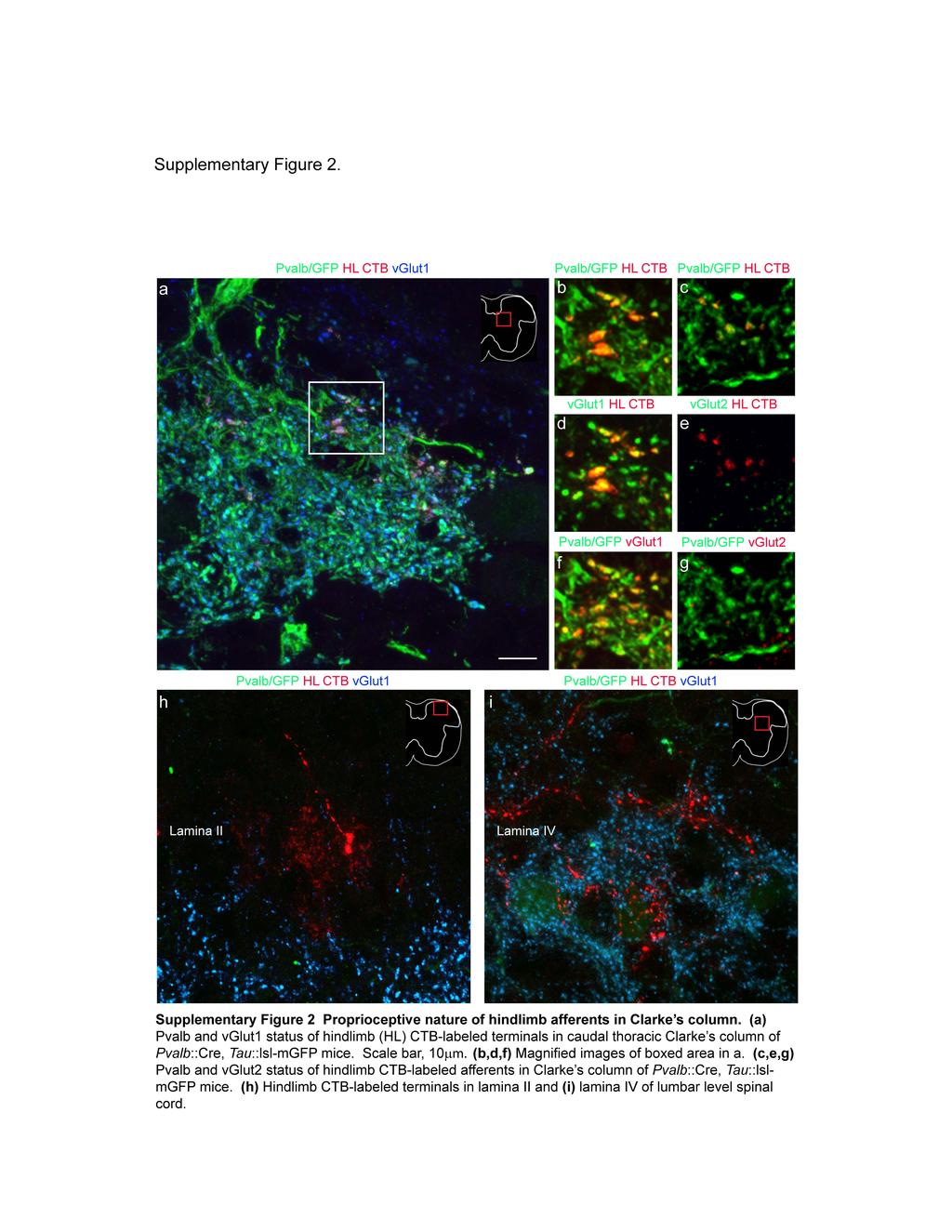
7 Supplementary Results Characterizing the origin of primary sensory inputs to Clarke s column neurons We used anatomical and genetic methods to define the origin of sensory terminals on Clarke's column dsc neurons. Different classes of primary sensory neurons in the dorsal root ganglia express the vesicular glutamate transporters vglut1and vglut2 27,42. Proprioceptors and low threshold mechanoreceptors express vglut1, whereas high threshold presumed nociceptive afferents express vglut2 27,42. In addition, all local excitatory spinal interneurons express vglut2 43. We therefore examined the status of vglut1/2 expression in identified sensory terminals on Clarke s column neurons. To provide a general label of sensory projections to Clarke s column we injected the hindlimbs of P5 7 mice with cholera toxin B (CTB) subunit and analyzed the synaptic status of CTB-labeled boutons in contact with fluorogold-labeled Clarke s column neurons 72h later. To label proprioceptive sensory terminals selectively, we examined GFP expression in terminal contacts on Clarke s column neurons in Pvalb::Cre, Tau::lsl-mGFP 23. In Pvalb::Cre, Tau::lsl-mGFP mice examined after P10 we found that >95% of CTBlabeled terminals in contact with fluorogold-labeled Clarke s column neurons co-expressed GFP and vglut1 and none of them expressed vglut2 (Supplementary Fig. 2a-e). Furthermore, >95% of vglut1 + sensory terminals in Clarke s column co-expressed GFP, independent of CTBlabeling status (Supplementary Fig. 2f,g). Together these findings suggest that proprioceptive afferents provide the predominant, and probably the sole, sensory synaptic input to Clarke s column dsc neurons. Stimulation of the ventral aspect of the cervical dorsal column selectively activates corticospinal axons. In experiments reported in this study, stimulation of the ventral aspect of the dorsal column has been equated with activation of the descending axons of corticospinal neurons. Below, we document several lines of evidence that support this conclusion and, in particular, 1
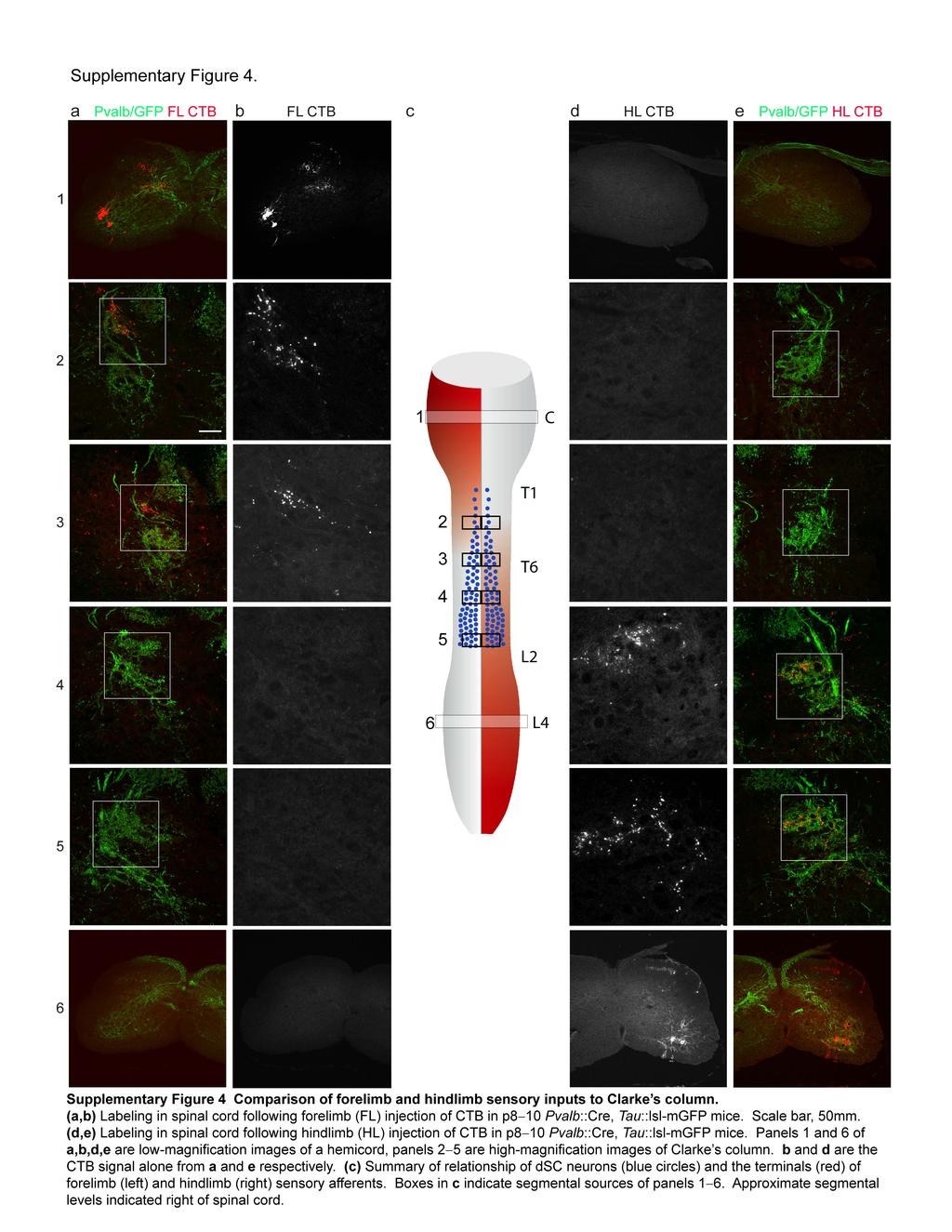
8 argue that responses of Clarke s column dsc neurons observed after dorsal column stimulation do not reflect the activation of descending or ascending sensory 26 or spinal 44 projection neurons that extend axons into the dorsal columns. Differential positioning of corticospinal, sensory, and spinal axons within the dorsal columns: The ventral aspect of the dorsal column is comprised almost exclusively of corticospinal axons (Fig. 1a; Supplementary Fig. 1) with axons of sensory 26 or spinal 45 origin restricted to more dorsal tracts within the dorsal columns (shown schematically in Supplementary Fig. 3). We found that placement of stimulating electrodes on the dorsal aspect of cervical level dorsal column failed to evoke monosynaptic excitatory input to thoraco-lumbar Clarke s column dsc neurons, whereas more ventral dorsal column stimulation in the same preparation elicited a high incidence of monosynaptic excitatory input (data not shown). This finding prompted us to examine in detail the issue of whether concentric bipolar electrodes placed in the ventral aspect of the dorsal columns activate corticospinal axons in a preferential manner. Monosynaptic input to dsc neurons following dorsal column stimulation does not result from orthodromic activation of descending sensory axons. We first considered the possibility that cervical dorsal column stimulation might activate the descending branches of primary sensory axons, and that these axons provide monosynaptic input to dsc neurons. Several lines of evidence argue against this possibility. First, anatomical tracing studies show that sensory axons located in cervical level dorsal columns do not project as far caudally as the thoraco-lumbar Clarke s column dsc neurons we examined physiologically. To evaluate this, CTB was injected into p7 mouse forelimb, and in separate experiments, sensory axons within cervical dorsal roots were labeled with rhodaminedextran (Rh-Dex). CTB-labeled sensory terminals were found at low density at rostral thoracic (T1 T6) levels of the spinal cord, and were absent from spinal levels caudal to T6 (Supplementary Fig. 4a-c). Moreover, most of the CTB-labeled terminals found at T4 T6 levels appear not to derive from proprioceptors, since few if any CTB-labeled sensory terminals at T4 T6 spinal levels expressed GFP, examined in Pvalb::Cre, Tau::lsl-mGFP mice (Supplementary 2
9 Fig. 4a-3). Similarly, few if any Rh-Dex labeled sensory axons and terminals were detected in the vicinity of Clarke s column at T6 L2 levels of the spinal cord (data not shown). As controls, we found that CTB-labeled sensory terminals were found in both the dorsal and ventral horns of cervical spinal cord (Supplementary Fig. 4a,b), indicating that this labeling method effectively fills both proprioceptive and cutaneous afferents. Based on these findings, we conclude that cervical level sensory afferents do not project to Clarke s column neurons located at caudal thoracic- and lumbar-levels (Supplementary Fig. 3). It is unlikely therefore that monosynaptic EPSCs recorded from Clarke s column dsc neurons after cervical level dorsal column stimulation reflect activation of the descending branches of sensory afferents. Monosynaptic input to dsc neurons following dorsal column stimulation does not result from antidromic activation of ascending sensory axons. We next asked whether hindlimb proprioceptive axons project as far rostrally as the cervical dorsal column. We were concerned that if this is the case, then cervical dorsal column stimulation could antidromically activate proprioceptive axons with collaterals that provide direct input to thoraco-lumbar Clarke s column dsc neurons. We mapped the ascending projections of sensory axons that provide input to Clarke's column by injecting CTB into the hindlimb. CTB-labeled sensory terminals were found at high density in lumbar spinal cord, in both dorsal and ventral gray matter (Supplementary Figs. 2h,i; 4d,e), but at progressively decreasing density at more rostral segmental levels, between T12 and T6. Very few CTB-labeled terminals were found rostral to segment T6 (Supplementary Fig. 4ce). We also monitored the status of GFP expression in the CTB-labeled sensory terminals of Pvalb::Cre, Tau::lsl-mGFP mice that had received hindlimb CTB injections. CTB +, GFP + terminals were not detected in the gray matter of the cervical spinal cord (Supplementary Fig. 4ce). Together, these findings indicate that hindlimb proprioceptive axons do not give rise to collaterals that enter the cervical gray matter. 3
10 We also considered whether hindlimb proprioceptive axons pass through the cervical dorsal columns en route to the brainstem, without sending collaterals into the cervical spinal cord. We therefore examined the origin of sensory axons that terminate in the cuneate, external cuneate, and the gracile nuclei. Consistent with prior studies 46, hindlimb-derived CTB-labeled terminals were found exclusively in the gracile nucleus (Supplementary Fig. 5a,b,f), whereas forelimb-derived CTB terminals were found in the cuneate and external cuneate nuclei (Supplementary Fig. 5c-f). Importantly, in Pvalb::Cre, Tau::lsl-mGFP mice, few if any hindlimb injection derived CTB-labeled terminals in the gracile nucleus expressed GFP (Supplementary Fig. 5a,b,f), indicating that these terminals derived from sensory neurons other than proprioceptors. The lack of GFP labeling was not the consequence of inadequate GFP expression at the more rostral brainstem sites, since in Pvalb::Cre, Tau::lsl-mGFP mice, many forelimb-derived CTB-labeled terminals in the external cuneate nucleus did express GFP (Supplementary Fig. 5c,e,f). We conclude that proprioceptive sensory axons do not project to brainstem termination zones nor do they form a terminal projection zone at cervical levels of the spinal cord. Thus, cervical level dorsal column stimulation does not result in antidromic activation of ascending proprioceptive axons (see Supplementary Fig. 3 for schematic diagram). Our data showing that hindlimb proprioceptive inputs do not ascend into the cervical spinal cord in the mouse is in agreement with physiological and anatomical studies of ascending proprioceptive pathways in other species 47,48. Cervical dorsal column stimulation does not antidromically activate sensory axons which activate inhibitory inputs to dsc neurons: The findings described above leave unresolved the issue of whether cervical dorsal column stimulation could antidromically activate the axons of non-proprioceptive sensory afferents of hindlimb origin. These sensory axons could, in principle, send caudal collaterals onto inhibitory interneurons that project to Clarke s column dsc neurons. In this view, inhibitory responses in dsc neurons recorded after cervical dorsal column stimulation could reflect antidromic sensory rather than orthodromic corticospinal axonal activation. Against this idea, there is extensive evidence that activation of cutaneous sensory inputs does not activate disynaptic inhibitory pathways that project to Clarke's column Instead, 4
11 cutaneous stimulation appears to facilitate muscle afferent-evoked inhibition of dsc neurons, likely by dis-inhibiting muscle afferents themselves 52. The fact that peripherally-evoked cutaneous input fails to recruit inhibitory neurons pre-synaptic to Clarke's column neurons and their location in the dorsal aspects of the dorsal column makes it unlikely that antidromic stimulation of cutaneous sensory axons underlies the dorsal column-evoked inhibition of dsc neurons observed in our studies (Supplementary Fig. 3). Dorsal column stimulation does not antidromically activate the ascending axons of post-synaptic dorsal column neurons which, in turn, activate inhibitory inputs to dsc neurons: The dorsal aspect of the dorsal columns also contains the axons of post-synaptic dorsal column (PSDC) pathway neurons 44. The main source of sensory input to PSDC neurons is from cutaneous afferents 44. But activation of cutaneous afferents is ineffective in inhibiting dsc neurons and so, adhering to the logic of the preceding section, it is unlikely that antidromic activation of the axons of PSDC neurons by cervical level dorsal column stimulation will recruit inhibitory inputs to Clarke's column (see Supplementary Fig. 3 for a schematic of PSDC pathways). From this series of control experiments, we conclude that under our focal stimulation conditions, stimulation of the ventral aspect of cervical dorsal columns results in selective activation of the axons of corticospinal projection neurons. 5
12 42. Todd, A. J. et al. The expression of vesicular glutamate transporters VGLUT1 and VGLUT2 in neurochemically defined axonal populations in the rat spinal cord with emphasis on the dorsal horn. Eur J Neurosci 17, (2003) 43. Persson, S. et al. Distribution of vesicular glutamate transporters 1 and 2 in the rat spinal cord, with a note on the spinocervical tract. J Comp Neurol 497, (2006) 44. Brown, A. G. & Fyffe, R. E. Form and function of dorsal horn neurones with axons ascending the dorsal columns in cat. J Physiol 321, (1981) 45. Brown, A. G. Organization in the spinal cord : the anatomy and physiology of identified neurones. 238 (Springer-Verlag, 1981). 46. Maslany, S., Crockett, D. P. & Egger, M. D. Somatotopic organization of the dorsal column nuclei in the rat: transganglionic labelling with B-HRP and WGA-HRP. Brain Res 564, (1991) 47. Lloyd, D. P. & Mc, I. A. Dorsal column conduction of group I muscle efferent impulses and their relay through Clarke's column. J Neurophysiol 13, (1950) 48. Perl, E. R., Whitlock, D. G. & Gentry, J. R. Cutaneous projection to second-order neurons of the dorsal column system. J Neurophysiol 25, (1962) 49. Hongo, T. et al. Inhibition of dorsal spinocerebellar tract cells by interneurones in upper and lower lumbar segments in the cat. J Physiol 342, (1983) 50. Hongo, T. et al. The same interneurones mediate inhibition of dorsal spinocerebellar tract cells and lumbar motoneurones in the cat. J Physiol 342, (1983) 51. Jankowska, E. & Puczynska, A. Interneuronal activity in reflex pathways from group II muscle afferents is monitored by dorsal spinocerebellar tract neurons in the cat. J Neurosci 28, (2008) 52. Jimenez, I., Rudomin, P. & Solodkin, M. PAD patterns of physiologically identified afferent fibres from the medial gastrocnemius muscle. Exp Brain Res 71, (1988) 6
Spinal Interneurons. Control of Movement
 Control of Movement Spinal Interneurons Proprioceptive afferents have a variety of termination patterns in the spinal cord. This can be seen by filling physiologically-identified fibers with HRP, so their
Medical Neuroscience Tutorial
 Pain Pathways Medical Neuroscience Tutorial Pain Pathways MAP TO NEUROSCIENCE CORE CONCEPTS 1 NCC1. The brain is the body's most complex organ. NCC3. Genetically determined circuits are the foundation
SENSORY (ASCENDING) SPINAL TRACTS
 SPINAL TRACTS") SENSORY (ASCENDING) SPINAL TRACTS Dr. Jamila El-Medany Dr. Essam Eldin Salama OBJECTIVES By the end of the lecture, the student will be able to: Define the meaning of a tract. Distinguish between the different
SENSORY (ASCENDING) SPINAL TRACTS Dr. Jamila El-Medany Dr. Essam Eldin Salama OBJECTIVES By the end of the lecture, the student will be able to: Define the meaning of a tract. Distinguish between the different
Spinal Cord Organization. January 12, 2011
 Spinal Cord Organization January 12, 2011 Spinal Cord 31 segments terminates at L1-L2 special components - conus medullaris - cauda equina no input from the face Spinal Cord, Roots & Nerves Dorsal root
Cortical Control of Movement
 Strick Lecture 2 March 24, 2006 Page 1 Cortical Control of Movement Four parts of this lecture: I) Anatomical Framework, II) Physiological Framework, III) Primary Motor Cortex Function and IV) Premotor
Table of Contents: Chapter 1 The organization of the spinal cord Charles Watson and Gulgun Kayalioglu
 Table of Contents: Chapter 1 The organization of the spinal cord Charles Watson and Gulgun Kayalioglu The gross anatomy of the spinal cord Spinal cord segments Spinal nerves Spinal cord gray and white
OVERVIEW. Today. Sensory and Motor Neurons. Thursday. Parkinsons Disease. Administra7on. Exam One Bonus Points Slides Online
 OVERVIEW Today Sensory and Motor Neurons Thursday Parkinsons Disease Administra7on Exam One Bonus Points Slides Online 7 major descending motor control pathways from Cerebral Cortex or Brainstem
Spinal Cord H. Ruth Clemo, Ph.D.
 Spinal Cord H. Ruth Clemo, Ph.D. OBJECTIVES After studying the material of this lecture, the student should be familiar with: 1. Surface anatomy of the spinal cord. 2. Internal structure and organization
Sensory coding and somatosensory system
 Sensory coding and somatosensory system Sensation and perception Perception is the internal construction of sensation. Perception depends on the individual experience. Three common steps in all senses
Department of Neurology/Division of Anatomical Sciences
 Spinal Cord I Lecture Outline and Objectives CNS/Head and Neck Sequence TOPIC: FACULTY: THE SPINAL CORD AND SPINAL NERVES, Part I Department of Neurology/Division of Anatomical Sciences LECTURE: Monday,
Last time we talked about the descending pathways of pain and the ALS. Today we will continue talking about these descending pathways.
 Last time we talked about the descending pathways of pain and the ALS. Today we will continue talking about these descending pathways. Each higher level will control the level under It. In controlling
Uncrossed actions of feline corticospinal tract neurones on lumbar interneurones evoked via ipsilaterally descending pathways
 J Physiol 580.1 (2007) pp 133 147 133 Uncrossed actions of feline corticospinal tract neurones on lumbar interneurones evoked via ipsilaterally descending pathways E. Jankowska and K. Stecina Department
Gross Anatomy of Lower Spinal Cord
 Chapter 13 Spinal Cord, Spinal Nerves and Somatic Reflexes Spinal cord Spinal nerves Somatic reflexes Gross Anatomy of Lower Spinal Cord Meninges of Vertebra & Spinal Cord Spina Bifida Congenital defect
Lecturer. Prof. Dr. Ali K. Al-Shalchy MBChB/ FIBMS/ MRCS/ FRCS 2014
 Lecturer Prof. Dr. Ali K. Al-Shalchy MBChB/ FIBMS/ MRCS/ FRCS 2014 Dorsal root: The dorsal root carries both myelinated and unmyelinated afferent fibers to the spinal cord. Posterior gray column: Long
The Nervous System: Neural Tissue Pearson Education, Inc.
 13 The Nervous System: Neural Tissue Introduction Nervous System Characteristics Controls and adjust the activity of the body Provides swift but brief responses The nervous system includes: Central Nervous
Lecture VIII. The Spinal Cord, Reflexes and Brain Pathways!
 Reflexes and Brain Bio 3411! Monday!! 1! Readings! NEUROSCIENCE 5 th ed: Review Chapter 1 pp. 11-21;!!Read Chapter 9 pp. 189-194, 198! THE BRAIN ATLAS 3 rd ed:! Read pp. 4-17 on class web site! Look at
THE BACK. Dr. Ali Mohsin. Spinal Cord
 Spinal Cord THE BACK Dr. Ali Mohsin The spinal cord is the elongated caudal part of the CNS. It starts as the inferior continuation of the medulla oblongata at the level of foramen magnum, & ends as an
Chapter 14: Integration of Nervous System Functions I. Sensation.
 Chapter 14: Integration of Nervous System Functions I. Sensation A. General Organization 1. General senses have receptors a. The somatic senses provide information about & 1. Somatic senses include: a.
Somatic Sensory System I. Background
 Somatic Sensory System I. Background A. Differences between somatic senses and other senses 1. Receptors are distributed throughout the body as opposed to being concentrated at small, specialized locations
susceptibility of either the axons in the dorsal and ventral roots, or the intramedullary
 213 J. Physiol. (31958) I40, 2I3-2I9 THE SITE OF ACTION OF PROCAINE ON THE ISOLATED SPINAL CORD OF THE FROG BY M. HARMEL AND J. L. MALCOLM From the Department of Physiology, State University of New York,
Posterior White Column-Medial Lemniscal Pathway
 Posterior White Column-Medial Lemniscal Pathway Modality: Discriminative Touch Sensation (include Vibration) and Conscious Proprioception Receptor: Most receptors except free nerve endings Ist Neuron:
Chapter 17 Nervous System
 Chapter 17 Nervous System 1 The Nervous System Two Anatomical Divisions Central Nervous System (CNS) Brain and Spinal Cord Peripheral Nervous System (PNS) Two Types of Cells Neurons Transmit nerve impulses
Medial View of Cerebellum
 Meds 5371 System Neuroscience D. L. Oliver CEREBELLUM Anterior lobe (spinal) Posterior lobe (cerebral) Flocculonodular lobe (vestibular) Medial View of Cerebellum 1 Ventral View of Cerebellum Flocculus
Brainstem. Steven McLoon Department of Neuroscience University of Minnesota
 Brainstem Steven McLoon Department of Neuroscience University of Minnesota 1 Course News Change in Lab Sequence Week of Oct 2 Lab 5 Week of Oct 9 Lab 4 2 Goal Today Know the regions of the brainstem. Know
performed. From the work of Lloyd & McIntyre (1950) it is known that some group progressively after entering the dorsal columns.
 it is known that some group progressively after entering the dorsal columns.") Journal of Physiology (1988), 401, pp. 97-113 97 With 7 text-figures Printed in Great Britain THE DORSAL COLUMN PROJECTION OF MUSCLE AFFERENT FIBRES FROM THE CAT HINDLIMB BY R. FERN, P. J. HARRISON AND
Journal of Physiology (1988), 401, pp. 97-113 97 With 7 text-figures Printed in Great Britain THE DORSAL COLUMN PROJECTION OF MUSCLE AFFERENT FIBRES FROM THE CAT HINDLIMB BY R. FERN, P. J. HARRISON AND
Reflexes. Handout on The Basic Reflex Arc and Stretch and Tendon Reflexes. -55 mv -70 mv EPSP. By Noel Ways
 Reflexes Handout on The Basic Reflex Arc and Stretch and Tendon Reflexes By Noel Ways Basic Reflex Arch 2. : s are always unipolar and will conduct and impulse to a control center. In this case the control
SOMATIC SENSATION PART I: ALS ANTEROLATERAL SYSTEM (or SPINOTHALAMIC SYSTEM) FOR PAIN AND TEMPERATURE
 FOR PAIN AND TEMPERATURE") Dental Neuroanatomy Thursday, February 3, 2011 Suzanne S. Stensaas, PhD SOMATIC SENSATION PART I: ALS ANTEROLATERAL SYSTEM (or SPINOTHALAMIC SYSTEM) FOR PAIN AND TEMPERATURE Reading: Waxman 26 th ed, :
Dental Neuroanatomy Thursday, February 3, 2011 Suzanne S. Stensaas, PhD SOMATIC SENSATION PART I: ALS ANTEROLATERAL SYSTEM (or SPINOTHALAMIC SYSTEM) FOR PAIN AND TEMPERATURE Reading: Waxman 26 th ed, :
Note: Waxman is very sketchy on today s pathways and nonexistent on the Trigeminal.
 Dental Neuroanatomy Thursday, February 3, 2011 Suzanne Stensaas, PhD Note: Waxman is very sketchy on today s pathways and nonexistent on the Trigeminal. Resources: Pathway Quiz for HyperBrain Ch. 5 and
Skin types: hairy and glabrous (e.g. back vs. palm of hand)
") Lecture 19 revised 03/10 The Somatic Sensory System Skin- the largest sensory organ we have Also protects from evaporation, infection. Skin types: hairy and glabrous (e.g. back vs. palm of hand) 2 major
Lecture 19 revised 03/10 The Somatic Sensory System Skin- the largest sensory organ we have Also protects from evaporation, infection. Skin types: hairy and glabrous (e.g. back vs. palm of hand) 2 major
Biological Bases of Behavior. 8: Control of Movement
 Biological Bases of Behavior 8: Control of Movement m d Skeletal Muscle Movements of our body are accomplished by contraction of the skeletal muscles Flexion: contraction of a flexor muscle draws in a
Pain classifications slow and fast
 Pain classifications slow and fast Fast Pain Slow Pain Sharp, pricking (Aδ) fiber Short latency Well localized Short duration Dull, burning (C) fiber Slower onset Diffuse Long duration Less emotional Emotional,
 Spinal Cord Tracts DESCENDING SPINAL TRACTS: Are concerned with somatic motor function, modification of ms. tone, visceral innervation, segmental reflexes. Main tracts arise form cerebral cortex and others
Introduction and Basic structural organization of the nervous system
 Introduction and Basic structural organization of the nervous system **the slides are in bold and the book is in red Done by : razan krishan & marah marahleh INTRODUCTION The nervous system, along with
PREPARED FOR: U.S. Army Medical Research and Materiel Command Fort Detrick, Maryland
 AWARD NUMBER: W81XWH-15-1-0595 TITLE: Enhancing Propriospinal Relays to Improve Functional Recovery after SCI PRINCIPAL INVESTIGATOR: George Smith CONTRACTING ORGANIZATION: Temple University of the Commonwealth
ANATOMY OF SPINAL CORD. Khaleel Alyahya, PhD, MEd King Saud University School of
 ANATOMY OF SPINAL CORD Khaleel Alyahya, PhD, MEd King Saud University School of Medicine @khaleelya OBJECTIVES At the end of the lecture, students should be able to: Describe the external anatomy of the
SOMATOSENSORY SYSTEMS: Conscious and Non-Conscious Proprioception Kimberle Jacobs, Ph.D.
 SOMATOSENSORY SYSTEMS: Conscious and Non-Conscious Proprioception Kimberle Jacobs, Ph.D. Divisions of Somatosensory Systems The pathways that convey sensory modalities from the body to consciousness are
Rewiring of hindlimb corticospinal neurons after spinal cord injury
 Rewiring of hindlimb corticospinal neurons after spinal cord injury Arko Ghosh, Florent Haiss, Esther Sydekum, Regula Schneider, Miriam Gullo, Matthias T. Wyss, Thomas Mueggler, Christof Baltes, Markus
Commissural interneurons with input from group I and II muscle afferents in feline lumbar segments: neurotransmitters, projections and target cells
 J Physiol 587.2 (2009) pp 401 418 401 Commissural interneurons with input from group I and II muscle afferents in feline lumbar segments: neurotransmitters, projections and target cells E. Jankowska 2,
POSTSYNAPTIC INHIBITION OF CRAYFISH TONIC FLEXOR MOTOR NEURONES BY ESCAPE COMMANDS
 J. exp. Biol. (1980), 85, 343-347 343 With a figures Printed in Great Britain POSTSYNAPTIC INHIBITION OF CRAYFISH TONIC FLEXOR MOTOR NEURONES BY ESCAPE COMMANDS BY J. Y. KUWADA, G. HAGIWARA AND J. J. WINE
diameter, i.e. the largest afferent fibres from PC. The motoneurones active stimuli for the reflex were evaluated. It was concluded that the receptors
 J. Physiol. (1971), 216, pp. 483-501 483 With 7 text- gure Printed in Great Britain THE PLANTAR CUSHION REFLEX CIRCUIT: AN OLIGOSYNAPTIC CUTANEOUS REFLEX BY M. DAVID EGGER* AND PATRICK D. WALL From the
General Sensory Pathways of the Trunk and Limbs
 General Sensory Pathways of the Trunk and Limbs Lecture Objectives Describe gracile and cuneate tracts and pathways for conscious proprioception, touch, pressure and vibration from the limbs and trunk.
Biology 218 Human Anatomy
 Chapter 21 Adapted form Tortora 10 th ed. LECTURE OUTLINE A. Overview of Sensations (p. 652) 1. Sensation is the conscious or subconscious awareness of external or internal stimuli. 2. For a sensation
Human Anatomy - Problem Drill 11: The Spinal Cord and Spinal Nerves
 Human Anatomy - Problem Drill 11: The Spinal Cord and Spinal Nerves Question No. 1 of 10 Instructions: (1) Read the problem statement and answer choices carefully, (2) Work the problems on paper as needed,
Nervous System. The Peripheral Nervous System Agenda Review of CNS v. PNS PNS Basics Cranial Nerves Spinal Nerves Reflexes Pathways
 Nervous System Agenda Review of CNS v. PNS PNS Basics Cranial Nerves Spinal Nerves Sensory Motor Review of CNS v. PNS Central nervous system (CNS) Brain Spinal cord Peripheral nervous system (PNS) All
Human Anatomy. Spinal Cord and Spinal Nerves
 Human Anatomy Spinal Cord and Spinal Nerves 1 The Spinal Cord Link between the brain and the body. Exhibits some functional independence from the brain. The spinal cord and spinal nerves serve two functions:
I: To describe the pyramidal and extrapyramidal tracts. II: To discuss the functions of the descending tracts.
 Descending Tracts I: To describe the pyramidal and extrapyramidal tracts. II: To discuss the functions of the descending tracts. III: To define the upper and the lower motor neurons. 1. The corticonuclear
Fig Cervical spinal nerves. Cervical enlargement C7. Dural sheath. Subarachnoid space. Thoracic. Spinal cord Vertebra (cut) spinal nerves
 spinal nerves") Fig. 13.1 C1 Cervical enlargement C7 Cervical spinal nerves Dural sheath Subarachnoid space Thoracic spinal nerves Spinal cord Vertebra (cut) Lumbar enlargement Medullary cone T12 Spinal nerve Spinal nerve
Fig. 13.1 C1 Cervical enlargement C7 Cervical spinal nerves Dural sheath Subarachnoid space Thoracic spinal nerves Spinal cord Vertebra (cut) Lumbar enlargement Medullary cone T12 Spinal nerve Spinal nerve
(Received 10 April 1956)
") 446 J. Physiol. (I956) I33, 446-455 A COMPARISON OF FLEXOR AND EXTENSOR REFLEXES OF MUSCULAR ORIGIN BY M. G. F. FUORTES AND D. H. HUBEL From the Department ofneurophysiology, Walter Reed Army Institute
446 J. Physiol. (I956) I33, 446-455 A COMPARISON OF FLEXOR AND EXTENSOR REFLEXES OF MUSCULAR ORIGIN BY M. G. F. FUORTES AND D. H. HUBEL From the Department ofneurophysiology, Walter Reed Army Institute
CHAPTER 10 THE SOMATOSENSORY SYSTEM
 CHAPTER 10 THE SOMATOSENSORY SYSTEM 10.1. SOMATOSENSORY MODALITIES "Somatosensory" is really a catch-all term to designate senses other than vision, hearing, balance, taste and smell. Receptors that could
Chapter 13! Chapter 13 Spinal Cord and Spinal Nerves! The Spinal Cord and Spinal Nerves!
 Chapter 13! The Spinal Cord and Spinal Nerves! SECTION 13-1! The brain and spinal cord make up the central nervous system, and the cranial nerves and spinal nerves constitute the peripheral nervous system!
Ting Ting Liu 1, B. Anne Bannatyne 1, Elzbieta Jankowska 2 and David J. Maxwell 1
 J Physiol 588.21 (2010) pp 4217 4233 4217 Properties of axon terminals contacting intermediate zone excitatory and inhibitory premotor interneurons with monosynaptic input from group I and II muscle afferents
HEAD AND NECK PART 2
 HEAD AND NECK PART 2 INTEGRATED CURRICULUM = Integrate Basic Science and Clinical Training 1- ENT PATIENT EXAM IN ICS COURSE - Today and next week - Review/Preview Anatomy underlying ENT exam 2- NEUROANATOMY/NEUROLOGY
Psychophysical laws. Legge di Fechner: I=K*log(S/S 0 )
") Psychophysical laws Legge di Weber: ΔS=K*S Legge di Fechner: I=K*log(S/S 0 ) Sensory receptors Vision Smell Taste Touch Thermal senses Pain Hearing Balance Proprioception Sensory receptors Table 21-1 Classification
Psychophysical laws Legge di Weber: ΔS=K*S Legge di Fechner: I=K*log(S/S 0 ) Sensory receptors Vision Smell Taste Touch Thermal senses Pain Hearing Balance Proprioception Sensory receptors Table 21-1 Classification
(SOCPs) ascending in the dorsal funiculi (DF) have the most extensive projections
 ascending in the dorsal funiculi (DF) have the most extensive projections") Journal of Physiology (1991), 441, pp. 275-284 275 With 5 figures Printed in Great Britain THE POSTSYNAPTIC DORSAL COLUMN PATHWAY MEDIATES CUTANEOUS NOCICEPTIVE INFORMATION TO CEREBELLAR CLIMBING FIBRES
Journal of Physiology (1991), 441, pp. 275-284 275 With 5 figures Printed in Great Britain THE POSTSYNAPTIC DORSAL COLUMN PATHWAY MEDIATES CUTANEOUS NOCICEPTIVE INFORMATION TO CEREBELLAR CLIMBING FIBRES
Nervous System C H A P T E R 2
 Nervous System C H A P T E R 2 Input Output Neuron 3 Nerve cell Allows information to travel throughout the body to various destinations Receptive Segment Cell Body Dendrites: receive message Myelin sheath
Chapter 12b. Overview
 Chapter 12b Spinal Cord Overview Spinal cord gross anatomy Spinal meninges Sectional anatomy Sensory pathways Motor pathways Spinal cord pathologies 1 The Adult Spinal Cord About 18 inches (45 cm) long
Lesson 33. Objectives: References: Chapter 16: Reading for Next Lesson: Chapter 16:
 Lesson 33 Lesson Outline: Nervous System Structure and Function Neuronal Tissue Supporting Cells Neurons Nerves Functional Classification of Neuronal Tissue Organization of the Nervous System Peripheral
The Nervous System PART A
 7 The Nervous System PART A PowerPoint Lecture Slide Presentation by Jerry L. Cook, Sam Houston University ESSENTIALS OF HUMAN ANATOMY & PHYSIOLOGY EIGHTH EDITION ELAINE N. MARIEB Structural Classification
συν together απτειν to clasp 2h Neuroscience with Pharmacology Functions and Mechanisms of Reflexes Cogito, ergo sum ( I think therefore I am ) Down
 Down") 2h Neuroscience with Pharmacology Functions and Mechanisms of Reflexes Neuroscience is studied at many different levels: from brain, to system, network, neurone, synapse, and molecule... Top Up Down René
2h Neuroscience with Pharmacology Functions and Mechanisms of Reflexes Neuroscience is studied at many different levels: from brain, to system, network, neurone, synapse, and molecule... Top Up Down René
Neuronal relays in double crossed pathways between feline motor cortex and ipsilateral hindlimb motoneurones
 J Physiol 575.2 (2006) pp 527 541 527 Neuronal relays in double crossed pathways between feline motor cortex and ipsilateral hindlimb motoneurones E. Jankowska 1, K. Stecina 1, A. Cabaj 1, L.-G. Pettersson
Degree of freedom problem
 KINE 4500 Neural Control of Movement Lecture #1:Introduction to the Neural Control of Movement Neural control of movement Kinesiology: study of movement Here we re looking at the control system, and what
KINE 4500 Neural Control of Movement. Lecture #1:Introduction to the Neural Control of Movement. Neural control of movement
 KINE 4500 Neural Control of Movement Lecture #1:Introduction to the Neural Control of Movement Neural control of movement Kinesiology: study of movement Here we re looking at the control system, and what
Laurie L. Wellman Ph.D.
 Laurie L. Wellman Ph.D. Theodore Tzavaras MD2015 Eastern Virginia Medical School Dr. Craig Goodmurphy Anatomy Guy 1. Discuss -- what is pain? 2. Outline the Anterolateral Quadrant (ALQ) pathway 3. Locate
Chapter 13: The Spinal Cord and Spinal Nerves
 Chapter 13: The Spinal Cord and Spinal Nerves Spinal Cord Anatomy Protective structures: Vertebral column and the meninges protect the spinal cord and provide physical stability. a. Dura mater, b. Arachnoid,
Somatosensory System. Steven McLoon Department of Neuroscience University of Minnesota
 Somatosensory System Steven McLoon Department of Neuroscience University of Minnesota 1 Course News Dr. Riedl s review session this week: Tuesday (Oct 10) 4-5pm in MCB 3-146B 2 Sensory Systems Sensory
Nervous system. The main regulation mechanism of organism's functions
 Nervous system The main regulation mechanism of organism's functions Questions Neuron The reflex arc The nervous centers Properties of the nervous centers The general principles of coordination Inhibition
The Nervous System: Sensory and Motor Tracts of the Spinal Cord
 15 The Nervous System: Sensory and Motor Tracts of the Spinal Cord PowerPoint Lecture Presentations prepared by Steven Bassett Southeast Community College Lincoln, Nebraska Introduction Millions of sensory
The Nervous System S P I N A L R E F L E X E S
 The Nervous System S P I N A L R E F L E X E S Reflexes Rapid, involuntary, predictable motor response to a stimulus Spinal Reflexes Spinal somatic reflexes Integration center is in the spinal cord Effectors
Motor tracts Both pyramidal tracts and extrapyramidal both starts from cortex: Area 4 Area 6 Area 312 Pyramidal: mainly from area 4 Extrapyramidal:
 Motor tracts Both pyramidal tracts and extrapyramidal both starts from cortex: Area 4 Area 6 Area 312 Pyramidal: mainly from area 4 Extrapyramidal: mainly from area 6 area 6 Premotorarea: uses external
The Nervous System: The
 C h a p t e r 14 The Nervous System: The Spinal Cord and Spinal Nerves PowerPoint Lecture Slides prepared by Jason LaPres North Harris College Houston, Texas Copyright 2009 Pearson Education, Inc., publishing
Brain Stem and cortical control of motor function. Dr Z Akbari
 Brain Stem and cortical control of motor function Dr Z Akbari Brain stem control of movement BS nuclear groups give rise to descending motor tracts that influence motor neurons and their associated interneurons
Neural Control of Lower Urinary Tract Function. William C. de Groat University of Pittsburgh Medical School
 Neural Control of Lower Urinary Tract Function William C. de Groat University of Pittsburgh Medical School Disclosures Current funding: NIH Grants, DK093424, DK-091253, DK-094905, DK-090006. Other financial
The Cerebellum. The Little Brain. Neuroscience Lecture. PhD Candidate Dr. Laura Georgescu
 The Cerebellum The Little Brain Neuroscience Lecture PhD Candidate Dr. Laura Georgescu Learning Objectives 1. Describe functional anatomy of the cerebellum - its lobes, their input and output connections
Gross Morphology of Spinal Cord
 Gross Morphology of Spinal Cord Lecture Objectives Describe the gross anatomical features of the spinal cord. Describe the level of the different spinal segments compared to the level of their respective
Nervous Systems: Diversity & Functional Organization
 Nervous Systems: Diversity & Functional Organization Diversity of Neural Signaling The diversity of neuron structure and function allows neurons to play many roles. 3 basic function of all neurons: Receive
Receptors and Neurotransmitters: It Sounds Greek to Me. Agenda. What We Know About Pain 9/7/2012
 Receptors and Neurotransmitters: It Sounds Greek to Me Cathy Carlson, PhD, RN Northern Illinois University Agenda We will be going through this lecture on basic pain physiology using analogies, mnemonics,
BIOH111. o Cell Module o Tissue Module o Integumentary system o Skeletal system o Muscle system o Nervous system o Endocrine system
 BIOH111 o Cell Module o Tissue Module o Integumentary system o Skeletal system o Muscle system o Nervous system o Endocrine system Endeavour College of Natural Health endeavour.edu.au 1 Textbook and required/recommended
Neural Integration I: Sensory Pathways and the Somatic Nervous System
 15 Neural Integration I: Sensory Pathways and the Somatic Nervous System PowerPoint Lecture Presentations prepared by Jason LaPres Lone Star College North Harris An Introduction to Sensory Pathways and
Can sense be made of spinal interneuron circuits?
 BEHAVIORAL AND BRAIN SCIENCES (1992) 15,633-643 Printed in the United States of America Can sense be made of spinal interneuron circuits? D. A. McCrea Department of Physiology, Faculty of Medicine, University
Module H NERVOUS SYSTEM
 Module H NERVOUS SYSTEM Topic from General functions of the nervous system Organization of the nervous system from both anatomical & functional perspectives Gross & microscopic anatomy of nervous tissue
Systems Neuroscience November 21, 2017 The autonomic nervous system
 Systems Neuroscience November 21, 2017 The autonomic nervous system Daniel C. Kiper kiper@ini.phys.ethz.ch http: www.ini.unizh.ch/~kiper/system_neurosci.html How is the organization of the autonomic nervous
Sympathetic Nervous System
 Sympathetic Nervous System Lecture Objectives Review the subdivisions of the nervous system. Review the general arrangement and compare the sympathetic and parasympathetic parts. Describe the following
HUMAN MOTOR CONTROL. Emmanuel Guigon
 HUMAN MOTOR CONTROL Emmanuel Guigon Institut des Systèmes Intelligents et de Robotique Université Pierre et Marie Curie CNRS / UMR 7222 Paris, France emmanuel.guigon@upmc.fr e.guigon.free.fr/teaching.html
The Motor Systems. What s the motor system? Plan
 The Motor Systems What s the motor system? Parts of CNS and PNS specialized for control of limb, trunk, and eye movements Also holds us together From simple reflexes (knee jerk) to voluntary movements
Differential presynaptic inhibition of actions of group II afferents in di- and polysynaptic pathways to feline motoneurones
 Journal of Physiology (2002), 542.1, pp. 287 299 DOI: 10.1113/jphysiol.2001.014068 The Physiological Society 2002 www.jphysiol.org Differential presynaptic inhibition of actions of group II afferents in
Chapter 14. The Nervous System. The Spinal Cord and Spinal Nerves. Lecture Presentation by Steven Bassett Southeast Community College
 Chapter 14 The Nervous System The Spinal Cord and Spinal Nerves Lecture Presentation by Steven Bassett Southeast Community College Introduction The Central Nervous System (CNS) consists of: The spinal
Sensory information processing, somato-sensory systems
 mm? Sensory information processing, somato-sensory systems Recommended literature 1. Kandel ER, Schwartz JH, Jessel TM (2000) Principles of Neural Science, McGraw-Hill, Ch. xx. 2. Berne EM, Levy MN, Koeppen
Nature Neuroscience: doi: /nn Supplementary Figure 1. Splenic atrophy and leucopenia caused by T3 SCI.
 Supplementary Figure 1 Splenic atrophy and leucopenia caused by T3 SCI. (a) Gross anatomy of representative spleens from control and T3 SCI mice at 28 days post-injury. (b and c) Hematoxylin and eosin
(From the Kerckhoff Laboratories of Biology, California Institute of Technology, Pasadena)
") Published Online: 20 November, 1950 Supp Info: http://doi.org/10.1085/jgp.34.2.137 Downloaded from jgp.rupress.org on January 12, 2019 THE INTERACTION BETWEEN THE SYNAPSES OF A SINGLE MOTOR FIBER BY C.
Published Online: 20 November, 1950 Supp Info: http://doi.org/10.1085/jgp.34.2.137 Downloaded from jgp.rupress.org on January 12, 2019 THE INTERACTION BETWEEN THE SYNAPSES OF A SINGLE MOTOR FIBER BY C.
SPINAL NEURONAL ACTIVITY DURING THE PECTORAL FIN REFLEX OF THE DOGFISH: PATHWAYS FOR REFLEX GENERATION AND CEREBELLAR CONTROL
 L exp. Biol. 148, 403-414 (1990) 403 Printed in Great Britain The Company of Biologists Limited 1990 SPINAL NEURONAL ACTIVITY DURING THE PECTORAL FIN REFLEX OF THE DOGFISH: PATHWAYS FOR REFLEX GENERATION
THE SOMATOSENSORY SYSTEM
 Daniel J. Simons, Ph.D. I. Peripheral Nervous System THE SOMATOSENSORY SYSTEM The somatosensory system consists of those components of the peripheral and central nervous systems that mediate sensations
The Spinal Cord. The Nervous System. The Spinal Cord. The Spinal Cord 1/2/2016. Continuation of CNS inferior to foramen magnum.
 The Nervous System Spinal Cord Continuation of CNS inferior to foramen magnum Simpler than the brain Conducts impulses to and from brain Two way conduction pathway Reflex actions Passes through vertebral
CHAPTER 3 THE STRUCTURE OF THE NERVOUS SYSTEM
 CHAPTER 3 THE STRUCTURE OF THE NERVOUS SYSTEM 3.1. THE BASIC STRUCTURE OF THE NERVOUS SYSTEM. The nervous system of all animals is made up of groups of neurons that receive information from sensory systems,
Dendrites Receive impulse from the axon of other neurons through synaptic connection. Conduct impulse towards the cell body Axon
 Dendrites Receive impulse from the axon of other neurons through synaptic connection. Conduct impulse towards the cell body Axon Page 22 of 237 Conduct impulses away from cell body Impulses arise from
Differences Between Right and Left Patellar Reflexes
 Differences Between Right and Left Patellar Reflexes Background: somatic senses: Miss School, Miss Out! Miss School, Miss Out! 7 1. Receptor region 2. Afferent neuron 3. Interneuron 4. Efferent neuron
Motor systems.... the only thing mankind can do is to move things... whether whispering or felling a forest. C. Sherrington
 Motor systems... the only thing mankind can do is to move things... whether whispering or felling a forest. C. Sherrington 1 Descending pathways: CS corticospinal; TS tectospinal; RS reticulospinal; VS
Cranial Nerves and Spinal Cord Flashcards
 1. Name the cranial nerves and their Roman numeral. 2. What is Cranial Nerve I called, and what does it 3. Scientists who are trying to find a way to make neurons divide to heal nerve injuries often study
Chapter 13. The Spinal Cord & Spinal Nerves. Spinal Cord. Spinal Cord Protection. Meninges. Together with brain forms the CNS Functions
 Spinal Cord Chapter 13 The Spinal Cord & Spinal Nerves Together with brain forms the CNS Functions spinal cord reflexes integration (summation of inhibitory and excitatory) nerve impulses highway for upward
STRUCTURAL ORGANIZATION OF THE NERVOUS SYSTEM
 STRUCTURAL ORGANIZATION OF THE NERVOUS SYSTEM STRUCTURAL ORGANIZATION OF THE BRAIN The central nervous system (CNS), consisting of the brain and spinal cord, receives input from sensory neurons and directs