The Cerebellum. The Little Brain. Neuroscience Lecture. PhD Candidate Dr. Laura Georgescu
|
|
|
- Stephen Jennings
- 5 years ago
- Views:
Transcription
1 The Cerebellum The Little Brain Neuroscience Lecture PhD Candidate Dr. Laura Georgescu
2 Learning Objectives 1. Describe functional anatomy of the cerebellum - its lobes, their input and output connections and their functions 2. Draw and label the circuitry of the cerebellum cortex, assign the functional role of each neuron type and give its synaptic action (excitatory/inhibitory) 3. Describe what is known about the role of the cerebellum in the regulation of skilled movement and in motor learning 4. Explain servo-control mechanisms as a model for cerebellar regulation of movements 5. Predict the neurological disturbances that can result from disease or damage in different regions of the cerebellum


3 Introduction The basal ganglia and cerebellum modify movement on a minute to minute basis The motor cortex sends information to both, and both structures send information back via the thalamus (no direct projections to lower motor neurons of skeletal muscles) The balance between these two systems allows for smooth, coordinated movement, and a disturbance in either system will show up as movement disorders

4 The Cerebellum Cerebellum (Latin, little brain)= little brain 10% total volume of the brain but more than half of all its neurons Arranged in a highly regular manner as repeating units but with input and outputs from virtually all motor cortical regions as well as the spinal cord - in particular proprioceptive input The cerebellum is provided with extensive information - 40 times more axons project into the cerebellum than exit from it The cerebellum is not necessary to basic elements of perception or movement Damage to the cerebellum disrupts the spatial accuracy and temporal coordination of movement It impairs balance and reduces muscle tone and motor learning and certain cognitive functions.
5 Functions of the Cerebellum Maintenance of balance and posture. Through its input from vestibular receptors and proprioceptors, it modulates commands to motor neurons Coordination of voluntary movements - a number of different muscle groups acting together in a temporally coordinated fashion Motor learning. The cerebellum - a major role in adapting and fine-tuning motor programs to make accurate movements through a trial-and-error process Cognitive functions. Involved in certain cognitive functions - language Thus, like the basal ganglia, the cerebellum is historically considered as part of the motor system, but its functions extend beyond motor control in ways that are not yet well understood.
6 The Cerebellum - Anatomy Shape: Oval shaped, with an approximate weight of 150 gm Location: Situated in the posterior cranial fossa, dorsal to the pons and medulla, separated from the overlying cerebral cortex by a tough flap in the dura, the cerebellar tentorium Outer mantle of grey matter Internal layer of white matter that contain deep nuclei

7 General view Gross features of the cerebellum, including the nuclei, cerebellar peduncles, lobes, folia, and fissures. (Adapted from Nieuwenhuys et al. 1988) A. Dorsal view. Part of the right hemisphere has been cut out to show the underlying cerebellar peduncles. B. Ventral view of the cerebellum detached from the brain stem. C. Midsagittal section through the brain stem and cerebellum, showing the branching structures of the cerebellum.
 - fastigial and interposed - nuclei - Control of limbs and trunk 3.")
8 Cerebellar Functional Anatomy 1. Vestibulocerebellum (flocculo-nodular lobe)- lateral vestibular nuclei- Control of eye&head movements, postural maintenance 2. Spinocerebellum (vermis and intermediate zone) - fastigial and interposed - nuclei - Control of limbs and trunk 3. Cerebrocerebellum (lateral zones)- dentate nuclei Planning and timing of movement+ cognitive functions

9 The Cerebellum - Three Functionally Distinct Regions The three functional regions of the cerebellum have different inputs and outputs.

10 Functional organization of cerebellum Cerebellar peduncles = Three fiber bundles carry the input and output of the cerebellum. 1. The inferior cerebellar peduncle primarily contains afferent fibers from the medulla, as well as efferents to the vestibular nuclei 2. The middle cerebellar peduncle primarily contains afferents from the pontine nuclei 3. The superior cerebellar peduncle primarily contains efferent fibers from the cerebellar nuclei, as well as some afferents from the spinocerebellar tract.
 2. Purkinje Cells (output cells) 3. Golgi Cells 4. Basket Cells 5. Stellate Cells (these are all inhibitory)")
11 Histology and Connectivity of Cerebellar Cortex Simple three layer arrangement: 1. Molecular 2. Purkinje 3. Granule Five cell types: 1. Granule Cells (excitatory) 2. Purkinje Cells (output cells) 3. Golgi Cells 4. Basket Cells 5. Stellate Cells (these are all inhibitory)
12 Functional organization of cerebellum All output is via the deep nuclei except flocculonodular lobe which outputs via lateral and medial vestibular nuclei 40 times more input than output fibers Cerebellar lesions lead to ipsilateral symptoms because of a double crossing of the output fibres The cerebellum projects to the contralateral motor cortex, via the thalamus, and the corticospinal pathway recrosses the midline at the lower medulla

13 Input to the cerebellum
14 Input to the cerebellum *The cerebellar cortex has a relatively simple, stereotyped connectivity pattern that is identical throughout the whole structure Cerebellar input can be divided into two distinct classes: 1. Mossy fibers originate in the pontine nuclei, the spinal cord, the brainstem reticular formation, and the vestibular nuclei, and they make excitatory projections onto the cerebellar nuclei and onto granule cells in the cerebellar cortex 2. Climbing fibers originate exclusively in the inferior olive and make excitatory projections onto the cerebellar nuclei and onto the Purkinje cells of the cerebellar cortex.
15 Cerebellar cortex Input to cerebellum is excitatory and via: 1. Mossy fibers - named because of the tufted appearance of their synaptic contacts with granule cells 2. Climbing fibers named because their axons climb and wrap around the dendrites of the Purkinje cell like a climbing vine Both inputs go first to the deep nuclei to excite them and then ascend upward into the cerebellum

16 Cerebellar cortex Climbing fibers branch in the sagittal dimension to excite 10 or so Purkinje cells anterior and posterior to the branch point. The transverse connections of the parallel fibers and the sagittal connections of the climbing fibers thus form an orthogonal matrix.

17 Geometrical Plan of Parallel and Climbing fibers The geometry of the mossy and parallel fiber system contrasts with that of the climbing fiber system. Mossy fibers excite granule cells whose parallel fibers branch transversely to excite hundreds of Purkinje cells several millimeters from the branch point, both medially and laterally.
 Very small, densely packed neurons that account for the huge")

18 Internal Circuitry 1. Granule cells Layer (innermost) Very small, densely packed neurons that account for the huge majority of neurons in the cerebellum-10 billion granule cells + interneurons known as Golgi cells These cells receive input from mossy fibers and project to the Purkinje cells Cerebellar glomeruli confluence of mossy fibers on granule cells and Golgi cells
 Consists of Purkinje cell bodies (50-80 micrometers) Purkinje cell is one of the most striking cell")

19 Internal Circuitry 2. The Purkinje cell Layer (middle) Consists of Purkinje cell bodies (50-80 micrometers) Purkinje cell is one of the most striking cell types in the mammalian brain. Its apical dendrites form a large fan of finely branched processes that extends into the outer molecular The dendritic tree is almost twodimensional; looked at from the side, the dendritic tree is flat. All Purkinje cells are oriented in parallel. This arrangement has important functional considerations Axons descend to the deep nuclei Release GABA
 Parallel fibers axons from granule cells which")
20 Internal Circuitry 3. Molecular Cell Layer (outermost) Parallel fibers axons from granule cells which synapse with Purkinje cells Interneurons basket cells and stellate cells

21 The Purkinje Cells Receive Excitatory Input From Two Afferent Fiber Systems and Are Inhibited by Three Local Interneurons Synaptic organization of the basic cerebellar circuit module. Mossy and climbing fibers convey output from the cerebellum via a main excitatory loop through the deep nuclei. This loop is modulated by an inhibitory side-loop passing through the cerebellar cortex. This figure shows the excitatory (+) and inhibitory (-) connections among the cell types.
22 Internal Circuitry Output from Purkinje neurons through deep nuclei, to: Cerebral cortex Brainstem Spinal cord Output of Purkinje cells is inhibitory Output of deep nuclei is excitatory

23 Descending outputs from the cerebellum

24 Input and output pathways of the cerebellum The cerebellar deep nuclei = the sole outputs of the cerebellum All cerebellar nuclei and all regions of cerebellum get special inputs from the inferior olive of the medulla The anatomical locations of the cerebellar nuclei correspond to the cerebellar cortex regions from which they receive input The medially located fastigial nucleus receives input from the medially located vermis; the slightly lateral interposed nuclei receive input from the slightly lateral intermediate zone The most lateral dentate nucleus receives input from the lateral hemispheres.

25 Ascending outputs from the cerebellum

26 Functions of the cerebellum The primary function of the cerebellum is to modulate motor output. It compares what will happen with what is intended to happen and makes adjustments. This is referred to as a feed-forward mechanism
27 Functions of the cerebellum 1. Smoothening postural and skilled movements resulting from skeletal muscle contraction (including eyeball movements) 2. Comparison of intent and action (ie., errors) and generates corrective signals 3. Motor learning and adaptation Plays a role in automating and optimizing behavior
28 Functions of the cerebellum Feedback systems Reactive Responds to the current state of affairs Depends on sensory signals Feed Forward Systems Anticipatory Depends on sensory information but also through experience Can modify the operation of negative feedback mechanisms Essential for rapid action
29 Functions of the cerebellum Referred to as a silent area as electrical stimulation produces: No movement No sensation However removal or lesions cause severe motor deficits: Movements become uncoordinated and erratic

30 Motor circuit
31 Mossy and Climbing Fibers Encode Peripheral and Descending Information Differently Simple and complex spikes recorded intracellularly from cerebellar Purkinje cells. Complex spikes (right bracket) are evoked by climbing fiber synapses, while simple spikes (left bracket) are produced by mossy fiber input. (From Martinez et al ) Prolonged depolarization (due to opening of voltage gated calcium channels) with an initial large amplitude spike followed by a high-frequency burst of smalleramplitude action potentials.
32 Climbing Fibers: Firing Patterns It is believed that during movement climbing fibers provide an error signal that depresses parallel fibers that are active concurrentently and generate the simple spikes. This allows the correct movements (without the error) to emerge. It is also supposed to be important for motor learning
33 Mossy Fibers Originate from nuclei in the spinal cord and brain stem as well as carrying sensory information from the periphery and cortex Terminate on the dendrites of granule cells and excite Purkinje cells via parallel fibers Each Purkinje cell can receive input from as many as 1 million granule cells

34 Summary of cerebellar organization
35 Vestibulocerebellum Vestibular organs, Superior colliculus, Striate (visual) cortex, via pons Vestibulocerebellum Vestibular nuclei (lateral & medial) Vestibulospinal/bulbar tracts

36 Vestibulocerebellum Functions: 1. Coordination of movements of head & eyes 2. Control of axial muscles & limb extensors Maintenance of balance/ posture Lesions: Nystagmus: loss of ability to focus Vertigo: loss of balance Ataxia: wide gait
 lesion: 1. Ataxia 2.")
37 Spinocerebellum There are body maps on the cerebellar cortex and deep nuclei. Functions: 1. Control of axial and proximal muscles (ongoing movements) 2. Maintenance of balance 3. Coordination of head & eye movements Lesions: Vermis (spinocerebellum or associated nucleus) lesion: 1. Ataxia 2. Titubation (head or truncal tremor)

38
39 Spinocerebellum - intermediate Dorsal spinocerebellar tract, ventral spinocerebellar tract- these provide cerebellum with information from the muscle& joint propriocetors Spinocerebellum (intermediate zone) Interposed nucleus (emboliform & globose) 1. Descending Dorsolateral system: Magnocelular red nucleus & rubrospinal tract 2. Motor cortex (distal limb areas) via ventrolateral thalamus
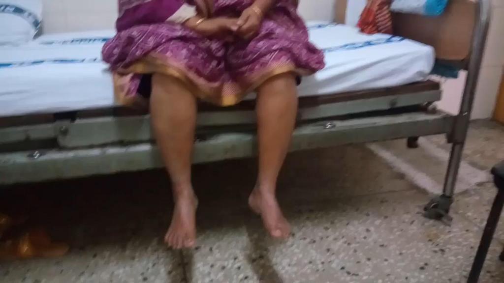
40 Spinocerebellum Lesions 1. Dysarthria - motor speech disorder 2. Dysmetria - loss of control of movement often characterized by overshooting of undershooting the target - results from loss of the feed-forward anticipatory control of movement
41
42 Spinocerebellum Lesions 3. Intention tremor broad coarse tremor that increases as target is aproached 4. Decreased muscle tone in limbs 5. Pendular movement of limbs
43
44 Cerebrocerebellum Receives fibers from the motor cortex, premotor cortex via the pontine nuclei Cerebrocerebellum Dentate nucleus 1. Red nucleus sends output to the ipsilateral inferior olivary nucleus which goes back to the cerebellum via the climbing fibers 2. Primary Motor cortex and premotor area via the thalamus
45 Cerebrocerebellum Functions: 1. Planning, initiation and timing of movements 2. Learning of motor skills 3.? Language Connections to prefrontal cortex (Brocca s area)via dentate & thalamus

46 Motor learning
47 Cerebrocerebellum - Lesions Many similar signs as for the spino-cerebellum intermediate zone Dysdiadochokinesia = inability to perform rapid alternating movements especially pronation and supination Decomposition of movement especially involving movement across multiple joints and in the distal limb
48
49 Thank you!
50
The Cerebellum. Little Brain. Neuroscience Lecture. Dr. Laura Georgescu
 The Cerebellum Little Brain Neuroscience Lecture Dr. Laura Georgescu Learning Objectives 1. Describe functional anatomy of the cerebellum- its lobes, their input and output connections and their functions.
Cerebellum. Steven McLoon Department of Neuroscience University of Minnesota
 Cerebellum Steven McLoon Department of Neuroscience University of Minnesota 1 Anatomy of the Cerebellum The cerebellum has approximately half of all the neurons in the central nervous system. The cerebellum
Unit VIII Problem 5 Physiology: Cerebellum
 Unit VIII Problem 5 Physiology: Cerebellum - The word cerebellum means: the small brain. Note that the cerebellum is not completely separated into 2 hemispheres (they are not clearly demarcated) the vermis
CASE 48. What part of the cerebellum is responsible for planning and initiation of movement?
 CASE 48 A 34-year-old woman with a long-standing history of seizure disorder presents to her neurologist with difficulty walking and coordination. She has been on phenytoin for several days after having
Located below tentorium cerebelli within posterior cranial fossa. Formed of 2 hemispheres connected by the vermis in midline.
 The Cerebellum Cerebellum Located below tentorium cerebelli within posterior cranial fossa. Formed of 2 hemispheres connected by the vermis in midline. Gray matter is external. White matter is internal,
The Cerebellum. Outline. Lu Chen, Ph.D. MCB, UC Berkeley. Overview Structure Micro-circuitry of the cerebellum The cerebellum and motor learning
 The Cerebellum Lu Chen, Ph.D. MCB, UC Berkeley 1 Outline Overview Structure Micro-circuitry of the cerebellum The cerebellum and motor learning 2 Overview Little brain 10% of the total volume of the brain,
Cerebellum John T. Povlishock, Ph.D.
 Cerebellum John T. Povlishock, Ph.D. OBJECTIVES 1. To identify the major sources of afferent inputs to the cerebellum 2. To define the pre-cerebellar nuclei from which the mossy and climbing fiber systems
Strick Lecture 3 March 22, 2017 Page 1
 Strick Lecture 3 March 22, 2017 Page 1 Cerebellum OUTLINE I. External structure- Inputs and Outputs Cerebellum - (summary diagram) 2 components (cortex and deep nuclei)- (diagram) 3 Sagittal zones (vermal,
Medial View of Cerebellum
 Meds 5371 System Neuroscience D. L. Oliver CEREBELLUM Anterior lobe (spinal) Posterior lobe (cerebral) Flocculonodular lobe (vestibular) Medial View of Cerebellum 1 Ventral View of Cerebellum Flocculus
Connection of the cerebellum
 CEREBELLUM Connection of the cerebellum The cerebellum has external layer of gray matter (cerebellar cortex ), & inner white matter In the white matter, there are 3 deep nuclei : (a) dentate nucleus laterally
Abdullah AlZibdeh. Dr. Maha ElBeltagy. Maha ElBeltagy
 19 Abdullah AlZibdeh Dr. Maha ElBeltagy Maha ElBeltagy Introduction In this sheet, we discuss the cerebellum; its lobes, fissures and deep nuclei. We also go into the tracts and connections in which the
The Cerebellum. Outline. Overview Structure (external & internal) Micro-circuitry of the cerebellum Cerebellum and motor learning
 Micro-circuitry of the cerebellum Cerebellum and motor learning") The Cerebellum P.T Ji Jun Cheol Rehabilitation Center 1 HansarangAsan Hospital. Outline Overview Structure (external & internal) Micro-circuitry of the cerebellum Cerebellum and motor learning 2 1 Cerebellum
The Cerebellum P.T Ji Jun Cheol Rehabilitation Center 1 HansarangAsan Hospital. Outline Overview Structure (external & internal) Micro-circuitry of the cerebellum Cerebellum and motor learning 2 1 Cerebellum
For more information about how to cite these materials visit
 Author(s): Peter Hitchcock, PH.D., 2009 License: Unless otherwise noted, this material is made available under the terms of the Creative Commons Attribution Non-commercial Share Alike 3.0 License: http://creativecommons.org/licenses/by-nc-sa/3.0/
THE CEREBELLUM SUDIVISIONS, STRUCTURE AND CONNECTIONS
 THE CEREBELLUM Damage to the cerebellum produces characteristic symptoms primarily with respect to the coordination of voluntary movements. The cerebellum receives information from the skin, joints, muscles,
Basal nuclei, cerebellum and movement
 Basal nuclei, cerebellum and movement MSTN121 - Neurophysiology Session 9 Department of Myotherapy Basal Nuclei (Ganglia) Basal Nuclei (Ganglia) Role: Predict the effects of various actions, then make
FUNCTION: It COORDINATES movement HOW IT WORKS
 CEREBELLUM Chris Cohan, Ph.D. Dept. of Pathology/Anat Sci University at Buffalo Objectives: Describe the anatomy of the cerebellum, its 3 functions and associated regions Describe how the cerebellum influences
Cerebellum: little brain. Cerebellum. gross divisions
 Cerebellum The anatomy of the cerebellum and its gross divisions Its principal input and output pathways The organization of the cerebellar cortex Role of climbing vs. mossy fibre input The parallel-fibre/
The Nervous System: Sensory and Motor Tracts of the Spinal Cord
 15 The Nervous System: Sensory and Motor Tracts of the Spinal Cord PowerPoint Lecture Presentations prepared by Steven Bassett Southeast Community College Lincoln, Nebraska Introduction Millions of sensory
Voluntary Movement. Ch. 14: Supplemental Images
 Voluntary Movement Ch. 14: Supplemental Images Skeletal Motor Unit: The basics Upper motor neuron: Neurons that supply input to lower motor neurons. Lower motor neuron: neuron that innervates muscles,
Motor System Hierarchy
 Motor Pathways Lectures Objectives Define the terms upper and lower motor neurons with examples. Describe the corticospinal (pyramidal) tract and the direct motor pathways from the cortex to the trunk
Motor systems III: Cerebellum April 16, 2007 Mu-ming Poo
 Motor systems III: Cerebellum April 16, 2007 Mu-ming Poo Population coding in the motor cortex Overview and structure of cerebellum Microcircuitry of cerebellum Function of cerebellum -- vestibulo-ocular
Cerebellum: little brain. Cerebellum. gross divisions
 Cerebellum The anatomy of the cerebellum and its gross divisions Its principal input and output pathways The organization of the cerebellar cortex Role of climbing vs. mossy fibre input The parallel-fibre/
Functions. Traditional view: Motor system. Co-ordination of movements Motor learning Eye movements. Modern view: Cognition
 The Cerebellum Involved in motor coordination and timing Is simple and well documented Only has one type of output cell (Purkinje) The cerebellum influences motor activity through inhibition The Cerebellum
Copy Right- Hongqi ZHANG-Department of Anatomy-Fudan University. Systematic Anatomy. Nervous system Cerebellum. Dr.Hongqi Zhang ( 张红旗 )
") Systematic Anatomy Nervous system Cerebellum Dr.Hongqi Zhang ( 张红旗 ) Email: zhanghq58@126.com 1 The Cerebellum Cerebellum evolved and developed with the complication of animal movement. Key points about
Systematic Anatomy Nervous system Cerebellum Dr.Hongqi Zhang ( 张红旗 ) Email: zhanghq58@126.com 1 The Cerebellum Cerebellum evolved and developed with the complication of animal movement. Key points about
Faculty of Dental Medicine and Surgery. Sem 4 Cerebellum Dr. Abbas
 Faculty of Dental Medicine and Surgery Sem 4 Cerebellum Dr. Abbas Anatomy of the cerebellum Cerebellum Configurations External - located in posterior cranial fossa - communicate with other structure via
Basal Nuclei (Ganglia)
") Doctor said he will not go deep within these slides because we will take them in physiology, so he will explain the anatomical structures, and he will go faster in the functions sheet in yellow Basal Nuclei
Doctor said he will not go deep within these slides because we will take them in physiology, so he will explain the anatomical structures, and he will go faster in the functions sheet in yellow Basal Nuclei
Cerebellum 1/20/2016. Outcomes you need to be able to demonstrate. MHD Neuroanatomy Module
 This power point is made available as an educational resource or study aid for your use only. This presentation may not be duplicated for others and should not be redistributed or posted anywhere on the
1/2/2019. Basal Ganglia & Cerebellum a quick overview. Outcomes you want to accomplish. MHD-Neuroanatomy Neuroscience Block. Basal ganglia review
 This power point is made available as an educational resource or study aid for your use only. This presentation may not be duplicated for others and should not be redistributed or posted anywhere on the
THE CEREBELLUM. - anatomy of the cerebellum cerebellar nuclei cerebellar inputs and neuronal structure of the Purkinje cells outputs cerebellum
 CHAPTER THE CEREBELLUM Key Terms - anatomy of the cerebellum cerebellar nuclei cerebellar inputs and neuronal structure of the Purkinje cells outputs cerebellum cerebellar disorders Figure 14.9 For each
Developmental sequence of brain
 Cerebellum Developmental sequence of brain Fourth week Fifth week Location of cerebellum Lies above and behind the medullar and pons and occupies posterior cranial fossa Location of cerebellum External
Biological Bases of Behavior. 8: Control of Movement
 Biological Bases of Behavior 8: Control of Movement m d Skeletal Muscle Movements of our body are accomplished by contraction of the skeletal muscles Flexion: contraction of a flexor muscle draws in a
The Motor Systems. What s the motor system? Plan
 The Motor Systems What s the motor system? Parts of CNS and PNS specialized for control of limb, trunk, and eye movements Also holds us together From simple reflexes (knee jerk) to voluntary movements
Brainstem. Steven McLoon Department of Neuroscience University of Minnesota
 Brainstem Steven McLoon Department of Neuroscience University of Minnesota 1 Course News Change in Lab Sequence Week of Oct 2 Lab 5 Week of Oct 9 Lab 4 2 Goal Today Know the regions of the brainstem. Know
PETER PAZMANY CATHOLIC UNIVERSITY Consortium members SEMMELWEIS UNIVERSITY, DIALOG CAMPUS PUBLISHER
 PETER PAZMANY CATHOLIC UNIVERSITY SEMMELWEIS UNIVERSITY Development of Complex Curricula for Molecular Bionics and Infobionics Programs within a consortial* framework** Consortium leader PETER PAZMANY
Chapter 14: Integration of Nervous System Functions I. Sensation.
 Chapter 14: Integration of Nervous System Functions I. Sensation A. General Organization 1. General senses have receptors a. The somatic senses provide information about & 1. Somatic senses include: a.
Biology 218 Human Anatomy
 Chapter 21 Adapted form Tortora 10 th ed. LECTURE OUTLINE A. Overview of Sensations (p. 652) 1. Sensation is the conscious or subconscious awareness of external or internal stimuli. 2. For a sensation
Auditory and Vestibular Systems
 Auditory and Vestibular Systems Objective To learn the functional organization of the auditory and vestibular systems To understand how one can use changes in auditory function following injury to localize
The Embryology and Anatomy of the Cerebellum
 The Embryology and Anatomy of the Cerebellum Maryam Rahimi Balaei, Niloufar Ashtari, and Hugo Bergen Abstract The cerebellum is an important structure in the central nervous system that controls and regulates
SENSORY (ASCENDING) SPINAL TRACTS
 SPINAL TRACTS") SENSORY (ASCENDING) SPINAL TRACTS Dr. Jamila El-Medany Dr. Essam Eldin Salama OBJECTIVES By the end of the lecture, the student will be able to: Define the meaning of a tract. Distinguish between the different
SENSORY (ASCENDING) SPINAL TRACTS Dr. Jamila El-Medany Dr. Essam Eldin Salama OBJECTIVES By the end of the lecture, the student will be able to: Define the meaning of a tract. Distinguish between the different
 Spinal Cord Tracts DESCENDING SPINAL TRACTS: Are concerned with somatic motor function, modification of ms. tone, visceral innervation, segmental reflexes. Main tracts arise form cerebral cortex and others
KINE 4500 Neural Control of Movement. Lecture #1:Introduction to the Neural Control of Movement. Neural control of movement
 KINE 4500 Neural Control of Movement Lecture #1:Introduction to the Neural Control of Movement Neural control of movement Kinesiology: study of movement Here we re looking at the control system, and what
skilled pathways: distal somatic muscles (fingers, hands) (brainstem, cortex) are giving excitatory signals to the descending pathway
 (brainstem, cortex) are giving excitatory signals to the descending pathway") L15 - Motor Cortex General - descending pathways: how we control our body - motor = somatic muscles and movement (it is a descending motor output pathway) - two types of movement: goal-driven/voluntary
L15 - Motor Cortex General - descending pathways: how we control our body - motor = somatic muscles and movement (it is a descending motor output pathway) - two types of movement: goal-driven/voluntary
Cortical Control of Movement
 Strick Lecture 2 March 24, 2006 Page 1 Cortical Control of Movement Four parts of this lecture: I) Anatomical Framework, II) Physiological Framework, III) Primary Motor Cortex Function and IV) Premotor
Degree of freedom problem
 KINE 4500 Neural Control of Movement Lecture #1:Introduction to the Neural Control of Movement Neural control of movement Kinesiology: study of movement Here we re looking at the control system, and what
Pathways of proprioception
 The Autonomic Nervous Assess Prof. Fawzia Al-Rouq Department of Physiology College of Medicine King Saud University Pathways of proprioception System posterior column& Spinocerebellar Pathways https://www.youtube.com/watch?v=pmeropok6v8
1. The cerebellum coordinates fine movement through interactions with the following motor-associated areas:
 DENT/OBHS 131 2009 Take-home test 4 Week 6: Take-home test (2/11/09 close 2/18/09) 1. The cerebellum coordinates fine movement through interactions with the following motor-associated areas: Hypothalamus
Lecturer. Prof. Dr. Ali K. Al-Shalchy MBChB/ FIBMS/ MRCS/ FRCS 2014
 Lecturer Prof. Dr. Ali K. Al-Shalchy MBChB/ FIBMS/ MRCS/ FRCS 2014 Dorsal root: The dorsal root carries both myelinated and unmyelinated afferent fibers to the spinal cord. Posterior gray column: Long
Neurophysiology of systems
 Neurophysiology of systems Motor cortex (voluntary movements) Dana Cohen, Room 410, tel: 7138 danacoh@gmail.com Voluntary movements vs. reflexes Same stimulus yields a different movement depending on context
Lecture : Basal ganglia & Cerebellum By : Zaid Al-Ghnaneem
 Lecture : Basal ganglia & Cerebellum By : Zaid Al-Ghnaneem Some notes in the beginning : #1 : there is a slides file contains the sheet info as notes for those who love slides more than word papers. #2
The Cerebellum. Physiology #13 #CNS1
 Physiology #13 #CNS1 The cerebellum consists of cortex and deep nuclei, it is hugely condensed with gray mater (condensed with neurons (1/3 of the neurons of the brain)). Cerebellum contains 30 million
Chapter 8. Control of movement
 Chapter 8 Control of movement 1st Type: Skeletal Muscle Skeletal Muscle: Ones that moves us Muscles contract, limb flex Flexion: a movement of a limb that tends to bend its joints, contraction of a flexor
Brain Stem and cortical control of motor function. Dr Z Akbari
 Brain Stem and cortical control of motor function Dr Z Akbari Brain stem control of movement BS nuclear groups give rise to descending motor tracts that influence motor neurons and their associated interneurons
I: To describe the pyramidal and extrapyramidal tracts. II: To discuss the functions of the descending tracts.
 Descending Tracts I: To describe the pyramidal and extrapyramidal tracts. II: To discuss the functions of the descending tracts. III: To define the upper and the lower motor neurons. 1. The corticonuclear
CN V! touch! pain! Touch! P/T!
 CN V! touch! pain! Touch! P/T! Visual Pathways! L! R! B! A! C! D! LT! E! F! RT! G! hypothalamospinal! and! ALS! Vestibular Pathways! 1. Posture/Balance!!falling! 2. Head Position! 3. Eye-Head Movements
Organization of The Nervous System PROF. MOUSAED ALFAYEZ & DR. SANAA ALSHAARAWY
 Organization of The Nervous System PROF. MOUSAED ALFAYEZ & DR. SANAA ALSHAARAWY Objectives At the end of the lecture, the students should be able to: List the parts of the nervous system. List the function
Lecture - Chapter 13: Central Nervous System
 Lecture - Chapter 13: Central Nervous System 1. Describe the following structures of the brain, what is the general function of each: a. Cerebrum b. Diencephalon c. Brain Stem d. Cerebellum 2. What structures
Motor tracts Both pyramidal tracts and extrapyramidal both starts from cortex: Area 4 Area 6 Area 312 Pyramidal: mainly from area 4 Extrapyramidal:
 Motor tracts Both pyramidal tracts and extrapyramidal both starts from cortex: Area 4 Area 6 Area 312 Pyramidal: mainly from area 4 Extrapyramidal: mainly from area 6 area 6 Premotorarea: uses external
Overview of the Nervous System (some basic concepts) Steven McLoon Department of Neuroscience University of Minnesota
 Steven McLoon Department of Neuroscience University of Minnesota") Overview of the Nervous System (some basic concepts) Steven McLoon Department of Neuroscience University of Minnesota 1 Coffee Hour Tuesday (Sept 11) 10:00-11:00am Friday (Sept 14) 8:30-9:30am Surdyk s
Overview of the Nervous System (some basic concepts) Steven McLoon Department of Neuroscience University of Minnesota 1 Coffee Hour Tuesday (Sept 11) 10:00-11:00am Friday (Sept 14) 8:30-9:30am Surdyk s
Motor Functions of Cerebral Cortex
 Motor Functions of Cerebral Cortex I: To list the functions of different cortical laminae II: To describe the four motor areas of the cerebral cortex. III: To discuss the functions and dysfunctions of
Brain anatomy and artificial intelligence. L. Andrew Coward Australian National University, Canberra, ACT 0200, Australia
 Brain anatomy and artificial intelligence L. Andrew Coward Australian National University, Canberra, ACT 0200, Australia The Fourth Conference on Artificial General Intelligence August 2011 Architectures
Medical Neuroscience Tutorial
 Pain Pathways Medical Neuroscience Tutorial Pain Pathways MAP TO NEUROSCIENCE CORE CONCEPTS 1 NCC1. The brain is the body's most complex organ. NCC3. Genetically determined circuits are the foundation
CNS MCQ 2 nd term. Select the best answer:
 Select the best answer: CNS MCQ 2 nd term 1) Vestibular apparatus: a) Represent the auditory part of the labyrinth. b) May help in initiating the voluntary movements. c) Contains receptors concerned with
Functional Distinctions
 Functional Distinctions FUNCTION COMPONENT DEFICITS Start Basal Ganglia Spontaneous Movements Move UMN/LMN Cerebral Cortex Brainstem, Spinal cord Roots/peripheral nerves Plan Cerebellum Ataxia Adjust Cerebellum
Nervous System C H A P T E R 2
 Nervous System C H A P T E R 2 Input Output Neuron 3 Nerve cell Allows information to travel throughout the body to various destinations Receptive Segment Cell Body Dendrites: receive message Myelin sheath
Cranial Nerve VIII (The Vestibulo-Cochlear Nerve)
") Cranial Nerve VIII (The Vestibulo-Cochlear Nerve) Please view our Editing File before studying this lecture to check for any changes. Color Code Important Doctors Notes Notes/Extra explanation Objectives
Cranial Nerve VIII (The Vestibulo-Cochlear Nerve) Please view our Editing File before studying this lecture to check for any changes. Color Code Important Doctors Notes Notes/Extra explanation Objectives
DEVELOPMENT OF BRAIN
 Ahmed Fathalla OBJECTIVES At the end of the lecture, students should: List the components of brain stem. Describe the site of brain stem. Describe the relations between components of brain stem & their
General Sensory Pathways of the Trunk and Limbs
 General Sensory Pathways of the Trunk and Limbs Lecture Objectives Describe gracile and cuneate tracts and pathways for conscious proprioception, touch, pressure and vibration from the limbs and trunk.
Cortical Organization. Functionally, cortex is classically divided into 3 general types: 1. Primary cortex:. - receptive field:.
 Cortical Organization Functionally, cortex is classically divided into 3 general types: 1. Primary cortex:. - receptive field:. 2. Secondary cortex: located immediately adjacent to primary cortical areas,
Organization of The Nervous System PROF. SAEED ABUEL MAKAREM
 Organization of The Nervous System PROF. SAEED ABUEL MAKAREM Objectives By the end of the lecture, you should be able to: List the parts of the nervous system. List the function of the nervous system.
SOMATOSENSORY SYSTEMS: Pain and Temperature Kimberle Jacobs, Ph.D.
 SOMATOSENSORY SYSTEMS: Pain and Temperature Kimberle Jacobs, Ph.D. Sensory systems are afferent, meaning that they are carrying information from the periphery TOWARD the central nervous system. The somatosensory
A3.1.7 Motor Control. 10 November 2016 Institute of Psychiatry,Psychology and Neuroscience Marinela Vavla
 A3.1.7 Motor Control 10 November 2016 Institute of Psychiatry,Psychology and Neuroscience Marinela Vavla marinela.vavla@kcl.ac.uk Learning objectives Motor systems: components & organization Spinal cord
By Dr. Saeed Vohra & Dr. Sanaa Alshaarawy
 By Dr. Saeed Vohra & Dr. Sanaa Alshaarawy 1 By the end of the lecture, students will be able to : Distinguish the internal structure of the components of the brain stem in different levels and the specific
Brainstem. Amadi O. Ihunwo, PhD School of Anatomical Sciences
 Brainstem Amadi O. Ihunwo, PhD School of Anatomical Sciences Lecture Outline Constituents Basic general internal features of brainstem External and Internal features of Midbrain Pons Medulla Constituents
PETER PAZMANY CATHOLIC UNIVERSITY Consortium members SEMMELWEIS UNIVERSITY, DIALOG CAMPUS PUBLISHER
 PETER PAZMANY CATHOLIC UNIVERSITY SEMMELWEIS UNIVERSITY Development of Complex Curricula for Molecular Bionics and Infobionics Programs within a consortial* framework** Consortium leader PETER PAZMANY
Ch 13: Central Nervous System Part 1: The Brain p 374
 Ch 13: Central Nervous System Part 1: The Brain p 374 Discuss the organization of the brain, including the major structures and how they relate to one another! Review the meninges of the spinal cord and
11/2/2011. Basic circuit anatomy (the circuit is the same in all parts of the cerebellum)
") 11/2/2011 Neuroscientists have been attracted to the puzzle of the Cerebellum ever since Cajal. The orderly structure, the size of the cerebellum and the regularity of the neural elements demands explanation.
11/2/2011 Neuroscientists have been attracted to the puzzle of the Cerebellum ever since Cajal. The orderly structure, the size of the cerebellum and the regularity of the neural elements demands explanation.
Neural Basis of Motor Control
 Neural Basis of Motor Control Central Nervous System Skeletal muscles are controlled by the CNS which consists of the brain and spinal cord. Determines which muscles will contract When How fast To what
Subcortical Motor Systems: cerebellum
 Outline Subcortical Motor Systems: cerebellum 陽明大學醫學院腦科所陳昌明副教授 Anatomy Cerebellar cortex Neuronal circuitry Cerebellar connections Vestibulocerebellum Spinocerebellum Neocerebellum Other cerebellar functions
b. The groove between the two crests is called 2. The neural folds move toward each other & the fuse to create a
 Chapter 13: Brain and Cranial Nerves I. Development of the CNS A. The CNS begins as a flat plate called the B. The process proceeds as: 1. The lateral sides of the become elevated as waves called a. The
The Central Nervous System I. Chapter 12
 The Central Nervous System I Chapter 12 The Central Nervous System The Brain and Spinal Cord Contained within the Axial Skeleton Brain Regions and Organization Medical Scheme (4 regions) 1. Cerebral Hemispheres
The neurvous system senses, interprets, and responds to changes in the environment. Two types of cells makes this possible:
 NERVOUS SYSTEM The neurvous system senses, interprets, and responds to changes in the environment. Two types of cells makes this possible: the neuron and the supporting cells ("glial cells"). Neuron Neurons
OVERVIEW. Today. Sensory and Motor Neurons. Thursday. Parkinsons Disease. Administra7on. Exam One Bonus Points Slides Online
 OVERVIEW Today Sensory and Motor Neurons Thursday Parkinsons Disease Administra7on Exam One Bonus Points Slides Online 7 major descending motor control pathways from Cerebral Cortex or Brainstem
Chapter 12b. Overview
 Chapter 12b Spinal Cord Overview Spinal cord gross anatomy Spinal meninges Sectional anatomy Sensory pathways Motor pathways Spinal cord pathologies 1 The Adult Spinal Cord About 18 inches (45 cm) long
Brainstem. By Dr. Bhushan R. Kavimandan
 Brainstem By Dr. Bhushan R. Kavimandan Development Ventricles in brainstem Mesencephalon cerebral aqueduct Metencephalon 4 th ventricle Mylencephalon 4 th ventricle Corpus callosum Posterior commissure
MODULE 6: CEREBELLUM AND BASAL GANGLIA
 MODULE 6: CEREBELLUM AND BASAL GANGLIA This module will summarize the important neuroanatomical and key clinical concepts from Chapters 15 and 16 of the textbook for the course. The first part of this
Department of Neurology/Division of Anatomical Sciences
 Spinal Cord I Lecture Outline and Objectives CNS/Head and Neck Sequence TOPIC: FACULTY: THE SPINAL CORD AND SPINAL NERVES, Part I Department of Neurology/Division of Anatomical Sciences LECTURE: Monday,
Human Anatomy - Problem Drill 11: The Spinal Cord and Spinal Nerves
 Human Anatomy - Problem Drill 11: The Spinal Cord and Spinal Nerves Question No. 1 of 10 Instructions: (1) Read the problem statement and answer choices carefully, (2) Work the problems on paper as needed,
Making Things Happen: Simple Motor Control
 Making Things Happen: Simple Motor Control How Your Brain Works - Week 10 Prof. Jan Schnupp wschnupp@cityu.edu.hk HowYourBrainWorks.net The Story So Far In the first few lectures we introduced you to some
Computational cognitive neuroscience: 8. Motor Control and Reinforcement Learning
 1 Computational cognitive neuroscience: 8. Motor Control and Reinforcement Learning Lubica Beňušková Centre for Cognitive Science, FMFI Comenius University in Bratislava 2 Sensory-motor loop The essence
Internal Organisation of the Brainstem
 Internal Organisation of the Brainstem Major tracts and nuclei of the brainstem (Notes) The brainstem is the major pathway for tracts and houses major nuclei, that contain sensory, motor and autonomics
Organization of Motor Functions 4.
 Organization of Motor Functions 4. Dr. Attila Nagy 2018 Sensory-motor system Limbic cortex Structure Subcortical Motivational sub areas Frontal cortex Task Motivation Sequence Plan Tim e Ascending system
Role of brainstem in somatomotor (postural) functions
 functions") Role of brainstem in somatomotor (postural) functions (vestibular apparatus) The muscle tone and its regulation VESTIBULAR SYSTEM (Equilibrium) Receptors: Otolith organs Semicircular canals Sensation (information):
Role of brainstem in somatomotor (postural) functions (vestibular apparatus) The muscle tone and its regulation VESTIBULAR SYSTEM (Equilibrium) Receptors: Otolith organs Semicircular canals Sensation (information):
Neocortex. Cortical Structures in the Brain. Neocortex Facts. Laminar Organization. Bark-like (cortical) structures: Shepherd (2004) Chapter 12
 structures: Shepherd (2004) Chapter 12") Neocortex Shepherd (2004) Chapter 12 Rodney Douglas, Henry Markram, and Kevan Martin Instructor: Yoonsuck Choe; CPSC 644 Cortical Networks Cortical Structures in the Brain Bark-like (cortical) structures:
Neocortex Shepherd (2004) Chapter 12 Rodney Douglas, Henry Markram, and Kevan Martin Instructor: Yoonsuck Choe; CPSC 644 Cortical Networks Cortical Structures in the Brain Bark-like (cortical) structures:
cortical and brain stem control of motor function
 cortical and brain stem control of motor function cortical and brain stem control of motor function most voluntary movements initiated by the cerebral cortex are achieved when the cortex activates patterns
Neural Basis of Motor Control. Chapter 4
 Neural Basis of Motor Control Chapter 4 Neurological Perspective A basic understanding of the physiology underlying the control of voluntary movement establishes a more comprehensive appreciation and awareness
Announcement. Danny to schedule a time if you are interested.
 Announcement If you need more experiments to participate in, contact Danny Sanchez (dsanchez@ucsd.edu) make sure to tell him that you are from LIGN171, so he will let me know about your credit (1 point).
Neural Integration I: Sensory Pathways and the Somatic Nervous System
 15 Neural Integration I: Sensory Pathways and the Somatic Nervous System PowerPoint Lecture Presentations prepared by Jason LaPres Lone Star College North Harris An Introduction to Sensory Pathways and
The Brain Worksheet Sections 5-7
 The Brain Worksheet Sections 5-7 1. neuroglia 2. autonomic nervous system 3. sensory neurons 4. oligodendrocytes 5. ascending tracts 6. descending tracts 7. saltatory propagation 8. continuous propagation
Histology of the CNS
 Histology of the CNS Lecture Objectives Describe the histology of the cerebral cortex layers. Describe the histological features of the cerebellum; layers and cells of cerebellar cortex. Describe the elements