Απεικονιστικές τεχνικές στην σύγκλειση του ωτίου του αριστερού κόλπου. Κ. Αγγέλη Α Πανεπιστημιακή Καρδιολογική Κλινική Ιπποκράτειο Νοσοκομείο
|
|
|
- Christiana Hicks
- 5 years ago
- Views:
Transcription

1 Απεικονιστικές τεχνικές στην σύγκλειση του ωτίου του αριστερού κόλπου Κ. Αγγέλη Α Πανεπιστημιακή Καρδιολογική Κλινική Ιπποκράτειο Νοσοκομείο


2

3


4 LAA Anatomy Koplay et al, European society of Radiology


5 LAA Anatomic Relation The LAA is lying in the left atrioventricular groove and in close relation with the left circumflex artery, with the left superior pulmonary vein posteriorly, with the mitral valve annulus medially, and with the left phrenic nerve laterally. Regazzoli et al. BioMed Research International Volume 2015

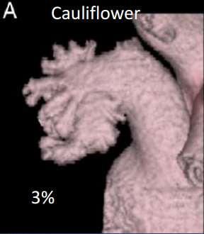
6 Anatomic Variants of LAA Morphology Sample images taken from explanted hearts Chicken wing Cauliflower Windsock Cactus Beigel et al, J Am Coll Cardiol Img 2014;7:

7 Anatomic Variants of LAA Morphology Morphologies and Modalities Cauliflower Windsock Cactus Chicken wing


8 LAA shape/morphology & size vary widely


9 Other LAA Shapes

10 Trabecular structures which might be misinterpreted as thrombi! Ernst et al, The Anatomical Record 242: (1995)


11 LAA Morphology and Risk of Stroke in AF Di Biase et al, J Am Coll Cardiol 2012;60:531 8)
12 Another Angiographic Classification Shi A, et al. J Interna Med Research 2012;40:1560-7

13 Orifice of the LAA and Pectinate Muscles Beigel et al, J Am Coll Cardiol Img 2014;7:

14 Ostium of the LA appendage Variant morphologies


15 MOrifice of the LAA and Pectinate Musclese The orifice of the appendage is usually oval, whereas round, triangular, and water-drop shapes are observed less frequently. The left lateral ridge separates the orifices of the left pulmonary veins from the LAA orifice. The smooth muscular wall of the LA vestibule separates the orifice from the mitral annulus. John P. Veinot et al. Circulation. 1997;96:

16 Role of LAA in LA function LAA: Useless or Priceless? Acts as: a reservoir during LV systole, a conduit for blood transiting from the PVs to the LV during early diastole, an active contractile chamber that augments left ventricular filling in late diastole, and a suction source that refills itself in early systole Regazzoli et al. BioMed Research International Volume 2015
; high")
 occurring during LAA filling.")
17 Role of LAA in LA function The profile is triphasic in a normal SR, which includes early atrial systole before P wave on ECG (above baseline); high amplitude late systolic wave (above the baseline) after P wave, and a late diastolic wave (below baseline) occurring during LAA filling. Abdelmoneim et al, JAFIB, 2014;7:1


18 Echo Guidance of LAA Closure Pre-Procedural Imaging


19 Assess LAA According to Device


20 Assess LAA Anatomy


21 Assess LAA Anatomy


22 Assess LAA Anatomy

23 Assess LAA Anatomy


24 Assess LAA Anatomy


25 Assess LAA Anatomy
, triangular (H), foot-like (I, J), water drop-like (K), and round type (L).")
26 LAA Studied by CT to Plan Closure Device Placement LAA could be classified into 4 types including ChickenWing (A,B), WindSock (C, D), Cauliflower (E), and Cactus type (F). The shape of the LAA orifice could be classified into 5 types including oval (G), triangular (H), foot-like (I, J), water drop-like (K), and round type (L). Wang et al, J Cardiovasc Electrophysiol, Vol. 21, pp , 2010

27 CT Imaging Techniques CT images Postmortem images Type I: smooth LAA origin with a short and poorly defined appendicular limbus. Type II: the limbus is defined and larger. Type III: the limbus originates at a low level and is short. Lopez-Miguez et al, Cardiovasc Electrophysiol,25; , 2014


28 LAA morphologies by cardiac CT, MRI, and autopsies LAA: The Missing Piece in the Puzzle Romero et al, J Cardiovasc Electrophysiol, Vol. 26, pp , September 2015.


29 Preprocedural ΤΕΕ 0 degrees 0 degrees First Department of Cardiology, University of Athens

30 Preprocedural ΤΕΕ 45 degrees 45 degrees First Department of Cardiology, University of Athens

31 Preprocedural ΤΕΕ 90 degrees 90 degrees First Department of Cardiology, University of Athens


32 Preprocedural ΤΕΕ 135 degrees 135 degrees First Department of Cardiology, University of Athens
33 3D LAA Imaging A Watchman 24mm was selected First Department of Cardiology, University of Athens

34 WATCHMAN: TEE measurements

35 ACP: TEE measurements

36 Echo Guidance of LAA Closure
37 Transseptal Puncture: TEE Access to the LA is via a transseptal puncture, performed under TEE in multiple imaging planes, principally 45 and 90, ensuring a posterior puncture and avoidance of the aortic root. Tenting of the atrial septum (arrow) due to pressure from the needle prior to puncture.


38 Transseptal Puncture

39 Pigtail in LUPV


40 First Department of Cardiology, University of Athens Catheter in LAA
41 Catheter in LAA 3D Imaging First Department of Cardiology, University of Athens
42 Device Deployment First Department of Cardiology, University of Athens


43 Device Deployment- Measurements First Department of Cardiology, University of Athens


44 Fluoroscopic Closure Assessment First Department of Cardiology, University of Athens


45 Ultrasound Closure Assessment First Department of Cardiology, University of Athens
46 3D Imaging of Watchman Device First Department of Cardiology, University of Athens

47 3D Imaging of Watchman Device First Department of Cardiology, University of Athens

48 Final Result Fluoroscopic First Department of Cardiology, University of Athens

49 Atrial Septum Integrity Evaluation First Department of Cardiology, University of Athens

50 WATCHMAN Release Criteria: PASS


51 ACP/Amulet Release Criteria: 5 signs

52

53 Summary LAA anatomy is complex and varies widely in size and morphology Pre-procedural, procedural and post-procedural TEE is the gold standard imaging modality for closure of the LAA Knowledge of TEE views to scrutinize LAA anatomy, accurate measurements and familiarity with steps of procedure are crucial for successful LAA closure
Congenital Absence of the Left Atrial Appendage Visualized by 3D Echocardiography in Two Adult Patients
 DOI: 10.1111/echo.12882 2015, Wiley Periodicals, Inc. Echocardiography CASE REPORT Congenital Absence of the Left Atrial Appendage Visualized by 3D Echocardiography in Two Adult Patients Mona Saleh, B.A.,*
Endocardial LAA Occlusion: Which Device for Which Patient?
 Endocardial LAA Occlusion: Which Device for Which Patient? Roy Beinart, MD Davidai Arrhythmia Center The Leviev Heart Center Sheba Medical Center Sheba Medical Center Tel Hashomer The Leviev Heart Center
Feel the rhythm of the beat! Imaging the left atrium: What the electophysiologist wants to know
 Feel the rhythm of the beat! Imaging the left atrium: What the electophysiologist wants to know Anna Shmukler MD, Anna S Bader MD MS, Narmadan Kumarasamy MD, Eric Bader MD, Linda B Haramati MD MS Disclosure
Role of cardiac imaging for catheterbased left atrial appendage closure
 Role of cardiac imaging for catheterbased left atrial appendage closure Ana G. Almeida, MD, PhD Cardiology University Hospital Santa Maria, Lisbon Ana G. Almeida, MD, PhD, FESC University Hospital Santa
Case Report Pulmonary Vein Compression After Implantation of a Left Atrial Appendage Occluder: Presentation and Discussion of a Case
 194 Case Report Pulmonary Vein Compression After Implantation of a Left Atrial Appendage Occluder: Presentation and Discussion of a Case Maryam Ayati MD, Feifan Ouyang MD, KH Kuck MD Department of cardiology,
Does the left atrial appendage morphology correlates with the risk of stroke in patients with atrial fibrillation? Result from a multicenter study.
 Does the left atrial appendage morphology correlates with the risk of stroke in patients with atrial fibrillation? Result from a multicenter study. Luigi Di Biase, MD, PhD, Fiorenzo Gaita, MD, Ilaria Salvetti,
NCVH Birmingham 2013 August 24, Michael S. Bailey, MD Birmingham Heart Clinic
 NCVH Birmingham 2013 August 24, 2013 Michael S. Bailey, MD Birmingham Heart Clinic NCVH Birmingham 2015 August 29, 2015 Michael S. Bailey, MD Birmingham Heart Clinic Left Atrial Anatomy Part of a larger
A Large prolapsing left Atrial Appendage Thrombus in Sinus Rhythm
 A Large prolapsing left Atrial Appendage Thrombus in Sinus Rhythm Sudish Lal Advanced Cardiology Trainee Dunedin Hospital Disclosures: none Case 69yr old male Past Medical History: OSA on CPAP, Asthma,
ATRIAL SEPTAL CLOSURE AND LEFT ATRIAL APPENDAGE OCCLUSION: INDICATIONS AND GUIDANCE ECHOCARDIOGRAPHY IN INTERVENTIONAL CARDIOLOGY
 ATRIAL SEPTAL CLOSURE AND LEFT ATRIAL APPENDAGE OCCLUSION: INDICATIONS AND GUIDANCE Aristides G. Panlilio, MD, FPCP, FPCC,FPSE, FASE Philippine Heart Center Chinese General Hospital and Medical Center
ΔΙΑΔΕΡΜΙΚΗ ΑΝΤΙΜΕΤΩΠΙΣΗ ΔΟΜΙΚΩΝ ΠΑΘΗΣΕΩΝ: Ο ΡΟΛΟΣ ΤΗΣ ΑΠΕΙΚΟΝΙΣΗΣ ΣΤΟ ΑΙΜΟΔΥΝΑΜΙΚΟ ΕΡΓΑΣΤΗΡΙΟ ΣΤΗΝ ΤΟΠΟΘΕΤΗΣΗ MITRACLIP
 ΔΙΑΔΕΡΜΙΚΗ ΑΝΤΙΜΕΤΩΠΙΣΗ ΔΟΜΙΚΩΝ ΠΑΘΗΣΕΩΝ: Ο ΡΟΛΟΣ ΤΗΣ ΑΠΕΙΚΟΝΙΣΗΣ ΣΤΟ ΑΙΜΟΔΥΝΑΜΙΚΟ ΕΡΓΑΣΤΗΡΙΟ ΣΤΗΝ ΤΟΠΟΘΕΤΗΣΗ MITRACLIP ΒΛΑΣΗΣ ΝΙΝΙΟΣ MD MRCP ΚΛΙΝΙΚΗ ΑΓΙΟΣ ΛΟΥΚΑΣ ΘΕΣΣΑΛΟΝΙΚΗ CONFLICT OF INTEREST PROCTOR
Patients selection criteria for LAA occlusion. Young Keun On, MD, PhD, FHRS Samsung Medical Center Sungkyunkwan University School of Medicine
 Patients selection criteria for LAA occlusion Young Keun On, MD, PhD, FHRS Samsung Medical Center Sungkyunkwan University School of Medicine Atrial Fibrillation The most common cardiac arrhythmia. Confers
the Cardiovascular System I
 the Cardiovascular System I By: Dr. Nabil A Khouri MD, MsC, Ph.D MEDIASTINUM 1. Superior Mediastinum 2. inferior Mediastinum Anterior mediastinum. Middle mediastinum. Posterior mediastinum Anatomy of
Coronary Anomalies & Hemodynamic Identification
 Coronary Anomalies & Hemodynamic Identification David Stultz, MD Cardiology Fellow, PGY 6 May 2, 2006 Anomaly #1 Anomaly #2 Anomaly #3 Figure 18-27 Anomalous origin of the left circumflex artery.
Left atrial function. Aliakbar Arvandi MD
 In the clinic Left atrial function Abstract The left atrium (LA) is a left posterior cardiac chamber which is located adjacent to the esophagus. It is separated from the right atrium by the inter-atrial
Left Atrial Structure When Expert Imaging Makes the Difference. Renewed Interest in Anatomy & Function. Feroze Mahmood MD, FASE Associate Professor
 Left Atrial Structure When Expert Imaging Makes the Difference Feroze Mahmood MD, FASE Associate Professor Center enewed Interest in Anatomy & Function Center Once considered a EDUNDANT structure Of no
Papel da imagem na estratificac ão de risco e na predic ão do risco tromboemboĺico
 Imagiologia na Fibrilhac ão Auricular Papel da imagem na estratificac ão de risco e na predic ão do risco tromboemboĺico Mª João Andrade Diagnosis and timely detection of atrial fibrillation Echo-machines
JOINT MEETING 2 Tricuspid club Chairpersons: G. Athanassopoulos, A. Avgeropoulou, M. Khoury, G. Stavridis
 JOINT MEETING 2 Tricuspid club Chairpersons: G. Athanassopoulos, A. Avgeropoulou, M. Khoury, G. Stavridis Similarities and differences in Tricuspid vs. Mitral Valve Anatomy and Imaging. Echo evaluation
Anatomy of the Heart. Figure 20 2c
 Anatomy of the Heart Figure 20 2c Pericardium & Myocardium Remember, the heart sits in it s own cavity, known as the mediastinum. The heart is surrounded by the Pericardium, a double lining of the pericardial
Intracardiac EchoCardiography (ICE) Common Views
 Common Views") Intracardiac EchoCardiography (ICE) Common Views Introduction What is ICE? Catheter with microscopic ultrasound transducer tip and doppler capabilities inserted into the heart via the IVC (typically) or
Intracardiac EchoCardiography (ICE) Common Views Introduction What is ICE? Catheter with microscopic ultrasound transducer tip and doppler capabilities inserted into the heart via the IVC (typically) or
Routine MitraClip. Image Guidance Step by Step
 Routine MitraClip Image Guidance Step by Step Douglas C. Shook, MD, FASE Director, Cardiothoracic Anesthesia Fellowship Director, Cardiac Interventional Anesthesia Department of Anesthesiology BRIGHAM
Left Atrial Appendage Closure: Techniques and Guidelines. Mohammad Shenasa, MD Heart & Rhythm Medical Group San Jose, CA
 Left Atrial Appendage Closure: Techniques and Guidelines Mohammad Shenasa, MD Heart & Rhythm Medical Group San Jose, CA May is Stroke Awareness Month 2 September is AF Awareness Month Lecture Highlights
Μαρία Δρακοπούλου, Σοφία Βαïνά
 Μαρία Δρακοπούλου, Σοφία Βαïνά Α Πανεπιστημιακή Καρδιολογική Κλινική Ιπποκράτειο Νοσοκομείο Spectrum of mitral regurgitation German Heart Report 2017 MitraClip implantations have numerically outperformed
Organic mitral regurgitation
 The best in heart valve disease Organic mitral regurgitation Ewa Szymczyk Department of Cardiology Medical University of Lodz, Poland I have nothing to declare Organic mitral regurgitation leaflet abnormality
We present the case of an asymptomatic, 75-year-old
 Images in Cardiovascular Medicine Asymptomatic Rupture of the Left Ventricle Lech Paluszkiewicz, MD; Stefan Ożegowski, MD; Mohammad Amin Parsa, MD; Jan Gummert, PhD, MD We present the case of an asymptomatic,
Atrial fibrillation (AF), one of the
, one of the") Hellenic J Cardiol 2013; 54: 408-412 Case Report Left Atrial Appendage Occlusion with the Amplatzer Amulet for Stroke Prevention in Atrial Fibrillation: The First Case in Greece Apostolos Tzikas, Lambros
Hellenic J Cardiol 2013; 54: 408-412 Case Report Left Atrial Appendage Occlusion with the Amplatzer Amulet for Stroke Prevention in Atrial Fibrillation: The First Case in Greece Apostolos Tzikas, Lambros
Left atrial appendage (LAA) closure is now a commercially
 closure is now a commercially") Assessing Anatomy for Left Atrial Appendage Closure The roles of TEE and CTA in anatomic assessment and device selection for LAA closure. BY DEE DEE WANG, MD, AND MARVIN H. ENG, MD Left atrial appendage
Assessing Anatomy for Left Atrial Appendage Closure The roles of TEE and CTA in anatomic assessment and device selection for LAA closure. BY DEE DEE WANG, MD, AND MARVIN H. ENG, MD Left atrial appendage
2D/3D in Evaluation of Atrial Septum
 2D/3D in Evaluation of Atrial Septum Roberto M Lang, MD OSTIUM SECUNDUM ASD: 2D AND 3D TNSESOPHAGEAL ECHO 1 Biplane views 90 0 3D Acquisi on Acquire 3D volume Lang RM et al. JASE 2012;25:3 46. Right atrial
The Atrial Septum: Opening the Septum Transseptal Needle Perforation, Radio Frequency Perforation, and Stent Placement
 The Atrial Septum: Opening the Septum Transseptal Needle Perforation, Radio Frequency Perforation, and Stent Placement Matthew J. Gillespie MD, FSCAI The Children s Hospital of Philadelphia SCAI Fellows
Prognostic Value of Left Atrial Size and Function
 Prognostic Value of Left Atrial Size and Function James D. Thomas, M.D., F.A.C.C. Cardiovascular Imaging Center Department of Cardiology Cleveland Clinic Foundation Cleveland, Ohio, USA Conflicts: None
Cleveland Clinic Policy
 Atrial Fibrillation: It is all about the Left Atrial Appendage LAA Occluder Device 3 Amigos Risk Stratification, Ablation, and LAA closure Allan L Klein M.D. Director of Pericardial Center Professor of
Trans-septal Catheterization. December 8, Jonathan Tobis, MD Professor of Medicine Interventional Cardiology, UCLA
 Trans-septal Catheterization December 8, 2015 Jonathan Tobis, MD Professor of Medicine Interventional Cardiology, UCLA No conflicts of interest for this talk BRK = Brockenbrough needle BRK may be easier
3D Printing & Echocardiography
 ASE SOTA Feb 19, 2018 3D Printing & Echocardiography Stephen H. Little, MD John S. Dunn Chair in Cardiovascular Research and Education, Associate professor, Weill Cornell Medicine Disclosures Personal
Percutaneous Mitral Valve Repair
 Percutaneous Mitral Valve Repair MitraClip: Procedure, Data, Patient Selection Chad Rammohan, MD FACC Director, Cardiac Cath Lab El Camino Hospital Mountain View, California Mitral Regurgitation MitraClip
Index. cardiology.theclinics.com. Note: Page numbers of article titles are in boldface type.
 Index Note: Page numbers of article titles are in boldface type. A AADs. See Antiarrhythmic drugs (AADs) ACE inhibitors. See Angiotensin-converting enzyme (ACE) inhibitors ACP in transseptal approach to
Watchman and Structural update..the next frontier. Ari Chanda, MD Cardiology Associates of Fredericksburg
 Watchman and Structural update..the next frontier Ari Chanda, MD Cardiology Associates of Fredericksburg Different Left Atrial Appendage (LAA) morphologies Watchman (the device) Fabric Anchors Device structure
ViewFlex Xtra ICE Catheter. Positioning Reference Manual
 ViewFlex Xtra ICE Catheter Positioning Reference Manual ViewFlex Xtra ICE Catheter Index The ViewFlex Xtra ICE Catheter, which is compatible with the ViewMate Z and ViewMate II ultrasound consoles, provides
Ch.15 Cardiovascular System Pgs {15-12} {15-13}
 Ch.15 Cardiovascular System Pgs {15-12} {15-13} E. Skeleton of the Heart 1. The skeleton of the heart is composed of rings of dense connective tissue and other masses of connective tissue in the interventricular
Case Report Hemostasis of Left Atrial Appendage Bleed With Lariat Device
 273 Case Report Hemostasis of Left Atrial Appendage Bleed With Lariat Device Amena Hussain MD, Muhamed Saric MD, Scott Bernstein MD, Douglas Holmes MD, Larry Chinitz MD NYU Langone Medical Center, United
Middle mediastinum---- heart & pericardium. Dep. of Human Anatomy Zhou Hongying
 Middle mediastinum---- heart & pericardium Dep. of Human Anatomy Zhou Hongying eaglezhyxzy@163.com Subdivisions of the mediastinum Contents of Middle mediastinum Heart Pericardium: a serous sac enclosing
Atrial Septal Defects
 Supplementary ACHD Echo Acquisition Protocol for Atrial Septal Defects The following protocol for echo in adult patients with atrial septal defects (ASDs) is a guide for performing a comprehensive assessment
Devices to Protect Against Stroke in Atrial Fibrillation
 Devices to Protect Against Stroke in Atrial Fibrillation Jonathan C. Hsu, MD, MAS Associate Clinical Professor Division of Cardiology, Section of Cardiac Electrophysiology June 2, 2018 Disclosures Honoraria
Normal TTE/TEE Examinations
 Normal TTE/TEE Examinations Geoffrey A. Rose, MD FACC FASE Sanger Heart & Vascular Institute Before you begin imaging... Obtain the patient s Height Weight BP PLAX View PLAX View Is apex @ 9-10 o clock?
Bogdan A. Popescu. University of Medicine and Pharmacy Bucharest, Romania. EAE Course, Bucharest, April 2010
 Bogdan A. Popescu University of Medicine and Pharmacy Bucharest, Romania EAE Course, Bucharest, April 2010 This is how it started Mitral stenosis at a glance 2D echo narrow diastolic opening of MV leaflets
LAA Occluders: The Right Device for the Right Patient ACC/SHA MEETING OCTOBER 31 ST 2015 JEDDAH, KSA OMER A. M. ELAMIN, MD, FACC
 LAA Occluders: The Right Device for the Right Patient ACC/SHA MEETING OCTOBER 31 ST 2015 JEDDAH, KSA OMER A. M. ELAMIN, MD, FACC INTERVENTIONAL CARDIOLOGIST DIRECTOR, ADULT CARDIOLOGY FELLOWSHIP TRAINING
Transseptal catheterization is an established
 Site-Specific Transseptal Puncture for Emerging Structural Heart Interventions Imaging-assisted techniques for accurate access. By Michael J. Rinaldi, MD, FACC, FSCAI; Markus Scherer, MD, FACC, FSCCT;
Thessaloniki October 9, Apostolos Tzikas MD, PhD, FESC
 7 th IICE Congress Thessaloniki October 9, 2014 Left atrial appendage occlusion with the Amplatzer Amulet: a case report Apostolos Tzikas MD, PhD, FESC Interventional Cardiologist Structural & Congenital
Cardiac ultrasound protocols
 Cardiac ultrasound protocols IDEXX Telemedicine Consultants Two-dimensional and M-mode imaging planes Right parasternal long axis four chamber Obtained from the right side Displays the relative proportions
Left Atrial Appendage Closure: The Rationale
 Left Atrial Appendage Closure: The Rationale JOHN D. HUMMEL, MD DIRECTOR OF CLINCAL ELECTROPHYSIOLOGY RESEARCH PROFESSOR OF CLINICAL INTERNAL MEDICINE OHIO STATE UNIVERSITY WEXNER MEDICAL CENTER 1 Disclosures
Hybrid Ablation of AF in the Operating Room: Is There a Need? MAZE III Procedure. Spectrum of Atrial Fibrillation
 Hybrid Ablation of AF in the Operating Room: Is There a Need? MAZE III Procedure Paul J. Wang, MD Amin Al-Ahmad, MD Gan Dunnington, MD Stanford University Cox J, et al. Ann Thorac Surg. 1993;55:578-580.
Revealing new insights. irotate electronic rotation and xplane adjustable biplane imaging. Ultrasound cardiology. irotate and xplane
 Ultrasound cardiology irotate and xplane Revealing new insights irotate electronic rotation and xplane adjustable biplane imaging Annemien van den Bosch and Jackie McGhie Department of Cardiology, Erasmus
10 ο ΣΥΝΕΔΡΙΟ ΕΠΕΜΒΑΤΙΚΗΣ ΚΑΡΔΙΟΛΟΓΙΑΣ ΚΑΙ ΗΛΕΚΤΡΟΦΥΣΙΟΛΟΓΙΑΣ Σεπτεμβρίου 2017 Electra Palace Θεσσαλονικη
 10 ο ΣΥΝΕΔΡΙΟ ΕΠΕΜΒΑΤΙΚΗΣ ΚΑΡΔΙΟΛΟΓΙΑΣ ΚΑΙ ΗΛΕΚΤΡΟΦΥΣΙΟΛΟΓΙΑΣ 14-16 Σεπτεμβρίου 2017 Electra Palace Θεσσαλονικη Ηχωκαρδιογραφία στην ένδειξη-προετοιμασία, διενέργεια, παρακολούθηση ασθενών που υποβάλλονται
3D Printing & Echocardiography
 Echo Hawaii Jan 18, 2018 3D Printing & Echocardiography Stephen H. Little, MD John S. Dunn Chair in Cardiovascular Research and Education, Associate professor, Weill Cornell Medicine Rapid Prototyping
The Normal Echocardiogram
 The Normal Echocardiogram Pravin V. Patil, MD FACC Lewis Katz School of Medicine at Temple University Acknowledgments Dr. Susan Wiegers Dr. Martin Keane Temple Cardiac Sonographers Disclosures No relevant
Radiofrequency Energy: Irrigation and Alternate Catheters. Andreas Pflaumer
 Radiofrequency Energy: Irrigation and Alternate Catheters Andreas Pflaumer Irrigated tip RF ablation Irrigated tip How does it work? Potential benefits? Potential risks? How is this relevant to pediatric
Chapter 18 - Heart. I. Heart Anatomy: size of your fist; located in mediastinum (medial cavity)
") Chapter 18 - Heart I. Heart Anatomy: size of your fist; located in mediastinum (medial cavity) A. Coverings: heart enclosed in double walled sac called the pericardium 1. Fibrous pericardium: dense connective
Chapter 18 - Heart I. Heart Anatomy: size of your fist; located in mediastinum (medial cavity) A. Coverings: heart enclosed in double walled sac called the pericardium 1. Fibrous pericardium: dense connective
Atrial fibrillation (AF) is the most common
 is the most common") Left Atrial Appendage Closure With the Watchman Device A review of trial results and clinical application. BY JAYANT KHITHA, MD, AND TANVIR K. BAJWA, MD Atrial fibrillation (AF) is the most common arrhythmia
Left Atrial Appendage Closure With the Watchman Device A review of trial results and clinical application. BY JAYANT KHITHA, MD, AND TANVIR K. BAJWA, MD Atrial fibrillation (AF) is the most common arrhythmia
When Does 3D Echo Make A Difference?
 When Does 3D Echo Make A Difference? Wendy Tsang, MD, SM Assistant Professor, University of Toronto Toronto General Hospital, University Health Network 1 Practical Applications of 3D Echocardiography Recommended
human anatomy 2016 lecture thirteen Dr meethak ali ahmed neurosurgeon
 Heart The heart is a hollow muscular organ that is somewhat pyramid shaped and lies within the pericardium in the mediastinum. It is connected at its base to the great blood vessels but otherwise lies
Adult Echocardiography Examination Content Outline
 Adult Echocardiography Examination Content Outline (Outline Summary) # Domain Subdomain Percentage 1 2 3 4 5 Anatomy and Physiology Pathology Clinical Care and Safety Measurement Techniques, Maneuvers,
Rotation: Echocardiography: Transthoracic Echocardiography (TTE)
") Rotation: Echocardiography: Transthoracic Echocardiography (TTE) Rotation Format and Responsibilities: Fellows rotate in the echocardiography laboratory in each clinical year. Rotations during the first
Rotation: Echocardiography: Transthoracic Echocardiography (TTE) Rotation Format and Responsibilities: Fellows rotate in the echocardiography laboratory in each clinical year. Rotations during the first
Percutaneous Epicardial LAA Closure: When Does it Make Sense?
 Percutaneous Epicardial LAA Closure: When Does it Make Sense? Petr Neuzil, MD,PhD, FESC Professor of Medicine Cardiology department Na Homolce Hospital, Prague, Czechia petr.neuzil@gmail.com Disclosures
Ο ΡOΛΟΣ ΤΗΣ ΑΠΕΙΚOΝΙΣΗΣ ΣΤΗΝ ΗΛΕΚΤΡΟΦΥΣΙΟΛΟΓIΑ - ΥΠΟΣΤΡΩΜΑ ΚΟΛΠΟΙ ΩΤΙΑ ΑΞΙΟΛΟΓΗΣΗ ΟΥΛΗΣ
 Ο ΡOΛΟΣ ΤΗΣ ΑΠΕΙΚOΝΙΣΗΣ ΣΤΗΝ ΗΛΕΚΤΡΟΦΥΣΙΟΛΟΓIΑ - ΥΠΟΣΤΡΩΜΑ ΚΟΛΠΟΙ ΩΤΙΑ ΑΞΙΟΛΟΓΗΣΗ ΟΥΛΗΣ Κώστας Παπαδόπουλος, Επιμ. Α Καρδιολογίας, Νοσοκομείο Ερυθρός Σταυρός ATRIA ANATOMY Complex anatomy Best knowledge
Cardiac Imaging in abnormal rhythm Role of MDCT
 Cardiac Imaging in abnormal rhythm Role of MDCT Cardiac Imaging in abnormal rhythm Role of MDCT Scope of the problem CT in Atrial Fibrillation CT and pacing Ventricular arrhythmia Other applications 1
Chapter 20 (1) The Heart
 The Heart") Chapter 20 (1) The Heart Learning Objectives Describe the location and structure of the heart Describe the path of a drop of blood from the superior vena cava or inferior vena cava through the heart out
Chapter 20 (1) The Heart Learning Objectives Describe the location and structure of the heart Describe the path of a drop of blood from the superior vena cava or inferior vena cava through the heart out
National Imaging Associates, Inc. Clinical guidelines CARDIAC CATHETERIZATION -LEFT HEART CATHETERIZATION. Original Date: October 2015 Page 1 of 5
 National Imaging Associates, Inc. Clinical guidelines CARDIAC CATHETERIZATION -LEFT HEART CATHETERIZATION CPT Codes: 93451, 93452, 93453, 93454, 93455, 93456, 93457, 93458, 93459, 93460, 93461 LCD ID Number:
Review Article Thromboembolism Prevention via Transcatheter Left Atrial Appendage Closure with Transeosophageal Echocardiography Guidance
 Hindawi Publishing Corporation rombosis Volume 2014, Article ID 832752, 6 pages http://dx.doi.org/10.1155/2014/832752 Review Article Thromboembolism Prevention via Transcatheter Left Atrial Appendage Closure
Advanced imaging of the left atrium - strain, CT, 3D, MRI -
 Advanced imaging of the left atrium - strain, CT, 3D, MRI - Monica Rosca, MD Carol Davila University of Medicine and Pharmacy, Bucharest, Romania Declaration of interest: I have nothing to declare Case
Imaging Evaluation of the Ventricular Septum
 Imaging Evaluation of the Ventricular Septum Craig E Fleishman, MD FACC FASE The Heart Center at Arnold Palmer Hospital for Children, Orlando SCAI Fall Fellows Course 2013 Las Vegas Disclosure Information
Anatomy of left ventricular outflow tract'
 Anatomy of left ventricular outflow tract' ROBERT WALMSLEY British Heart Journal, 1979, 41, 263-267 From the Department of Anatomy and Experimental Pathology, The University, St Andrews, Scotland SUMMARY
Successful Percutaneous Closure of Mitral Bioprosthetic Paravalvular Leak Using Figulla ASD Occluder
 Hans R. Figulla, M.D., PhD ; Ali Hamadanchi, M.D. Medicine, Pneumology Universitity Hospital, Jena, Germany Successful Percutaneous Closure of Mitral Bioprosthetic Paravalvular Leak Using Figulla ASD Occluder
Left Atrial Appendage Closure Devices. Atrial Fibrillation 10/11/2017
 Left Atrial Appendage Closure Devices Emile Daoud, MD Chief, Cardiac Electrophysiology Wexner Medical Center, The Ohio State University Atrial Fibrillation 1 Adjusted Annual Stroke Risk Using CHA 2 DS
Different transseptal puncture for different procedures: Optimization of left atrial catheterization guided by transesophageal echocardiography
 Original Article Different transseptal puncture for different procedures: Optimization of left atrial catheterization guided by transesophageal echocardiography Andrea Radinovic 1, Patrizio Mazzone 1,
Basic Approach to the Echocardiographic Evaluation of Ventricular Diastolic Function
 Basic Approach to the Echocardiographic Evaluation of Ventricular Diastolic Function J A F E R A L I, M D U N I V E R S I T Y H O S P I T A L S C A S E M E D I C A L C E N T E R S T A F F C A R D I O T
Watchman a Stroke Prevention Technology for Patients with Atrial Fibrillation
 Watchman a Stroke Prevention Technology for Patients with Atrial Fibrillation Scripps hospital,la Jolla, CA Atrial fibrillation is a major source of cardiogenic embolic related stroke 500,000 strokes per
Lab Activity 23. Cardiac Anatomy. Portland Community College BI 232
 Lab Activity 23 Cardiac Anatomy Portland Community College BI 232 Cardiac Muscle Histology Branching cells Intercalated disc: contains many gap junctions connecting the adjacent cell cytoplasm, creates
Critical role of multi-modality planning in Transcatheter Mitral Valve Replacement
 Critical role of multi-modality planning in Transcatheter Mitral Valve Replacement Dee Dee Wang, MD, FACC, FASE, FSCCT Director Structural Heart Imaging Medical Director 3D Printing Henry Ford Innovations
Francesco Fulvio Faletra Gila Perk Natesa G. Pandian Hans-Joachim Nesser Itzhak Kronzon. Real-Time 3D Interventional Echocardiography
 Francesco Fulvio Faletra Gila Perk Natesa G. Pandian Hans-Joachim Nesser Itzhak Kronzon Real-Time 3D Interventional Echocardiography 123 Real-Time 3D Interventional Echocardiography Francesco Fulvio Faletra
The Emerging Atrial Fibrillation Epidemic: Treat It, Leave It or Burn It. Chandra Kumbar MD FACC FHRS The Heart Group, Evansville IN
 The Emerging Atrial Fibrillation Epidemic: Treat It, Leave It or Burn It Chandra Kumbar MD FACC FHRS The Heart Group, Evansville IN Disclosures Consultant Advisory Board, Medtronic Atrial fibrillation
Atrial fibrillation (AF) is associated with a high
 is associated with a high") Echo Essentials for Endoluminal LAA Closure Echocardiography is the imaging modality of choice for this new interventional technique. By Nina C. Wunderlich, MD; Martin J. Swaans, MD; Harald Küx, MD; Roy
Echo Essentials for Endoluminal LAA Closure Echocardiography is the imaging modality of choice for this new interventional technique. By Nina C. Wunderlich, MD; Martin J. Swaans, MD; Harald Küx, MD; Roy
Transvenous Pacemaker Implantation 22 years after the Mustard Procedure
 Case Report Transvenous Pacemaker Implantation 22 years after the Mustard Procedure Masato Sakamoto MD, Yoshie Ochiai MD, Yutaka Imoto MD, Akira Sese MD, Mamie Watanabe MD, Kunitaka Joo MD Department of
Peripheral and Cardiology Coder 2018
 Peripheral and Cardiology Coder 2018 Cardiovascular Services and Procedures Prepared and Published By: MedLearn Publishing A Division of MedLearn Media, Inc. 445 Minnesota Street, Suite 514 St. Paul, MN
Objectives. Diastology: What the Radiologist Needs to Know. LV Diastolic Function: Introduction. LV Diastolic Function: Introduction
 Objectives Diastology: What the Radiologist Needs to Know. Jacobo Kirsch, MD Cardiopulmonary Imaging, Section Head Division of Radiology Cleveland Clinic Florida Weston, FL To review the physiology and
Structural Heart Disease: Setting the Stage for Success
 Structural Heart Disease: Setting the Stage for Success Brenda McCulloch, RN MSN RCIS Cardiovascular Clinical Nurse Specialist, Interventional & Medical Cardiology Sutter Medical Center, Sacramento mccullb@sutterhealth.org
COMPREHENSIVE EVALUATION OF FETAL HEART R. GOWDAMARAJAN MD
 COMPREHENSIVE EVALUATION OF FETAL HEART R. GOWDAMARAJAN MD Disclosure No Relevant Financial Relationships with Commercial Interests Fetal Echo: How to do it? Timing of Study -optimally between 22-24 weeks
Επιπλοκές κατάλυσης πνευµονικών φλεβών
 Επιπλοκές κατάλυσης πνευµονικών φλεβών Παναγιώτης Ιωαννίδης Διευθυντής Τµήµατος Αρρυθµιών & Επεµβατικής Ηλεκτροφυσιολογίας Βιοκλινικής Αθηνών ΣΕΜΙΝΑΡΙΑ ΟΜΑΔΩΝ ΕΡΓΑΣΙΑΣ Ιωάννινα, 27-2-2015 Solving an equation
8/31/2016. Mitraclip in Matthew Johnson, MD
 Mitraclip in 2016 Matthew Johnson, MD 1 Abnormal Valve Function Valve Stenosis Obstruction to valve flow during that phase of the cardiac cycle when the valve is normally open. Hemodynamic hallmark - pressure
Conflict of Interests
 Introduction to Interventional Echocardiography Roberto M Lang, MD Tomtec Conflict of Interests Research Grants Philips Medical Imaging Research Grants Speakers bureau Advisory bureau 1 Structural Heart
The radial procedure was developed as an outgrowth
 The Radial Procedure for Atrial Fibrillation Takashi Nitta, MD The radial procedure was developed as an outgrowth of an alternative to the maze procedure. The atrial incisions are designed to radiate from
Case 47 Clinical Presentation
 93 Case 47 C Clinical Presentation 45-year-old man presents with chest pain and new onset of a murmur. Echocardiography shows severe aortic insufficiency. 94 RadCases Cardiac Imaging Imaging Findings C
Cardiac Masses. Cardiac Masses: Considerations. Dennis A. Tighe, MD, FASE. University of Massachusetts Medical School Worcester, MA 4/16/2018
 Cardiac Masses Dennis A. Tighe, MD, FASE University of Massachusetts Medical School Worcester, MA Cardiac Masses: Considerations Definition of the mass Nature Location Benign or malignant Presentation
Catheter ablation: the recovery process and what to expect
 Catheter ablation: the recovery process and what to expect Mark O Neill DPhil FRCP FHRS Consultant Cardiologist & Professor of Cardiac Electrophysiology Division of Imaging and Biomedical Engineering &
Simultaneous Double Clipping Delivery Guide Strategy for Treatment of Severe Coaptation Failure in Functional Mitral Regurgitation
 Heart, Lung and Circulation (2015) 24, 98 102 1443-9506/04/$36.00 http://dx.doi.org/10.1016/j.hlc.2014.09.008 HOW-TO-DO-IT Simultaneous Double Clipping Delivery Guide Strategy for Treatment of Severe Coaptation
THE HEART. A. The Pericardium - a double sac of serous membrane surrounding the heart
 THE HEART I. Size and Location: A. Fist-size weighing less than a pound (250 to 350 grams). B. Located in the mediastinum between the 2 nd rib and the 5 th intercostal space. 1. Tipped to the left, resting
Concomitant procedures using minimally access
 Surgical Technique on Cardiac Surgery Concomitant procedures using minimally access Nelson Santos Paulo Cardiothoracic Surgery, Centro Hospitalar de Vila Nova de Gaia, Oporto, Portugal Correspondence to:
How to Approach the Patient with CRT and Recurrent Heart Failure
 How to Approach the Patient with CRT and Recurrent Heart Failure Byron K. Lee MD Associate Professor of Medicine Electrophysiology and Arrhythmia Section UCSF Update in Electrocardiography and Arrhythmias
Dad needed to get off his blood thinner. His doctor told us about an alternative. It s called
 Dad needed to get off his blood thinner. His doctor told us about an alternative. It s called A one-time procedure that may reduce stroke risk for a lifetime in people with AFib not caused by a heart valve
CV Anatomy Quiz. Dr Ella Kim Dr Pip Green
 CV Anatomy Quiz Dr Ella Kim Dr Pip Green Q1 The location of the heart is correctly described as A) lateral to the lungs. B) medial to the sternum. C) superior to the diaphragm. D) posterior to the spinal
EAE RECOMMENDATIONS FOR TRANSESOPHAGEAL ECHO. Cardiac Sources of Embolism. Luigi P. Badano, MD, FESC
 EAE RECOMMENDATIONS FOR TRANSESOPHAGEAL ECHO. Cardiac Sources of Embolism Luigi P. Badano, MD, FESC Background Stroke is the 3 cause of death in several industrial countries; Embolism accounts for 15-30%
Image Assistance in TAVI Why CT? Won-Jang Kim, MD, PhD Clinical Assistant Professor of Medicine, Heart Institute, Asan Medical Center, Seoul, Korea
 Image Assistance in TAVI Why CT? Won-Jang Kim, MD, PhD Clinical Assistant Professor of Medicine, Heart Institute, Asan Medical Center, Seoul, Korea Major Uses of CT in TAVI Ileofemoral Patient Arterial
THE LEFT ATRIUM HOW CAN ECHO HELP US?
 THE LEFT ATRIUM HOW CAN ECHO HELP US? Dr. Dragos COZMA BACKGROUND Left atrium (LA) dilation can occur in a broad spectrum of cardiovascular diseases including hypertension, left ventricular dysfunction,