The Normal Echocardiogram
|
|
|
- Leon Wilcox
- 5 years ago
- Views:
Transcription
1 The Normal Echocardiogram Pravin V. Patil, MD FACC Lewis Katz School of Medicine at Temple University

2 Acknowledgments Dr. Susan Wiegers Dr. Martin Keane Temple Cardiac Sonographers
3 Disclosures No relevant financial disclosures
4 Question 1 Which aortic cusp is noted by the arrow? A. Right B. Left C. Non-Coronary D. Can t tell
5 Question 2 Which myocardial segment is denoted by the arrow? A. Mid Anterior B. Mid Anteroseptum C. Mid Inferoseptum D. Basal Anteroseptum E. Mid Inferolateral
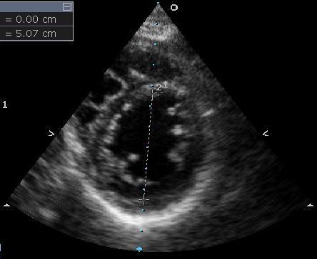
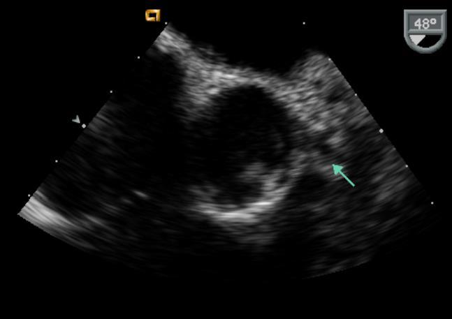
6 Question 3 Which scallop is noted by the arrow? A. Non-coronary B. A2 C. P1 D. A1 E. A3
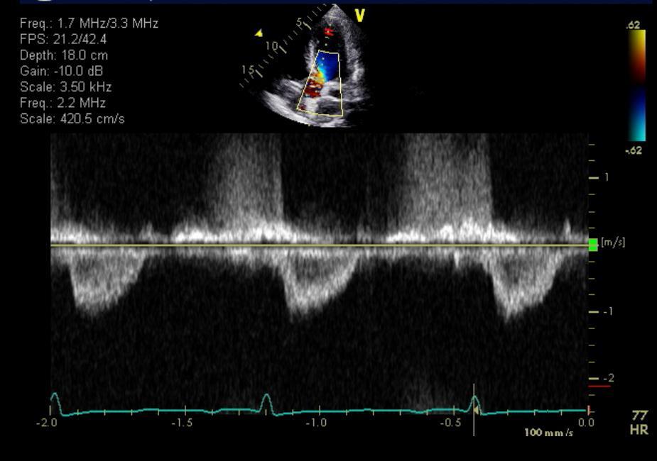
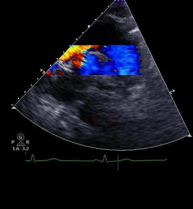
7 Question 4 How do you optimize this acquisition? A. Raise baseline B. Lower baseline C. Increase scale D. Decrease scale E. Pedoff transducer
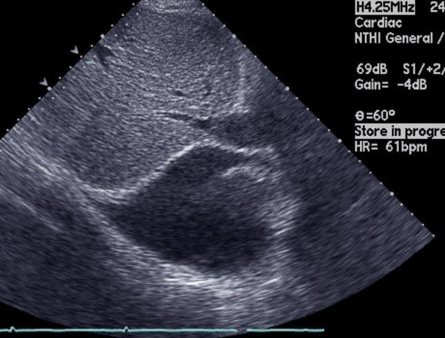
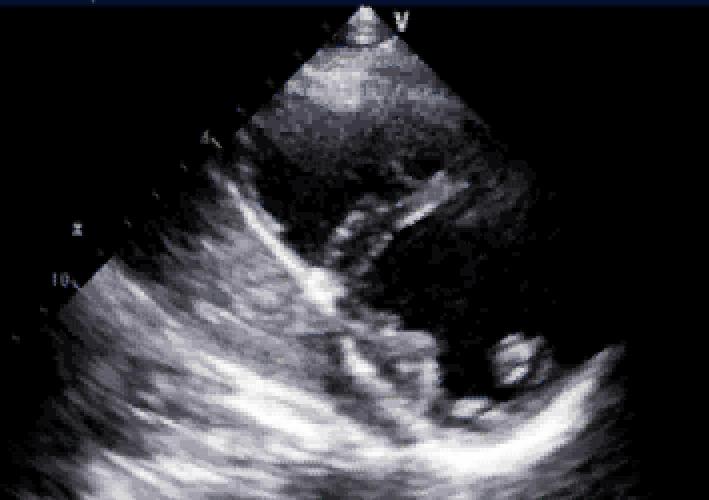
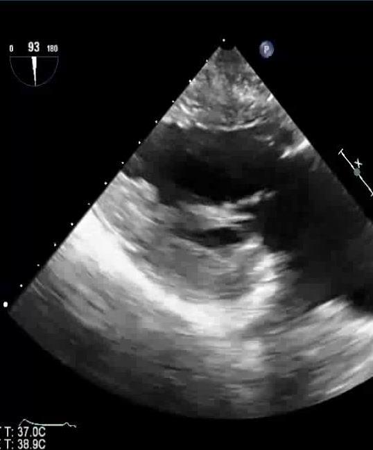
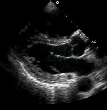
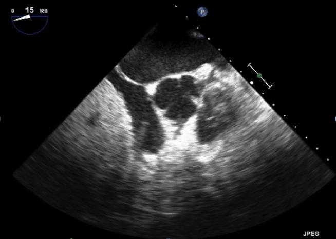
8 Question 5 What does the arrow indicate? A. Eustachian Valve B. RA thrombus C. Chiari Network D. Catheter in RA
9 Transthoracic Echocardiography
10 Normal Echocardiography Why do we need to review this? Recognize pathology when it exists ASCeXAM Standardized image acquisition Image optimization Anatomic identification Chamber quantification Less known normal structures AUC/Indications/Contraindications

11 Appropriate Use Criteria Appropriate Use Criteria for Echocardiography J Am Soc Echocardiogr 2011;24:

12 TEE Guidelines TEE Indications & Contraindications J Am Soc Echocardiogr 2013;26:
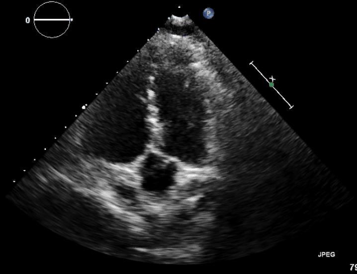
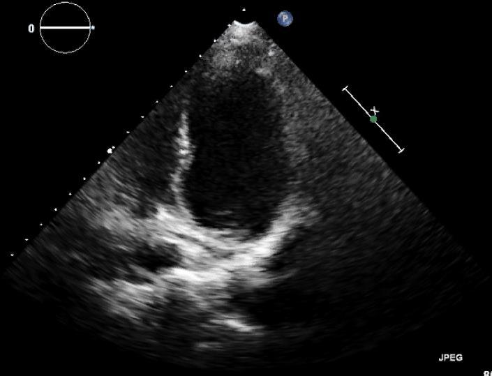
13 Parasternal Long Axis


14 Depth Matters LPE LPE RPE RPE
15 Down One Interspace
16 Up One Interspace
17 Off-Axis Measurements

18 On-Axis Measurements

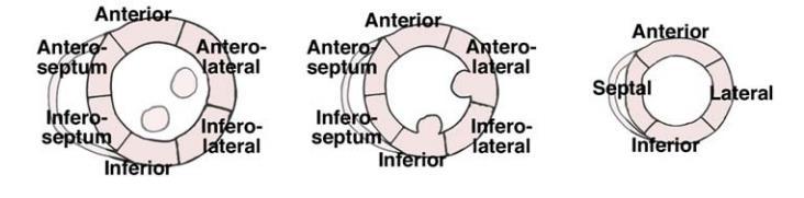
19 ASE/AHA 17 Segment Model ASE Chamber Quant Guidelines, JASE 2015

20 Short Axis LV Parasternal Subcostal
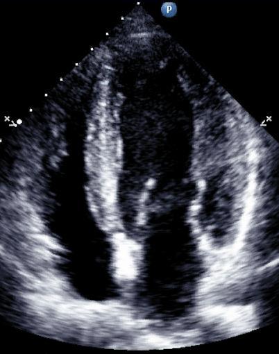
21 Apical Four Chamber
22 Tail Down
23 Tail Up


24 Apical 4 Chamber Off-Axis On-Axis

25 ASE/AHA 17 Segment Model ASE Chamber Quant Guidelines, JASE 2015


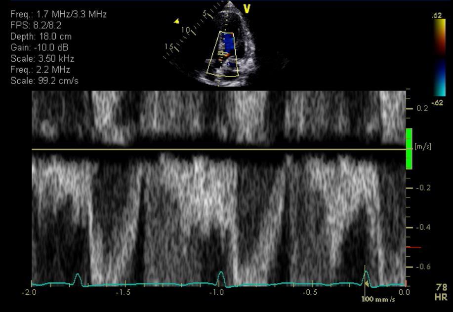
26 Normal? Apical Foreshortening Poor Endocardial Border Definition

27 Contrast for LV Opacification Commercial Contrast Improve endocardial border definition Eliminate foreshortening Evaluate for mural thrombi Restore diagnostic quality
28 ASCeXAM Focus How do you fix this image? Recognize off-axis views Imaging from wrong interspace Foreshorten cardiac structure Contrast use and optimization Anatomical identification Myocardial segment identification Extracardiac findings recognition Common Artifacts
29 Signal Amplitude Tissue Harmonic Imaging Non-linear distortion of acoustic signal in tissue generates harmonics SNR 1.5 Tissue Noise Noise/artifacts generate no significant harmonic SNR 3 Tissue Harmonic Imaging takes advantage of increased SNR f 2f


30 Tissue Harmonic Imaging Fundamental Tissue Harmonic

31 Bubbles Have Harmonics too.. Harmonics 1.3/2.6 MHz Fundamental 1.6 MHz
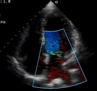
32 Doppler Echocardiography Optimal 2D images when ultrasound beam is perpendicular to structures Optimal Doppler imaging when ultrasound beam is parallel to flow Apical views allow alignment with most cardiac flows (i.e. aortic, mitral and tricuspid valves)
33 Doppler Echocardiography Color Doppler Pulse wave modality that cannot resolve high velocities Turbulence/variance maps can help define jet, direction and turbulence Pulse Wave Spectral Doppler Range specific Subject to aliasing at high velocities like CFD Continuous Wave Spectral Doppler Able to resolve high velocities Range ambiguous

34 Color Flow Doppler Pay attention to the baseline Make note of the Nyquist limit Color scales vary Variance maps Optimize size and sector for frame rate
35 Doppler Optimization Scale Optimized

36 Doppler Optimization Baseline Adjusted
37 ASCeXAM Focus Effects of harmonic imaging Appropriate indications for contrast Contrast Physics and optimization Types of Doppler and technique limitations Spectral Doppler signal optimization Color Flow Doppler optimization
38 Transesophageal Echocardiography

39 Left Atrial Appendage


40 Hahn et al. TEE Guidelines. JASE 2013;26: Mitral Valve
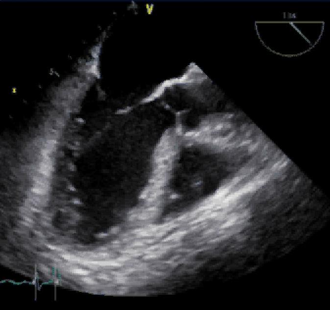
41 3D Mitral Valve

42 Aortic Valve

43 3D Aortic Valve

44 Bi-Caval View
45 Transgastric LV
46 ASCeXAM Focus TEE not heavily tested AUC, Indications and Contraindications for TEE Anatomical identification Standard 3D views of Mitral and Aortic valves Correlative anatomic imaging with TTE
47 Normal Anatomic Structures Transthoracic and Transesophageal Echocardiography
48 Persistent Venous Valves Chiari Network No known function Not present in every patient Netlike structure that is highly mobile Usually arises from the vicinity of the IVC not attached to the septum Eustachian Valve Directs IVC flow across fossa in fetus Present in every fetus Ridge of tissue - rarely mobile at all Arises from the IVC and runs to the fossa


49 Chiari Network RV Inflow Apical Four PSAX
50 Eustachian Valve

51 Crista Terminalis Normal structure Often confused for a right atrial mass Smooth myocardial ridge from RA-SVC junction along posterolateral RA wall

52 Moderator Band

53 Coronary Sinus
54 Pericardial Sinuses
55 Transverse Sinus
56 Subcostal SAX Aortic Valve N R L


57 Coronary Arteries RCA LMCA

58 Coronary Arteries

59 Coronary Arteries
60 TEE Aortic Valve
61 LAD or Circumflex? TEE Aortic Valve


62 Papillary Muscles PSAX TTE Transgastric TEE PM PM AL

63 Pulmonary Veins


64 Pulmonary Veins LUPV TEE RUPV TEE
65 Suprasternal Notch
66 Suprasternal Notch
67 ASCeXAM Focus Normal Anatomic Structures Right Heart Persistent Venous Valve Crista Terminalis Coronary Sinus Moderator Band Left Heart Pericardial Sinuses Pulmonary Veins Coronary Arteries Papillary Muscles Suprasternal Notch
68 Question 1 Which aortic cusp is noted by the arrow? A. Right B. Left C. Non-Coronary D. Can t tell


69 Question 1 - Followup Answer: B. Left Coronary Cusp N R L N L R

70 Question 2 Which myocardial segment is denoted by the arrow? A. Mid Anterior B. Mid Anteroseptum C. Mid Inferoseptum D. Basal Anteroseptum E. Mid Inferolateral

71 Question 2 - Followup Answer: C. Mid Inferoseptum MI MIS MAS MA
72 Question 3 Which scallop is noted by the arrow? A. Non-coronary B. A2 C. P1 D. A1 E. A3

73 Question 3 - Followup Answer: E. A3 Scallop
74 Question 4 How do you optimize this acquisition? A. Raise baseline B. Lower baseline C. Increase scale D. Decrease scale E. Pedoff transducer

75 Question 4 - Followup Answer: D. Decrease Scale

76 Question 5 What does the arrow indicate? A. Eustachian Valve B. RA thrombus C. Chiari Network D. Catheter in RA
77 Question 5 - Followup Answer: C. Chiari Network Chiari Network No known function Not present in every patient Eustachian Valve Directs IVC flow across fossa in fetus Present in every fetus Netlike structure that is highly mobile Usually arises from the vicinity of the IVC not attached to the septum Ridge of tissue - rarely mobile at all Arises from the IVC and runs to the fossa
78 Thank You!
Normal TTE Examination, Doppler Echocardiography and Normal Antegrade Flow Patterns
 Normal TTE Examination, Doppler Echocardiography and Normal Antegrade Flow Patterns Pravin Patil, MD FACC FASE Associate Professor of Medicine Director, Cardiovascular Disease Training Program Lewis Katz
Concepts of Imaging and Knobology
 Concepts of Imaging and Knobology Pravin Patil, MD FACC FASE Associate Professor of Medicine Director, Cardiovascular Disease Training Program Lewis Katz School of Medicine at Temple University Disclosures
Normal TTE/TEE Examinations
 Normal TTE/TEE Examinations Geoffrey A. Rose, MD FACC FASE Sanger Heart & Vascular Institute Before you begin imaging... Obtain the patient s Height Weight BP PLAX View PLAX View Is apex @ 9-10 o clock?
PART II ECHOCARDIOGRAPHY LABORATORY OPERATIONS ADULT TRANSTHORACIC ECHOCARDIOGRAPHY TESTING
 PART II ECHOCARDIOGRAPHY LABORATORY OPERATIONS ADULT TRANSTHORACIC ECHOCARDIOGRAPHY TESTING STANDARD - Primary Instrumentation 1.1 Cardiac Ultrasound Systems SECTION 1 Instrumentation Ultrasound instruments
ASCeXAM / ReASCE. Practice Board Exam Questions Monday Morning
 ASCeXAM / ReASCE Practice Board Exam Questions Monday Morning Ultrasound Physics Artifacts Doppler Physics Imaging, Knobology, and Artifacts Echocardiographic Evaluation of the RV Tricuspid and Pulmonary
British Society of Echocardiography
 British Society of Echocardiography Affiliated to the British Cardiac Society A Minimum Dataset for a Standard Adult Transthoracic Echocardiogram From the British Society of Echocardiography Education
JOINT MEETING 2 Tricuspid club Chairpersons: G. Athanassopoulos, A. Avgeropoulou, M. Khoury, G. Stavridis
 JOINT MEETING 2 Tricuspid club Chairpersons: G. Athanassopoulos, A. Avgeropoulou, M. Khoury, G. Stavridis Similarities and differences in Tricuspid vs. Mitral Valve Anatomy and Imaging. Echo evaluation
Welcome, Intro & Goals
 Welcome, Intro & Goals PP16 Imaging Conference Bicol Hospital, Legaspi City, Philippines July 2016 David Adams, ACS, RCS, RDCS, FASE Duke University Medical Center Echocardiography The Anatomy Lesson of
Adult Echocardiography Examination Content Outline
 Adult Echocardiography Examination Content Outline (Outline Summary) # Domain Subdomain Percentage 1 2 3 4 5 Anatomy and Physiology Pathology Clinical Care and Safety Measurement Techniques, Maneuvers,
Back to Basics: Common Errors In Quantitation In Everyday Practice
 Back to Basics: Common Errors In Quantitation In Everyday Practice Deborah Agler, ACS, RDCS, FASE October 9, 2017 ASE: Echo Florida Rebecca T. Hahn, MD Director of Interventional Echocardiography Professor
Cardiac Mass and Mass-like Structures
 KSE 2017 Basic Echo Review Course (4) Nov 26, 2017 Cardiac Mass and Mass-like Structures Sun Hwa Lee, MD, PhD Chonbuk National University Hospital & Medical School Introduction Although cardiac tumors
Breakout Session: Transesophageal Echocardiography
 Breakout Session: Transesophageal Echocardiography Doris Ockert, MD Andrew Schroeder, MD University of Wisconsin School of Medicine and Public Health Jutta Novalija, MD, PhD Medical College of Wisconsin
Atrial Septal Defects
 Supplementary ACHD Echo Acquisition Protocol for Atrial Septal Defects The following protocol for echo in adult patients with atrial septal defects (ASDs) is a guide for performing a comprehensive assessment
2D/3D in Evaluation of Atrial Septum
 2D/3D in Evaluation of Atrial Septum Roberto M Lang, MD OSTIUM SECUNDUM ASD: 2D AND 3D TNSESOPHAGEAL ECHO 1 Biplane views 90 0 3D Acquisi on Acquire 3D volume Lang RM et al. JASE 2012;25:3 46. Right atrial
Appendix II: ECHOCARDIOGRAPHY ANALYSIS
 Appendix II: ECHOCARDIOGRAPHY ANALYSIS Two-Dimensional (2D) imaging was performed using the Vivid 7 Advantage cardiovascular ultrasound system (GE Medical Systems, Milwaukee) with a frame rate of 400 frames
Since the introduction of transesophageal echocardiography
 ASE/SCA Guidelines for Performing a Comprehensive Intraoperative Multiplane Transesophageal Echocardiography Examination: Recommendations of the American Society of Echocardiography Council for Intraoperative
Pediatric Echocardiography Examination Content Outline
 Pediatric Echocardiography Examination Content Outline (Outline Summary) # Domain Subdomain Percentage 1 Anatomy and Physiology Normal Anatomy and Physiology 10% 2 Abnormal Pathology and Pathophysiology
Right Heart Evaluation ASE Guidelines Review. Chris Mann RDCS, RCS, FASE Faculty, Echocardiography Pitt Community College Greenville, NC
 Right Heart Evaluation ASE Guidelines Review Chris Mann RDCS, RCS, FASE Faculty, Echocardiography Pitt Community College Greenville, NC Objectives Briefly review right atrial and right ventricular anatomy
Doppler Basic & Hemodynamic Calculations
 Doppler Basic & Hemodynamic Calculations August 19, 2017 Smonporn Boonyaratavej MD Division of Cardiology, Department of Medicine Chulalongkorn University Cardiac Center, King Chulalongkorn Memorial Hospital
Pulmonary arteriovenous fistula
 International Journal of Medical Imaging 2014; 2(2): 34-38 Published online April 10, 2014 (http://www.sciencepublishinggroup.com/j/ijmi) doi: 10.11648/j.ijmi.20140202.16 Pulmonary arteriovenous fistula
Revealing new insights. irotate electronic rotation and xplane adjustable biplane imaging. Ultrasound cardiology. irotate and xplane
 Ultrasound cardiology irotate and xplane Revealing new insights irotate electronic rotation and xplane adjustable biplane imaging Annemien van den Bosch and Jackie McGhie Department of Cardiology, Erasmus
Introduction to TEE using Heartworks Echocardiography Simulator
 Introduction to TEE using Heartworks Echocardiography Simulator Steven M. Ewer, MD Assistant Professor Division of Cardiovascular Medicine University of Wisconsin School of Medicine & Public Health Version
ECHOCARDIOGRAPHY SERVICE OBJECTIVES FOR ECHOCARDIOGRAPHY IN THE McGILL CARDIOLOGY TRAINING PROGRAM
 ECHOCARDIOGRAPHY SERVICE OBJECTIVES FOR ECHOCARDIOGRAPHY IN THE McGILL CARDIOLOGY TRAINING PROGRAM As stipulated by Royal College training requirements, residents undergo a minimum of 6 months of training
Cardiac Masses. Cardiac Masses: Considerations. Dennis A. Tighe, MD, FASE. University of Massachusetts Medical School Worcester, MA 4/16/2018
 Cardiac Masses Dennis A. Tighe, MD, FASE University of Massachusetts Medical School Worcester, MA Cardiac Masses: Considerations Definition of the mass Nature Location Benign or malignant Presentation
Diagnostic approach to heart disease
 Diagnostic approach to heart disease Initial work up History Physical exam Chest radiographs ECG Special studies Echocardiography Cardiac catheterization Echocardiography principles Technique of producing
ARTIFACTS: THEORY AND ILLUSTRATIVE EXAMPLES
 ARTIFACTS: THEORY AND ILLUSTRATIVE EXAMPLES Robert A. Levine, M.D. Marielle Scherrer-Crosbie, M.D. Eric M. Isselbacher, M.D. No conflicts of interest Philippe Bertrand, Pieter Vendervoort, Hasselt and
Hemodynamic Assessment. Assessment of Systolic Function Doppler Hemodynamics
 Hemodynamic Assessment Matt M. Umland, RDCS, FASE Aurora Medical Group Milwaukee, WI Assessment of Systolic Function Doppler Hemodynamics Stroke Volume Cardiac Output Cardiac Index Tei Index/Index of myocardial
Ultrasound 10/1/2014. Basic Echocardiography for the Internist. Mechanical (sector) transducer Piezoelectric crystal moved through a sector sweep
 transducer Piezoelectric crystal moved through a sector sweep") Ultrasound Basic Echocardiography for the Internist Carol Gruver, MD, FACC UT Erlanger Cardiology Mechanical wave of compression and rarefaction Requires a medium for transmission Ultrasound frequency
Ultrasound Basic Echocardiography for the Internist Carol Gruver, MD, FACC UT Erlanger Cardiology Mechanical wave of compression and rarefaction Requires a medium for transmission Ultrasound frequency
Cardiac ultrasound protocols
 Cardiac ultrasound protocols IDEXX Telemedicine Consultants Two-dimensional and M-mode imaging planes Right parasternal long axis four chamber Obtained from the right side Displays the relative proportions
POST GRADUATE DIPLOMA IN CLINICAL CARDIOLOGY (PGDCC) MCC-002 : FUNDAMENTALS OF CARDIOVASCULAR SYSTEM-II
 MCC-002 : FUNDAMENTALS OF CARDIOVASCULAR SYSTEM-II") 13 MCC-002 Total No. of Questions : 90] [Total No. of Printed Pages : 16 POST GRADUATE DIPLOMA IN CLINICAL CARDIOLOGY (PGDCC) Term-End Examination June, 2014 MCC-002 : FUNDAMENTALS OF CARDIOVASCULAR SYSTEM-II
13 MCC-002 Total No. of Questions : 90] [Total No. of Printed Pages : 16 POST GRADUATE DIPLOMA IN CLINICAL CARDIOLOGY (PGDCC) Term-End Examination June, 2014 MCC-002 : FUNDAMENTALS OF CARDIOVASCULAR SYSTEM-II
Adopted by Council March 2006, Revised March 2012, September 2015
 Guidelines, Policies and Statements E4 Education Protocol: Competences Required Of Cardiac Sonographers Who Practice Adult Transthoracic Cardiac Ultrasound Examinations Adopted by Council March 2006, Revised
Cardiac Masses. Dennis A. Tighe, MD, FASE. University of Massachusetts Medical School Worcester, MA
 Cardiac Masses Dennis A. Tighe, MD, FASE University of Massachusetts Medical School Worcester, MA Cardiac Masses: Considerations Definition of the mass Nature Location Benign or malignant Presentation
TAVR TTE INTERROGATION BY ALAN MATTHEWS
 TAVR TTE INTERROGATION BY ALAN MATTHEWS KEYS TO ACCURATE ASSESSMENT EDWARDS SAPIEN VALVE 3 PHASES OF TAVR TTE Evaluation (Qualifying) Placement (Intraoperative) Follow-up (Post-Op) GOALS High quality TTE
Top 10 Facts in Contrast Echocardiography. Pamela R. Burgess, BS, RDCS, RDMS, RVT, FASE
 Top 10 Facts in Contrast Echocardiography Pamela R. Burgess, BS, RDCS, RDMS, RVT, FASE Presenter Disclosure The following relationship exist related to this presentation: Pamela R. Burgess, BS, RDCS, RDMS,
Echocardiography as a diagnostic and management tool in medical emergencies
 Echocardiography as a diagnostic and management tool in medical emergencies Frank van der Heusen MD Department of Anesthesia and perioperative Care UCSF Medical Center Objective of this presentation Indications
Cardiology Fellowship Manual. Goals & Objectives -Cardiac Imaging- 1 P a g e
 Cardiology Fellowship Manual Goals & Objectives -Cardiac Imaging- 1 P a g e UNIV. OF NEBRASKA CHILDREN S HOSPITAL & MEDICAL CENTER DIVISION OF CARDIOLOGY FELLOWSHIP PROGRAM CARDIAC IMAGING ROTATION GOALS
Copyright 2017 American College of Emergency Physicians. All rights reserved.
 POLICY Approved April 2017 Guidelines for the Use of Transesophageal Echocardiography (TEE) in the ED for Cardiac Arrest Approved by the ACEP Board of Directors April 2017 1. Introduction The American
TEE Outside of the Cardiac OR
 TEE Outside of the Cardiac OR STEVE GIBSON MD PHD OU DEPARTMENT OF ANESTHEIOLOGY I have no financial relationships or conflicts of interest to disclose TRANSESOPHAGEAL ECHOCARDIOGRAPHY Basic principles
ΔΙΑΔΕΡΜΙΚΗ ΑΝΤΙΜΕΤΩΠΙΣΗ ΔΟΜΙΚΩΝ ΠΑΘΗΣΕΩΝ: Ο ΡΟΛΟΣ ΤΗΣ ΑΠΕΙΚΟΝΙΣΗΣ ΣΤΟ ΑΙΜΟΔΥΝΑΜΙΚΟ ΕΡΓΑΣΤΗΡΙΟ ΣΤΗΝ ΤΟΠΟΘΕΤΗΣΗ MITRACLIP
 ΔΙΑΔΕΡΜΙΚΗ ΑΝΤΙΜΕΤΩΠΙΣΗ ΔΟΜΙΚΩΝ ΠΑΘΗΣΕΩΝ: Ο ΡΟΛΟΣ ΤΗΣ ΑΠΕΙΚΟΝΙΣΗΣ ΣΤΟ ΑΙΜΟΔΥΝΑΜΙΚΟ ΕΡΓΑΣΤΗΡΙΟ ΣΤΗΝ ΤΟΠΟΘΕΤΗΣΗ MITRACLIP ΒΛΑΣΗΣ ΝΙΝΙΟΣ MD MRCP ΚΛΙΝΙΚΗ ΑΓΙΟΣ ΛΟΥΚΑΣ ΘΕΣΣΑΛΟΝΙΚΗ CONFLICT OF INTEREST PROCTOR
New 3D Quantification of Mitral Regurgitation Severity. Judy Hung, MD Cardiac Ultrasound Laboratory Massachusetts General Hospital Boston, MA
 New 3D Quantification of Mitral Regurgitation Severity Judy Hung, MD Cardiac Ultrasound Laboratory Massachusetts General Hospital Boston, MA No Financial Disclosures No off label discussion of devices
ARTIFACTS: THEORY AND ILLUSTRATIVE EXAMPLES
 ARTIFACTS: THEORY AND ILLUSTRATIVE EXAMPLES Robert A. Levine, M.D. Marielle Scherrer-Crosbie, M.D. Eric M. Isselbacher, M.D. 60 year old man Cardiac source of embolus? NAME THAT MASS! 1 NAME THAT MASS!
Certificate in Clinician Performed Ultrasound (CCPU) Syllabus. Rapid Cardiac Echo (RCE)
 Syllabus. Rapid Cardiac Echo (RCE)") Certificate in Clinician Performed Ultrasound (CCPU) Syllabus Rapid Cardiac Echo (RCE) Purpose: Rapid Cardiac Echocardiography (RCE) This unit is designed to cover the theoretical and practical curriculum
Certificate in Clinician Performed Ultrasound (CCPU) Syllabus Rapid Cardiac Echo (RCE) Purpose: Rapid Cardiac Echocardiography (RCE) This unit is designed to cover the theoretical and practical curriculum
Rotation: Echocardiography: Transthoracic Echocardiography (TTE)
") Rotation: Echocardiography: Transthoracic Echocardiography (TTE) Rotation Format and Responsibilities: Fellows rotate in the echocardiography laboratory in each clinical year. Rotations during the first
Rotation: Echocardiography: Transthoracic Echocardiography (TTE) Rotation Format and Responsibilities: Fellows rotate in the echocardiography laboratory in each clinical year. Rotations during the first
RIGHT VENTRICULAR SIZE AND FUNCTION
 RIGHT VENTRICULAR SIZE AND FUNCTION Edwin S. Tucay, MD, FPCC, FPCC, FPSE Philippine Society of Echocardiography Quezon City, Philippines Echo Mission, BRTTH, Legaspi City, July 1-2, 2016 NO DISCLOSURE
Adult Congenital Heart Disease: What All Echocardiographers Should Know Sharon L. Roble, MD, FACC Echo Hawaii 2016
 1 Adult Congenital Heart Disease: What All Echocardiographers Should Know Sharon L. Roble, MD, FACC Echo Hawaii 2016 DISCLOSURES I have no disclosures relevant to today s talk 2 Why should all echocardiographers
Ins and Outs of the Atria
 Ins and Outs of the Atria San Antonio Echocardiography Society July 2007 Joe M. Moody, Jr, MD UTHSCSA and STVAHCS Atrial Echo: The Ins and Outs Atrial Anatomic review Atrial size Atrial function by echo
Ιπποκράτειες μέρες καρδιολογίας Θεσσαλονίκη, 9-10 Μαρτίου Φωτεινή Α. Λαζαρίδου Επιμελήτρια Α Γενικό Νοσοκομείο Αγιος Παύλος, Θεσσαλονίκη
 Ιπποκράτειες μέρες καρδιολογίας Θεσσαλονίκη, 9-10 Μαρτίου 2018 Φωτεινή Α. Λαζαρίδου Επιμελήτρια Α Γενικό Νοσοκομείο Αγιος Παύλος, Θεσσαλονίκη RV shape Triangular shape in frontal plane crescent shape in
Index. K Knobology, TTE artifact, image resolution, ultrasound, 14
 A Acute aortic regurgitation (AR), 124 128 Acute aortic syndrome (AAS) classic aortic dissection diagnosis, 251 263 evolutive patterns, 253 255 pathology, 250 251 classifications, 247 248 incomplete aortic
PROSTHETIC VALVE BOARD REVIEW
 PROSTHETIC VALVE BOARD REVIEW The correct answer D This two chamber view shows a porcine mitral prosthesis with the typical appearance of the struts although the leaflets are not well seen. The valve
ECHOCARDIOGRAPHY. Patient Care. Goals and Objectives PF EF MF LF Aspirational
 Patient Care Be able to: Perform and interpret basic TTE and X cardiac Doppler examinations Perform and interpret a comprehensive X TTE and cardiac Doppler examination Perform and interpret a comprehensive
Echocardiography in the Adult with Congenital Heart Disease
 1 1 Echocardiography in the Adult with Congenital Heart Disease Julie A. Kovach Indications for Echocardiography in the Evaluation of the Adult with Congenital Heart Disease........ 279 Indications and
ISUOG Basic Training. Obtaining & Interpreting Heart Views Correctly Alfred Abuhamad, USA. Basic training. Editable text here
 ISUOG Basic Training Obtaining & Interpreting Heart Views Correctly Alfred Abuhamad, USA Learning Objectives 6, 7 & 8 At the end of the lecture you will be able to: describe how to assess cardiac situs
Pulsed Wave Doppler and Color Flow Doppler Evaluation in Healthy Dogs and Dogs with Cardiac Disease
 Cloud Publications International Journal of Advanced Veterinary Science and Technology 2016, Volume 5, Issue 2, pp. 256-265, Article ID Sci-446 ISSN 2320-3595 Research Article Open Access Pulsed Wave Doppler
Echocardiography Conference
 Echocardiography Conference David Stultz, MD Cardiology Fellow, PGY-6 September 20, 2005 Atrial Septal Aneurysm Bulging of Fossa Ovalis Associated commonly with Atrial septal defect or small perforations
The Fontan circulation. Folkert Meijboom
 The Fontan circulation Folkert Meijboom What to expect? Why a Fontan-circulation Indications How does it work Types of Fontan circulation Historical overview Role of echocardiography What to expect? Why
Comprehensive Transoesophageal Echocardiography Examination
 IJUTPC PICTORIAL REVIEW Comprehensive Transoesophageal Echocardiography Examination Comprehensive Transoesophageal Echocardiography Examination 1 Ravi Hebballi, 2 Ann Ngui 1 Consultant in Cardiothoracic
VMS Quick Reference Guide
 VMS Quick Reference Guide Connecting to the VMS Workstation Ensure Ultrasound system is up and running prior to starting VMS. Connect the video cable from the ultrasound machine to the VMS Integrated Station.
Introduction to Fetal Doppler Echocardiography
 Chapter 32 Introduction to Fetal Doppler Echocardiography Dev Maulik Introduction Evaluation of the fetal heart constitutes one of the critical areas of prenatal diagnosis. Advances in diagnostic medical
Case # 1. Page: 8. DUKE: Adams
 Case # 1 Page: 8 1. The cardiac output in this patient is reduced because of: O a) tamponade physiology O b) restrictive physiology O c) coronary artery disease O d) left bundle branch block Page: 8 1.
Data Collected: June 17, Reported: June 30, Survey Dates 05/24/ /07/2010
 Job Task Analysis for ARDMS Pediatric Echocardiography Data Collected: June 17, 2010 Reported: Analysis Summary For: Pediatric Echocardiography Exam Survey Dates 05/24/2010-06/07/2010 Invited Respondents
Adel Hasanin Ahmed 1
 Adel Hasanin Ahmed 1 PERICARDIAL DISEASE The pericardial effusion ends anteriorly to the descending aorta and is best visualised in the PLAX. PSAX is actually very useful sometimes for looking at posterior
COMPREHENSIVE EVALUATION OF FETAL HEART R. GOWDAMARAJAN MD
 COMPREHENSIVE EVALUATION OF FETAL HEART R. GOWDAMARAJAN MD Disclosure No Relevant Financial Relationships with Commercial Interests Fetal Echo: How to do it? Timing of Study -optimally between 22-24 weeks
The Doppler Examination. Katie Twomley, MD Wake Forest Baptist Health - Lexington
 The Doppler Examination Katie Twomley, MD Wake Forest Baptist Health - Lexington OUTLINE Principles/Physics Use in valvular assessment Aortic stenosis (continuity equation) Aortic regurgitation (pressure
Index. radiologic.theclinics.com. Note: Page numbers of article titles are in boldface type.
 Index Note: Page numbers of article titles are in boldface type. A ALCAPA. See Anomalous left coronary artery from the pulmonary artery. Angiosarcoma computed tomographic assessment of, 809 811 Anomalous
LV FUNCTION ASSESSMENT: WHAT IS BEYOND EJECTION FRACTION
 LV FUNCTION ASSESSMENT: WHAT IS BEYOND EJECTION FRACTION Jamilah S AlRahimi Assistant Professor, KSU-HS Consultant Noninvasive Cardiology KFCC, MNGHA-WR Introduction LV function assessment in Heart Failure:
Anatomy of left ventricular outflow tract'
 Anatomy of left ventricular outflow tract' ROBERT WALMSLEY British Heart Journal, 1979, 41, 263-267 From the Department of Anatomy and Experimental Pathology, The University, St Andrews, Scotland SUMMARY
The background of the Cardiac Sonographer Network News masthead is a diagnostic image:
 Number 5 Welcome Number 5 Welcome to the newsletter created just for you: sonographers who perform pediatric echocardiograms in primarily adult echo labs. Each issue features tips on echocardiography of
Imaging Guide Echocardiography
 Imaging Guide Guide to Small Animal Echocardiography using the Vevo Imaging Systems System Compatibility: This guide contains instructions and suggestions for work on the Vevo2100, VevoLAZR, Vevo 3100
Blood supply of the Heart & Conduction System. Dr. Nabil Khouri
 Blood supply of the Heart & Conduction System Dr. Nabil Khouri Arterial supply of Heart Right coronary artery Left coronary artery 3 Introduction: Coronary arteries - VASAVASORUM arising from aortic sinuses
evicore cardiology procedures and services requiring prior authorization
 evicore cardiology procedures and services requiring prior authorization Moda Health Commercial Group and Individual Members* *Check EBT to verify member enrollment in evicore program Radiology Advanced
Codes Requiring Authorization from MedSolutions (MSI): Updated 3/2014
: Updated 3/2014") s Requiring Authorization from MedSolutions (): Updated 3/2014 0042T Cerebral Perfusion Analysis using CT with contrast 0159T CAD, including computer algorithm analysis, BREAST MRI 0195T prepare interspace,
s Requiring Authorization from MedSolutions (): Updated 3/2014 0042T Cerebral Perfusion Analysis using CT with contrast 0159T CAD, including computer algorithm analysis, BREAST MRI 0195T prepare interspace,
Echo in Pulmonary HTN
 Echo in Pulmonary HTN Steven A. Goldstein MD FACC FASE Professor of Medicine Georgetown University Medical Center MedStar Heart Institute Washington Hospital Center Monday, October 10, 2017 Pulmonary Artery
Evaluation of Left Ventricular Diastolic Dysfunction by Doppler and 2D Speckle-tracking Imaging in Patients with Primary Pulmonary Hypertension
 ESC Congress 2011.No 85975 Evaluation of Left Ventricular Diastolic Dysfunction by Doppler and 2D Speckle-tracking Imaging in Patients with Primary Pulmonary Hypertension Second Department of Internal
Handbook of Contrast Echocardiography Left ventricular function and myocardial perfusion
 Harald Becher. Peter N Burns Handbook of Contrast Echocardiography Left ventricular function and myocardial perfusion Peter N Burns Professor of Medical Biophysics and Radiology University of Toronto Imaging
Pericardial Diseases. Smonporn Boonyaratavej, MD. Division of Cardiology, Department of Medicine Chulalongkorn University
 Pericardial Diseases Smonporn Boonyaratavej, MD Division of Cardiology, Department of Medicine Chulalongkorn University Cardiac Center, King Chulalongkorn Memorial Hospital 21 AUGUST 2016 Pericardial
L ecocardiografia nello Scompenso Cardiaco Acuto e cronico: vecchi dogmi e nuovi trends.
 V SESSIONE SCOMPENSO CARDIACO 2015 Genova, 13-14 Novembre 2015 L ecocardiografia nello Scompenso Cardiaco Acuto e cronico: vecchi dogmi e nuovi trends. Gian Paolo Bezante, MD, FACC UOC Clinica di Malattie
Transposition of the Great Arteries Preoperative Diagnostic Considerations. John Simpson Evelina Children s Hospital London, UK
 Transposition of the Great Arteries Preoperative Diagnostic Considerations John Simpson Evelina Children s Hospital London, UK Areas to be covered Definitions Scope of occurrence of transposition of the
Middle mediastinum---- heart & pericardium. Dep. of Human Anatomy Zhou Hongying
 Middle mediastinum---- heart & pericardium Dep. of Human Anatomy Zhou Hongying eaglezhyxzy@163.com Subdivisions of the mediastinum Contents of Middle mediastinum Heart Pericardium: a serous sac enclosing
Diastolic Function: What the Sonographer Needs to Know. Echocardiographic Assessment of Diastolic Function: Basic Concepts 2/8/2012
 Diastolic Function: What the Sonographer Needs to Know Pat Bailey, RDCS, FASE Technical Director Beaumont Health System Echocardiographic Assessment of Diastolic Function: Basic Concepts Practical Hints
MSRS 6473 Vascular Noninvasive Imaging Procedures
 MSRS 6473 Vascular Noninvasive Imaging Procedures Rex T. Christensen MHA RT (R) (MR) (CT) (ARRT) CIIP Basic Physics Equipment Cardiac Positioning Perfusion Pathology MRI 1 Animal Magnetism MRI Basic Physics
Percutaneous Mitral Valve Repair. Transthoracic Echo Acquisition Guide
 TTE Screening MitraClip Percutaneous Mitral Valve Repair Transthoracic Echo Acquisition Guide Settings and General Comments n Digital archived images should include three (3) or more cardiac cycles unless
Intracardiac EchoCardiography (ICE) Common Views
 Common Views") Intracardiac EchoCardiography (ICE) Common Views Introduction What is ICE? Catheter with microscopic ultrasound transducer tip and doppler capabilities inserted into the heart via the IVC (typically) or
Intracardiac EchoCardiography (ICE) Common Views Introduction What is ICE? Catheter with microscopic ultrasound transducer tip and doppler capabilities inserted into the heart via the IVC (typically) or
B-Mode measurements protocols:
 Application Note How to Perform the Most Commonly Used Measurements from the Cardiac Measurements Package associated with Calculations of Cardiac Function using the Vevo Lab Objective The Vevo LAB offline
Doppler Color Flow Imaging #4
 Doppler Color Flow Imaging #4 Joseph A. Kisslo, MD David B. Adams, RDCS INTRODUCTION Doppler color flow imaging is a method for noninvasively imaging blood flow through the heart by displaying flow data
Φωτεινή Α. Λαζαρίδου Επιμελήτρια Α Γενικό Νοσοκομείο Αγιος Παύλος, Θεσσαλονίκη
 17 ο Πανελλήνιο Καρδιολογικό Συνέδριο ΚΕΒΕ Θεσσαλονίκη, 24-26 Μαϊου 2018 Στρογγυλό τραπέζι: Κλινικά προβλήματα στην πνευμονική υπέρταση Φωτεινή Α. Λαζαρίδου Επιμελήτρια Α Γενικό Νοσοκομείο Αγιος Παύλος,
A Guide to Performing a Complete Standardised Echocardiographic Examination
 Great Ape TAG - endorsed A Guide to Performing a Complete Standardised Echocardiographic Examination In Great Apes April 2015 Victoria Strong BVSc BSc (Hons) MRCVS Mike Martin MVB DVC MRCVS, RCVS Specialist
2/14/2018 CASE STUDIES: COMPLICATIONS OF MYOCARDIAL INFARCTION. Judy Hung, MD Cardiology Division Massachusetts General Hospital Boston, MA CASE 1
 CASE STUDIES: COMPLICATIONS OF MYOCARDIAL INFARCTION Judy Hung, MD Cardiology Division Massachusetts General Hospital Boston, MA CASE 1 1 PRESENTATION 57 yo male with a past medical history of hypertension
University of Zurich. Normal valves. Zurich Open Repository and Archive. Bettex, D; Chassot, P G. Year: 2010
 University of Zurich Zurich Open Repository and Archive Winterthurerstr. 190 CH-8057 Zurich http://www.zora.uzh.ch Year: 2010 Normal valves Bettex, D; Chassot, P G Bettex, D; Chassot, P G (2010). Normal
Fig.1 Normal appearance of RV in SAX:
 Tutorial 7 - Assessment of the right heart Assessment of the Right heart The right heart assessment clinically and echocardiographically is not a very important part of mainstream cardiology. In the ICU,
Uncommon Doppler Echocardiographic Findings of Severe Pulmonic Insufficiency
 Uncommon Doppler Echocardiographic Findings of Severe Pulmonic Insufficiency Rahul R. Jhaveri, MD, Muhamed Saric, MD, PhD, FASE, and Itzhak Kronzon, MD, FASE, New York, New York Background: Two-dimensional
Aortic Stenosis: Spectrum of Disease, Low Flow/Low Gradient and Variants
 Aortic Stenosis: Spectrum of Disease, Low Flow/Low Gradient and Variants Martin G. Keane, MD, FASE Professor of Medicine Lewis Katz School of Medicine at Temple University Basic root structure Parasternal
Transthoracic Echocardiographic
 Transthoracic Echocardiographic Findings of Mitral Regurgitation Caused by Commissural Prolapse 1 Hyue Mee Kim, 1 Kyung-Jin Kim, 1 Hyung-Kwan Kim*, 1 Jun-Bean Park, 2 Ho-Young Hwang, 3 Yeonyee E. Yoon,
ECHOCARDIOGRAPHY DATA REPORT FORM
 Patient ID Patient Study ID AVM - - Date of form completion / / 20 Initials of person completing the form mm dd yyyy Study period Preoperative Postoperative Operative 6-month f/u 1-year f/u 2-year f/u
CARDIAC AND CORONARY ARTERY ANATOMY NO DISCLOSURES. Axial Anatomy of Heart. Axial Anatomy of Heart. Axial Anatomy of Heart
 CARDIAC AND CORONARY ARTERY ANATOMY NO DISCLOSURES NASCI MEETING, ORLANDO FLORIDA 2009 KOSTAKI G. BIS, MD, FACR DEPARTMENT OF RADIOLOGY WILLIAM BEAUMONT HOSPITAL Royal Oak, Michigan OBJECTIVES CARDIAC
human anatomy 2016 lecture thirteen Dr meethak ali ahmed neurosurgeon
 Heart The heart is a hollow muscular organ that is somewhat pyramid shaped and lies within the pericardium in the mediastinum. It is connected at its base to the great blood vessels but otherwise lies
AIMI-HF PROCEDURE MANUAL TECHNICAL GUIDE FOR ECHOCARDIOGRAPHY. MHI Core Laboratory E. O Meara - J.C. Tardif J. Vincent, G. Grenier, C.
 AIMI-HF PROCEDURE MANUAL TECHNICAL GUIDE FOR ECHOCARDIOGRAPHY MHI Core Laboratory E. O Meara - J.C. Tardif J. Vincent, G. Grenier, C. Roy February 2016 Montreal Heart Institute HF Research Aude Turgeon,
Cardiac Chamber Quantification by Echocardiography
 Cardiac Chamber Quantification by Echocardiography Maryam Bokhamseen, RCS, RCDS, EACVI Echotechnologist ǁ, Non invasive Cardiac Laboratory King Abdulaziz Cardiac Center. Outline: Introduction. Background
Introduction to Echocardiography
 5 Introduction to Echocardiography Raymond F. Stainback Historical Perspective............................. 93 Indications....................................... 96 Two-Dimensional Imaging.........................
Giovanni Di Salvo MD, PhD, FESC Second University of Naples Monaldi Hospital
 Giovanni Di Salvo MD, PhD, FESC Second University of Naples Monaldi Hospital VSD is one of the most common congenital cardiac abnormalities in the newborn. It can occur as an isolated finding or in combination
You Won t Believe What I Saw on. Disclosures. Goals. Dimensions 2013 October 18 th Michael Pfeiffer, MD. No Financial Disclosures
 You Won t Believe What I Saw on that ECHO! Dimensions 2013 October 18 th Michael Pfeiffer, MD Disclosures No Financial Disclosures Goals Review unusual and unique echocardiographic images. Briefly present