Haemodynamic derangements
|
|
|
- Abigayle Bailey
- 5 years ago
- Views:
Transcription
1 Haemodynamic derangements Dr Durga Moratuwagam MD,FRCPath(Haem) 1 Faculty of Medicine-Ragama
2 Contents Edema Hyperaemia and congestion Haemorrhage Haemostasis & Thrombosis -Normal haemostasis -Thrombosis -DIC Embolism Infarction Shock 2

3 A 35 year old woman (P 4 C 4 )became breathless 1 hour after delivery She was pale and tachycardic. 10/24/2016 3
4 HEMOSTASIS Definition Hemostasis: drives from the Greek meaning The stoppage of blood flow. Components involved in haemostasis *Blood vessel *Platelets *Coagulation factors *Coagulation inhibitors *Fibrinolysis 4


5 Vessel wall, Blood flow & Coagulation Substances In Case if there is an Endothelial Injury (Bleeding must be prevented at site of injury) 5

6 Normal Haemostasis 6

7 Normal Haemostasis 7
8 Normal Haemostasis -Platelets adhere to damaged vessel wall adhere to each other form a platelet plug platelet release reaction Platelet aggregation 8

9 Normal Haemostasis 9
10 Normal Haemostasis-Coagulation Cascade Series of inactive components converted to active components Prothrombin Thrombin Fibrinogen Fibrin 10

11 Clotting cascade 11

12 12

13 Coagulation factor inhibitors-tfpi/at/pr C/S Fibrinolytic pathway-plasminogen/tpa/tafi/ Blood flow 13
14 The Author [2013]. Published by Oxford University Press on behalf of the British Journal of Anaesthesia. All rights reserved. For Permissions, please Anticlotting mechanisms. J. Adanma Ezihe-Ejiofor, and Nevil Hutchinson Contin Educ Anaesth Crit Care Pain 2013;bjaceaccp.mks061
15 The Author [2013]. Published by Oxford University Press on behalf of the British Journal of Anaesthesia. All rights reserved. For Permissions, please The anticoagulant system. J. Adanma Ezihe-Ejiofor, and Nevil Hutchinson Contin Educ Anaesth Crit Care Pain 2013;bjaceaccp.mks061
16 The Author [2013]. Published by Oxford University Press on behalf of the British Journal of Anaesthesia. All rights reserved. For Permissions, please The fibrinolytic system (modified version of diagram by Rijken et al.).6 u-pa, urokinase-type plasminogen activator; t-pa, tissue-type plasminogen activator; PAI-1, plasminogen activator inhibitor 1; FDP, fibrin degradation products; α2-ap, α2 antiplasmin; TAFI, thrombin activatable fibrinolysis inhibitor. J. Adanma Ezihe-Ejiofor, and Nevil Hutchinson Contin Educ Anaesth Crit Care Pain 2013;bjaceaccp.mks061
17 Which one of the following statements about the platelet phase of hemostasis is TRUE? X X X X A. B. C. D. Platelets secrete factors that promote primary hemostasis. Most clotting factors circulate as inactive precursors. Platelets can adhere to collagen via the Von Willebrand Factor. ADP, thrombin, and thromboxane A 2 can cause platelets to aggregate. E. All of the above. 17
18 Which ONE of the following does NOT contribute to clot formation? X Ạ Calcium. X Ḅ Phospholipids. X C. Thrombin. X Ḍ Tissue factor. Ẹ Heparin. 18
19 What does von Willebrand factor do? A. Binds platelets to each other B. Binds platelets to the subendothelium C. Binds platelets to the phospholipid surface D. Carries factor VII E. Cleaves factor V 19
20 Which of the following dissolves clots: 1. Fibrinogen 2. Plasmin 3. Thrombin 4. Tissue Factor 5. vwf 20
21 Deficiency of which of the following is likely to result in thrombosis? 1. Factor IX 2. Fibrinogen 3. Plasmin 4. Protein C 5. Thrombin 6. (1, 3) 7. (3,4) 21
22 . Thrombosis 22
23 Objectives Describe the pathogenesis of thrombosis. Describe the fate of a thrombus. Co-relate pathogenesis of thrombosis to clinical conditions Ex: pregnancy and thrombosis Cancer and thrombosis 23
24 Thrombosis Definition Thrombosis is the formation of a solid mass of blood within the circulatory system in a living being 24


25 Virchow triad 1.Endothelial injury 2.Alteration of the blood flow 3. Blood hypercoagulability ( ) 25

26 1.Endothelial injury Endothelial injury itself can lead to thrombosis. Important in arterial system and in the heart. Exposure of subendothelial ECM Adhesion of platelets Release of TF 26
27 2.Alteration in the normal blood flow Alteration of blood flow could be a. Turbulence b. Stasis 27
28 Normal blood flow is laminar. Platelets flow centrally in the vessel lumen. This is separated from the vessel wall by a zone of clear plasma. Turbulence and stasis disrupt this laminar flow. 1.brings platelets into contact with the endothelium 2.prevent dilution of the clotting factors from the fresh flowing blood 3.retard inflow of clotting factor inhibitors 4.promote endothelial cell activation Turbulence is important in arterial thrombosis where as stasis is more important in venous thrombosis. 28
29 3.Hypercoagulability Any alteration in the coagulation pathways that predisposes to thrombosis. Causes primary(genetic) secondary(acquired). 29
30 Hypercoagulability 30
31 Causes-Thrombosis Primary(genetic) Factor V gene mutation (factor V leiden) Prothrombin gene mutation Antithrombin III deficiency Protein C deficiency Protein S deficiency Secondary(acquired) Common - Prolonged immobilization Atrial fibrillation Cancer Tissue damage; surgery, fractures; burns Prosthetic heart valves Anti phospholipid syndrome Nephrotic syndrome Contraceptive pills Smoking Heparin induced thrombocytopenia 31

32 32
33 Arterial thrombiocclusive Common sites-coronary,cerebral, femoral Formed on atherosclerotic plaques Firmly adherent, Grey white,friable Venous thrombi Occlusive Common sites-lower extremities Red Heart-Mural thrombi Causes- arrhythmias, Myocardial infarction 33
34 Effects of thrombosis Arterial ischaemia infarction depends on site and collateral circulation Venous congestion oedema ischaemia infarction 34
35 Fate of thrombus 1.Propagation (progression) 2.Embolization 3.Dissolution 4.Organization and recanalization (inflammation and fibrosis) 35

36 36
37 Question 1. Describe the pathogenesis of thrombosis. 2. Enumerate the fate of thrombus. 3. Explain how pregnancy becomes a thrombogenic state? 37
38 Thrombosis and pregnancy Pregnant women are at an increased risk for venous thromboembolic disease (VTE) 1 in 1000 pregnancies 2-4 fold increase compared to non-pregnant state Cesarian delivery > vaginal delivery 2/3 of DVT occur antepartum (equally distributed among all three trimesters) 43-60% of PE occur 4-6 weeks after delivery Daily risk of PE and DVT highest following delivery than antepartum PE is the major non-obstetric cause of maternal mortality 38
39 Pathogenesis of thrombosis in pregnancy Marked by the presence of all three components of Virchow's triad: venous stasis, endothelial injury hypercoagulable state All features likely contribute to the increased risk of VTE in pregnancy. 39
40 Venous stasis of the lower extremities two factors: pregnancy-associated changes in venous capacitance. Compression of large veins by the gravid uterus. 40

41 The lower extremity veins appear to be subject to increased stasis even before the uterus has enlarged substantially. The linear flow velocity in the lower extremity veins is decreased due to hormonally induced dilation of capacitance veins, leading to venous pooling and valvular incompetence These changes are amplified by IVC and iliac vein compression by the gravid uterus 41

42 Compression of the left iliac vein by the right iliac artery is thought to contribute to the predilection of left-sided DVT during pregnancy 42
43 Endothelial injury Delivery is associated with vascular injury and changes at the uteroplacental surfacecontribute to the increased risk of VTE in the immediate postpartum period. Forceps, vacuum extraction, or surgical delivery can exaggerate vascular intimal injury 43

44 Hypercoagulability PAI-1/2 44
45 Thrombosis & pregnancy Hypercoagulability factors VII, VIII, X and von Willebrand factor, fibrinogen. Free protein S-decreased during pregnancy. Plasminogen activator inhibitor type 1 (PAI-1) levels are increased fivefold. Levels of PAI-2, produced by the placenta, increase dramatically in T3. Acquired protein C resistance Markers of thrombin generation are also increased. Begin with conception May not return to baseline until more than 8 weeks postpartum 45
46 Thrombosis & pregnancy Physiological changes: *decreased mobility Hyperemesis-dehydration 46
47 Which of the following contribute to increased risk of thrombosis in pregnancy? a)increased fibrinogen level b)decreased protein C level c) Increased PAI-1 d)compression of IVC e)decreased vascular tone t,f,t,t,t 47
48 Line of Zahn is seen in: A. Venous thrombi. B. Pulmonary congestion. C. Postmortum clot. D. Arterial Thrombi. E. Amniotic fluid embolism. D 48
49 Endothelial cell injury is the principal mechanism for production of thrombosis in case of: A. Thrombosis occurring in post-partum women. B. Thrombosis associated with pancreatic cancer. C. Thrombosis of atherosclerotic coronary arteries. D. Protein C deficiency. E. Left atrial dilatation 49

50 50
51 Definition Embolism Detached intravascular solid, liquid or gaseous mass that is carried by the blood to a site distant from its point of origin. >90% of emboli are thrombo-emboli 51

52 Rare forms of emboli Fat embolism Air embolism Amniotic fluid embolism Tumor emboli Cholesterol Bone marrow 52 Foreign bodies
53 Effect of emboli Occlusion of vessels Ischaemic necrosis of the tissue supplied by the vessel 53


54 Embolism Arterial Venous 54
55 Thrombo-emboli from systemic veins pass to the lungs = pulmonary emboli from the heart pass via the aorta to renal, mesenteric, and other femoral arteries from atheromatous carotid arteries pass to the brain from atheromatous abdominal aorta pass to arteries of the legs 55
56 Pulmonary embolism A pulmonary thromboembolus travels from a large vein in the leg up the inferior vena cava, through the right side of the heart, and to the main pulmonary arteries as they branch. Such thrombi embolize most often from large veins in the legs and pelvis where thrombi may form with stasis and/or inflammation 56
57 Pulmonary Thromboembolism 95% coming from DVT (above knee) may occlude main pulmonary artery (Saddle embolus) or in small branches of vessels (multiple) 57
58 Pulmonary Thromboembolism 60-80% are asymptomatic sudden death Right hear failure Cardiovascular collapse >60% reduction in BF Pulmonary hemorrhage Pulmonary infarct Multiple emboli may lead to pulmonary hypertension 58

59 This pulmonary thromboembolus is occluding the main pulmonary artery. The patient can experience sudden onset of shortness of breath. Death may occur within minutes. 59
60 Amniotic fluid embolism Sudden SOB,cyanosis,hypotensive shock,seizures,comma Pulmonary oedema,dic Infusion of AF or fetal tissue in to the maternal circulation Fetal Squamous cells,lanugo hair,fat vernix caseosa,mucin-rt/git Diffuse alveolar damage,fibrin thrombi 60

61 61
62 62
63 Differential diagnosis of amniotic fluid embolism Pulmonary thromboembolism Air embolism Anesthetic complications (total spinal or high epidural block) Drug-induced allergic anaphylaxis Myocardial infarction Cardiac arrhythmia Peripartum cardiomyopathy Aortic dissection Aspiration of gastric contents Reaction to local anesthetic drugs Blood transfusion reaction Sepsis Postpartum hemorrhage Uterine rupture Placental abruption Eclampsia 63
64 Fat Embolism Syndrome may present with: (a) Hypotension. (b) Hypoxaemia. (c) Confusion. (d) Petechial rash. (e) Hypoventilation 64

65 Fat embolism Pulmonary insufficiency Neurologic symptoms Anaemia Thrombocytopenia 65


66 66

67 Define an infarct. Infarction-Objectives List the causes of infarction. Describe macroscopy and microscopy of an infarct Describe the factors that determine the development of an infarct. 67
68 Infarction Area of ischemic necrosis caused by occlusion of arterial supply or venous drainage Example: MI, cerebral infarction, pulmonary infarct, bowel infract 68
69 Causes 99%-thrombotic or embolic events Others-vasospasm extrinsic compression twisting compression-oedema Hyperviscosity Spasm 69


70 Classification Red vs Pale Solid vs liquid Septic vs bland 70
71 Infarction Red infarct: o Due to venous occlusion ex:ovarian torsion o In loose tissue ex. Lung o Organs with dual circulation o In tissues that have been previously congested o Reestablishment of flow Venous infarct occurs in organs with single venous outflow. Ex: Testis, ovary White infarct o Arterial occlusion of solid organs, ex: Heart, kidneys, spleen 71
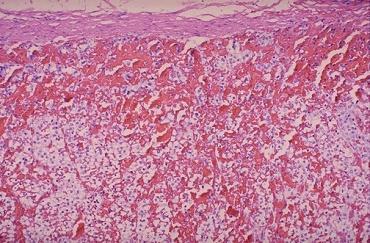
72 Infarction Infarction is usually wedge shape surrounded by rim of hyperemia Necrosis is of coagulative type (except brain: liquifactive) Inflammation within few hours Repair process 72


73 White infarct Red infarct 73

74 Hemorrhagic infarction may be seen in places where some collateral flow can occur, as in the bowel, shown here at autopsy with marked dark red ischemic small bowel. Ordinarily, it is difficult to infarct the small or large bowel, because of an extensive anastomosing blood supply. Typically, severe compromise of at least 2 of the 3 major arterial supplies (celiac trunk, superior mesenteric, inferior mesenteric) is required for infarction to occur. Such arterial compromise is most likely to occur with severe atherosclerosis (often with diabetes mellitus) and with vasculitis (as with classic polyarteritis nodosa).cardiac failure with severe 74 hypotension can produce a similar result.
75 Factors influencing the development of an infarct Nature of the vascular supply Rate of development of the occlusion Vulnerability of the organ to hypoxia Blood oxygen content 75
76 Question Define an infarct. (10 marks) 3.2. List five causes of infarction. (20 marks) 3.3 Describe the factors that determine the development of an infarct.(40 marks) 3.4. Outline with examples, the different mechanisms through which healing occurs in tissue following infraction. (30 marks) 76
77 Where are Red infarcts not typically seen: A. Intestines B. Testes C. Liver D. Lungs E. Kidneys 77
 coagulation(dic) H.M.D.Moratuwagama 78")
78 Disseminated Intravascular Coagulation(DIC) coagulation(dic) H.M.D.Moratuwagama 78
79 DIC-Objectives Define DIC List the causes of DIC Describe the pathogenesis of DIC (DIC & obstetrical and gynaecological conditions) List the investigations to confirm DIC Describe the principles of management of DIC 79
80 Discuss disseminated intra-vascular coagulation (D.I.C) with special reference to different -"obstetrical and gynaecological conditions. 80
81 DIC Acquired bleeding disorder Widespread inappropriate intravascular deposition of fibrin Due to Increased procoagulant material Widespread endothelial damage Platelet aggregation 81
82 DIC -Etiology H/W Severe sepsis Disseminated malignancy Obstetric complications- amniotic fluid embolism / septic abortions/eclampsia/help/placental abruption/iud/pph/aflp Widespread tissue damage-surgery /trauma / burns Incompatible blood transfusions Massive blood loss... 82
83 A case of fatal hemorrhagic diathesis with premature detachment of the placenta. Am J Obstet Gynecol... (Am. J. Obstet. 44; 785, 1901). 83

84 Increased thrombin generation Procoagulant material /consumption 84
85 Three main triggers Endothelial injury Thromboplastin release Phospholipid exposure End result = generation of thrombin with fibrin deposition Many pathologies overlap 85


86 DIC-Bleeding DIC-Thrombosis 86

87 Laboratory findings FBC+BP+retic count Coagulation tests-pt/aptt/tt Fibrinogen FDP/D-dimers 87
88 88
89 Management Treat the underlying condition Blood and blood product support Don't treat the lab reports Thrombosis-?anticoagulation 89

90 Summary-DIC Stimuli Thrombosis Diffuse Bleeding 90

91 91
92 Shock-Objectives Definition of shock Classification of shock Pathophysiology of shock Macroscopy and Microscopy of affected organs 92

93 Definition of shock Systemic hypo perfusion caused by reduction either in CO or in the effective circulating blood volume. 93
94 Physiologic Determinants Global tissue perfusion is determined by: Cardiac output (CO) CO = Heart rate (HR) times Stroke Volume (SV) SV = function of Preload, Afterload, Contractility Systemic vascular resistance (SVR) 94
95 Pathophysiology of shock Inadequate tissue perfusion Decreased oxygen supply Anaerobic metabolism Accumulation metabolic waste Cellular failure 95
96 Types of Shock Cardiogenic Hypovolemic Distributive Sepsis neurogenic (spinal shock) anaphylaxis 96
97 Cardiogenic Shock Intracardiac Arrhythmias Valvular lesions AMI Severe CHF Hypertrophic Cardiomyopathy Extracardiac Pulmonary Embolism Cardiac Tamponade pump failure or SV pump failure or SV 97
98 Hypovolemic Shock Reduced circulating blood volume with secondary decreased cardiac output Acute hemorrhage-ex-pph Vomiting/Diarrhea Dehydration Burns from preload 98
99 Distributive Shock Peripheral Vasodilatation secondary to disruption of cellular metabolism by the effects of inflammatory mediators. Gram negative or other overwhelming infection. Results in decreased Peripheral Vascular Resistance. 99
100 Pathogenesis of Septic shock Inflammatory mediators Endothelial cell activation-thrombosis / increased vascular permiability/vasodilatation Metabolic abnormalities Immunosuppression Organ dysfunction 100

101 101
102 Stages of shock Non progressive Progressive Irreversible 102

103 103
104 Morphology of organs Those of hypoxic injury Any organ Specially in Brain,Heart,Lungs,Kidneys,adrenals,GIT 104
105 Heart Macroscopy: petechial hemorrhages of the epicardiumn, the endocardium, especially the left outflow tract. 105
 The affected fibres stain a deep red with eosin and the nuclei become pyknotic.")
106 Microscopically: necrotic foci in the myocardium, ( loss of single fibres to large areas of necrosis.) The affected fibres stain a deep red with eosin and the nuclei become pyknotic. Prominent contraction bands-better seen by EM 106
 that span the short axis of the")
107 contraction bands are thick intensely eosinophilic staining bands (typically 4-5 micrometres wide) that span the short axis of the myocyte. 107
108 Kidney Acute renal failure- kidney is large, swollen,congested,cortex may be pale. Cross section-blood pooling in the outer strip of the medulla. 108

109 Acute Tubular Necrosis: Gross ; swollen cortex secondary to body burn 109

110 Microscopically: acute tubular necrosis; dilatation of the proximal tubules and focal necrosis of cells. Acute tubular necrosis 110
111 Lung Following the onset of severe and prolonged shock, injury to the alveolar wall results in focal or generalized interstitial pneumonitis (shock lung). The sequence of changes is mediated by acute inflammatory cells and includes interstitial edema, necrosis of endothelial cells, microthrombi, and necrosis of the alveolar epithelium. 111


112 Lung cont. Grossly: lung is firm and congested. Frothy fluid exudes from the cut surface. Normal lung The lungs are large and dusky red. Firm and airless by palpation. 112

113 Lung cont. The frothy liquid oozing from the cut surface of the lung is caused by air moving through water in the respiratory tract. 113
114 Lung cont Shock-induced lung injury leads to the appearance of hyaline membranes in the alveoli, which are frequently expelled into the alveolar ducts and terminal bronchioles. These lung changes may heal entirely, but in half of the patients the repair processes progress and cause a thickening of the alveolar wall. 114


115 115
 from cardiac failure, from marked blood loss, or from loss of blood supply from mechanical obstruction (as with the bowel incarcerated in a")
116 Intestine The small intestinal mucosa demonstrates marked hyperemia as a result of ischemic enteritis. Such ischemia most often results from hypotension (shock) from cardiac failure, from marked blood loss, or from loss of blood supply from mechanical obstruction (as with the bowel incarcerated in a hernia or with volvulus or intussusception). If the blood supply is not quickly restored, the bowel will infarct. 116

117 Intestine cont. Early ischemic enteritis involves the tips of the villi. A colonoscopic view of ischemic colitis with minimal overlying exudate is shown above. Bowel is hard to infarct from atherosclerotic vascular narrowing or thromboembolization because of the widely anastomosing blood supply. Thus, most cases of bowel ischemia and infarction result from generalized hypotension and decreased cardiac output. 117

118 Intestine cont. Mucosal surface of the bowel- early necrosis with hyperemia extending all the way from mucosa to submucosal and muscular wall vessels. The submucosa and muscularis, however, are still intact. 118
119 Liver Macroscopy: liver is heavy and enlarged and has a mottled cut surface that reflects marked centrilobular pooling of blood. 119

120 Liver cont. Microscopy: centrilobular zonal necrosis The cells in the centre of the lobule are the most distant from the blood supply that comes from the portal tracts and are, therefore, presumably more vulnerable to circulatory disturbances. Centri-lobular hepatic necrosis 120

121 Brain lesions are rare. Brain Occasionally, microscopic hemorrhages are seen In severe cases, hemorrhage and necrosis may appear in the overlapping region between the terminal distributions of major arteries; watershed zone.. 121
122 Adrenals In severe shock the adrenal glands may exhibit conspicuous hemorrhage in the inner cortex. Frequently, this hemorrhage is only focal, but it can be massive and accompanied by hemorrhagic necrosis of the entire gland, as seen in the Waterhouse-Friderichsen syndrome. 122

123 Adrenal 123
124 1.1 Describe the pathogenesis of septic shock including its systemic effects. (50 marks) 124

125 THANK YOU THANK YOU 125
Hemodynamic Disorders, Thrombosis, and Shock. Richard A. McPherson, M.D.
 Hemodynamic Disorders, Thrombosis, and Shock Richard A. McPherson, M.D. Edema The accumulation of abnormal amounts of fluid in intercellular spaces of body cavities. Inflammation and release of mediators
Disturbance of Circulation Hemodynamic Disorder
 Disturbance of Circulation Hemodynamic Disorder 2/17/2017 By Dr. Hemn Hassan Othman PhD, Pathology Fall 2016 1 Thrombosis Definition: Thrombosis is the formation of solid or semisolid blood clot within
Circulatory Disturbances 5: Thrombosis, Embolism, Infarction, Shock
 Circulatory Disturbances 5: Thrombosis, Embolism, Infarction, Shock Shannon Martinson, Feb 2016 http://people.upei.ca/smartinson/ VPM 152 General Pathology Thrombosis, Embolism, Infarction, Shock Learning
HEMODYNAMIC DISORDERS
 HEMODYNAMIC DISORDERS Normal fluid homeostasis requires vessel wall integrity as well as maintenance of intravascular pressure and osmolarity within certain physiologic ranges. Increases in vascular volume
Thrombosis and emboli. Peter Nagy
 Thrombosis and emboli Peter Nagy A thrombus is any solid object developing from the blood in vivo within the vascular system or heart. Thrombosis is hemostasis in the wrong place. Major components, forms:
Hemodynamic Disorders Thrombosis and Shock. 1. Interstitial, between the cells, but outside of the vascular system. - water making up the blood and
 Hemodynamic Disorders Thrombosis and Shock I. Body water, where is it and what keeps it there? A. Intracellular B. Extracellular (intercellular) 1. Interstitial, between the cells, but outside of the vascular
Pathophysiology. Tutorial 3 Hemodynamic Disorders
 Pathophysiology Tutorial 3 Hemodynamic Disorders ILOs Recall different causes of thrombosis. Explain different types of embolism and their predisposing factors. Differentiate between hemorrhage types.
THROMBOSIS. Dr. Nisreen Abu Shahin Assistant Professor of Pathology Pathology Department University of Jordan
 THROMBOSIS Dr. Nisreen Abu Shahin Assistant Professor of Pathology Pathology Department University of Jordan NORMAL BLOOD VESSEL HISTOLOGY THROMBOSIS Pathogenesis (called Virchow's triad): 1. Endothelial*
Shock, Hemorrhage and Thrombosis
 Shock, Hemorrhage and Thrombosis 1 Shock Systemic hypoperfusion due to: Reduction in cardiac output Reduction in effective circulating blood volume Hypotension Impaired tissue perfusion Cellular hypoxia
Pathology of pulmonary vascular disease. Dr.Ashraf Abdelfatah Deyab. Assistant Professor of Pathology Faculty of Medicine Almajma ah University
 Pathology of pulmonary vascular disease Dr.Ashraf Abdelfatah Deyab Assistant Professor of Pathology Faculty of Medicine Almajma ah University Pulmonary vascular disease Type of pulmonary circulation: Types
What are blood clots?
 What are blood clots? Dr Matthew Fay GP Principal The Willows Medical Practice- Queensbury GPwSI and Co-Founder Westcliffe Cardiology Service GP Partner Westcliffe Medical Group Created 5/31/18 Dr. Matthew
ATHEROSCLEROSIS. Secondary changes are found in other coats of the vessel wall.
 ATHEROSCLEROSIS Atherosclerosis Atherosclerosis is a disease process affecting the intima of the aorta and large and medium arteries, taking the form of focal thickening or plaques of fibrous tissue and
Thromboembolismand Shock 血管栓塞和休克
 Thromboembolismand Shock 血管栓塞和休克 Major Hemodynamic Disorders Edema Hypermia and Congestion 充血 Haemorrhage Hemostasis 止血 and Blood Coagulation 血液凝固 Thrombosis 血栓形成 Embolism 栓塞 Infarction 梗死 Disseminated
Bachelor of Chinese Medicine Shock
 BCM Year 2 Dr. Irene Ng Jan 28, 2003 9:30 am 1:00 pm Rm 004 UPB Bachelor of Chinese Medicine 2002 2003 Shock Learning objectives Be able to: know the definition of shock know the classification and causes
HYPEREMIA AND CONGESTION
 HYPEREMIA AND CONGESTION Learning Objectives Define congestion and hyperemia Differentiate between the two with regard to: Mechanisms / underlying causes Appearance (gross and histologic) Effects Differentiate
Ischemic heart disease
 Ischemic heart disease Introduction In > 90% of cases: the cause is: reduced coronary blood flow secondary to: obstructive atherosclerotic vascular disease so most of the time it is called: coronary artery
Thrombosis. Dr. László Terézia
 Thrombosis Dr. László Terézia HYPERCOAGULABILITY THROMBOSIS BLOODFLOW ENDOTHEL VIRCHOW ENDOTHEL INJURY L. ventricle: Arteries: surgery infection prosthetic valve hypertension irradiation chemical: cigarette
1- Thromboembolism. 2- fat embolism. 3- air embolism. 4- amniotic fluid embolism.
 Embolism Definition:- An embolus is a detached intravascular solid, liquid or gaseous mass that is carried by blood to sites distant from its point of origin. After traveling via the blood, the embolus
Aneurysms & a Brief Discussion on Embolism
 Aneurysms & a Brief Discussion on Embolism Aneurysms, overview = congenital or acquired dilations of blood vessels or the heart True aneurysms -involve all three layers of the artery (intima, media, and
Hemodynamic Disorders, Thromboembolic Disease, and Shock
 Hemodynamic Disorders, Thromboembolic Disease, and Shock Kumar et al: Robbins & Cotran Pathologic Basis of Disease 7E Figure 4-1 Factors affecting fluid balance across capillary walls. Capillary hydrostatic
Pathophysiology of Cardiovascular System. Dr. Hemn Hassan Othman, PhD
 Pathophysiology of Cardiovascular System Dr. Hemn Hassan Othman, PhD hemn.othman@univsul.edu.iq What is the circulatory system? The circulatory system carries blood and dissolved substances to and from
Chapter 4: Haemodynamic disorders, shock
 Chapter 4: Haemodynamic disorders, shock 1. Regarding platelets (2006) (a) They are the main source of thrombin (b) they number 150-300 x10 3 per microlitre (c) They contain a nucleus (d) They are biconcave
HEME 10 Bleeding Disorders
 HEME 10 Bleeding Disorders When injury occurs, three mechanisms occur Blood vessels Primary hemostasis Secondary hemostasis Diseases of the blood vessels Platelet disorders Thrombocytopenia Functional
HEART HEALTH WEEK 2 SUPPLEMENT. A Beginner s Guide to Cardiovascular Disease ATHEROSCLEROSIS. Fatty deposits can narrow and harden the artery
 WEEK 2 SUPPLEMENT HEART HEALTH A Beginner s Guide to Cardiovascular Disease ATHEROSCLEROSIS FIGURE 1 Atherosclerosis is an inflammatory process where cholesterol is deposited in the wall of arteries and
Thrombosis, Embolism and Infarction
 Thrombosis, Embolism and Infarction THROMBOSIS Thrombus formation (called Virchow's triad): (1) endothelial injury, (2) stasis or turbulent blood flow (3) hypercoagulability of the blood Endothelial Injury
Disseminated intravascular coagulation (DIC) Dr. Klara Vezendi Szeged University Transfusiology Department
 Dr. Klara Vezendi Szeged University Transfusiology Department") Disseminated intravascular coagulation (DIC) Dr. Klara Vezendi Szeged University Transfusiology Department Disseminated intravascular coagulation (DIC, consumptive coagulopathy) is a clinicopathologic
Disseminated intravascular coagulation (DIC) Dr. Klara Vezendi Szeged University Transfusiology Department Disseminated intravascular coagulation (DIC, consumptive coagulopathy) is a clinicopathologic
PHYSIOLOGICAL CHANGES IN PREGNANCY AND OBSTETRIC EMERGENCIES
 PHYSIOLOGICAL CHANGES IN PREGNANCY AND OBSTETRIC EMERGENCIES Shankari Arulkumaran BSc MSc MD MRCOG Consultant Obstetrician and Gynaecologist St Mary s Hospital Imperial College NHS Trust OVERVIEW Obstetric
Haemodynamic Disorders
 Haemodynamic Disorders ZHANG WEI 张伟 Ph.D., A.P. Institute of Pathology & Forensic Medicine Department of Pathology & Patho-physiology Zhejiang University School of Medicine Email:zwei72@zju.edu.cn Thrombosis
SHOCK AETIOLOGY OF SHOCK (1) Inadequate circulating blood volume ) Loss of Autonomic control of the vasculature (3) Impaired cardiac function
 Inadequate circulating blood volume ) Loss of Autonomic control of the vasculature (3) Impaired cardiac function") SHOCK Shock is a condition in which the metabolic needs of the body are not met because of an inadequate cardiac output. If tissue perfusion can be restored in an expeditious fashion, cellular injury may
SHOCK Shock is a condition in which the metabolic needs of the body are not met because of an inadequate cardiac output. If tissue perfusion can be restored in an expeditious fashion, cellular injury may
Part IV Antithrombotics, Anticoagulants and Fibrinolytics
 Part IV Antithrombotics, Anticoagulants and Fibrinolytics "The meaning of good and bad, of better and worse, is simply helping or hurting" Emerson Chapter 16: Blood Coagulation and Fibrinolytic System
Thrombosis. Jeffrey Jhang, M.D.
 Thrombosis Jeffrey Jhang, M.D. Introduction The human hemostatic system has evolved to maintain blood flow under normal physiologic conditions while remaining primed to rapidly respond to vascular injury
Chapter 3 Disorder of Local Blood Circulation
 Chapter 3 Disorder of Local Blood Circulation Disorder of Circulation Disorder of vascular flow may be divided into general and local categories. The local disorders contain: 1Derangement of local blood
Dr. Rami M. Adil Al-Hayali Assistant Professor in Medicine
 Dr. Rami M. Adil Al-Hayali Assistant Professor in Medicine Venous thromboembolism: pulmonary embolism (PE) deep vein thrombosis (DVT) 1% of all patients admitted to hospital 5% of in-hospital mortality
PE and DVT. Dr Anzo William Adiga WatsApp or Call Medical Officer/RHEMA MEDICAL GROUP
 PE and DVT Dr Anzo William Adiga WatsApp or Call +256777363201 Medical Officer/RHEMA MEDICAL GROUP OBJECTIVES DEFINE DVT AND P.E PATHOPHYSIOLOGY OF DVT CLINICAL PRESENTATION OF DVT/PE INVESTIGATE DVT MANAGEMENT
Disseminated Intravascular Coagulation. M.Bahmanpour MD Assistant professor IUMS
 به نام خدا Disseminated Intravascular Coagulation M.Bahmanpour MD Assistant professor IUMS Algorithm for Diagnosis of DIC DIC Score factor score Presence of known underlying disorder No= 0 yes=2 Coagolation
Approach to disseminated intravascular coagulation
 Approach to disseminated intravascular coagulation Khaire Ananta Shankarrao 1, Anil Burley 2, Deshmukh 3 1.MD Scholar, [kayachikitsa] 2.Professor,MD kayachikitsa. 3.Professor and HOD,Kayachikitsa. CSMSS
Chapter 19. Hemostasis
 Chapter 19 Hemostasis Hemostasis Hemostasis is the cessation of bleeding stopping potentially fatal leaks important in small blood vessels not effective in hemorrhage excessive bleeding from large blood
Disseminated Intravascular Coagulation (DIC) Seminar. Ron Kopilov 4 th year Medical Student, Tel Aviv University Internal Medicine A 8.3.
 Seminar. Ron Kopilov 4 th year Medical Student, Tel Aviv University Internal Medicine A 8.3.") Disseminated Intravascular Coagulation (DIC) Seminar Ron Kopilov 4 th year Medical Student, Tel Aviv University Internal Medicine A 8.3.2012 1 Our plan: Understand the pathophysiology Identify risk factors
Disseminated Intravascular Coagulation (DIC) Seminar Ron Kopilov 4 th year Medical Student, Tel Aviv University Internal Medicine A 8.3.2012 1 Our plan: Understand the pathophysiology Identify risk factors
Hemodynamic Disorders Thrombosis and Shock
 Hemodynamic Disorders Thrombosis and Shock SCPA 202 Basic Pathology Somphong Narkpinit, M.D. Department of Pathobiology, Faculty of Science, Mahidol University Email : somphong.nar@mahidol.ac.th Hemodynamic
Physiology of. The Blood hemostasis. By prof. Israa f. jaafar
 Physiology of The Blood hemostasis By prof. Israa f. jaafar Learning objectives Understand the Platelet structure and function Explane the Platelet production Understand the phases of hemostasis: vascular
Intended Learning Outcomes
 2011 Acute Limb Ischemia Definition, Etiology & Pathophysiology Clinical Evaluation Management Ali SABBOUR Prof. of Vascular Surgery, Ain Shams University Acute Limb Ischemia Intended Learning Outcomes
Wheater: Part 1: Thrombosis, embolism and infarction. Laboratory assignment: C601/C602 Histopathology manual, hemodynamic unit.
 Pathology C 601 Hemodynamic Derangements Assignment page. Reading: Robbins: Chapter 4 Clinical Lab Source: - Protime (PT) Know about INR - Activated partial thrmboplastin time (APTT) - Activated coagulation
This slide belongs to iron lecture and it is to clarify the iron cycle in the body and the effect of hypoxia on erythropoitein secretion
 This slide belongs to iron lecture and it is to clarify the iron cycle in the body and the effect of hypoxia on erythropoitein secretion Topics of today lectures: Hemostasis Meaning of hemostasis Mechanisms
Disorder of Local Blood Circulation. Pathology Department, Zhejiang University School of Medicine,
 Disorder of Local Blood Circulation Pathology Department, Zhejiang University School of Medicine, maliqin198@zju.edu.cn Hyperemia Hemorrhage Thrombosis Embolism Infarction Edema Conception: Disorder of
Results of Ischemic Heart Disease
 Ischemic Heart Disease: Angina and Myocardial Infarction Ischemic heart disease; syndromes causing an imbalance between myocardial oxygen demand and supply (inadequate myocardial blood flow) related to
UNIT VI. Chapter 37: Platelets Hemostasis and Blood Coagulation Presented by Dr. Diksha Yadav. Copyright 2011 by Saunders, an imprint of Elsevier Inc.
 UNIT VI Chapter 37: Platelets Hemostasis and Blood Coagulation Presented by Dr. Diksha Yadav Hemostasis: Prevention of Blood Loss Vascular constriction Formation of a platelet plug Formation of a blood
Coagulation Disorders. Dr. Muhammad Shamim Assistant Professor, BMU
 Coagulation Disorders Dr. Muhammad Shamim Assistant Professor, BMU 1 Introduction Local Vs. General Hematoma & Joint bleed Coagulation Skin/Mucosal Petechiae & Purpura PLT wound / surgical bleeding Immediate
Mabel Labrada, MD Miami VA Medical Center
 Mabel Labrada, MD Miami VA Medical Center *1-Treatment for acute DVT with underlying malignancy is for 3 months. *2-Treatment of provoked acute proximal DVT can be stopped after 3months of treatment and
Myocardial Infarction
 Myocardial Infarction MI = heart attack Defined as necrosis of heart muscle resulting from ischemia. A very significant cause of death worldwide. of these deaths, 33% -50% die before they can reach the
Topics of today lectures: Hemostasis
 Topics of today lectures: Hemostasis Meaning of hemostasis Mechanisms of hemostasis - Vascular contraction - Platelets plug - Blood coagulation (clotting) - Structure and functions of platelets - Blood
Maternal Collapse Guideline
 Maternal Collapse Guideline Guideline Number: 664 Supersedes: Classification Clinical Version No: Date of EqIA: Approved by: Date Approved: Date made active: Review Date: 1 Obstetric Written Documentation
1) Severe, crushing substernal chest pain 2) radiate to the neck, jaw, epigastrium, or left arm. 3- rapid and weak pulse 4- nausea (posterior MI).
 Severe, crushing substernal chest pain 2) radiate to the neck, jaw, epigastrium, or left arm. 3- rapid and weak pulse 4- nausea (posterior MI).") 1) Severe, crushing substernal chest pain 2) radiate to the neck, jaw, epigastrium, or left arm. 3- rapid and weak pulse 4- nausea (posterior MI). 5- cardiogenic shock (massive MIs >40% of the left ventricle)
1) Severe, crushing substernal chest pain 2) radiate to the neck, jaw, epigastrium, or left arm. 3- rapid and weak pulse 4- nausea (posterior MI). 5- cardiogenic shock (massive MIs >40% of the left ventricle)
Thursday, February 26, :00 am. Regulation of Coagulation/Disseminated Intravascular Coagulation HEMOSTASIS/THROMBOSIS III
 REGULATION OF COAGULATION Introduction HEMOSTASIS/THROMBOSIS III Regulation of Coagulation/Disseminated Coagulation necessary for maintenance of vascular integrity Enough fibrinogen to clot all vessels
Lung diseases of Vascular Origin. By: Shefaa Qa qqa
 Lung diseases of Vascular Origin By: Shefaa Qa qqa Pulmonary Hypertension Pulmonary hypertension is defined as a mean pulmonary artery pressure greater than or equal to 25 mm Hg at rest. Based on underlying
Moath Darweesh. Omar Sami. Saleem Khreisha. 1 P a g e
 7 Moath Darweesh Omar Sami Saleem Khreisha 1 P a g e -First of all, I want to give a quick revision to simplify the whole hemostasis mechanism, it will be much easier here with me. Enjoy (you can skip
Thrombosis. By Dr. Sara Mohamed Abuelgasim
 Thrombosis By Dr. Sara Mohamed Abuelgasim 1 Thrombosis Unchecked, blood coagulation would lead to dangerous occlusion of blood vessels if the protective mechanisms of coagulation factor inhibitors, blood
The Cardiovascular System. The Structure of Blood Vessels. The Structure of Blood Vessels. The Blood Vessels. Blood Vessel Review
 The Cardiovascular System The Blood Vessels The Structure of Blood Vessels Blood Vessel Review Arteries carry blood away from the heart Pulmonary trunk to lungs Aorta to everything else Microcirculation
Supplemental Digital Content: Definitions Based on the International Classification of Diseases, Ninth Revision, Clinical Modification
 Supplemental Digital Content: Definitions Based on the International Classification of Diseases, Ninth Revision, Clinical Modification (ICD-9-CM) Diagnose and Procedures Codes 1. ICD-9-CM definition of
1. Which of the following blood vessels has a thin elastic layer? A. Aorta. B. Pulmonary artery. C. Posterior vena cava. D. Mesenteric capillary.
 CIRCULATORY SYSTEM 1. Which of the following blood vessels has a thin elastic layer? A. Aorta. B. Pulmonary artery. C. Posterior vena cava. D. Mesenteric capillary. 2. Capillary beds are equipped with
Circulatory shock. Types, Etiology, Pathophysiology. Physiology of Circulation: The Vessels. 600,000 miles of vessels containing 5-6 liters of blood
 Circulatory shock Types, Etiology, Pathophysiology Blagoi Marinov, MD, PhD Pathophysiology Dept. Physiology of Circulation: The Vessels 600,000 miles of vessels containing 5-6 liters of blood Vessel tone
Cover Page. The handle holds various files of this Leiden University dissertation.
 Cover Page The handle http://hdl.handle.net/1887/19768 holds various files of this Leiden University dissertation. Author: Langevelde, Kirsten van Title: Are pulmonary embolism and deep-vein thrombosis
Shock Management. Seyed Tayeb Moradian MSc, Critical Care Nursing Ph.D Candidate. PDF created with pdffactory Pro trial version
 Shock Management Seyed Tayeb Moradian MSc, Critical Care Nursing Ph.D Candidate Definition of Shock The definition of shock does not involve low blood pressure, rapid pulse or cool clammy skin - these
DVT Pathophysiology and Prophylaxis in Medically Hospitalized Patients. David Liff MD Oklahoma Heart Institute Vascular Center
 DVT Pathophysiology and Prophylaxis in Medically Hospitalized Patients David Liff MD Oklahoma Heart Institute Vascular Center Overview Pathophysiology of DVT Epidemiology and risk factors for DVT in the
DIC. Bert Vandewiele Fellow Critical Care 23 May 2011
 DIC Bert Vandewiele Fellow Critical Care 23 May 2011 Dissiminated Intravascular Coagulopathie 11/3/2011 Dr. Bert Vandewiele 2 Dissiminated Intravascular Coagulopathie = Consumption coagulopathie = Defibrination
BUSINESS. Articles? Grades Midterm Review session
 BUSINESS Articles? Grades Midterm Review session REVIEW Cardiac cells Myogenic cells Properties of contractile cells CONDUCTION SYSTEM OF THE HEART Conduction pathway SA node (pacemaker) atrial depolarization
Physiological Response to Hypovolemic Shock Dr Khwaja Mohammed Amir MD Assistant Professor(Physiology) Objectives At the end of the session the
 Objectives At the end of the session the") Physiological Response to Hypovolemic Shock Dr Khwaja Mohammed Amir MD Assistant Professor(Physiology) Objectives At the end of the session the students should be able to: List causes of shock including
Physiological Response to Hypovolemic Shock Dr Khwaja Mohammed Amir MD Assistant Professor(Physiology) Objectives At the end of the session the students should be able to: List causes of shock including
SESSION IV: MECHANISMS OF HUMAN DISEASE: LABORATORY SESSIONS PULMONARY PATHOLOGY I. December 5, 2012
 SESSION IV: MECHANISMS OF HUMAN DISEASE: LABORATORY SESSIONS PULMONARY PATHOLOGY I December 5, 2012 FACULTY COPY GOAL: Describe the basic morphologic and pathophysiologic changes in various conditions
Cardiovascular Disorders. Heart Disorders. Diagnostic Tests for CV Function. Bio 375. Pathophysiology
 Cardiovascular Disorders Bio 375 Pathophysiology Heart Disorders Heart disease is ranked as a major cause of death in the U.S. Common heart diseases include: Congenital heart defects Hypertensive heart
OPEN ACCESS TEXTBOOK OF GENERAL SURGERY
 OPEN ACCESS TEXTBOOK OF GENERAL SURGERY MESENTERIC ISCHAEMIA P Zwanepoel INTRODUCTION Mesenteric ischaemia results from hypoperfusion of the gut, most commonly due to occlusion, thrombosis or vasospasm.
Acquired: Not genetic, produced by influences such as illness, treatment, and lifestyle.
 GLOSSARY OF TERMS A Acquired: Not genetic, produced by influences such as illness, treatment, and lifestyle. Activated partial thromboplastin time (aptt): A test to measure inhibition of coagulation, specifically,
-Cardiogenic: shock state resulting from impairment or failure of myocardium
 Shock chapter Shock -Condition in which tissue perfusion is inadequate to deliver oxygen, nutrients to support vital organs, cellular function -Affects all body systems -Classic signs of early shock: Tachycardia,tachypnea,restlessness,anxiety,
Haemostasis & Coagulation disorders Objectives:
 Haematology Lec. 1 د.ميسم مؤيد علوش Haemostasis & Coagulation disorders Objectives: - Define haemostasis and what are the major components involved in haemostasis? - How to assess the coagulation status?
VENOUS THROMBOEMBOLISM AND CORONARY ARTERY DISEASE: IS THERE A LINK?
 VENOUS THROMBOEMBOLISM AND CORONARY ARTERY DISEASE: IS THERE A LINK? Ayman El-Menyar (1), MD, Hassan Al-Thani (2),MD (1)Clinical Research Consultant, (2) Head of Vascular Surgery, Hamad General Hospital
5 DISTURBANCES IN CIRCULATION. Congestion / Hyperemia Haemorrhage Thrombosis Embolism Ischemia Infarction Oedema Shock Sludged blood Model Questions
 5 DISTURBANCES IN CIRCULATION Congestion / Hyperemia Haemorrhage Thrombosis Embolism Ischemia Infarction Oedema Shock Sludged blood Model Questions CONGESTION/ HYPEREMIA Hyperemia is increased amount of
The Circulatory System. The Heart, Blood Vessels, Blood Types
 The Circulatory System The Heart, Blood Vessels, Blood Types The Closed Circulatory System Humans have a closed circulatory system, typical of all vertebrates, in which blood is confined to vessels and
UPMC Critical Care
 UPMC Critical Care www.ccm.pitt.edu Shock and Monitoring Samuel A. Tisherman, MD, FACS, FCCM Professor Departments of CCM and Surgery University of Pittsburgh Shock Anaerobic metabolism Lactic acidosis
SPECIAL PATHOPHYSIOLOGY SHOCK
 SPECIAL PATHOPHYSIOLOGY SHOCK 1. How do we call blood pressure values below the reference range? 1.Hypovolemia. 2. Hypothermia. 3. Hypooncia. 4. Hypoosmia. 5. Hypotension. 2. What is acute circulatory
Blood Vessels. Dr. Nabila Hamdi MD, PhD
 Blood Vessels Dr. Nabila Hamdi MD, PhD ILOs Understand the structure and function of blood vessels. Discuss the different mechanisms of blood pressure regulation. Compare and contrast the following types
Approach to Thrombosis
 Approach to Thrombosis Theera Ruchutrakool, M.D. Division of Hematology Department of Medicine Siriraj Hospital Faculty of Medicine Mahidol University Approach to Thrombosis Thrombosis: thrombus formation
ACQUIRED COAGULATION ABNORMALITIES
 ACQUIRED COAGULATION ABNORMALITIES ACQUIRED COAGULATION ABNORMALITIES - causes 1. Liver disease 2. Vitamin K deficiency 3. Increased consumption of the clotting factors (disseminated intravascular coagulation
Imaging abdominal vascular emergencies. V.Stoynova
 Imaging abdominal vascular emergencies V.Stoynova Abdominal vessels V. Stoynova 2 Acute liver bleeding trauma anticoagulant therapy liver disease : HCC, adenoma, meta, FNH, Hemangioma Diagnosis :CT angiography
Cardiac Ischemia (is-kē-mē-uh)
") Chapter 21 Cardiac Ischemia (is-kē-mē-uh) By: Alejandra & Lindsay I. Cardiac Ischemia =the most common cause of death in Western Culture ~35% of deaths. -Suddenly from acute coronary occlusion or fibrillation
Chapter 21 Cardiac Ischemia (is-kē-mē-uh) By: Alejandra & Lindsay I. Cardiac Ischemia =the most common cause of death in Western Culture ~35% of deaths. -Suddenly from acute coronary occlusion or fibrillation
BLEEDING DISORDERS Simple complement:
 BLEEDING DISORDERS Simple complement: 1. Select the statement that describe the thrombocytopenia definition: A. Marked decrease of the Von Willebrandt factor B. Absence of antihemophilic factor A C. Disorder
Hemodynamic derangement. Komson Wannasai, M.D.,FRCPath. Department of Pathology Faculty of Medicine Chiang Mai University
 Hemodynamic derangement Komson Wannasai, M.D.,FRCPath. Department of Pathology Faculty of Medicine Chiang Mai University Objective The students should be able to Explain normal body fluid homeostasis Explain
1 Functions of endothelial cells include all the following EXCEPT. 2 Response to vascular injury is characterised by
 airns ase Hospital mergency epartment Part 1 FM MQs 1 Functions of endothelial cells include all the following XPT Formation of von-willebrand factor Formation of collagen and proteoglycans Formation of
Jessica Bryan, Natalia Evans, Karlyn Henderson, & Whitney Parks
 Jessica Bryan, Natalia Evans, Karlyn Henderson, & Whitney Parks 1. What is the most common cause of death in hospitalized patients? 1. Hospital-acquired infection 2. Pulmonary embolism 3. Myocardial infarction
HYPERTENSIVE VASCULAR DISEASE
 HYPERTENSIVE VASCULAR DISEASE Cutoffs in diagnosing hypertension in clinical practice sustained diastolic pressures >90 mm Hg, or sustained systolic pressures >140 mm Hg Malignant hypertension A small
Fourth Practical Pathology. Circulatory disturbances
 Fourth Practical Pathology Circulatory disturbances 12.12.2018 1 Organ: Lung (40X, low power) 1) The blood capillaries within the alveolar septa are engorged with blood 2) Pinkish proteinaceous fluid,
Primary Exam Physiology lecture 5. Haemostasis
 Primary Exam Physiology lecture 5 Haemostasis Haemostasis Body s response for the prevention and cessation of bleeding. Broadly consists of: Primary Haemostasis - vascular spasm and platlet plug formation
Anatomy and Physiology
 Anatomy and Physiology For The First Class 2 nd Semester Thrombocytes = Platelets Thrombocytes = Platelets Blood platelets are non-nucleated disc like cell fragments 2-4 µm in diameter. Platelets are not
Heart disease remains the leading cause of morbidity and mortality in industrialized nations. It accounts for nearly 40% of all deaths in the United
 Heart disease remains the leading cause of morbidity and mortality in industrialized nations. It accounts for nearly 40% of all deaths in the United States, totaling about 750,000 individuals annually
Cardiac Pathology & Rehabilitation
 Cardiac Pathology & Rehabilitation Which of the following best describes the physical activity performed in my leisure time? A. I perform vigorous physical activity 3X/week for 20 minutes each time B.
When the learner has completed this module, she/he will be able to:
 Thrombolytics and Myocardial Infarction WWW.RN.ORG Reviewed September 2017, Expires September 2019 Provider Information and Specifics available on our Website Unauthorized Distribution Prohibited 2017
Acute arterial embolism
 Acute arterial embolism Definition Thrombus come from heart or blood vessel or other embolus such as tumor,air gas or fat flow with blood stream and occlude distal limb or visceral arteries which causes
An aneurysm is a localized abnormal dilation of a blood vessel or the heart Types: 1-"true" aneurysm it involves all three layers of the arterial
 An aneurysm is a localized abnormal dilation of a blood vessel or the heart Types: 1-"true" aneurysm it involves all three layers of the arterial wall (intima, media, and adventitia) or the attenuated
Anticoagulants. Pathological formation of a haemostatic plug Arterial associated with atherosclerosis Venous blood stasis e.g. DVT
 Haemostasis Thrombosis Phases Endogenous anticoagulants Stopping blood loss Pathological formation of a haemostatic plug Arterial associated with atherosclerosis Venous blood stasis e.g. DVT Vascular Platelet
MESENTERIC ISCHEMIA THE FORGOTTEN DIAGNOSIS. Richard M. Gore, MD North Shore University Health System University of Chicago Evanston, Illinois
 MESENTERIC ISCHEMIA THE FORGOTTEN DIAGNOSIS Richard M. Gore, MD North Shore University Health System University of Chicago Evanston, Illinois SCBT/MR 2010 San Diego, California March 8, 2010 16:00-16:10
Disclosures. DVT: Diagnosis and Treatment. Questions To Ask. Dr. Susanna Shin - DVT: Diagnosis and Treatment. Acute Venous Thromboembolism (VTE) None
 None") Disclosures DVT: Diagnosis and Treatment None Susanna Shin, MD, FACS Assistant Professor University of Washington Acute Venous Thromboembolism (VTE) Deep Venous Thrombosis (DVT) Pulmonary Embolism (PE)
Disclosures DVT: Diagnosis and Treatment None Susanna Shin, MD, FACS Assistant Professor University of Washington Acute Venous Thromboembolism (VTE) Deep Venous Thrombosis (DVT) Pulmonary Embolism (PE)