Chapter 3 Disorder of Local Blood Circulation
|
|
|
- Archibald Bennett
- 5 years ago
- Views:
Transcription
1 Chapter 3 Disorder of Local Blood Circulation
2 Disorder of Circulation Disorder of vascular flow may be divided into general and local categories. The local disorders contain: 1Derangement of local blood volume: hyperemia and ischemia; 2Derangement of blood properties and content: thrombosis, embolism and infarction; 3Derangements of vascular permeability and anatomic integrity: edema, hemorrhage.

3 hyperemia and ischemia
4 Section 1 Hyperemia Local increased volume of blood caused by dilatation of the small vessels. 1. Arterial hyperemia (active hyperemia) It results from an augmented arterial inflow. Examples: the muscles during exercise, at sites of inflammation, and in the pleasing neurovascular dilatation termed blushing. Active hyperemia causes increased metabolism and function of the organ. It is beneficial to the organism. Active hyperemia of body surface shows red in color and increase in local temperature.
5 Hyperemia 2. Venous Hyperemia (passive hyperemia, congestion) It results from diminished venous drainage such as follows cardiac failure or obstructive venous disease. Also called venous hyperemia. When body surface is involved, the skin show red-blue color owing to deoxgenation of impounded red cells cyanosis. Etiology (1) Compression on vein (2) Intravenous obstruction (3) Cardiac failure Morphology Congested organ increases in size and weight, and shows dark-red color.
6 Congestion The consequences of congestion: 1. Congestive edema and hemorrhage. Congestion of capillary beds is closely related to the development of edema. If the edematous fluids contain lots of red bloods cells, is referred to congestive hemorrhage. 2. Congestive sclerosis. Chronic passive congestion may lead to hypoxic atrophy or degeneration or death of parenchymal cells, followed by proliferation of fibrous tissue. 3. Atrophy of parenchymal cells. 4. Formation collateral blood circulation. Examples: collateral circulation formed in cirrhosis.

7 Pulmonary edema edematous fluid

8 Congestive sclerosis of the live

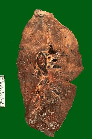
9 Congestion of Main Organs (1) Pulmonary Congestion Chronic lungs congestion: congestion and edema seen mainly in left ventricular failure. Normal Congestion Fibrosis of alveolar septa

10 Acute Pulmonary Congestion and edema pink edema fluid
11 Chronic pulmonary congestion Chronic pulmonary congestion G: The lung is harder in consistency, dark brown in gross view, which is referred to brown induration of the lung. H: The alveolar septa have become thickened and fibrotic, and the alveolar spaces contain hemosiderin or heart failure cell ( hemosiderinladen macrophage). Hemosiderin-laden macrophage appear in alveolar space in chronic (left) heart failure, is called heart failure cell


12 Chronic pulmonary congestion G: brown induration of the lung. M: the septa have become thickened and fibrotic, and the alveolar spaces contain hemosiderin, heart failure cells can be seen.

13 Chronic pulmonary congestion
14 (2) Chronic congestion of liver It is caused by chronic right heart failure. The central hepatocytes degenerated; the periportal hepatocytes, better oxygenated because of their proximity to hepatic arterioles, experience less severe hypoxia and may only develop fatty change. G: yellow stripes alternated with red stripes on the section surface of the liver (nutmeg liver). H: red blood cell filled in dilated hepatic sinusoid and central vein, some hepatocytes were atrophied and fatty changed.
.")

15 G: yellow stripes alternated with red stripes on the section surface of the liver (nutmeg liver). H: red blood cell filled in dilated hepatic sinusoid and central vein, some hepatocytes were atrophied and fatty changed. Chronic congestion of liver

16 Congestion of liver Sinusoid dilated and filled red blood cells
17 Section 2 Hemorrhage Extravasation of blood caused by rupture of blood vessel. Hematoma: Accumulation of large amount of blood within the tissue of the body. Hemothorax, hemopericardium, hemoperitoneum, hemarthrosis: blood accumulation in body cavities as thoracic cavity, pericardial cavity, peritoneal cavity and arthritic cavity respectively. Petechiae: Minute hemorrhage into skin, mucous membrane or serosal surface. Purpura: Slight larger hemorrhage. Ecchymosis: A large subcutaneous hematoma.


18 Cerebral Hemorrhage It is a common complication of hypertension Petechia & Ecchymosis
19 Section 3 Thrombosis Thrombosis refers to the process of formation of an adherent clotted mass of blood within the cardiovascular system in a living body. The clotted mass is thrombus. *Three required factors for thrombosis: 1. endothelial injury; 2. alterations in normal blood flow; 3. blood hypercoagulability
20 Thrombosis Normal hemostasis depends on balance between coagulative and procoagulant system or activation and inactivation of clotting factors. Three key contributors to hemostasis : 1. The vascular wall, particularly endothelium and underlying collogen. 2. Platelets. 3. Clotting factors.
21 Factors for thrombosis (1) Endothelial injury On the one hand, endothelial cells possess antiplatelet, anticoagulant, and fibrinolytic properties; On the other hand, they exert procogulant functions. Intact endothelium insulates the blood platelets and coagulation proteins from the highly thrombogenic subendothelial components, principally collagen. Damaged endothelium has a dominant influence to thrombogenesis, and the only one factor may lead to thrombosis. Examples: ulcerated atherosclerotic plaques; vascular traumatic or inflammatory injury; endocardium in the site of myocardial infarction or myocarditis.

22 Favor & inhibit thrombosis

23 Coagulation cascade (HMWK: high molecular-weight kininogin )
24 (2) Alteration of blood flow (stasis and turbulence) In normal laminar blood flow, the formed elements are separated from the endothelial surface by plasmatic zone. Normal laminar blood flow disrupt laminar flow in stasis and turbulence conditions. 1disrupt laminar flow permit platelets to contact with the endothelium; 2prevent dilution of activated clotting factors; Factors for thrombosis 3 the turbulence is a injury factors for endotheliuml 4 promote endothelial cell hypoxia and injury, predisposing to platelet and fibrin deposition as well as reducing release of t-pa; Examples: thrombus are likely formed in vein than in artery.
25 (3) Hypercoagulability An alteration of the blood or, specifically, the clotting mechanism that in some way predisposes to thrombosis. Hypercoagulability can be seen in many clinical settings such as severe trauma, burns, disseminated cancer, long term use of oral contraceptives.
26 Morphogenesis of thrombi 1 Endotbelial injury platelets adhesion, release reaction and aggregation. Platelets activation release reaction (ADP, TxA2, Ca++) platelet aggregation. 2 Intrinsic and extrinsic coagulation sequence: prothrombin thrombin fibrinogen fibrin


27
28 Types of thrombi 1. pale thrombus: dry, friable gray mass transection: darker gray lines of platelets (Zahn s line) H. Platelet trabeculae formation (head). vegatation, arterial thrombus, mural thrombi 2. mixed thrombus: white thrombus intermingled with red thrombus (body). Venous thrombus, globular thrombus in aneurysms 3. red thrombus : dark red in color, laminations are not well developed and composed of rbc and fibrin (tail). Venous thrombus 4. Hyaline thrombus: DIC(microthrombus)
29 Types of thrombi Components of thrombi: Pale thrombus: plt Mixed thrombus: plt.+rbc.+wbc+ fibrin Red thrombus: Rbc+fibrin Hyaline thrombus: fibrin

30 WBC Plt trabeculae RBC Mixed thrombus: plt.+rbc.+wbc+ fibrin
31 Classification according morphology or relationship to walls of vessel or heart: 1Mural thrombi: arterial thrombi arise in heart chambers or in the aortic lumen, they are usually adhere to the wall. 2Ball thrombi: thrombus at atrium or ball-shaped. 3Occlusive thrombus: Thrombus block the vescular lumen. 4Vegetations: Thrombus at cardiac valve. Types of thrombi

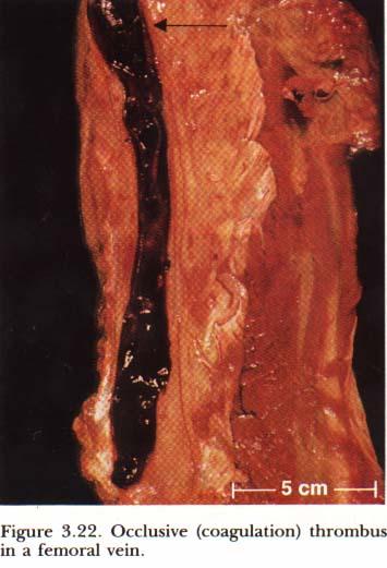
32 Mural & Occlusive thrombus


33 Vegetations

34 Coronary artery thrombi forming with hemorrhage

35 Hyaline thrombus
36 Outcome of thrombus 4. The outcome of thrombus (1) Softening, resolution and absorption: due to the fibrinolysis, small thrombi may be completely absorpted; large thrombi may be sources of emboli. (2) Organization and recanalization. Thrombi replaced by granulation tissue; The blocked vessel lumen be reconnected. (3) Calcification: leading formation of phlebolith, arteriolith.


37 thrombi cap. wall of vessel Organization & Recanalization
38 Microcirculatory Thrombosis Disseminated intravascular coagulation (DIC) DIC is the sudden or insidious onset of widespread fibrin thrombi in the microcirculation, and subsequent hemorrhage due to depletion of coagulatic factors. Etiology and mechanism: it is a complication of some diseases (trauma, severe infection, shock, carcinoma etc.) which activate the blood clotting processes. The simplest mechanism involves the release of tissue factor into the circulation (obstetric complication. carcinomas). Morphology: The microthrombi are composed of fibrin, and found in the arterioles and capillaries of the kidney, adrenals, brain and heart.

39 Microcirculatory Thrombosis
40 The influence of thrombus Advantages: hemostasis in injurious vessel. Disadvantages: (1) obstruction of arteries and veins. (2) sources of emboli. (3) heart valves malformation. (4) hemorrhage: DIC also called as consumption coagulopathy.
41 Section 4 Embolism Embolism refers to occlusion of some part of the cardiovascular system by any abnormal mass carried there in the blood stream. The transported mass is embolus. It is a detached intravascular solid, liquid, or gaseous mass.
42 1. Routes of embolus transportation (1) Venous embolus --- pulmonary embolization and infarction (2) Arteria embolus --- the important organs of body, such as coronary, cerebal, liver and kidney artery. (3) Digestive system embolus--- intrahepatic portal vein embolus embolism. (4) Venous embolus from right heart to left heart cause arteria system embolism, known as crossed embolism. (5) Large venous embolism may travel against the direction of blood flow to obstruct small vein known as retrograde emblism. The pathways of embolism
43 (1) Thromboembolism 2. Types of embolism 1Pulmonary embolism most commonly originates from thrombus of lower limb. The prognosis depends on the size of the occluded vessel and the status of the patient s cardiovascular system. * Large embolus: fatal. (sudden death) Mechanical obstruction + severe resistant pressue on the right heart (acute cor pulmonale) + reflexive vascular and bronchial spasm. Small emboli obstruct small branches of pulmonary artery: The thrombi can be resolved and so of little significance. If bronchial circulation is insufficiency (chronic passive congestion of lung), small emboli may produce either pulmonary hemorrhagic infarction.

44 pulmonary embolization
45 2 Systemic Embolism Emboli traveling within the arterial circulation. Thromboembolism Emboli usually arise from mural thrombi in the left ventricle or atrium, from vegetations on the left side valves, and occasionally from thrombi on the aorta (atherosclerotic plaque). They produce occlusion of blood stream most frequently in the lower extremities, brain, intestine, spleen, and the kidneys, often result to infarcts or gangrene. The consequences of systemic emboli depend on the extent of collateral vascular supply in the affected tissue, the tissue s vulnerability to ischemia, and the caliber of the vessel occluded.
46 (2) Fat embolism Fat droplets appear in the circulation and obstruct small vessel. The causes include: fracture of long bone, severe soft tissue trauma and burns. Although traumatic fat embolism can be demonstrated anatomically in some 90% of individuals with severe bone injury, only 1% of such patients show clinical findings. Fat embolism syndrome is characterized by pulmonary insufficiency, and is fatal in about 10% of cases. Typically, the symptoms appear 1 to 3 days after injury, with sudden onset of tachypnea, dyspnea, and tachycardia. Sequences: The prognosis of fat embolism depends on the quantity and site of embolism.


47 Fat embolism in lung Alveolar septal capillaries and small arteries are full of dark red stained fat microglobule (Sudan stain )
48 Fat embolism 1 Pulmonary fat embolism 9~20g fat 75% pulmonary arteries are occluded pulmonary edema, hemorrhage, atelectasis asphyxia, right sided heart failure. 2 Lung-brain-kidney-skin embolism syndrome <20μm fat droplets may enter systemic circulation and result in embolism. Neurologic symptoms, skin ecchymosis may appear.,
49 (3) Gas embolism Embolism Gas bubbles within the circulation obstruct blood flow. 1Air embolism As a complication of trauma (chest), cardio-thoracic surgery, various diagnostic or therapeutic procedures, or rupture of veins during delivery. Sequences: Small volume: absorbed >100ml: acute distress. >300ml: fatal due to pulmonary embolism.
50 Gas embolism 2 Decompression sickness (cassion disease) Decompression sickness occurs when individuals encounter sudden changes in atmospheric pressure. When decompressed rapidly from higher atmospheric pressure to a lower one, dissolved gas may dissociate and bubble out of the blood, tissue fluid, and fat. The nitrogen is of low solubility and persists as minute bubbles to obstruct the blood vessel. Examples: in deep sea divers, in underwater construction workers, in aviators Cassion disease may cause ache of muscle, articulation or infarction.

51 Pulmonary air embolism
52 (4)Amniotic fluid embolism Embolism A grave but fortunately uncommon complication of labor and the immediate postpartum period (1 per 50,000 deliveries). Etiology and mechanism: After rupture of membranes, some of the amniotic fluid may be forced to maternal circulation by vigorous uterine contractions via teared uterine sinusoids. It is now suspected that vasoactive substances within the amniotic fluid such as prostaglandins may be the cause of pulmonary vasoconstriction. Morphology: The classic findings in the pulmonary arteries and capillaries at autopsy are epithelial squames from fetal skin, lanugo hairs, fat from vernix caseosa, and mucin, presumed to be from the fetal GIT.

53 Amniotic fluid embolism Epithelial squames in myocarcial vessels

54 (5) Other types of embolism Embolism Tumor cells, parasites and cholesterol crystal may cause embolism Tumor cells embolus
55 Section 5 Infarction Ischemic necrosis caused by occlusion of either its arterial supply or its venous drainage. The process of infarct formation is called as infarction. 1. Causes and factors that determine the development of infarct (1)Nature of vascular supply. 1Effective anastomosis 2Double blood supply (liver, lung etc) (2)Rate of development of occlusion (3)Vulnerability of the organ or tissue to hypoxia (nerve cells, myocardial muscle>fibroblasts) (4)Oxygen carrying capacity of blood: The anemic or cyanotic patient tolerates arterial insufficiency less well than normal person.
56 2. Types of infarct (1) White infarct (anemic infarct): It result from arterial occlusion, commonly seen in compact, solid organs with less collateral circulation (kidneys, spleen and heart). The solidity of the tissue limits the amount of hemorrhage that can seep into the area of ischemic necrosis from adjoining capillary beds. (2) Red infarct (hemorrhagic infarct): Red infarct of intestine: Limited venous outflow due to vascular obstruction + by pass channels cannot develop. It occurs in: 1) loose tissues (lung) that allow blood to accumulate in the infarcted zone; 2) tissues with dual circulations ( lung and small intestine), permitting flow of blood from the unobstructed vascular channel into the necrotic area); (3) Septic infarct: infarct caused by septic emboli.


57 Anemic infarct

58 Myocardial infarct

59 Anemic infarct of kidney Hyperemic and hemorrhagic zone on the border ( )

60 Anemic infarct of spleen
61 Infarct 3. Morphology of infarct G: All infarcts (red and white), tend to be wedgeshaped, the occluded vessel at the apex and the periphery of the organ forming the base. H. Necrotic tissue surrounding with hyperemic and hemorrhagic zone. Compact organs have relative little hemorrhage, the necrotic area is gradually decolorized (rbc are lysed and removed from the area), and appears pale or yellow-white (white infarct). Organs of soft, loose tissue with double supply, infarcts tend to remain hemorrhagic (Red infarct) and show dark red colour.
 of")
62 Hemorrhagic (Red) infarct) of intestine


63 Hemorrhagic infarct of the intestine Pulmonary hemorrhagic infarct
64 4. Fate of infarct (1) Small infarct: softened, absorbed (2) Large infarct: organization and scar formation (3) Septic infarct: abscess formation (4) Infarction of brain, myocardium or lung: sometimes may cause sudden death.
65 Edema Pitting edema Pulmonary edema: Occur in left heart failure, ARDS, allergy, etc. Cerebral edema: may cause hernia cerebri.
Disorder of Local Blood Circulation. Pathology Department, Zhejiang University School of Medicine,
 Disorder of Local Blood Circulation Pathology Department, Zhejiang University School of Medicine, maliqin198@zju.edu.cn Hyperemia Hemorrhage Thrombosis Embolism Infarction Edema Conception: Disorder of
Hemodynamic Disorders, Thrombosis, and Shock. Richard A. McPherson, M.D.
 Hemodynamic Disorders, Thrombosis, and Shock Richard A. McPherson, M.D. Edema The accumulation of abnormal amounts of fluid in intercellular spaces of body cavities. Inflammation and release of mediators
HEMODYNAMIC DISORDERS
 HEMODYNAMIC DISORDERS Normal fluid homeostasis requires vessel wall integrity as well as maintenance of intravascular pressure and osmolarity within certain physiologic ranges. Increases in vascular volume
Disturbance of Circulation Hemodynamic Disorder
 Disturbance of Circulation Hemodynamic Disorder 2/17/2017 By Dr. Hemn Hassan Othman PhD, Pathology Fall 2016 1 Thrombosis Definition: Thrombosis is the formation of solid or semisolid blood clot within
Haemodynamic Disorders
 Haemodynamic Disorders ZHANG WEI 张伟 Ph.D., A.P. Institute of Pathology & Forensic Medicine Department of Pathology & Patho-physiology Zhejiang University School of Medicine Email:zwei72@zju.edu.cn Thrombosis
Thrombosis and emboli. Peter Nagy
 Thrombosis and emboli Peter Nagy A thrombus is any solid object developing from the blood in vivo within the vascular system or heart. Thrombosis is hemostasis in the wrong place. Major components, forms:
Circulatory Disturbances 5: Thrombosis, Embolism, Infarction, Shock
 Circulatory Disturbances 5: Thrombosis, Embolism, Infarction, Shock Shannon Martinson, Feb 2016 http://people.upei.ca/smartinson/ VPM 152 General Pathology Thrombosis, Embolism, Infarction, Shock Learning
ATHEROSCLEROSIS. Secondary changes are found in other coats of the vessel wall.
 ATHEROSCLEROSIS Atherosclerosis Atherosclerosis is a disease process affecting the intima of the aorta and large and medium arteries, taking the form of focal thickening or plaques of fibrous tissue and
Hemodynamic Disorders Thrombosis and Shock. 1. Interstitial, between the cells, but outside of the vascular system. - water making up the blood and
 Hemodynamic Disorders Thrombosis and Shock I. Body water, where is it and what keeps it there? A. Intracellular B. Extracellular (intercellular) 1. Interstitial, between the cells, but outside of the vascular
HYPEREMIA AND CONGESTION
 HYPEREMIA AND CONGESTION Learning Objectives Define congestion and hyperemia Differentiate between the two with regard to: Mechanisms / underlying causes Appearance (gross and histologic) Effects Differentiate
Pathology of pulmonary vascular disease. Dr.Ashraf Abdelfatah Deyab. Assistant Professor of Pathology Faculty of Medicine Almajma ah University
 Pathology of pulmonary vascular disease Dr.Ashraf Abdelfatah Deyab Assistant Professor of Pathology Faculty of Medicine Almajma ah University Pulmonary vascular disease Type of pulmonary circulation: Types
Pathophysiology. Tutorial 3 Hemodynamic Disorders
 Pathophysiology Tutorial 3 Hemodynamic Disorders ILOs Recall different causes of thrombosis. Explain different types of embolism and their predisposing factors. Differentiate between hemorrhage types.
Hemodynamic Disorders Thrombosis and Shock
 Hemodynamic Disorders Thrombosis and Shock SCPA 202 Basic Pathology Somphong Narkpinit, M.D. Department of Pathobiology, Faculty of Science, Mahidol University Email : somphong.nar@mahidol.ac.th Hemodynamic
Aneurysms & a Brief Discussion on Embolism
 Aneurysms & a Brief Discussion on Embolism Aneurysms, overview = congenital or acquired dilations of blood vessels or the heart True aneurysms -involve all three layers of the artery (intima, media, and
5 DISTURBANCES IN CIRCULATION. Congestion / Hyperemia Haemorrhage Thrombosis Embolism Ischemia Infarction Oedema Shock Sludged blood Model Questions
 5 DISTURBANCES IN CIRCULATION Congestion / Hyperemia Haemorrhage Thrombosis Embolism Ischemia Infarction Oedema Shock Sludged blood Model Questions CONGESTION/ HYPEREMIA Hyperemia is increased amount of
Pathophysiology of Cardiovascular System. Dr. Hemn Hassan Othman, PhD
 Pathophysiology of Cardiovascular System Dr. Hemn Hassan Othman, PhD hemn.othman@univsul.edu.iq What is the circulatory system? The circulatory system carries blood and dissolved substances to and from
Hemodynamic derangement. Komson Wannasai, M.D.,FRCPath. Department of Pathology Faculty of Medicine Chiang Mai University
 Hemodynamic derangement Komson Wannasai, M.D.,FRCPath. Department of Pathology Faculty of Medicine Chiang Mai University Objective The students should be able to Explain normal body fluid homeostasis Explain
Fourth Practical Pathology. Circulatory disturbances
 Fourth Practical Pathology Circulatory disturbances 12.12.2018 1 Organ: Lung (40X, low power) 1) The blood capillaries within the alveolar septa are engorged with blood 2) Pinkish proteinaceous fluid,
Shock, Hemorrhage and Thrombosis
 Shock, Hemorrhage and Thrombosis 1 Shock Systemic hypoperfusion due to: Reduction in cardiac output Reduction in effective circulating blood volume Hypotension Impaired tissue perfusion Cellular hypoxia
According to the etiology, edema may be:
 What is edema? Edema : It refers to the accumulation of excess liquid in the interstitial (extracellular) spaces of a tissue or in pre-existing cavities. It may affect any organ, but most often it appears
THROMBOSIS. Dr. Nisreen Abu Shahin Assistant Professor of Pathology Pathology Department University of Jordan
 THROMBOSIS Dr. Nisreen Abu Shahin Assistant Professor of Pathology Pathology Department University of Jordan NORMAL BLOOD VESSEL HISTOLOGY THROMBOSIS Pathogenesis (called Virchow's triad): 1. Endothelial*
Ischemic heart disease
 Ischemic heart disease Introduction In > 90% of cases: the cause is: reduced coronary blood flow secondary to: obstructive atherosclerotic vascular disease so most of the time it is called: coronary artery
1- Thromboembolism. 2- fat embolism. 3- air embolism. 4- amniotic fluid embolism.
 Embolism Definition:- An embolus is a detached intravascular solid, liquid or gaseous mass that is carried by blood to sites distant from its point of origin. After traveling via the blood, the embolus
Hemodynamic Disorders, Thromboembolic Disease, and Shock
 Hemodynamic Disorders, Thromboembolic Disease, and Shock Kumar et al: Robbins & Cotran Pathologic Basis of Disease 7E Figure 4-1 Factors affecting fluid balance across capillary walls. Capillary hydrostatic
What are blood clots?
 What are blood clots? Dr Matthew Fay GP Principal The Willows Medical Practice- Queensbury GPwSI and Co-Founder Westcliffe Cardiology Service GP Partner Westcliffe Medical Group Created 5/31/18 Dr. Matthew
Cardiac Ischemia (is-kē-mē-uh)
") Chapter 21 Cardiac Ischemia (is-kē-mē-uh) By: Alejandra & Lindsay I. Cardiac Ischemia =the most common cause of death in Western Culture ~35% of deaths. -Suddenly from acute coronary occlusion or fibrillation
Chapter 21 Cardiac Ischemia (is-kē-mē-uh) By: Alejandra & Lindsay I. Cardiac Ischemia =the most common cause of death in Western Culture ~35% of deaths. -Suddenly from acute coronary occlusion or fibrillation
Thromboembolismand Shock 血管栓塞和休克
 Thromboembolismand Shock 血管栓塞和休克 Major Hemodynamic Disorders Edema Hypermia and Congestion 充血 Haemorrhage Hemostasis 止血 and Blood Coagulation 血液凝固 Thrombosis 血栓形成 Embolism 栓塞 Infarction 梗死 Disseminated
Thrombosis, Embolism and Infarction
 Thrombosis, Embolism and Infarction THROMBOSIS Thrombus formation (called Virchow's triad): (1) endothelial injury, (2) stasis or turbulent blood flow (3) hypercoagulability of the blood Endothelial Injury
Ischaemia It means local anemia, it is characterized by a decrease amount of blood in an organ or region. Causes of Ischemia: *1.
 المرحلة الثالثة م. هالة عباس ناجي Ischaemia It means local anemia, it is characterized by a decrease amount of blood in an organ or region. Causes of Ischemia: *1.External pressure upon an artery e.g:
Lung diseases of Vascular Origin. By: Shefaa Qa qqa
 Lung diseases of Vascular Origin By: Shefaa Qa qqa Pulmonary Hypertension Pulmonary hypertension is defined as a mean pulmonary artery pressure greater than or equal to 25 mm Hg at rest. Based on underlying
Circulatory disorders
 Circulatory disorders Edema = fluid in interstitium transudate s.w.1.012, exudate 1.020) generalized x local prominent cavities hydrothorax, hydropericardium, ascites subcutaneous tissue (pitting edema)
Myocardial Infarction
 Myocardial Infarction MI = heart attack Defined as necrosis of heart muscle resulting from ischemia. A very significant cause of death worldwide. of these deaths, 33% -50% die before they can reach the
Thrombosis. Dr. László Terézia
 Thrombosis Dr. László Terézia HYPERCOAGULABILITY THROMBOSIS BLOODFLOW ENDOTHEL VIRCHOW ENDOTHEL INJURY L. ventricle: Arteries: surgery infection prosthetic valve hypertension irradiation chemical: cigarette
Thrombosis, Embolism, and Infarction
 Topic 6: Introduction Thrombosis Formation of Thrombi Causes of Thrombosis Events Occurring in Thrombosis The Gross and Microscopic Appearance of Thrombi Embolism Composition of Emboli Thrombotic Emboli
General Pathology. Hemorrhage (Web)
") General Pathology Hemorrhage (Web) Paul Hanna Feb 2015 Hemorrhage escape of blood from the cardiovascular system may be external or internal Hemorrhage Causes Trauma Sepsis, viruses or toxins Coagulation
General Pathology Hemorrhage (Web) Paul Hanna Feb 2015 Hemorrhage escape of blood from the cardiovascular system may be external or internal Hemorrhage Causes Trauma Sepsis, viruses or toxins Coagulation
Wheater: Part 1: Thrombosis, embolism and infarction. Laboratory assignment: C601/C602 Histopathology manual, hemodynamic unit.
 Pathology C 601 Hemodynamic Derangements Assignment page. Reading: Robbins: Chapter 4 Clinical Lab Source: - Protime (PT) Know about INR - Activated partial thrmboplastin time (APTT) - Activated coagulation
HEME 10 Bleeding Disorders
 HEME 10 Bleeding Disorders When injury occurs, three mechanisms occur Blood vessels Primary hemostasis Secondary hemostasis Diseases of the blood vessels Platelet disorders Thrombocytopenia Functional
HEART HEALTH WEEK 2 SUPPLEMENT. A Beginner s Guide to Cardiovascular Disease ATHEROSCLEROSIS. Fatty deposits can narrow and harden the artery
 WEEK 2 SUPPLEMENT HEART HEALTH A Beginner s Guide to Cardiovascular Disease ATHEROSCLEROSIS FIGURE 1 Atherosclerosis is an inflammatory process where cholesterol is deposited in the wall of arteries and
Chapter 4: Haemodynamic disorders, shock
 Chapter 4: Haemodynamic disorders, shock 1. Regarding platelets (2006) (a) They are the main source of thrombin (b) they number 150-300 x10 3 per microlitre (c) They contain a nucleus (d) They are biconcave
Blood Vessels. Dr. Nabila Hamdi MD, PhD
 Blood Vessels Dr. Nabila Hamdi MD, PhD ILOs Understand the structure and function of blood vessels. Discuss the different mechanisms of blood pressure regulation. Compare and contrast the following types
1) Severe, crushing substernal chest pain 2) radiate to the neck, jaw, epigastrium, or left arm. 3- rapid and weak pulse 4- nausea (posterior MI).
 Severe, crushing substernal chest pain 2) radiate to the neck, jaw, epigastrium, or left arm. 3- rapid and weak pulse 4- nausea (posterior MI).") 1) Severe, crushing substernal chest pain 2) radiate to the neck, jaw, epigastrium, or left arm. 3- rapid and weak pulse 4- nausea (posterior MI). 5- cardiogenic shock (massive MIs >40% of the left ventricle)
1) Severe, crushing substernal chest pain 2) radiate to the neck, jaw, epigastrium, or left arm. 3- rapid and weak pulse 4- nausea (posterior MI). 5- cardiogenic shock (massive MIs >40% of the left ventricle)
Thrombosis (lec#1) * Pathogenesis (called Virchow's triad ): Endothelial Injury Stasis Blood Hypercoagulability. stimulated Stasis:
 * Pathogenesis (called Virchow's triad ): Endothelial Injury Stasis Blood Hypercoagulability. stimulated Stasis:") Thrombosis (lec#1) * Pathogenesis (called Virchow's triad): Endothelial Injury ( Heart, Arteries), Stasis (abnormal blood flow),blood Hypercoagulability. * Endothelial cells can be stimulated by direct
Thrombosis (lec#1) * Pathogenesis (called Virchow's triad): Endothelial Injury ( Heart, Arteries), Stasis (abnormal blood flow),blood Hypercoagulability. * Endothelial cells can be stimulated by direct
Bachelor of Chinese Medicine Shock
 BCM Year 2 Dr. Irene Ng Jan 28, 2003 9:30 am 1:00 pm Rm 004 UPB Bachelor of Chinese Medicine 2002 2003 Shock Learning objectives Be able to: know the definition of shock know the classification and causes
Pathology of blood circulation. (Edema, hyperemia, ischemia, hemorrhage, thrombosis, embolism).
.") Lecture # 4 General medicine Faculty Pathology of blood circulation. (Edema, hyperemia, ischemia, hemorrhage, thrombosis, embolism). Prepared by: Associate Professor, Ph.D. R. Deev Kazan, 2018 M.Mavlikeev.
Lecture # 4 General medicine Faculty Pathology of blood circulation. (Edema, hyperemia, ischemia, hemorrhage, thrombosis, embolism). Prepared by: Associate Professor, Ph.D. R. Deev Kazan, 2018 M.Mavlikeev.
THE BLOOD VESSELS. Manar hajeer, MD University of Jordan Faculty of medicine, pathology department.
 THE BLOOD VESSELS Manar hajeer, MD University of Jordan Faculty of medicine, pathology department. Vascular pathology: 1- Narrowing or complete obstruction of vessel lumina, either progressively (e.g.,
Coagulative Necrosis of Myocardium. Dr Rodney Itaki Division of Pathology
 Coagulative Necrosis of Myocardium Dr Rodney Itaki Division of Pathology Coagulative Necrosis Gross pathology: 3 day old infarct: Yellow necrosis surrounded by hyperemic borders. Arrow points to a transmural
Disturbances of Circulation, Lab 1: Edema and Congestion/Hyperemia. Shannon Martinson, Feb
 Disturbances of Circulation, Lab 1: Edema and Congestion/Hyperemia Shannon Martinson, Feb 2017 http://people.upei.ca/smartinson/ Case #1 Signalment and History: 6-month old feeder lamb found dead on pasture
The Circulatory System
 The Circulatory System Dr. Sami Zaqout The circulatory system Circulatory system Blood vascular systems Lymphatic vascular systems Blood vascular systems Blood vascular systems The circulatory system Circulatory
MI Acute occlusion of the proximal left anterior descending (LAD) artery is the cause of 40% to 50% of all MIs. *
 artery is the cause of 40% to 50% of all MIs. *") MI *33% -50% die before hospital lethal arrhythmia Sudden Cardiac Death. * Arrhythmias are caused by electrical abnormalities of the ischemic myocardium and conduction system. *Acute occlusion of the proximal
MI *33% -50% die before hospital lethal arrhythmia Sudden Cardiac Death. * Arrhythmias are caused by electrical abnormalities of the ischemic myocardium and conduction system. *Acute occlusion of the proximal
Chapter 19. Hemostasis
 Chapter 19 Hemostasis Hemostasis Hemostasis is the cessation of bleeding stopping potentially fatal leaks important in small blood vessels not effective in hemorrhage excessive bleeding from large blood
Moath Darweesh. Omar Sami. Saleem Khreisha. 1 P a g e
 7 Moath Darweesh Omar Sami Saleem Khreisha 1 P a g e -First of all, I want to give a quick revision to simplify the whole hemostasis mechanism, it will be much easier here with me. Enjoy (you can skip
UNIT VI. Chapter 37: Platelets Hemostasis and Blood Coagulation Presented by Dr. Diksha Yadav. Copyright 2011 by Saunders, an imprint of Elsevier Inc.
 UNIT VI Chapter 37: Platelets Hemostasis and Blood Coagulation Presented by Dr. Diksha Yadav Hemostasis: Prevention of Blood Loss Vascular constriction Formation of a platelet plug Formation of a blood
Histopathology: Vascular pathology
 Histopathology: Vascular pathology These presentations are to help you identify basic histopathological features. They do not contain the additional factual information that you need to learn about these
يجان سابع ةلاه. م ةثلاثلا ةلحرملا
 المرحلة الثالثة م. هالة عباس ناجي فرط الدم واالحتقان Congestion: Hyperemia and Both terms hyperemia and congestion indicate an increased volume of blood in the vessels of particular tissue. Hyperemia:
MESENTERIC ISCHEMIA THE FORGOTTEN DIAGNOSIS. Richard M. Gore, MD North Shore University Health System University of Chicago Evanston, Illinois
 MESENTERIC ISCHEMIA THE FORGOTTEN DIAGNOSIS Richard M. Gore, MD North Shore University Health System University of Chicago Evanston, Illinois SCBT/MR 2010 San Diego, California March 8, 2010 16:00-16:10
Disturbances of Circulation. Histopathology Lab #2 (Web)
") Disturbances of Circulation Histopathology Lab #2 (Web) Paul Hanna Winter 2015 Slide #96 History: pig was fine in the morning & found dead in the afternoon there was ~100 mls of clear fluid in the pericardial
Disturbances of Circulation Histopathology Lab #2 (Web) Paul Hanna Winter 2015 Slide #96 History: pig was fine in the morning & found dead in the afternoon there was ~100 mls of clear fluid in the pericardial
Heart Disorders. Cardiovascular Disorders (Part B-1) Module 5 -Chapter 8. Overview Heart Disorders Vascular Disorders
 Module 5 -Chapter 8. Overview Heart Disorders Vascular Disorders") Cardiovascular Disorders (Part B-1) Module 5 -Chapter 8 Overview Heart Disorders Vascular Disorders Susie Turner, MD 1/7/13 Heart Disorders Coronary Artery Disease Cardiac Arrhythmias Congestive Heart
Cardiovascular Disorders (Part B-1) Module 5 -Chapter 8 Overview Heart Disorders Vascular Disorders Susie Turner, MD 1/7/13 Heart Disorders Coronary Artery Disease Cardiac Arrhythmias Congestive Heart
Heart disease remains the leading cause of morbidity and mortality in industrialized nations. It accounts for nearly 40% of all deaths in the United
 Heart disease remains the leading cause of morbidity and mortality in industrialized nations. It accounts for nearly 40% of all deaths in the United States, totaling about 750,000 individuals annually
SESSION IV: MECHANISMS OF HUMAN DISEASE: LABORATORY SESSIONS PULMONARY PATHOLOGY I. December 5, 2012
 SESSION IV: MECHANISMS OF HUMAN DISEASE: LABORATORY SESSIONS PULMONARY PATHOLOGY I December 5, 2012 FACULTY COPY GOAL: Describe the basic morphologic and pathophysiologic changes in various conditions
Acute arterial embolism
 Acute arterial embolism Definition Thrombus come from heart or blood vessel or other embolus such as tumor,air gas or fat flow with blood stream and occlude distal limb or visceral arteries which causes
Imaging abdominal vascular emergencies. V.Stoynova
 Imaging abdominal vascular emergencies V.Stoynova Abdominal vessels V. Stoynova 2 Acute liver bleeding trauma anticoagulant therapy liver disease : HCC, adenoma, meta, FNH, Hemangioma Diagnosis :CT angiography
Cardiothoracic and Cardiothoracic Surgery ICD-10-CM 2014: Reference Mapping Card
 2014: Reference Mapping Card 162.3 Malignant neoplasm upper lobe lung 162.5 Malignant neoplasm lower lobe lung 162.9 lung/bronchus 396.2 396.3 Mitral insufficiency, aortic stenosis Mitral aortic valve
Disseminated Intravascular Coagulation (DIC) Seminar. Ron Kopilov 4 th year Medical Student, Tel Aviv University Internal Medicine A 8.3.
 Seminar. Ron Kopilov 4 th year Medical Student, Tel Aviv University Internal Medicine A 8.3.") Disseminated Intravascular Coagulation (DIC) Seminar Ron Kopilov 4 th year Medical Student, Tel Aviv University Internal Medicine A 8.3.2012 1 Our plan: Understand the pathophysiology Identify risk factors
Disseminated Intravascular Coagulation (DIC) Seminar Ron Kopilov 4 th year Medical Student, Tel Aviv University Internal Medicine A 8.3.2012 1 Our plan: Understand the pathophysiology Identify risk factors
HEMODYNAMIC DISORDERS. J v = ([Pc Pi] σ[πc πi])
![HEMODYNAMIC DISORDERS. J v = ([Pc Pi] σ[πc πi])](/thumbs/89/98227455.jpg "HEMODYNAMIC DISORDERS. J v = ([Pc Pi] σ[πc πi])") HEMODYNAMIC DISORDERS J v = ([Pc Pi] σ[πc πi]) Hemodynamic Disorders Thromboembolic Disease Shock Overview Edema Hyperemia Congestion Hemorrhage Hemostasis Thrombosis Embolism Infarction Shock EDEMA ONLY
HEMODYNAMIC DISORDERS J v = ([Pc Pi] σ[πc πi]) Hemodynamic Disorders Thromboembolic Disease Shock Overview Edema Hyperemia Congestion Hemorrhage Hemostasis Thrombosis Embolism Infarction Shock EDEMA ONLY
Results of Ischemic Heart Disease
 Ischemic Heart Disease: Angina and Myocardial Infarction Ischemic heart disease; syndromes causing an imbalance between myocardial oxygen demand and supply (inadequate myocardial blood flow) related to
Hemodynamic Disorders, Thromboembolic Disease and Shock (part 1)
") Hemodynamic Disorders, Thromboembolic Disease and Shock (part 1) Lilla Madaras Semmelweis University 2 nd Department of Pathology 17 th September 2018 1 Normal fluid homeostasis Vessel wall integrity Intravascular
Hemodynamic Disorders, Thromboembolic Disease and Shock (part 1) Lilla Madaras Semmelweis University 2 nd Department of Pathology 17 th September 2018 1 Normal fluid homeostasis Vessel wall integrity Intravascular
CIRCULATORY DISTURBANCES
 CIRCULATORY DISTURBANCES Shannon Martinson, January 2017 Office: 418N Email: smartinson@upei.ca All lecture notes and slide shows are available online: http://people.upei.ca/smartinson REFERENCE TEXTS:
Topics of today lectures: Hemostasis
 Topics of today lectures: Hemostasis Meaning of hemostasis Mechanisms of hemostasis - Vascular contraction - Platelets plug - Blood coagulation (clotting) - Structure and functions of platelets - Blood
HEMODYNAMIC DISORDERS
 CHAPTER 3 HEMODYNAMIC DISORDERS Ivan Damjanov, MD, PhD EDEMA 1. How is body water distributed? Body water is divided into two main compartments: & Intracellular, comprising two thirds of total body fluid.
Coagulation Disorders. Dr. Muhammad Shamim Assistant Professor, BMU
 Coagulation Disorders Dr. Muhammad Shamim Assistant Professor, BMU 1 Introduction Local Vs. General Hematoma & Joint bleed Coagulation Skin/Mucosal Petechiae & Purpura PLT wound / surgical bleeding Immediate
Disseminated Intravascular Coagulation. M.Bahmanpour MD Assistant professor IUMS
 به نام خدا Disseminated Intravascular Coagulation M.Bahmanpour MD Assistant professor IUMS Algorithm for Diagnosis of DIC DIC Score factor score Presence of known underlying disorder No= 0 yes=2 Coagolation
The Cardiovascular System Part I: Heart Outline of class lecture After studying part I of this chapter you should be able to:
 The Cardiovascular System Part I: Heart Outline of class lecture After studying part I of this chapter you should be able to: 1. Describe the functions of the heart 2. Describe the location of the heart,
CEREBROVASCULAR DISEASES. By: Shifaa AlQa qa
 CEREBROVASCULAR DISEASES By: Shifaa AlQa qa Cerebrovascular diseases Brain disorders caused by pathologic processes involving blood vessels 3 pathogenic mechanisms (1) thrombotic occlusion, (2) embolic
Cellular Pathology. Histopathology Lab #2 (web) Paul Hanna Jan 2018
 Paul Hanna Jan 2018") Cellular Pathology Histopathology Lab #2 (web) Paul Hanna Jan 2018 Slide #91 Clinical History: a necropsy was performed on an aged cat the gross pathological changes included: widespread subcutaneous edema
Cellular Pathology Histopathology Lab #2 (web) Paul Hanna Jan 2018 Slide #91 Clinical History: a necropsy was performed on an aged cat the gross pathological changes included: widespread subcutaneous edema
Cardiac Pathology 2: Heart Failure, Ischemic Heart Disease and other assorted stuff. Kris=ne Kra>s, M.D.
 Cardiac Pathology 2: Heart Failure, Ischemic Heart Disease and other assorted stuff Kris=ne Kra>s, M.D. Cardiac Pathology Outline Blood Vessels Heart I Heart II Cardiac Pathology Outline Blood Vessels
Approximately the size of your fist Location Superior surface of diaphragm Left of the midline in mediastinum Anterior to the vertebral column,
 Dr. Gary Mumaugh Approximately the size of your fist Location Superior surface of diaphragm Left of the midline in mediastinum Anterior to the vertebral column, posterior to the sternum Posteriorly the
CIRCULATORY DISTURBANCES
 CIRCULATORY DISTURBANCES Shannon Martinson, January 2016 Email: smartinson@upei.ca All lecture notes and slide shows are available online: http://people.upei.ca/smartinson Office: 418N REFERENCE TEXTS:
Cardiovascular Anatomy Dr. Gary Mumaugh
 Cardiovascular Anatomy Dr. Gary Mumaugh Location of Heart Approximately the size of your fist Location o Superior surface of diaphragm o Left of the midline in mediastinum o Anterior to the vertebral column,
Ischemic Heart Diseases. Dr. Nabila Hamdi MD, PhD
 Ischemic Heart Diseases Dr. Nabila Hamdi MD, PhD ILOs Compare and contrast the different types of angina regarding their pathogenesis, clinical manifestations and evolution. Discuss myocardial infarct,
An aneurysm is a localized abnormal dilation of a blood vessel or the heart Types: 1-"true" aneurysm it involves all three layers of the arterial
 An aneurysm is a localized abnormal dilation of a blood vessel or the heart Types: 1-"true" aneurysm it involves all three layers of the arterial wall (intima, media, and adventitia) or the attenuated
Arteriosclerosis & Atherosclerosis
 Arteriosclerosis & Atherosclerosis Arteriosclerosis = hardening of arteries = arterial wall thickening + loss of elasticity 3 types: -Arteriolosclerosis -Monckeberg medial sclerosis -Atherosclerosis Arteriosclerosis,
Branch of medicine that deals with blood, its formation and disorders is called. Three main functions of cardiovascular system are,, and.
 Chapter 19 The Blood Human body must maintain a balance called. Body fluid inside the cells is called fluid; that outside is called or fluid. Two major fluid networks that help in connecting cells are
Diagnosis and Management of Acute Myocardial Infarction
 Diagnosis and Management of Acute Myocardial Infarction Acute Myocardial Infarction (AMI) occurs as a result of prolonged myocardial ischemia Atherosclerosis leads to endothelial rupture or erosion that
AN ATOMY OF THE CARDIOVASCULAR SYSTEM
 Student Name CHAPTER 18 AN ATOMY OF THE CARDIOVASCULAR SYSTEM T he heart is actually two pumps one moves blood to the lungs, the other pushes it out into the body. These two functions seem rather elementary
This slide belongs to iron lecture and it is to clarify the iron cycle in the body and the effect of hypoxia on erythropoitein secretion
 This slide belongs to iron lecture and it is to clarify the iron cycle in the body and the effect of hypoxia on erythropoitein secretion Topics of today lectures: Hemostasis Meaning of hemostasis Mechanisms
Cardiovascular Disorders. Heart Disorders. Diagnostic Tests for CV Function. Bio 375. Pathophysiology
 Cardiovascular Disorders Bio 375 Pathophysiology Heart Disorders Heart disease is ranked as a major cause of death in the U.S. Common heart diseases include: Congenital heart defects Hypertensive heart
Blood Vessel Disorders
 Blood Vessel Disorders Objectives: Hyperlipidemia Arterioschlerosis Disorders associated with arteries Disorders associated with veins Hypertension and hypotension Part 1: Diseases Associated with Arterial
Heart failure congestive heart failure, or CHF
 Heart failure Heart failure (also called congestive heart failure, or CHF) is a frequent end point of many of the conditions In the United States alone, CHF affects nearly 5 million individuals annually,
Canine Liver Eneku Wilfred Bovine Pathology
 2012-1-3 Canine Liver Eneku Wilfred Bovine Pathology Contributor: New Mexico Department of Agriculture Veterinary Diagnostic Services Signalment: 5 month old male Weimaraner dog (Canis familiaris) History:
Dr Rodney Itaki Lecturer Anatomical Pathology Discipline. University of Papua New Guinea School of Medicine & Health Sciences Division of Pathology
 Arterial Diseases Dr Rodney Itaki Lecturer Anatomical Pathology Discipline University of Papua New Guinea School of Medicine & Health Sciences Division of Pathology Disease Spectrum Arteriosclerosis Atherosclerosis
Cover Page. The handle holds various files of this Leiden University dissertation.
 Cover Page The handle http://hdl.handle.net/1887/19768 holds various files of this Leiden University dissertation. Author: Langevelde, Kirsten van Title: Are pulmonary embolism and deep-vein thrombosis
Blood flows away from the heart in arteries, to the capillaries and back to the heart in the veins
 Cardiovascular System Summary Notes The cardiovascular system includes: The heart, a muscular pump The blood, a fluid connective tissue The blood vessels, arteries, veins and capillaries Blood flows away
Supplemental Digital Content: Definitions Based on the International Classification of Diseases, Ninth Revision, Clinical Modification
 Supplemental Digital Content: Definitions Based on the International Classification of Diseases, Ninth Revision, Clinical Modification (ICD-9-CM) Diagnose and Procedures Codes 1. ICD-9-CM definition of
Unit 1: Human Systems. The Circulatory System
 Unit 1: Human Systems The Circulatory System nourish all cells with oxygen, glucose, amino acids and other nutrients and carry away carbon dioxide, urea and other wastes Purposes Transport chemical messengers
Thrombosis. Jeffrey Jhang, M.D.
 Thrombosis Jeffrey Jhang, M.D. Introduction The human hemostatic system has evolved to maintain blood flow under normal physiologic conditions while remaining primed to rapidly respond to vascular injury
Arterial Diseases & Grafts What Can Go Wrong and How to Fix It
 Arterial Diseases & Grafts What Can Go Wrong and How to Fix It Lecture #9 Ref: Harloff, Jan, Are Biomaterials the Limiting Factor in the Progress of Arterial Prosthesis? Termpaper, BE 512, introduction
EDEMA. Learning Objectives
 EDEMA Learning Objectives Define edema Recognize and be able to describe the gross and microscopic appearance of edema Know the four pathophysiological mechanisms by which edema develops Understand the
Pathology of Coronary Artery Disease
 Pathology of Coronary Artery Disease Seth J. Kligerman, MD Pathology of Coronary Artery Disease Seth Kligerman, MD Assistant Professor Medical Director of MRI University of Maryland Department of Radiology