Cardiovascular System L- 3 Physiology of the Heart and Blood Circulation
|
|
|
- Junior Daniels
- 6 years ago
- Views:
Transcription
1 Cardiovascular System L- 3 Physiology of the Heart and Blood Circulation Dr Than Kyaw 20 Feb 2012
2 Goals Understanding:- Functional anatomy of the heart, - pericardium, myocardium, cardiac muscle. Excitability and conductive pathway of the heart, heart beat Explain the functioning of the valves of the heart and how they relate to the heart sounds. Blood flow, blood pressure
3 Cardiovascular System (CVS) The system consists of: - Fluid (blood) - Vessels (arteries, veins, capillaries) - A pump (heart)
4 Plasma and its constituents In Lecture 2 a little about plasma has been discussed Plasma = Blood minus formed elements (remember hematocrit) Serum = plasma minus most of cloting factors (supernatant yellow fluid that remains after a clot forms; contains antbody fractions of the blood)
5 Some typical plasma constituents Constituents Typical range Units of measurement Sodium meq/l Potassium meq/l Chloride meq/l Bicarbonate meq/l Total protein 6-8 g/dl Albumin 3-4 g/dl Blood urea nitrogen mg/dl Creatinine mg/dl Glucose mg/dl
6 Plasma and its constituents Plasma - 92% water; 8% other substances Osmolality = 290mOsm/kg at normal body temperature Na + and Cl ions contribute the most to total osmolality Albumin and globulin, 2 major proteins synthesized in liver - many small compounds and electrolytes bind to albumin, circulate in bound forms preventing their rapid loss in urine Albumin and other large molecules do not readily pass capillary walls; provide an effective osmotic force - prevent excessive fluid loss from capillaries to outside
7 Plasma and its constituents - and -globulins - functions as albumins - body defense - as precursor enzymes in blood clotting -globulin (synthesized in cells of immune system) - immunoglobulins: IgG, IgE, IgA, IgM, IgD - IgG most abundant In some animals immunoglobulins pass placenta; give immunity to the newborns Some animals through colostrum to newborns
8 The Heart and circulation

9 Location of the Heart Hollow muscular structure located in the thorax Large arteries & veins continuous with the heart at its base Base directed upward and forward; apex, the opposite end, directed downward and backward in pericardial space (sac)
 adheres to the heart surface 4.")
10 Covering of the heart - Pericardium 1. Fibrous pericardium - tough, collagenous 2. Parietal pericardium (lines fibrous pericardium) 3. Visceral pericardium (epicardium) adheres to the heart surface 4. Pericardial space filled with small amount of pericardial fluid important for lubrication of the heart for near-continuous motion
11 Structure of Heart Wall (myocardium) Epicardium = visceral Pericardium (serosa) Myocardium: muscle tissue + C/T + blood vessels +? Endocardium: - simple squamous epithelium continuous with endothelia of blood vessels - because of its smooth surface - reduce friction - minimize resistance for blood flow and thus lower energy requirement What will happen when - Endocarditis inflammation of endothelial lining of the heart Valvular endocarditis endocarditis involving inflammation of endothelium of the valves
12 Myocardium Myocardium - forms the wall + compartments (chambers) Muscles - arranged in a manner so that when they contract the blood is ejected from the chambers Right side and left side chambers; each side has an atrium and a ventricle Each atrium has an extension k/s auricle (to conserve space) Atria receive blood from the veins and the ventricles receive blood from the atria Right and left ventricles pump blood from the heart through the pulmonary trunk and aorta, respectively
 Right: low pressure pump right chamber is thinner walled than left Ventricles separated")
13 Left vs. Right Ventricle Muscles Left: high pressure pump (Why?) Right: low pressure pump right chamber is thinner walled than left Ventricles separated by interventricular septum

14 Cardiac Muscle Striated, aerobic, interwoven, autorhythmic Intercalated discs - gap junctions, strong junctional complex (desmosomes)
15 Properties of the cardiac muscle Excitability It is the ability of the cardiac muscle to respond to a stimulus. The index for excitability is Chronaxie. the minimum time required for excitation of a nerve or muscle when the stimulus is double the minimum (threshold) necessary to elicit a basic response.
16 Properties of the cardiac muscle Electrophysiology of the heart: I. Resting membrane potential (RMP) = - 60 MV This is because cardiac muscle is more permeable to Na+ ions Cardiac muscle, being less excitable, has higher Chronaxie than skeletal muscles (RMP of skeletal muscle = - 90 mv and smooth muscle = - 70 mv) II. Prolonged action potential Due to the plateau caused by opening of slow Ca+ channels which prolongs depolarization and thus mechanical shortening of the cardiac muscle occurs.
17 Properties of the cardiac muscle Contractility - The cardiac muscle has the ability to contract isometrically and isotonically Isometric contraction: The length remains constant but the tension increases e.g. early phase of ventricular systole Isotonic contraction: The tension remains constant while the length shortens e.g. late phase of ventricular contraction Contractility obeys "All or None Law" and "Starling Law" All or None Law: the strength by which a nerve or muscle fiber responds to a stimulus is not dependent on the strength of the stimulus. If the stimulus is any strength above threshold, the nerve or muscle fiber will give a complete response or otherwise no response at all.

18 A. Resting B. Isometric contraction; m/s no change in length, sarcomeres shorten, stretching the series elastic elements C. isotonic contraction; the contractile elements shorten, stretching the series elastic elelments, before they develop tension to lift the load. D. Muscle begins to shorten when contractile elements shorten further.
19 Starling Law: - The further the stretch of the muscle fibers, the stronger is the contraction. - Up to a certain limit beyond which the muscle fibers can no longer contract stronger even with greater stretch of the muscle fibers. Conductivity -- Ability of the myocardial fibers to spread conduction along the conduction system all over the heart. -- Conductivity myocardial fibers vary but generally have a high conductive velocity.
20 Metabolism of cardiac muscle Cardiac Muscle Heart metabolism is different from skeletal muscle in 3 ways. Heart muscle can function only under aerobic conditions. Heart muscle cells are rich in mitochondria facilitating aerobic respiration. Heart muscle - unable to store glycogen. Fatty acids are the preferred fuel of the heart. Glucose is the least favored fuel. Ketone bodies and lactate are used under stress when the energy demand is high.
21 Rythmicity -- Ability of the heart to contract with regular intervals of relaxation -- the distance between consecutive beats or myocardial contractions is almost equal or the duration of relaxation is almost the same Autorythmicity -- the ability to conduct and contract regularly without an external stimulus. -- In spite of that, the heart is still being adjusted by ANS and hormones to adjust cardiac muscle function in certain conditions e.g. stress
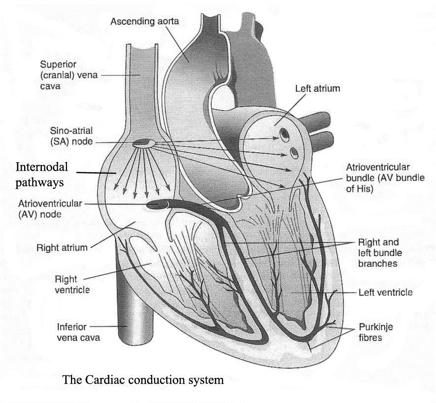
22 Autorythmicity - Sinoatrial Node (S.A): charge/min (natural pacemaker of the heart) - Atrioventricular Node (A.V): charges/min - Bundle of His: charges/min - Purkinje fibers: 15 charges /min (incompatible with life) - If S-A node fails to initiate a stimulus, any of the other areas will initiate the stimulus and will be considered an ectopic pacemaker for the heart.
23
24 Heart valves Atrioventricular (A-V) valves - Right side = tricuspid valve - Left side = bicuspid (mitral) valve - A-V valves prevent expulsion of ventricular blood into the atria when the ventricles contract. Tendency of A-V valves eversion is prevented by cords (chordae tendineae) attached to the free margin of the cusps at one end and to small muscles (papillary m/s) at the other end that extend from the myocardium Papillary m/s contraction is synchronized with the myocardial contraction so that tension to the chordae tendineae is approximately timed
25
26 Heart valves Back flow of blood just ejected from the ventricles is prevented by valves located at the exits of the arteries from the ventricles Pulmonary semilunar valve - for pulmonary trunk Aortic semilunar valve for aorta All heart valves ensure unidirectional blood flow

27 The Mammalian Heart Showing direction of blood flow

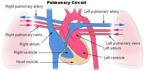
28 Blood flow through the Heart Systemic and Pulmonary
29 Blood flow through the Heart Venous Blood Pulmonary vein Blood from tissues in forward part of the body Caudal vena cava Cranial vena cava R - A R - V L - A L - V Blood from tissues in rear part of the body Lung Aorta Fig 21.6

30 Structure and Function of Valves = Mitral valve 4 sets of valves Prevent backflow of blood Close passively under blood pressure Heart sounds produced by valve closure

31 Support for AV valves: valves are restrained by chordae tendinae which are in turn attached to papillary muscles (prevention of backflow!) picture taken from R ventricle, looking toward R atrium
 Mitral valve cusps do not close properly Regurgitation during left ventricular")
32 Heart Valve Prolapse Mitral Valve Prolapse Most common cardiac variation (5-10% of population) Mitral valve cusps do not close properly Regurgitation during left ventricular systole
33 Special circulation: coronary, pulmonary & cerebral Blood from body enters right atrium Passes tricuspid valve into right ventricle Leaves by passing pulmonary semilunar valves into pulmonary trunk and to the lungs to be oxygenated Leaves left ventricle past aortic semilunar valves into aorta Returns from the lung by way of pulmonary veins into the left atrium Distributed to rest of the body From left atrium past bicuspid valve into left ventricle
34
35 Coronary circulation - circulation of blood in the blood vessels of the heart muscle (the myocardium) - vessels - deliver oxygen-rich blood to the myocardium - coronary arteries - cardiac veins. - Superficial coronary arteries - epicardial coronary arteries - when healthy - capable of autoregulation to maintain coronary blood flow at levels appropriate to the needs of the heart muscle. -relatively narrow vessels; commonly affected by atherosclerosis ; become blocked; cause - angina or a heart attack. - Deep coronary arteries - subendocardial. -Coronary arteries k/s "end circulation - because the only source of blood supply to the myocardium - very little redundant blood supply - blockage of these vessels can be so critical

36 Coronary Circulation Coronary arteries: first branches off the ascending aorta.
")
37 coronary veins coronary sinus right atrium (inferior to opening of inferior vena cava) posterior view
38 Coronary heart disease Coronary artery disease (CAD) Arteriosclerotic heart disease (CHD) a narrowing of the small blood vessels supply of blood and oxygen to the heart slow down or stop
 due to?")

39 Coronary Artery Disease (CAD) PET scan (Positron Emission Tomography ) due to? consequences? the brighter the color, the greater the blood flow through tissue
 Ischemic tissue")
40 Myocardial Infarction (MI) ~ 1.3 mil MIs / year in US Most commonly due to severe CAD (coronary thrombosis) Ischemic tissue degenerates nonfunctional area = infarct Predisposing factors?
41
42 Conducting System of the Heart Specialized muscle cells (autorhythmic cells) conduct APs to time and synchronize the action of the chambers SA node Also k/s pacemaker, located on the wall of right atrium - Initial stimulus - spontaneously depolarizes most rapidly - initiate heart beat - transmits action potential across the right and left atria - contraction of atria emptying the contents (blood) into the ventricles
43 Conducting System of the Heart AV node - when the impulse reaches AV node, it travels down along the AV bundle (bundle of His) within the septum Bundle of His branches, one of which supplies each ventricle where they branch into Purkinje fibers Purkinje fibers reflect up external walls of ventricles and stimulate contraction of cardiac muscle cells as a unit. The blood in the ventricles pumped into pulmonary artery and aorta to circulate to pulmonary and systemic circuits.
44 Conducting System of the Heart - Atria and ventricles - separated by a fibrous ring surrounding the AV valves. - It acts as an insulator. - Therefore the impulse that spreads throughout the atria does not spread to the ventricles and vice versa - This permit independent contractions of atria and ventricles - i.e, the ventricles are filled during their relaxation by the contraction and emptying of the atria. - AV bundle fibers - smaller diameter than other Purkinje fibers - conductivity - 10% slower than cardiac muscle fibers. - Ventricular m/s - thicker than atria and conduction distance is greater therefore to achieve coordinated contraction of muscle fibers of ventricles, it is essential to have a greater velocity of conduction which is provided by Purkinje fibers.
45 Conducting System of the Heart Cardiac muscle contract more slowly than skeletal m/s - longer refractory period (the period during repolarization when a stimulus cannot evoke another depolarization. Autorhythmic stimulation may be modified by sympathetic and parasympathetic nerves changing the nature of heart beat in terms of frequency and force of contraction

46 Great Vessels
47 Cardiac cycle and Heart Beat Cardiac cycle = the sequence of events that occurs during one complete heart beat. 2 phases systole (contraction) diastole (relaxation) 2 phases are continuous - assigned periods are arbitrary - The two atria are in systole and diastole together as are the two ventricles. After ventricular systole and during ventricular diastole: the following sequence of events occurs
48 Cardiac cycle 1. Atria filled by blood from Venae cavae and pulmonary veins; vol. & pressure (occurs during ventricular systole) AV valves open when arterial pressure exceeds the ventricular pressure (occurs at the beginning of ventricular diastole) 2. Blood flows into relaxed ventricles (abt 70% filled) 3. Atria contract (filling of ventricles complete) 4. Atria relax and begin filling. 5. Ventricles begin contraction; AV valves closed b/c ventricle pressures exceeds atrial pressures 6. Continued contraction of ventricles 7. Semilunar valves open 8. Blood ejected from ventricles. 9. Ventricles begin to relax. 10. Atrial pressures begin to exceed the ventricular pressures and semilunar valves closed.
49 Heart sounds Two heart sounds by auscultation: - 1 st sound = Lub = closure of aterioventricular valves when ventricles contract 2 nd sound = dub = closure of semilunar valves when ventricles start to fill following contraction (3 rd and 4 th sound may be detected on ECG; 3 rd sound due to flowing of blood into ventricles and 4 th sound due to atrial contraction) Murmurs: abnormal heart sounds resulted from valve disorders.
50 Factors that affect heart rate Physiological - excitement - muscular exercise (activity, work, exercise) - high environmental temperature - digestion - sleep - high altitude - smoking - some food or drinks e.g. caffeine (coffee, tea) - respiration (slight changes) - metabolic rate
51 Heart rate control Metabolic rate - HR - small animal > large animal - small animal has larger surface area/unit body mass - higher metabolic rate and O 2 consumption HR in adult resting animals Animal Beats/min Animal Beats/min Horse Cat Horse (Thoroughbred) Chicken Dairy cow Elephant 30 Sheep and goats Mouse 670 Pig Rat 420 Small dog 80 guinea pig 280 Large dog 120 Human 60-90
52 Heart rate control Pathological - cardiac diseases - infections (fever) - toxins - medication
53 Cardiac output the volume of blood being pumped by the heart, in particular by a left or right ventricle in the time interval of one minute. Stroke volume: The amount of blood pumped by the left ventricle of the heart in one contraction. -stroke volume is not all of the blood contained in the left ventricle. The heart does not pump all the blood out of the ventricle. Only about 2/3 of the blood in the ventricle is put out with each beat. - The cardiac output is therefore: Q = Stroke Volume Heart rate/min
Ch 19: Cardiovascular System - The Heart -
 Ch 19: Cardiovascular System - The Heart - Give a detailed description of the superficial and internal anatomy of the heart, including the pericardium, the myocardium, and the cardiac muscle. Trace the
the Cardiovascular System I
 the Cardiovascular System I By: Dr. Nabil A Khouri MD, MsC, Ph.D MEDIASTINUM 1. Superior Mediastinum 2. inferior Mediastinum Anterior mediastinum. Middle mediastinum. Posterior mediastinum Anatomy of
The Heart. Size, Form, and Location of the Heart. 1. Blunt, rounded point; most inferior part of the heart.
 12 The Heart FOCUS: The heart is composed of cardiac muscle cells, which are elongated, branching cells that appear striated. Cardiac muscle cells behave as a single electrical unit, and the highly coordinated
Cardiovascular system
 BIO 301 Human Physiology Cardiovascular system The Cardiovascular System: consists of the heart plus all the blood vessels transports blood to all parts of the body in two 'circulations': pulmonary (lungs)
The Heart. Happy Friday! #takeoutyournotes #testnotgradedyet
 The Heart Happy Friday! #takeoutyournotes #testnotgradedyet Introduction Cardiovascular system distributes blood Pump (heart) Distribution areas (capillaries) Heart has 4 compartments 2 receive blood (atria)
Collin County Community College. ! BIOL Anatomy & Physiology! WEEK 5. The Heart
 Collin County Community College! BIOL. 2402 Anatomy & Physiology! WEEK 5 The Heart 1 (1578-1657) A groundbreaking work in the history of medicine, English physician William Harvey s Anatomical Essay on
Chapter 20: Cardiovascular System: The Heart
 Chapter 20: Cardiovascular System: The Heart I. Functions of the Heart A. List and describe the four functions of the heart: 1. 2. 3. 4. II. Size, Shape, and Location of the Heart A. Size and Shape 1.
Cardiovascular System
 Cardiovascular System The Heart Cardiovascular System The Heart Overview What does the heart do? By timed muscular contractions creates pressure gradients blood moves then from high pressure to low pressure
10. Thick deposits of lipids on the walls of blood vessels, called, can lead to serious circulatory issues. A. aneurysm B. atherosclerosis C.
 Heart Student: 1. carry blood away from the heart. A. Arteries B. Veins C. Capillaries 2. What is the leading cause of heart attack and stroke in North America? A. alcohol B. smoking C. arteriosclerosis
2. right heart = pulmonary pump takes blood to lungs to pick up oxygen and get rid of carbon dioxide
 A. location in thorax, in inferior mediastinum posterior to sternum medial to lungs superior to diaphragm anterior to vertebrae orientation - oblique apex points down and to the left 2/3 of mass on left
11/10/2014. Muscular pump Two atria Two ventricles. In mediastinum of thoracic cavity 2/3 of heart's mass lies left of midline of sternum
 It beats over 100,000 times a day to pump over 1,800 gallons of blood per day through over 60,000 miles of blood vessels. During the average lifetime, the heart pumps nearly 3 billion times, delivering
The Cardiovascular System
 Essentials of Human Anatomy & Physiology Elaine N. Marieb Seventh Edition Chapter 11 The Cardiovascular System Slides 11.1 11.19 Lecture Slides in PowerPoint by Jerry L. Cook The Cardiovascular System
Human Anatomy, First Edition
 Human Anatomy, First Edition McKinley & O'Loughlin Chapter 22 : Heart 1 Functions of the Heart Center of the cardiovascular system, the heart. Connects to blood vessels that transport blood between the
Anatomy of the Heart. Figure 20 2c
 Anatomy of the Heart Figure 20 2c Pericardium & Myocardium Remember, the heart sits in it s own cavity, known as the mediastinum. The heart is surrounded by the Pericardium, a double lining of the pericardial
Heart. Structure Physiology of blood pressure and heartbeat
 Heart Structure Physiology of blood pressure and heartbeat Location and Anatomy Location and Anatomy Pericardial cavity: surrounds, isolates, and anchors heart Parietal pericardium lined with serous membrane
Heart Anatomy. 7/5/02 Stephen G Davenport 1
 Heart Anatomy Copyright 1999, Stephen G. Davenport, No part of this publication may be reproduced, stored in a retrieval system, or transmitted, in any form without prior written permission. 7/5/02 Stephen
The HEART. What is it???? Pericardium. Heart Facts. This muscle never stops working It works when you are asleep
 This muscle never stops working It works when you are asleep The HEART It works when you eat It really works when you exercise. What is it???? Located between the lungs in the mid thoracic region Apex
10/23/2017. Muscular pump Two atria Two ventricles. In mediastinum of thoracic cavity 2/3 of heart's mass lies left of midline of sternum
 It beats over 100,000 times a day to pump over 1,800 gallons of blood per day through over 60,000 miles of blood vessels. During the average lifetime, the heart pumps nearly 3 billion times, delivering
The Cardiovascular System
 The Cardiovascular System The Cardiovascular System A closed system of the heart and blood vessels The heart pumps blood Blood vessels allow blood to circulate to all parts of the body The function of
CARDIOVASCULAR SYSTEM
 CARDIOVASCULAR SYSTEM Overview Heart and Vessels 2 Major Divisions Pulmonary Circuit Systemic Circuit Closed and Continuous Loop Location Aorta Superior vena cava Right lung Pulmonary trunk Base of heart
CV Anatomy Quiz. Dr Ella Kim Dr Pip Green
 CV Anatomy Quiz Dr Ella Kim Dr Pip Green Q1 The location of the heart is correctly described as A) lateral to the lungs. B) medial to the sternum. C) superior to the diaphragm. D) posterior to the spinal
Chapter 18 - Heart. I. Heart Anatomy: size of your fist; located in mediastinum (medial cavity)
") Chapter 18 - Heart I. Heart Anatomy: size of your fist; located in mediastinum (medial cavity) A. Coverings: heart enclosed in double walled sac called the pericardium 1. Fibrous pericardium: dense connective
Chapter 18 - Heart I. Heart Anatomy: size of your fist; located in mediastinum (medial cavity) A. Coverings: heart enclosed in double walled sac called the pericardium 1. Fibrous pericardium: dense connective
The Heart. The Heart A muscular double pump. The Pulmonary and Systemic Circuits
 C H A P T E R 19 The Heart The Heart A muscular double pump circuit takes blood to and from the lungs Systemic circuit vessels transport blood to and from body tissues Atria receive blood from the pulmonary
The Cardiovascular System
 11 PART A The Cardiovascular System PowerPoint Lecture Slide Presentation by Jerry L. Cook, Sam Houston University ESSENTIALS OF HUMAN ANATOMY & PHYSIOLOGY EIGHTH EDITION ELAINE N. MARIEB The Cardiovascular
Chapter 14. The Cardiovascular System
 Chapter 14 The Cardiovascular System Introduction Cardiovascular system - heart, blood and blood vessels Cardiac muscle makes up bulk of heart provides force to pump blood Function - transports blood 2
Heart. Heart 2-Tunica media: middle layer (media ='middle') muscle fibers (smooth or cardiac).
 muscle fibers (smooth or cardiac).") t. innermost lumenal General Circulatory system heart and blood vessels walls have 3 layers (inside to outside) 1-Tunica interna: aka tunica intima layer--lumenal layer epithelium--endothelium simple squamous
t. innermost lumenal General Circulatory system heart and blood vessels walls have 3 layers (inside to outside) 1-Tunica interna: aka tunica intima layer--lumenal layer epithelium--endothelium simple squamous
4. The two inferior chambers of the heart are known as the atria. the superior and inferior vena cava, which empty into the left atrium.
 Answer each statement true or false. If the statement is false, change the underlined word to make it true. 1. The heart is located approximately between the second and fifth ribs and posterior to the
Chapter 13 The Cardiovascular System: Cardiac Function
 Chapter 13 The Cardiovascular System: Cardiac Function Overview of the Cardiovascular System The Path of Blood Flow through the Heart and Vasculature Anatomy of the Heart Electrical Activity of the Heart
The Heart. C h a p t e r. PowerPoint Lecture Slides prepared by Jason LaPres Lone Star College - North Harris
 C h a p t e r 20 The Heart PowerPoint Lecture Slides prepared by Jason LaPres Lone Star College - North Harris Copyright 2009 Pearson Education, Inc., publishing as Pearson Benjamin Cummings Introduction
THE HEART. A. The Pericardium - a double sac of serous membrane surrounding the heart
 THE HEART I. Size and Location: A. Fist-size weighing less than a pound (250 to 350 grams). B. Located in the mediastinum between the 2 nd rib and the 5 th intercostal space. 1. Tipped to the left, resting
Cardiovascular System
 Cardiovascular System Purpose Transport oxygen and nutrients Take waste products away from tissues & organs Things we learned Blood pressure: the force of blood pushing against the walls of blood vessels
The Cardiovascular System
 Essentials of Human Anatomy & Physiology Elaine N. Marieb Slides 11.1 11.19 Seventh Edition Chapter 11 The Cardiovascular System Functions of the Cardiovascular system Function of the heart: to pump blood
Chapter 20 (1) The Heart
 The Heart") Chapter 20 (1) The Heart Learning Objectives Describe the location and structure of the heart Describe the path of a drop of blood from the superior vena cava or inferior vena cava through the heart out
Chapter 20 (1) The Heart Learning Objectives Describe the location and structure of the heart Describe the path of a drop of blood from the superior vena cava or inferior vena cava through the heart out
Approximately the size of your fist Location. Pericardial physiology
 Heart Anatomy Approximately the size of your fist Location Superior surface of diaphragm Left of the midline Anterior to the vertebral column, posterior to the sternum Wednesday, March 28, 2012 Muscle
Cardiac physiology. b. myocardium -- cardiac muscle and fibrous skeleton of heart
 I. Heart anatomy -- general gross. A. Size/orientation - base/apex B. Coverings D. Chambers 1. parietal pericardium 2. visceral pericardium 3. Layers of heart wall a. epicardium Cardiac physiology b. myocardium
THE CARDIOVASCULAR SYSTEM. Part 1
 THE CARDIOVASCULAR SYSTEM Part 1 CARDIOVASCULAR SYSTEM Blood Heart Blood vessels What is the function of this system? What other systems does it affect? CARDIOVASCULAR SYSTEM Functions Transport gases,
THE CARDIOVASCULAR SYSTEM. Heart 2
 THE CARDIOVASCULAR SYSTEM Heart 2 PROPERTIES OF CARDIAC MUSCLE Cardiac muscle Striated Short Wide Branched Interconnected Skeletal muscle Striated Long Narrow Cylindrical PROPERTIES OF CARDIAC MUSCLE Intercalated
Cardiovascular System: The Heart
 Cardiovascular System: The Heart I. Anatomy of the Heart (See lab handout for terms list) A. Describe the size, shape and location of the heart B. Describe the structure and function of the pericardium
BIOL 4350 Cardiovascular Physiology Dr. Hamilton. Using the figure above, match the following: 1. Purkinje fibers. 2. SA node. 3. AV node.
 BIOL 4350 Cardiovascular Physiology Dr. Hamilton Using the figure above, match the following: 1. Purkinje fibers. 2. SA node. 3. AV node. 1 Using the figure above, match the following: 4. Atrial depolarization.
Section 5.1 The heart and heart disease
 Section 5.1 The heart and heart disease Mammals are too large to rely on diffusion. They need a circulatory system to move substances around the body. Blood moves down pressure gradients, from high to
Lab Activity 23. Cardiac Anatomy. Portland Community College BI 232
 Lab Activity 23 Cardiac Anatomy Portland Community College BI 232 Cardiac Muscle Histology Branching cells Intercalated disc: contains many gap junctions connecting the adjacent cell cytoplasm, creates
Principles of Biomedical Systems & Devices. Lecture 8: Cardiovascular Dynamics Dr. Maria Tahamont
 Principles of Biomedical Systems & Devices Lecture 8: Cardiovascular Dynamics Dr. Maria Tahamont Review of Cardiac Anatomy Four chambers Two atria-receive blood from the vena cave and pulmonary veins Two
IB TOPIC 6.2 THE BLOOD SYSTEM
 IB TOPIC 6.2 THE BLOOD SYSTEM THE BLOOD SYSTEM TERMS TO KNOW circulation ventricle artery vein 6.2.U1 - Arteries convey blood at high pressure from the ventricles to the tissues of the body Circulation
THE HEART OBJECTIVES: LOCATION OF THE HEART IN THE THORACIC CAVITY CARDIOVASCULAR SYSTEM
 BIOLOGY II CARDIOVASCULAR SYSTEM ACTIVITY #3 NAME DATE HOUR THE HEART OBJECTIVES: Describe the anatomy of the heart and identify and give the functions of all parts. (pp. 356 363) Trace the flow of blood
The Heart and Cardiovascular System
 The Heart and Cardiovascular System What you will learn The location of the heart 3 layers and covering of the heart Explain the function of the heart as 2 separate pumps Identify the 4 chambers of the
The cardiovascular system is composed of the heart and blood vessels that carry blood to and from the body s organs. There are 2 major circuits:
 1 The cardiovascular system is composed of the heart and blood vessels that carry blood to and from the body s organs. There are 2 major circuits: pulmonary and systemic. The pulmonary goes out to the
The Circulatory System. The Heart, Blood Vessels, Blood Types
 The Circulatory System The Heart, Blood Vessels, Blood Types The Closed Circulatory System Humans have a closed circulatory system, typical of all vertebrates, in which blood is confined to vessels and
The Cardiovascular System: The Heart
 PowerPoint Lecture Slides prepared by Meg Flemming Austin Community College C H A P T E R 12 The Cardiovascular System: The Heart Chapter 12 Learning Outcomes 12-1 12-2 Describe the anatomy of the heart,
37 1 The Circulatory System
 H T H E E A R T 37 1 The Circulatory System The circulatory system and respiratory system work together to supply cells with the nutrients and oxygen they need to stay alive. a) The respiratory system:
LECTURE 5. Anatomy of the heart
 LECTURE 5. Anatomy of the heart Main components of the CVS: Heart Blood circulatory system arterial compartment haemomicrocirculatory (=microvascular) compartment venous compartment Lymphatic circulatory
The Cardiovascular System
 PowerPoint Lecture Slide Presentation by Patty Bostwick-Taylor, Florence-Darlington Technical College The Cardiovascular System 11 PART A The Cardiovascular System A closed system of the heart and blood
IB TOPIC 6.2 THE BLOOD SYSTEM
 IB TOPIC 6.2 THE BLOOD SYSTEM TERMS TO KNOW circulation ventricle artery vein THE BLOOD SYSTEM 6.2.U1 - Arteries convey blood at high pressure from the ventricles to the tissues of the body Circulation
The Cardiovascular System (Heart)
") The Cardiovascular System The Cardiovascular System (Heart) A closed system of the heart and blood vessels The heart pumps blood Blood vessels allow blood to circulate to all parts of the body The function
The Cardiovascular System The Cardiovascular System (Heart) A closed system of the heart and blood vessels The heart pumps blood Blood vessels allow blood to circulate to all parts of the body The function
Cardiovascular System Notes: Heart Disease & Disorders
 Cardiovascular System Notes: Heart Disease & Disorders Interesting Heart Facts The Electrocardiograph (ECG) was invented in 1902 by Willem Einthoven Dutch Physiologist. This test is still used to evaluate
The Cardiovascular System Part I: Heart Outline of class lecture After studying part I of this chapter you should be able to:
 The Cardiovascular System Part I: Heart Outline of class lecture After studying part I of this chapter you should be able to: 1. Describe the functions of the heart 2. Describe the location of the heart,
- what other structures, besides the heart, does the mediastinum contain?
 Basic A & P II Dr. L. Bacha Chapter Outline (Martini & Nath 2010) An Introduction to the Cardiovascular System - read the paragraphs under this heading on page 580 The Heart is a Four Chambered Organ describe
Cardiovascular Physiology. Heart Physiology. Introduction. The heart. Electrophysiology of the heart
 Cardiovascular Physiology Heart Physiology Introduction The cardiovascular system consists of the heart and two vascular systems, the systemic and pulmonary circulations. The heart pumps blood through
Electrical Conduction
 Sinoatrial (SA) node Electrical Conduction Sets the pace of the heartbeat at 70 bpm AV node (50 bpm) and Purkinje fibers (25 40 bpm) can act as pacemakers under some conditions Internodal pathway from
The Cardiovascular System
 The Cardiovascular System The Manila Times College of Subic Prepared by: Stevens B. Badar, RN, MANc THE HEART Anatomy of the Heart Location and Size approx. the size of a person s fist, hollow and cone-shaped,
Principles of Anatomy and Physiology
 Principles of Anatomy and Physiology 14 th Edition CHAPTER 20 The Cardiovascular System: The Heart Introduction The purpose of the chapter is to: 1. Learn about the components of the cardiovascular system
The ancient Babylonians, Egyptians, Indians and Chinese believed the heart was the centre of thinking and emotions
 The Concept of Mind The ancient Babylonians, Egyptians, Indians and Chinese believed the heart was the centre of thinking and emotions Hippocrates 460 BC 370 BC - Thoughts, ideas, and feelings come from
Figure ) The specific chamber of the heart that is indicated by letter A is called the. Diff: 1 Page Ref: 364
 The specific chamber of the heart that is indicated by letter A is called the. Diff: 1 Page Ref: 364") Essentials of Anatomy and Physiology, 9e (Marieb) Chapter 11 The Cardiovascular System Short Answer Figure 11.1 Using Figure 11.1, identify the following: 1) The Purkinje fibers are indicated by label.
Essentials of Anatomy and Physiology, 9e (Marieb) Chapter 11 The Cardiovascular System Short Answer Figure 11.1 Using Figure 11.1, identify the following: 1) The Purkinje fibers are indicated by label.
Major Function of the Cardiovascular System. Transportation. Structures of the Cardiovascular System. Heart - muscular pump
 Structures of the Cardiovascular System Heart - muscular pump Blood vessels - network of tubes Blood - liquid transport vehicle brachiocephalic trunk superior vena cava right pulmonary arteries right pulmonary
CIRCULATORY SYSTEM BLOOD VESSELS
 Name: Block: CIRCULATORY SYSTEM Multicellular organisms (above the level of roundworms) rely on a circulatory system to bring nutrients to, and take wastes away from, cells. In higher organisms such as
BIOLOGY 2060 LECTURE NOTES ANATOMY & PHYSIOLOGY II (A. IMHOLTZ) HEART P1 OF 5
 HEART P1 OF 5") BIOLOGY 2060 LECTURE NOTES ANATOMY & PHYSIOLOGY II (A. IMHOLTZ) HEART P1 OF 5 1. Heart Functions a. Generates pressure that propels blood thru blood vessels. (Tissue perfusion.) b. Separates oxygenated
BIOLOGY 2060 LECTURE NOTES ANATOMY & PHYSIOLOGY II (A. IMHOLTZ) HEART P1 OF 5 1. Heart Functions a. Generates pressure that propels blood thru blood vessels. (Tissue perfusion.) b. Separates oxygenated
Cardiovascular System
 Cardiovascular System I. Structure of the Heart A. Average adult heart is 14 cm long and 9 cm wide. B. Lies in the mediastinum. C. Enclosed in the pericardium. 1. Fibrous pericardium- Outer, tough connective
Cardiovascular Anatomy Dr. Gary Mumaugh
 Cardiovascular Anatomy Dr. Gary Mumaugh Location of Heart Approximately the size of your fist Location o Superior surface of diaphragm o Left of the midline in mediastinum o Anterior to the vertebral column,
BME 5742 Bio-Systems Modeling and Control. Lecture 41 Heart & Blood Circulation Heart Function Basics
 BME 5742 Bio-Systems Modeling and Control Lecture 41 Heart & Blood Circulation Heart Function Basics Dr. Zvi Roth (FAU) 1 Pumps A pump is a device that accepts fluid at a low pressure P 1 and outputs the
Practice Exercises for the Cardiovascular System
 Practice Exercises for the Cardiovascular System On the diagram below, color the oxygen-rich blood red and the oxygen-poor blood blue. Label the parts: Continued on the next page... Label the parts on
Circulatory system. Terminology. Ventricles and resistance. Pressure gradients move blood through the heart and vessels.
 Circulatory system Pressure gradients move blood through the heart and vessels. Pulmonary circulation vs. systemic circulation (to pulmonary circuit) liver head and arms heart aorta diaphragm (from pulmonary
The Cardiovascular System. Chapter 15. Cardiovascular System FYI. Cardiology Closed systemof the heart & blood vessels. Functions
 Chapter 15 Cardiovascular System FYI The heart pumps 7,000 liters (4000 gallons) of blood through the body each day The heart contracts 2.5 billion times in an avg. lifetime The heart & all blood vessels
Pearson's Comprehensive Medical Assisting Administrative and Clinical Competencies
 Pearson's Comprehensive Medical Assisting Administrative and Clinical Competencies THIRD EDITION CHAPTER 27 The Cardiovascular System Lesson 1: Overview of the Cardiovascular System Lesson Objectives Upon
Heart Dissection. 5. Locate the tip of the heart or the apex. Only the left ventricle extends all the way to the apex.
 Heart Dissection Page 1 of 6 Background: The heart is a four-chambered, hollow organ composed primarily of cardiac muscle tissue. It is located in the center of the chest in between the lungs. It is the
Cardiovascular System. I. Structures of the heart A. : Pericardium sack that surrounds the heart
 Cardiovascular System I. Structures of the heart A. : Pericardium sack that surrounds the heart 1. : Pericardial Cavity serous fluid filled space between the heart and the pericardium B. Heart Wall 1.
Chp. 5 The cardiovascular system. What are the function of the cardiovascular system? Arteries and arterioles:
 5.1 Overview of the cardiovascular system Chp. 5 The cardiovascular system Includes the heart and blood vessels Brings nutrients to cells and helps get rid of wastes Blood is refreshed in the lung, kidneys,
Cardiovascular System Notes: Physiology of the Heart
 Cardiovascular System Notes: Physiology of the Heart Interesting Heart Fact Capillaries are so small it takes ten of them to equal the thickness of a human hair. Review What are the 3 parts of the cardiovascular
The Heart and Heart Disease
 The Heart and Heart Disease Illustration of the heart by Leonardo DaVinci heart-surgeon.com/ history.html 2/14/2010 1 I. Location, Size and Position of the Heart A. Triangular organ located 1. of mass
BIOLOGY 2060 LECTURE NOTES ANATOMY & PHYSIOLOGY II (A. IMHOLTZ) HEART P1 OF 7
 HEART P1 OF 7") BIOLOGY 2060 LECTURE NOTES ANATOMY & PHYSIOLOGY II (A. IMHOLTZ) HEART P1 OF 7 1. Heart a. Generates the pressure that propels blood thru blood vessels. b. Separates oxygenated and deoxygenated blood separate.
BIOLOGY 2060 LECTURE NOTES ANATOMY & PHYSIOLOGY II (A. IMHOLTZ) HEART P1 OF 7 1. Heart a. Generates the pressure that propels blood thru blood vessels. b. Separates oxygenated and deoxygenated blood separate.
Introduction to Anatomy. Dr. Maher Hadidi. Bayan Yanes. April/9 th /2013
 Introduction to Anatomy Dr. Maher Hadidi Bayan Yanes 27 April/9 th /2013 KEY POINTS: 1) Right side of the heart 2) Papillary muscles 3) Left side of the heart 4) Comparison between right and left sides
Anatomy of the Heart
 Biology 212: Anatomy and Physiology II Anatomy of the Heart References: Saladin, KS: Anatomy and Physiology, The Unity of Form and Function 8 th (2018). Required reading before beginning this lab: Chapter
Introduction. Circulation
 Introduction Circulation 1- Systemic (general) circulation 2- Pulmonary circulation carries oxygenated blood to all parts of the body carries deoxygenated blood to the lungs From Lt. ventricle aorta From
THE HEART Dr. Ali Ebneshahidi
 THE HEART Dr. Ali Ebneshahidi Functions is of the heart & blood vessels 1. The heart is an essential pumping organ in the cardiovascular system where the right heart pumps deoxygenated blood (returned
Unit 6: Circulatory System. 6.2 Heart
 Unit 6: Circulatory System 6.2 Heart Functions of Circulatory System 1. The heart is the pump necessary to circulate blood to all parts of the body 2. Arteries, veins and capillaries are the structures
Circulation. Circulation = is a process used for the transport of oxygen, carbon! dioxide, nutrients and wastes through-out the body
 Circulation Circulation = is a process used for the transport of oxygen, carbon! dioxide, nutrients and wastes through-out the body Heart = muscular organ about the size of your fist which pumps blood.
QUIZ/TEST REVIEW NOTES SECTION 1 CARDIAC MYOCYTE PHYSIOLOGY [CARDIOLOGY]
![QUIZ/TEST REVIEW NOTES SECTION 1 CARDIAC MYOCYTE PHYSIOLOGY [CARDIOLOGY]](/thumbs/96/126998162.jpg "QUIZ/TEST REVIEW NOTES SECTION 1 CARDIAC MYOCYTE PHYSIOLOGY [CARDIOLOGY]") QUIZ/TEST REVIEW NOTES SECTION 1 CARDIAC MYOCYTE PHYSIOLOGY [CARDIOLOGY] Learning Objectives: Describe the ionic basis of action potentials in cardiac contractile and autorhythmic cells Explain the relationship
QUIZ/TEST REVIEW NOTES SECTION 1 CARDIAC MYOCYTE PHYSIOLOGY [CARDIOLOGY] Learning Objectives: Describe the ionic basis of action potentials in cardiac contractile and autorhythmic cells Explain the relationship
Cardiovascular System. Heart Anatomy
 Cardiovascular System Heart Anatomy 1 The Heart Location & general description: Atria vs. ventricles Pulmonary vs. systemic circulation Coverings Walls The heart is found in the mediastinum, the medial
The Mammalian Circulatory System
 The Mammalian Heart The Mammalian Circulatory System Recall: What are the 3 cycles of the mammalian circulatory system? What are their functions? What are the three main vessel types in the mammalian circulatory
human anatomy 2016 lecture thirteen Dr meethak ali ahmed neurosurgeon
 Heart The heart is a hollow muscular organ that is somewhat pyramid shaped and lies within the pericardium in the mediastinum. It is connected at its base to the great blood vessels but otherwise lies
Department of medical physiology 7 th week and 8 th week
 Department of medical physiology 7 th week and 8 th week Semester: winter Study program: Dental medicine Lecture: RNDr. Soňa Grešová, PhD. Department of medical physiology Faculty of Medicine PJŠU Cardiovascular
1. What kind of blood is found in the rt. atrium? (oxygenated or deoxygenated)
") Carl Christennsen, PhD Chap. 19, 20, & 21 - Circulatory System Bio. 2304 Human Anatomy HEART 1. What kind of blood is found in the rt. atrium? (oxygenated or deoxygenated) Where does this blood come from?
Carl Christennsen, PhD Chap. 19, 20, & 21 - Circulatory System Bio. 2304 Human Anatomy HEART 1. What kind of blood is found in the rt. atrium? (oxygenated or deoxygenated) Where does this blood come from?
Ch.15 Cardiovascular System Pgs {15-12} {15-13}
 Ch.15 Cardiovascular System Pgs {15-12} {15-13} E. Skeleton of the Heart 1. The skeleton of the heart is composed of rings of dense connective tissue and other masses of connective tissue in the interventricular
Circulatory System Function Move circulatory fluid (blood) around body Gas Transport Nutrient Transport Excretory Product Transport
 around body Gas Transport Nutrient Transport Excretory Product Transport") Lecture 37 Introduction to Circulation BY DR QAZI IMTIAZ RASOOL OBJECTIVES Functions of the Heart Generating blood pressure Routing blood: separates pulmonary and systemic circulations Ensuring one-way
Lecture 37 Introduction to Circulation BY DR QAZI IMTIAZ RASOOL OBJECTIVES Functions of the Heart Generating blood pressure Routing blood: separates pulmonary and systemic circulations Ensuring one-way
Ch. 12 The Circulatory System. The heart. The heart is a double pump. A quick note on arteries vs. veins. = the muscular pump of the CV system
 Ch. 12 The Circulatory System The heart A.k.a. the cardiovascular system Blood was discussed in Ch. 11 Focus of Ch. 12: heart and blood vessels = the muscular pump of the CV system ~ 100,000 heartbeats/day!
Blood must move! 4/15/2014. Heart Basics
 What is the CARDIOVASCULAR system? The cardiovascular system carries blood and dissolved substances to and from different places in the body. The Heart has the job of pumping these things around the body.
Anatomy Review: The Heart Graphics are used with permission of A.D.A.M. Software, Inc. and Benjamin/Cummings Publishing Co.
 Anatomy Review: The Heart Graphics are used with permission of A.D.A.M. Software, Inc. and Benjamin/Cummings Publishing Co. Anatomy Views Label the diagrams of the heart below: Interactive Physiology Study
Anatomy & Physiology of Cardiovascular System. Chapter 18 & 19
 Anatomy & Physiology of Cardiovascular System Chapter 18 & 19 Objectives..cont 1. Discuss the physiological stages of cardiac muscle contraction. 2. Trace a typical ECG and label each wave or complex 3.
Functions of the Heart
 Cardiovascular System The Heart What is the Cardiovascular System? Blood circulated in Arteries, veins, and capillaries by the Pumping action of the heart Functions of the Heart Generating blood pressure
Read Me. covering the Heart Anatomy. Labs. textbook. use. car: you
 Heart Anatomy Lab Pre-Lab Exercises Read Me These exercises should be done before coming to lab, after watching the videos covering the Heart Anatomy Labs. Answer the questions in this guide using the
Circulatory System: Introduction. Dr. Carmen E. Rexach Anatomy 35 Mt. San Antonio College
 Circulatory System: Introduction Dr. Carmen E. Rexach Anatomy 35 Mt. San Antonio College Components Cardiovascular system Lymphatic system Cardiovascular system Heart, blood vessels, blood Functions: transport