Anatomy & Physiology of Cardiovascular System. Chapter 18 & 19
|
|
|
- Warren Flowers
- 6 years ago
- Views:
Transcription
1 Anatomy & Physiology of Cardiovascular System Chapter 18 & 19
2 Objectives..cont 1. Discuss the physiological stages of cardiac muscle contraction. 2. Trace a typical ECG and label each wave or complex 3. Define the terms systole and diastole. 4. Outline the phases of the cardiac cycle. 5. Discuss heart sounds in terms of what they represent. 6. Define the terms cardiac output (CO), heart rate (HR), and stroke volume (SV). 7. Discuss the factors the regulate heart rate.
3 Objectives 1. List the organs that compose the cardiovascular system and discuss the general functions of this system. 2. Describe the location, size, and orientation of the human heart. 3. Define the term cardiology. 4. Describe the structure of the heart in terms of its coverings, layers, chambers, valves, and blood vessels. 5. Explain why the atria are passive chambers, while the ventricles are active. 6. Name the function of heart valves. 7. Distinguish between AV and SL valves in terms of location, structure.
4 INTRODUCTION The major function of the cardiovascular systems is to circulate substances throughout the body. In other words, its organs function to supply cells & tissues with oxygen & nutrients and also to remove wastes (CO2 & urea) from cells and tissues. If cells do not receive O2 & nutrients and wastes accumulate, cells will die! Cardiology is the study of the heart and the diseases associated with it.
5 Functions Assist in gas transport. Deliver nutrients, hormones Remove waste products from the cells Assist in temperature regulation Assist in balancing body fluids and helps prevent dehydration
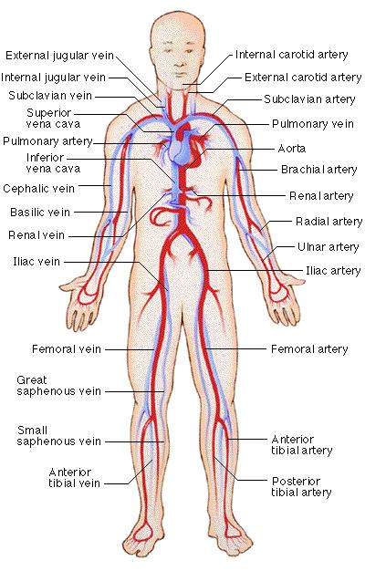
6 Major Components Pump (heart) Continuously circulates blood Network of tubes Arteries- blood away from heart Veins- blood back to the heart Blood Fluid that fills the circulatory system
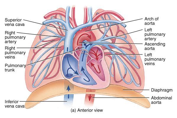
7 Specific parts Pulmonary arteries- transport blood to lungs Pulmonary veins- transport oxygenated blood to hear Aortic arch and trunk- main arteries from heart Common carotid artery- carries blood to brain Renal vein and artery- connects to kidneys Mesenteric veins- connects to intestine
8
9 Accessory Organs Lungs- organ where oxygen is taken up and CO 2 is released Kidneys- organ where wastes are removed from blood; critical in regulating fluids in the body Small intestine- digestion Large intestine- water absorption
10 The Heart The human heart has four chambers Left and right ventricle Left and right atrium The left side of the heart pumps oxygenated blood to the body. the right side of the heart pumps deoxygenated blood to the lungs where oxygen can be absorbed by the hemoglobin.
11 The heart functions to contract and propel blood through the two blood transport systems. 1. Pulmonary system 2. Systemic system
12
13 LOCATION/SIZE OF HEART A. Location = within mediastinum behind the body of the sternum between the points of attachment of ribs 2 through 6. B. Size = closed fist; 300g (adult). C. Base = wide superior border; D. Apex = inferior point.

14 LOCATION OF HEART
15 Surface Anatomy of the Heart (anterior view)
16 Structure of the heart Wall of the heart made up of three distinct layers pericardium : parietal layer lies inside fibrous, and visceral layer (epicardium) adheres to outside of the heart; pericardial space with pericardial fluid separates the two layers. Epicardium: visceral layer Myocardium: thick, contractile middle layer of heart wall; compresses the heart cavities, and the blood within them, with great force Endocardium: delicate inner layer of endothelial tissue

17 Wall of the heart
18 Cardiac muscle Each cardiac muscle contians two types of intracellular contractile protiens, actin & myosin. The myosin appears thick where as actin appears thin and connected to Z band on end.

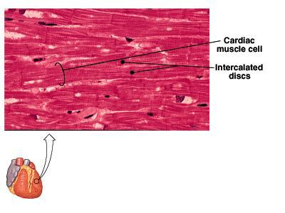
19 Cardiac Muscle
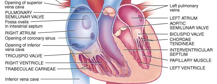
20 Chambers of the heart divided into four cavities with the right and left chambers separated by the septum 1. Atria Two superior chambers, known as receiving chambers, because they receive blood from veins Atria alternately contract and relax to receive blood and then push it into ventricles Myocardial wall of each atrium is not very thick, because little pressure is needed to move blood such a small distance Auricle earlike flap protruding from each atrium
21 Chambers of the heart..cont Ventricles Two lower chambers, known as pumping chambers, because they push blood into the large network of vessels Ventricular myocardium is thicker than myocardium of the atria, because great force must be generated to pump blood a large distance; myocardium of left ventricle is thicker than the right, because it must push blood much further
22
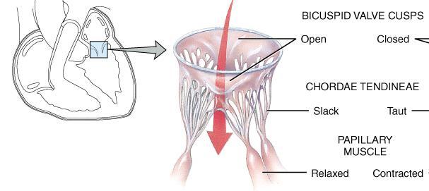
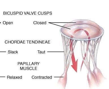
23 Valves of the heart mechanical devices that permit the flow of blood in one direction only. Atrioventricular (AV) valves prevent blood from flowing back into the atria from the ventricles when the ventricles contract 1.Tricuspid valve guards the right atrioventricular orifice; free edges of three flaps of endocardium are attached to papillary muscles by chordae tendineae 2. Bicuspid, or mitral, valve similar in structure to tricuspid valve except only two flaps present
24 Valves of the heart..cont Semilunar (SL) valves half moon shaped flaps growing out from the lining of the pulmonary artery and aorta; prevent blood from flowing back into ventricles from aorta and pulmonary artery 1. Pulmonary valve at entrance of pulmonary artery 2. Aortic valve at entrance of aorta


25 Heart valves 1. Mitral valve 2. Tricuspid valve 3. Aortic valve 4. Pulmonary valve
26
27 Blood supply of heart tissue Coronary arteries: myocardial cells receive blood from right and left coronary arteries First branches to come off aorta Ventricles receive blood from branches of both right and left coronary arteries Each ventricle receives blood only from a small branch of corresponding coronary artery Most abundant blood supply goes to myocardium of left ventricle The right coronary artery is dominant in approximately 50% of all hearts. Few anastomoses exist between the larger branches of the coronary arteries
28 Blood supply of heart tissue (cont.) Veins of the coronary circulation As a rule, veins follow a course that closely parallels that of coronary arteries After going through cardiac veins, blood enters coronary sinus to drain into right atrium Several veins drain directly into right atrium

29 Coronary arteries:

30 Skeleton of the Heart
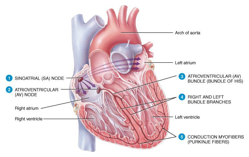
31 Conduction system of the heart 1. Sinoatrial (SA) node, 2. Atrioventricular (AV) node, 3. AV bundle 4. Purkinje fibers Sinoatrial node (SA node or pacemaker): in right atrial wall near opening of superior vena cava Atrioventricular node (AV node): small mass of special cardiac muscle in right atrium along lower part of interatrial septum Atrioventricular bundle (AV bundle or bundle of His) and Purkinje fibers extends by two branches down the two sides of the interventricular septum, and continues as Purkinje fibers Purkinje fibers extend out to papillary muscles and lateral walls of ventricles
32
33 Conduction system..cont Conduction system structures are more highly specialized than ordinary cardiac muscle tissue and permit only rapid conduction of an action potential through the heart SA node (pacemaker) Initiates each heartbeat and sets its pace Specialized pacemaker cells in the node possess an intrinsic rhythm
34 Sequence of cardiac stimulation After being generated by the SA node, each impulse travels throughout the muscle fibers of both atria, and the atria begin to contract As the action potential enters the AV node from the right atrium, its conduction slows to allow complete contraction of both atrial chambers before the impulse reaches the ventricles After the AV node, conduction velocity increases as the impulse is relayed through the AV bundle into the ventricles Right and left branches of the bundle fibers and Purkinje fibers conduct the impulses throughout the muscles of both ventricles, stimulating them to contract almost simultaneously
35 Conduction System
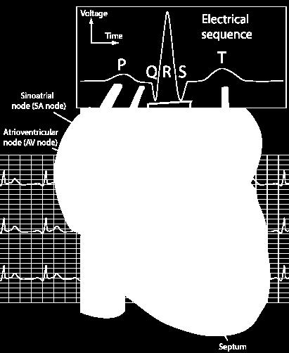
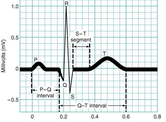
36 Electrocardiogram Normal ECG is composed of the following: P wave represents depolarization of the atria QRS complex represents depolarization of the ventricles and repolarization of the atria T wave represents repolarization of the ventricles; may also have a U wave that represents repolarization of the papillary muscle Measurement of the intervals between P, QRS, and T waves can provide information about the rate of conduction of an action potential through the heart P QRS T
37
38 Nerve supply of the heart Cardiac plexuses located near arch of aorta, made up of the combination of sympathetic and parasympathetic fibers Most fibers end in the SA node, but some end in the AV node and in the atrial myocardium Sympathetic nerves accelerator nerves Vagus fibers inhibitory, or depressor, nerves

39 Nerve supply of the heart

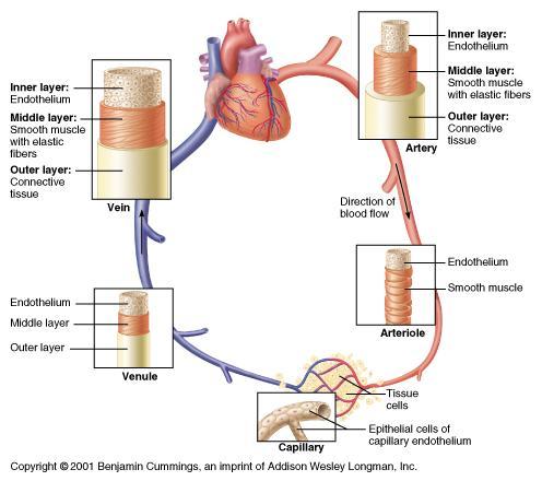
40 Nerve supply of the heart
41 Blood vessels Types of blood vessels Arteries Carry blood away from heart all arteries except pulmonary artery carry oxygenated blood Elastic arteries largest in body Examples: aorta and its major branches Able to stretch without injury Accommodate surge of blood when heart contracts and able to recoil when ventricles relax
42

43 Capillary Bed Arterioles Metarteriole Capallaries Venules
44 Capillaries primary exchange vessels Microscopic vessels Carry blood from arterioles to venules together, arterioles, capillaries and venules constitute the microcirculation Veins Carry blood toward the heart Act as collectors and as reservoir vessels; called capacitance vessels

45 Capillary Exchange

46 Venous valves
47 Layers of blood vessels Layers Tunica adventitia found in arteries and veins Tunica media found in arteries and veins Tunica intima found in all blood vessels; only layer present in capillaries

48
49
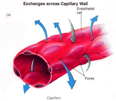
50 Structure of blood vessels 1. Endothelial cells Only lining found in capillary Line entire vascular tree Provide a smooth luminal surface protects against intravascular coagulation Intercellular clefts, cytoplasmic pores, and allow exchange to occur between blood and tissue fluid Capable of secreting a number of substances Capable of reproduction
51 Structure of blood vessels..cont 2. Collagen fibers Formed from protein molecules that aggregate into fibers Have only a limited ability to stretch Function to strengthen and keep lumen of vessel open 3. Elastic fibers Composed of insoluble protein called elastin Fibers can stretch over 100% under physiological conditions Play important role in creating passive tension to help regulate blood pressure throughout cardiac cycle 4. Smooth muscle fibers Present in all segments of vascular system except capillaries
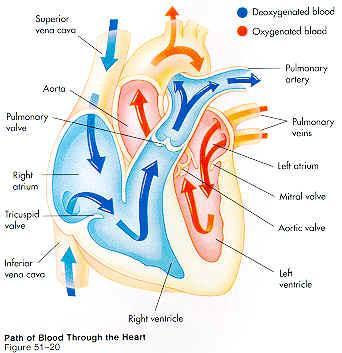
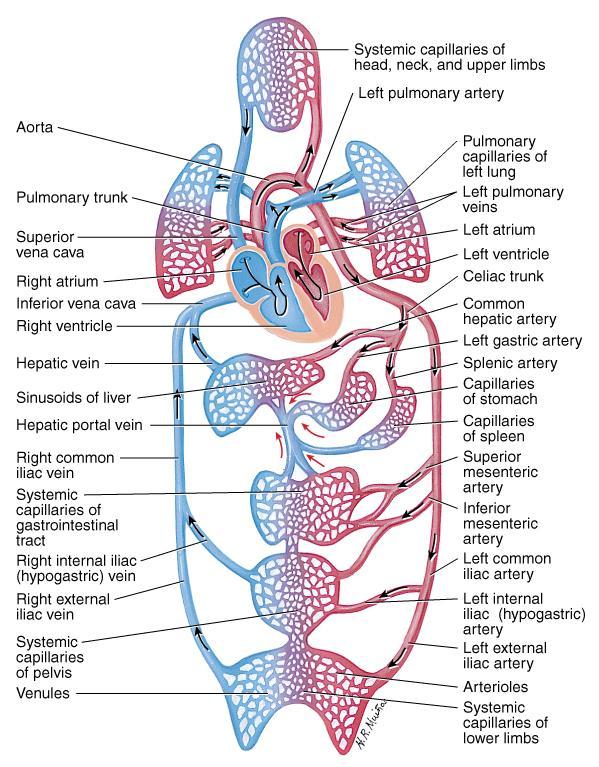
52 Types Circulation Systemic circulation blood flows from the left ventricle of the heart through blood vessels to all parts of the body (except gas exchange tissues of lungs) and back to right atrium Pulmonary circulation: venous blood moves from right atrium to right ventricle to pulmonary artery to lung arterioles and capillaries where gases are exchanged; oxygenated blood returns to left atrium via pulmonary veins; from left atrium, blood enters left ventricle
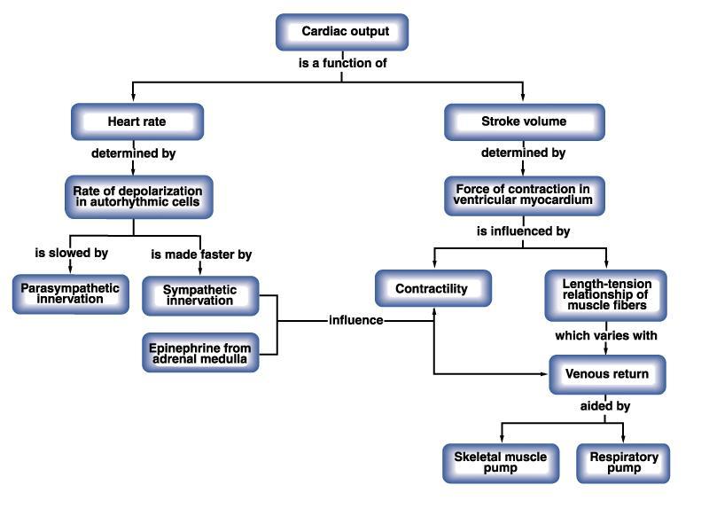
53
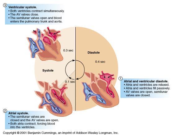
54 The Heart As a Pump Cardiac cycle a complete heartbeat consisting of contraction (systole) and relaxation (diastole) of both atria and both ventricles; the cycle is often divided into time intervals Atrial systole Contraction of atria completes emptying blood out of the atria into the ventricles AV valves are open; semilunar (SL) valves are closed Ventricles are relaxed and filling with blood This cycle begins with the P wave of the ECG
55 Cardiac cycle (cont.) ventricular contraction Occurs between the start of ventricular systole and the opening of the SL valves Ventricular volume remains constant as the pressure increases rapidly Onset of ventricular systole coincides with the R wave of the ECG and the appearance of the first heart sound
56 Ejection SL valves open and blood is ejected from the heart when the pressure gradient in the ventricles exceeds the pressure in the pulmonary artery and aorta Rapid ejection initial, short phase is characterized by a marked increase in ventricular and aortic pressure and in aortic blood flow
57 Cardiac cycle (cont.) ventricular relaxation Ventricular diastole begins with this phase Occurs between closure of the SL valves and opening of the AV valves A dramatic fall in intraventricular pressure but no change in volume The second heart sound is heard during this period
58
59 Contraction: systole Relaxation: Diastole The heart sounds Heart sound lub closing of AV valves dub closing of semilunar valves Heart sounds have clinical significance because they give information about the functioning of the valves of the heart
60 Cardiac out put Cardiac Output - the amount of blood pumped by a ventricle per minute. Units may be in milliliters or Liters per minute. Heart Rate - number of cardiac cycles per minute. Average Stroke Volume - amount of blood pumped out of a ventricle each beat. Average resting stroke volume = 70 ml.
61 COP Determined by: Cardiac output = Stroke Volume x Rate 5040ml = 70ml x 72 bpm Peripheral resistance Diameter of blood vessels Viscosity of blood Peripheral resistance resistance to blood flow Factors that influence peripheral resistance Blood viscosity Diameter of arterioles
62 Frank Starling Law of the Heart = Increased blood volume = increased stretch of myocardium Increased force to pump blood out.
63
64 Factors that affect heart rate 1. SA node normally initiates each heartbeat. 2. Anxiety, fear, and anger often increase heart rate 3. Grief tends to decrease heart rate 4. Emotions 5. Exercise 6. Increased blood temperature 7. Decreased blood temperature
65 Venous Return to Heart Venous return: amount of blood returned to the heart by the veins 1. Stress-relaxation effect : occurs when a change in blood pressure causes a change in vessel diameter 2. Gravity: the pull of gravity on venous blood while sitting or standing 3. Venous pumps: Respirations Skeletal muscle contractions Semilunar valves in veins
66 Blood pressure Systolic blood pressure : while ventricles are contracting Diastolic blood pressure: when ventricles are relaxed Pulse pressure: difference between systolic and diastolic Relation to arterial and venous bleeding 1. Arterial bleeding: blood escapes from artery 2. Venous bleeding: blood flows slowly and steadily due to low, practically constant pressure

67 Blood pressure
68 Factors influencing blood pressure Blood Pressure = Blood Volume Peripheral Resistance Blood volume loss due to injuries, hemorrhages, use of diuretics, etc. = BP Blood volume increases due to increased water retention from increased ADH production, IVs or transfusions = BP
69 Factors influencing blood pressure Blood Pressure = Blood Volume Peripheral Resistance Peripheral Resistance affected by: blood viscosity (thickness) (Polycythemia) diameter of vessels (vasoconstriction/vasodilation) Vasoconstriction = diameter = resistance = BP Vasodilation = diameter = resistance = BP elasticity of arterial walls Elastic Arterial Walls = BP
70 Blood pressure Hypotension: abnormal low blood pressure. Sustained systolic blood pressure of below 100 mm Hg. Hypertension : High blood pressure arterial blood pressure of 140/90 mm Hg or above
71 Pulse Pulse: alternate expansion and recoil of an artery Alternating increase and decrease of pressure in the vessel Elasticity of arterial walls allows walls to expand with increased pressure and recoil with decreased pressure Pulse site: Temporal radial carotid apical brachial posterior tibial Dorsal spedis pulse. Popliteal Femoral
72 Any Question??? Good luck Dr. Yassin Abobaker Abdulghani
10. Thick deposits of lipids on the walls of blood vessels, called, can lead to serious circulatory issues. A. aneurysm B. atherosclerosis C.
 Heart Student: 1. carry blood away from the heart. A. Arteries B. Veins C. Capillaries 2. What is the leading cause of heart attack and stroke in North America? A. alcohol B. smoking C. arteriosclerosis
Major Function of the Cardiovascular System. Transportation. Structures of the Cardiovascular System. Heart - muscular pump
 Structures of the Cardiovascular System Heart - muscular pump Blood vessels - network of tubes Blood - liquid transport vehicle brachiocephalic trunk superior vena cava right pulmonary arteries right pulmonary
The Cardiovascular System
 The Cardiovascular System The Cardiovascular System A closed system of the heart and blood vessels The heart pumps blood Blood vessels allow blood to circulate to all parts of the body The function of
Cardiovascular Anatomy Dr. Gary Mumaugh
 Cardiovascular Anatomy Dr. Gary Mumaugh Location of Heart Approximately the size of your fist Location o Superior surface of diaphragm o Left of the midline in mediastinum o Anterior to the vertebral column,
Cardiovascular System
 Cardiovascular System Purpose Transport oxygen and nutrients Take waste products away from tissues & organs Things we learned Blood pressure: the force of blood pushing against the walls of blood vessels
Chapter 14. The Cardiovascular System
 Chapter 14 The Cardiovascular System Introduction Cardiovascular system - heart, blood and blood vessels Cardiac muscle makes up bulk of heart provides force to pump blood Function - transports blood 2
Figure ) The specific chamber of the heart that is indicated by letter A is called the. Diff: 1 Page Ref: 364
 The specific chamber of the heart that is indicated by letter A is called the. Diff: 1 Page Ref: 364") Essentials of Anatomy and Physiology, 9e (Marieb) Chapter 11 The Cardiovascular System Short Answer Figure 11.1 Using Figure 11.1, identify the following: 1) The Purkinje fibers are indicated by label.
Essentials of Anatomy and Physiology, 9e (Marieb) Chapter 11 The Cardiovascular System Short Answer Figure 11.1 Using Figure 11.1, identify the following: 1) The Purkinje fibers are indicated by label.
Cardiovascular System Notes: Physiology of the Heart
 Cardiovascular System Notes: Physiology of the Heart Interesting Heart Fact Capillaries are so small it takes ten of them to equal the thickness of a human hair. Review What are the 3 parts of the cardiovascular
The Cardiovascular System
 Essentials of Human Anatomy & Physiology Elaine N. Marieb Seventh Edition Chapter 11 The Cardiovascular System Slides 11.1 11.19 Lecture Slides in PowerPoint by Jerry L. Cook The Cardiovascular System
Pearson's Comprehensive Medical Assisting Administrative and Clinical Competencies
 Pearson's Comprehensive Medical Assisting Administrative and Clinical Competencies THIRD EDITION CHAPTER 27 The Cardiovascular System Lesson 1: Overview of the Cardiovascular System Lesson Objectives Upon
Chapter 18 - Heart. I. Heart Anatomy: size of your fist; located in mediastinum (medial cavity)
") Chapter 18 - Heart I. Heart Anatomy: size of your fist; located in mediastinum (medial cavity) A. Coverings: heart enclosed in double walled sac called the pericardium 1. Fibrous pericardium: dense connective
Chapter 18 - Heart I. Heart Anatomy: size of your fist; located in mediastinum (medial cavity) A. Coverings: heart enclosed in double walled sac called the pericardium 1. Fibrous pericardium: dense connective
11/10/2014. Muscular pump Two atria Two ventricles. In mediastinum of thoracic cavity 2/3 of heart's mass lies left of midline of sternum
 It beats over 100,000 times a day to pump over 1,800 gallons of blood per day through over 60,000 miles of blood vessels. During the average lifetime, the heart pumps nearly 3 billion times, delivering
The Heart. Happy Friday! #takeoutyournotes #testnotgradedyet
 The Heart Happy Friday! #takeoutyournotes #testnotgradedyet Introduction Cardiovascular system distributes blood Pump (heart) Distribution areas (capillaries) Heart has 4 compartments 2 receive blood (atria)
The Cardiovascular System. Chapter 15. Cardiovascular System FYI. Cardiology Closed systemof the heart & blood vessels. Functions
 Chapter 15 Cardiovascular System FYI The heart pumps 7,000 liters (4000 gallons) of blood through the body each day The heart contracts 2.5 billion times in an avg. lifetime The heart & all blood vessels
Ch 19: Cardiovascular System - The Heart -
 Ch 19: Cardiovascular System - The Heart - Give a detailed description of the superficial and internal anatomy of the heart, including the pericardium, the myocardium, and the cardiac muscle. Trace the
Approximately the size of your fist Location Superior surface of diaphragm Left of the midline in mediastinum Anterior to the vertebral column,
 Dr. Gary Mumaugh Approximately the size of your fist Location Superior surface of diaphragm Left of the midline in mediastinum Anterior to the vertebral column, posterior to the sternum Posteriorly the
CIRCULATORY SYSTEM BLOOD VESSELS
 Name: Block: CIRCULATORY SYSTEM Multicellular organisms (above the level of roundworms) rely on a circulatory system to bring nutrients to, and take wastes away from, cells. In higher organisms such as
The Cardiovascular System
 11 PART A The Cardiovascular System PowerPoint Lecture Slide Presentation by Jerry L. Cook, Sam Houston University ESSENTIALS OF HUMAN ANATOMY & PHYSIOLOGY EIGHTH EDITION ELAINE N. MARIEB The Cardiovascular
The Heart. Size, Form, and Location of the Heart. 1. Blunt, rounded point; most inferior part of the heart.
 12 The Heart FOCUS: The heart is composed of cardiac muscle cells, which are elongated, branching cells that appear striated. Cardiac muscle cells behave as a single electrical unit, and the highly coordinated
4. The two inferior chambers of the heart are known as the atria. the superior and inferior vena cava, which empty into the left atrium.
 Answer each statement true or false. If the statement is false, change the underlined word to make it true. 1. The heart is located approximately between the second and fifth ribs and posterior to the
Cardiovascular System
 Cardiovascular System I. Structure of the Heart A. Average adult heart is 14 cm long and 9 cm wide. B. Lies in the mediastinum. C. Enclosed in the pericardium. 1. Fibrous pericardium- Outer, tough connective
Heart. Heart 2-Tunica media: middle layer (media ='middle') muscle fibers (smooth or cardiac).
 muscle fibers (smooth or cardiac).") t. innermost lumenal General Circulatory system heart and blood vessels walls have 3 layers (inside to outside) 1-Tunica interna: aka tunica intima layer--lumenal layer epithelium--endothelium simple squamous
t. innermost lumenal General Circulatory system heart and blood vessels walls have 3 layers (inside to outside) 1-Tunica interna: aka tunica intima layer--lumenal layer epithelium--endothelium simple squamous
The HEART. What is it???? Pericardium. Heart Facts. This muscle never stops working It works when you are asleep
 This muscle never stops working It works when you are asleep The HEART It works when you eat It really works when you exercise. What is it???? Located between the lungs in the mid thoracic region Apex
2. right heart = pulmonary pump takes blood to lungs to pick up oxygen and get rid of carbon dioxide
 A. location in thorax, in inferior mediastinum posterior to sternum medial to lungs superior to diaphragm anterior to vertebrae orientation - oblique apex points down and to the left 2/3 of mass on left
The Cardiovascular System (Heart)
") The Cardiovascular System The Cardiovascular System (Heart) A closed system of the heart and blood vessels The heart pumps blood Blood vessels allow blood to circulate to all parts of the body The function
The Cardiovascular System The Cardiovascular System (Heart) A closed system of the heart and blood vessels The heart pumps blood Blood vessels allow blood to circulate to all parts of the body The function
Cardiovascular System. I. Structures of the heart A. : Pericardium sack that surrounds the heart
 Cardiovascular System I. Structures of the heart A. : Pericardium sack that surrounds the heart 1. : Pericardial Cavity serous fluid filled space between the heart and the pericardium B. Heart Wall 1.
The Heart and Cardiovascular System
 The Heart and Cardiovascular System What you will learn The location of the heart 3 layers and covering of the heart Explain the function of the heart as 2 separate pumps Identify the 4 chambers of the
Health Science 20 Circulatory System Notes
 Health Science 20 Circulatory System Notes Functions of the Circulatory System The circulatory system functions mainly as the body s transport system. It transports: o Oxygen o Nutrients o Cell waste o
Anatomy Review: The Heart Graphics are used with permission of A.D.A.M. Software, Inc. and Benjamin/Cummings Publishing Co.
 Anatomy Review: The Heart Graphics are used with permission of A.D.A.M. Software, Inc. and Benjamin/Cummings Publishing Co. Anatomy Views Label the diagrams of the heart below: Interactive Physiology Study
THE CARDIOVASCULAR SYSTEM. Part 1
 THE CARDIOVASCULAR SYSTEM Part 1 CARDIOVASCULAR SYSTEM Blood Heart Blood vessels What is the function of this system? What other systems does it affect? CARDIOVASCULAR SYSTEM Functions Transport gases,
1. Which of the following blood vessels has a thin elastic layer? A. Aorta. B. Pulmonary artery. C. Posterior vena cava. D. Mesenteric capillary.
 CIRCULATORY SYSTEM 1. Which of the following blood vessels has a thin elastic layer? A. Aorta. B. Pulmonary artery. C. Posterior vena cava. D. Mesenteric capillary. 2. Capillary beds are equipped with
10/23/2017. Muscular pump Two atria Two ventricles. In mediastinum of thoracic cavity 2/3 of heart's mass lies left of midline of sternum
 It beats over 100,000 times a day to pump over 1,800 gallons of blood per day through over 60,000 miles of blood vessels. During the average lifetime, the heart pumps nearly 3 billion times, delivering
Cardiovascular System
 Cardiovascular System The Heart Cardiovascular System The Heart Overview What does the heart do? By timed muscular contractions creates pressure gradients blood moves then from high pressure to low pressure
Human Anatomy, First Edition
 Human Anatomy, First Edition McKinley & O'Loughlin Chapter 22 : Heart 1 Functions of the Heart Center of the cardiovascular system, the heart. Connects to blood vessels that transport blood between the
STRUCTURES OF THE CARDIOVASCULAR SYSTEM
 STRUCTURES OF THE CARDIOVASCULAR SYSTEM CARDIOVASCULAR SYSTEM Also called the circulatory system Consists of the heart, arteries, veins, and capillaries Main function is to pump/circulate oxygenated blood
The Cardiovascular and Lymphatic Systems Cardiovascular System Blood Vessels Blood Vessels Arteries Arteries Arteries
 CH 12 The Cardiovascular and s The Cardiovascular and s OUTLINE: Cardiovascular System Blood Vessels Blood Pressure Cardiovascular System The cardiovascular system is composed of Blood vessels This system
Cardiovascular system
 BIO 301 Human Physiology Cardiovascular system The Cardiovascular System: consists of the heart plus all the blood vessels transports blood to all parts of the body in two 'circulations': pulmonary (lungs)
The Heart. The Heart A muscular double pump. The Pulmonary and Systemic Circuits
 C H A P T E R 19 The Heart The Heart A muscular double pump circuit takes blood to and from the lungs Systemic circuit vessels transport blood to and from body tissues Atria receive blood from the pulmonary
37 1 The Circulatory System
 H T H E E A R T 37 1 The Circulatory System The circulatory system and respiratory system work together to supply cells with the nutrients and oxygen they need to stay alive. a) The respiratory system:
Anatomy of the Heart. Figure 20 2c
 Anatomy of the Heart Figure 20 2c Pericardium & Myocardium Remember, the heart sits in it s own cavity, known as the mediastinum. The heart is surrounded by the Pericardium, a double lining of the pericardial
Chapter 13 The Cardiovascular System: Cardiac Function
 Chapter 13 The Cardiovascular System: Cardiac Function Overview of the Cardiovascular System The Path of Blood Flow through the Heart and Vasculature Anatomy of the Heart Electrical Activity of the Heart
THE HEART. A. The Pericardium - a double sac of serous membrane surrounding the heart
 THE HEART I. Size and Location: A. Fist-size weighing less than a pound (250 to 350 grams). B. Located in the mediastinum between the 2 nd rib and the 5 th intercostal space. 1. Tipped to the left, resting
The Cardiovascular and Lymphatic Systems
 BIOLOGY OF HUMANS Concepts, Applications, and Issues Fifth Edition Judith Goodenough Betty McGuire 12 The Cardiovascular and Lymphatic Systems Lecture Presentation Anne Gasc Hawaii Pacific University and
Ch. 12 The Circulatory System. The heart. The heart is a double pump. A quick note on arteries vs. veins. = the muscular pump of the CV system
 Ch. 12 The Circulatory System The heart A.k.a. the cardiovascular system Blood was discussed in Ch. 11 Focus of Ch. 12: heart and blood vessels = the muscular pump of the CV system ~ 100,000 heartbeats/day!
- what other structures, besides the heart, does the mediastinum contain?
 Basic A & P II Dr. L. Bacha Chapter Outline (Martini & Nath 2010) An Introduction to the Cardiovascular System - read the paragraphs under this heading on page 580 The Heart is a Four Chambered Organ describe
Chapter 20: Cardiovascular System: The Heart
 Chapter 20: Cardiovascular System: The Heart I. Functions of the Heart A. List and describe the four functions of the heart: 1. 2. 3. 4. II. Size, Shape, and Location of the Heart A. Size and Shape 1.
the Cardiovascular System I
 the Cardiovascular System I By: Dr. Nabil A Khouri MD, MsC, Ph.D MEDIASTINUM 1. Superior Mediastinum 2. inferior Mediastinum Anterior mediastinum. Middle mediastinum. Posterior mediastinum Anatomy of
Cardiovascular System. Biology 105 Lecture 15 Chapter 12
 Cardiovascular System Biology 105 Lecture 15 Chapter 12 Outline I. Functions of cardiovascular system II. Components of the cardiovascular system: I. Blood vessels II. Heart III. Regulation of the heartbeat
The Circulatory System. The Heart, Blood Vessels, Blood Types
 The Circulatory System The Heart, Blood Vessels, Blood Types The Closed Circulatory System Humans have a closed circulatory system, typical of all vertebrates, in which blood is confined to vessels and
THE HEART OBJECTIVES: LOCATION OF THE HEART IN THE THORACIC CAVITY CARDIOVASCULAR SYSTEM
 BIOLOGY II CARDIOVASCULAR SYSTEM ACTIVITY #3 NAME DATE HOUR THE HEART OBJECTIVES: Describe the anatomy of the heart and identify and give the functions of all parts. (pp. 356 363) Trace the flow of blood
Collin County Community College. ! BIOL Anatomy & Physiology! WEEK 5. The Heart
 Collin County Community College! BIOL. 2402 Anatomy & Physiology! WEEK 5 The Heart 1 (1578-1657) A groundbreaking work in the history of medicine, English physician William Harvey s Anatomical Essay on
Chapter 13. Cardiovascular System
 Chapter 13 Cardiovascular System 1 Introduction A. The cardiovascular system consists of the heart and vessels (arteries, capillaries and veins.) B. A functional cardiovascular system is vital for supplying
Approximately the size of your fist Location. Pericardial physiology
 Heart Anatomy Approximately the size of your fist Location Superior surface of diaphragm Left of the midline Anterior to the vertebral column, posterior to the sternum Wednesday, March 28, 2012 Muscle
UNIT 11: THE CARDIOVASCULAR SYSTEM
 UNIT 11: THE CARDIOVASCULAR SYSTEM Functions of the Heart PUMPS Blood Transports Oxygen and Nutrients Removes Carbon Dioxide and Metabolic Wastes Thermoregulation Immunological Function Clotting Mechanisms
CV Anatomy Quiz. Dr Ella Kim Dr Pip Green
 CV Anatomy Quiz Dr Ella Kim Dr Pip Green Q1 The location of the heart is correctly described as A) lateral to the lungs. B) medial to the sternum. C) superior to the diaphragm. D) posterior to the spinal
Function: Transportation of. Oxygen Nutrients Waste Hormones gases
 Function: Transportation of Oxygen Nutrients Waste Hormones gases Pericardium: double sac of serous membrane filled with fluid (pericardial fluid to be exact) that surrounds the heart. Parietal pericardium:
IB TOPIC 6.2 THE BLOOD SYSTEM
 IB TOPIC 6.2 THE BLOOD SYSTEM TERMS TO KNOW circulation ventricle artery vein THE BLOOD SYSTEM 6.2.U1 - Arteries convey blood at high pressure from the ventricles to the tissues of the body Circulation
Heart. Structure Physiology of blood pressure and heartbeat
 Heart Structure Physiology of blood pressure and heartbeat Location and Anatomy Location and Anatomy Pericardial cavity: surrounds, isolates, and anchors heart Parietal pericardium lined with serous membrane
Circulatory System Review
 Circulatory System Review 1. Know the diagrams of the heart, internal and external. a) What is the pericardium? What is myocardium? What is the septum? b) Explain the 4 valves of the heart. What is their
The Cardiovascular System
 The Cardiovascular System The Manila Times College of Subic Prepared by: Stevens B. Badar, RN, MANc THE HEART Anatomy of the Heart Location and Size approx. the size of a person s fist, hollow and cone-shaped,
Circulation. Circulation = is a process used for the transport of oxygen, carbon! dioxide, nutrients and wastes through-out the body
 Circulation Circulation = is a process used for the transport of oxygen, carbon! dioxide, nutrients and wastes through-out the body Heart = muscular organ about the size of your fist which pumps blood.
LECTURE 5. Anatomy of the heart
 LECTURE 5. Anatomy of the heart Main components of the CVS: Heart Blood circulatory system arterial compartment haemomicrocirculatory (=microvascular) compartment venous compartment Lymphatic circulatory
Cardiovascular System. Heart Anatomy
 Cardiovascular System Heart Anatomy 1 The Heart Location & general description: Atria vs. ventricles Pulmonary vs. systemic circulation Coverings Walls The heart is found in the mediastinum, the medial
Cardiovascular. Function of the cardiovascular system is to transport blood containing: Nutrients Waste Hormones Immune cells Oxygen
 Cardiovascular The Cardiovascular System - Arteries Arteries Cardiovascular System Function of the cardiovascular system is to transport blood containing: Carry blood away from heart Carotid arteries Deliver
P215 SPRING 2019: CIRCULATORY SYSTEM Chaps 13, 14 & 15: pp , , , I. Major Functions of the Circulatory System
 P215 SPRING 2019: CIRCULATORY SYSTEM Chaps 13, 14 & 15: pp 360-390, 395-404, 410-428 433-438, 441-445 I. Major Functions of the Circulatory System 1. 2. 3. 4. II. Structure of the Heart 1. atria 2. ventricles
Cardiac Conduction System
 Cardiac Conduction System What causes the Heart to Beat? Heart contracts by electrical signals! Cardiac muscle tissue contracts on its own an electrical signal is sent out by the heart so that all cells
Chapter 20 (1) The Heart
 The Heart") Chapter 20 (1) The Heart Learning Objectives Describe the location and structure of the heart Describe the path of a drop of blood from the superior vena cava or inferior vena cava through the heart out
Chapter 20 (1) The Heart Learning Objectives Describe the location and structure of the heart Describe the path of a drop of blood from the superior vena cava or inferior vena cava through the heart out
BIOL 4350 Cardiovascular Physiology Dr. Hamilton. Using the figure above, match the following: 1. Purkinje fibers. 2. SA node. 3. AV node.
 BIOL 4350 Cardiovascular Physiology Dr. Hamilton Using the figure above, match the following: 1. Purkinje fibers. 2. SA node. 3. AV node. 1 Using the figure above, match the following: 4. Atrial depolarization.
The Cardiovascular System
 Essentials of Human Anatomy & Physiology Elaine N. Marieb Slides 11.1 11.19 Seventh Edition Chapter 11 The Cardiovascular System Functions of the Cardiovascular system Function of the heart: to pump blood
d) Cardiovascular System Higher Human Biology
 Cardiovascular System Higher Human Biology") d) Cardiovascular System Higher Human Biology What can your remember about the heart and blood vessels? What is the Cardiovascular System? The cardiovascular system, also known as the circulatory system,
d) Cardiovascular System Higher Human Biology What can your remember about the heart and blood vessels? What is the Cardiovascular System? The cardiovascular system, also known as the circulatory system,
CARDIOVASCULAR SYSTEM
 CARDIOVASCULAR SYSTEM Overview Heart and Vessels 2 Major Divisions Pulmonary Circuit Systemic Circuit Closed and Continuous Loop Location Aorta Superior vena cava Right lung Pulmonary trunk Base of heart
The Heart. C h a p t e r. PowerPoint Lecture Slides prepared by Jason LaPres Lone Star College - North Harris
 C h a p t e r 20 The Heart PowerPoint Lecture Slides prepared by Jason LaPres Lone Star College - North Harris Copyright 2009 Pearson Education, Inc., publishing as Pearson Benjamin Cummings Introduction
Cardiovascular Physiology. Heart Physiology. Introduction. The heart. Electrophysiology of the heart
 Cardiovascular Physiology Heart Physiology Introduction The cardiovascular system consists of the heart and two vascular systems, the systemic and pulmonary circulations. The heart pumps blood through
Cardiovascular System Notes: Heart Disease & Disorders
 Cardiovascular System Notes: Heart Disease & Disorders Interesting Heart Facts The Electrocardiograph (ECG) was invented in 1902 by Willem Einthoven Dutch Physiologist. This test is still used to evaluate
The cardiovascular system is composed of the heart and blood vessels that carry blood to and from the body s organs. There are 2 major circuits:
 1 The cardiovascular system is composed of the heart and blood vessels that carry blood to and from the body s organs. There are 2 major circuits: pulmonary and systemic. The pulmonary goes out to the
Anatomy and Physiology, Spring 2015 Exam II: Form A April 9, Name Student Number
 Anatomy and Physiology, Spring 2015 Exam II: Form A April 9, 2015 Name Student Number For Questions 1 2 refer to the following table. 1 Ventricular pressure is greater than aortic 6 AV valve is open 2
Test Review Circulatory System Chapters
 Test Review Circulatory System Chapters 13-2010 1. The tissue that forms the tight fitting sac around the heart is the a. parietal pericardium c. myocardium b. visceral pericardium d. endocardium 2. Which
The Cardiovascular System. Preview of Heart Action. The CV system provides oxygen & nutrients to tissues-removes wastes.
 The Cardiovascular System BIO 250 Human Anatomy & Physiology Preview of Heart Action http://www.youtube.com/watch?v=d3zdj gfddk0&nr=1 The CV system provides oxygen & nutrients to tissues-removes wastes.
Blood Functions. Blood and the Cardiovascular System. Blood. Plasma. Erythrocytes (RBCs) Erythrocytes (RBCs) 4/7/2017
 Erythrocytes (RBCs) 4/7/2017") Blood Functions Blood and the Cardiovascular System Distribution Delivery of oxygen and nutrients to all body cells; Transport of wastes to lungs and excretory organs; Transport of hormones Regulation
Blood Functions Blood and the Cardiovascular System Distribution Delivery of oxygen and nutrients to all body cells; Transport of wastes to lungs and excretory organs; Transport of hormones Regulation
The ancient Babylonians, Egyptians, Indians and Chinese believed the heart was the centre of thinking and emotions
 The Concept of Mind The ancient Babylonians, Egyptians, Indians and Chinese believed the heart was the centre of thinking and emotions Hippocrates 460 BC 370 BC - Thoughts, ideas, and feelings come from
Principles of Biomedical Systems & Devices. Lecture 8: Cardiovascular Dynamics Dr. Maria Tahamont
 Principles of Biomedical Systems & Devices Lecture 8: Cardiovascular Dynamics Dr. Maria Tahamont Review of Cardiac Anatomy Four chambers Two atria-receive blood from the vena cave and pulmonary veins Two
Circulation. Sinoatrial (SA) Node. Atrioventricular (AV) Node. Cardiac Conduction System. Cardiac Conduction System. Linked to the nervous system
 Node. Atrioventricular (AV) Node. Cardiac Conduction System. Cardiac Conduction System. Linked to the nervous system") Circulation Cardiac Conduction System AHS A H S Your body resembles a large roadmap. There are routes or arteries that take you downtown to the heart of the city and veins that take you to the outskirts
Circulation Cardiac Conduction System AHS A H S Your body resembles a large roadmap. There are routes or arteries that take you downtown to the heart of the city and veins that take you to the outskirts
Unit 11 - The Cardiovascular System 1
 Unit 11 - The Cardiovascular System 1 I. Unit 11: The Cardiovascular System A. The Cardiovascular System 1. A closed system of the heart and blood vessels a) The heart pumps blood b) Blood vessels allow
Anatomy of the Heart
 Biology 212: Anatomy and Physiology II Anatomy of the Heart References: Saladin, KS: Anatomy and Physiology, The Unity of Form and Function 8 th (2018). Required reading before beginning this lab: Chapter
The Circulatory System (p )
") The Circulatory System (p. 268-281) How Does Gravity Affect Blood Circulation? As with all land animals, the giraffe and the corn snake are constantly subject to the force of gravity The circulatory system
The Circulatory System (p. 268-281) How Does Gravity Affect Blood Circulation? As with all land animals, the giraffe and the corn snake are constantly subject to the force of gravity The circulatory system
A closed system of the heart/blood. Function: The heart pumps blood. Blood vessels allow blood to circulate throughout the body
 A closed system of the heart/blood The heart pumps blood It is no more than a transportation pump Blood vessels allow blood to circulate throughout the body MILES of blood vessels intricate network At
IB TOPIC 6.2 THE BLOOD SYSTEM
 IB TOPIC 6.2 THE BLOOD SYSTEM THE BLOOD SYSTEM TERMS TO KNOW circulation ventricle artery vein 6.2.U1 - Arteries convey blood at high pressure from the ventricles to the tissues of the body Circulation
BIOL 219 Spring Chapters 14&15 Cardiovascular System
 1 BIOL 219 Spring 2013 Chapters 14&15 Cardiovascular System Outline: Components of the CV system Heart anatomy Layers of the heart wall Pericardium Heart chambers, valves, blood vessels, septum Atrioventricular
The Heart and Heart Disease
 The Heart and Heart Disease Illustration of the heart by Leonardo DaVinci heart-surgeon.com/ history.html 2/14/2010 1 I. Location, Size and Position of the Heart A. Triangular organ located 1. of mass
Cardiovascular System
 Hole s Essentials of Human Anatomy & Physiology David Shier Jackie Butler Ricki Lewis Created by Dr. Melissa Eisenhauer Head Athletic Trainer/Assistant Professor Trevecca Nazarene University Chapter 13
1. What kind of blood is found in the rt. atrium? (oxygenated or deoxygenated)
") Carl Christennsen, PhD Chap. 19, 20, & 21 - Circulatory System Bio. 2304 Human Anatomy HEART 1. What kind of blood is found in the rt. atrium? (oxygenated or deoxygenated) Where does this blood come from?
Carl Christennsen, PhD Chap. 19, 20, & 21 - Circulatory System Bio. 2304 Human Anatomy HEART 1. What kind of blood is found in the rt. atrium? (oxygenated or deoxygenated) Where does this blood come from?
Biology Unit 3 The Human Heart P
 Biology 2201 Unit 3 The Human Heart P 314-321 Structure and Function of the Human Heart Structure of the Human Heart Has four Chambers (2 Atria and 2 Ventricles) Made of Cardiac Muscle Found in Chest Cavity
Large Arteries of Heart
 Cardiovascular System (Part A-2) Module 5 -Chapter 8 Overview Arteries Capillaries Veins Heart Anatomy Conduction System Blood pressure Fetal circulation Susie Turner, M.D. 1/5/13 Large Arteries of Heart
Mr. Epithelium s Anatomy and Physiology Test SSSS
 Mr. Epithelium s Anatomy and Physiology Test SSSS You have 50 minutes to complete this test packet. One 8.5 x 11 cheat sheet is allowed, along with 1 non-programmable calculator dedicated to computation.
Unit 8 THE CARDIOVASCULAR SYSTEM
 Unit 8 THE CARDIOVASCULAR SYSTEM 1 HEART FACTS Put your hand on your heart. Did you place your hand on the leftside of your chest? Many people do, but the heart is actually located almost in the center
Principles of Anatomy and Physiology
 Principles of Anatomy and Physiology 14 th Edition CHAPTER 20 The Cardiovascular System: The Heart Introduction The purpose of the chapter is to: 1. Learn about the components of the cardiovascular system
The Cardiovascular System
 PowerPoint Lecture Slide Presentation by Patty Bostwick-Taylor, Florence-Darlington Technical College The Cardiovascular System 11PART A The Cardiovascular System A closed system of the heart and blood
The Cardiovascular System Part I: Heart Outline of class lecture After studying part I of this chapter you should be able to:
 The Cardiovascular System Part I: Heart Outline of class lecture After studying part I of this chapter you should be able to: 1. Describe the functions of the heart 2. Describe the location of the heart,
Circulatory System 10.1
 1 Circulatory System 10.1 2 ARTERIES Arteries-blood vessels that carry blood away from the heart Thick walls Inner & Outer layers: connective tissue Middle layers are muscle and elastic connective tissue