Atlantic Health System
|
|
|
- Rosanna Nicholson
- 6 years ago
- Views:
Transcription


1 Atlantic Health System Morristown Medical Center Newton Medical Center Overlook Medical Center Basic Dysrhythmia Course Day 1 1
2 2
3 Chapter 1 Anatomy and Physiology
4 Learning Objectives 1) Identify electrophysiology of the heart. 2) Identify the electrical conduction structures and pathways of the heart. 3) Label an ECG complex identifying the waveforms and intervals for segments: PR, QRS, R-R, P-P, QT. 4) Recognize the features of normal sinus rhythm. 5) Identify atrial rhythms. 6) Identify junctional rhythms. 4
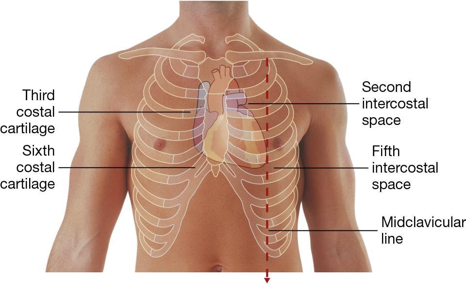
5 Location of the Heart Hollow muscular organ Lies in the space between the lungs (mediastinum) in the chest 2/3 lies to left of midline of sternum 5
6 6
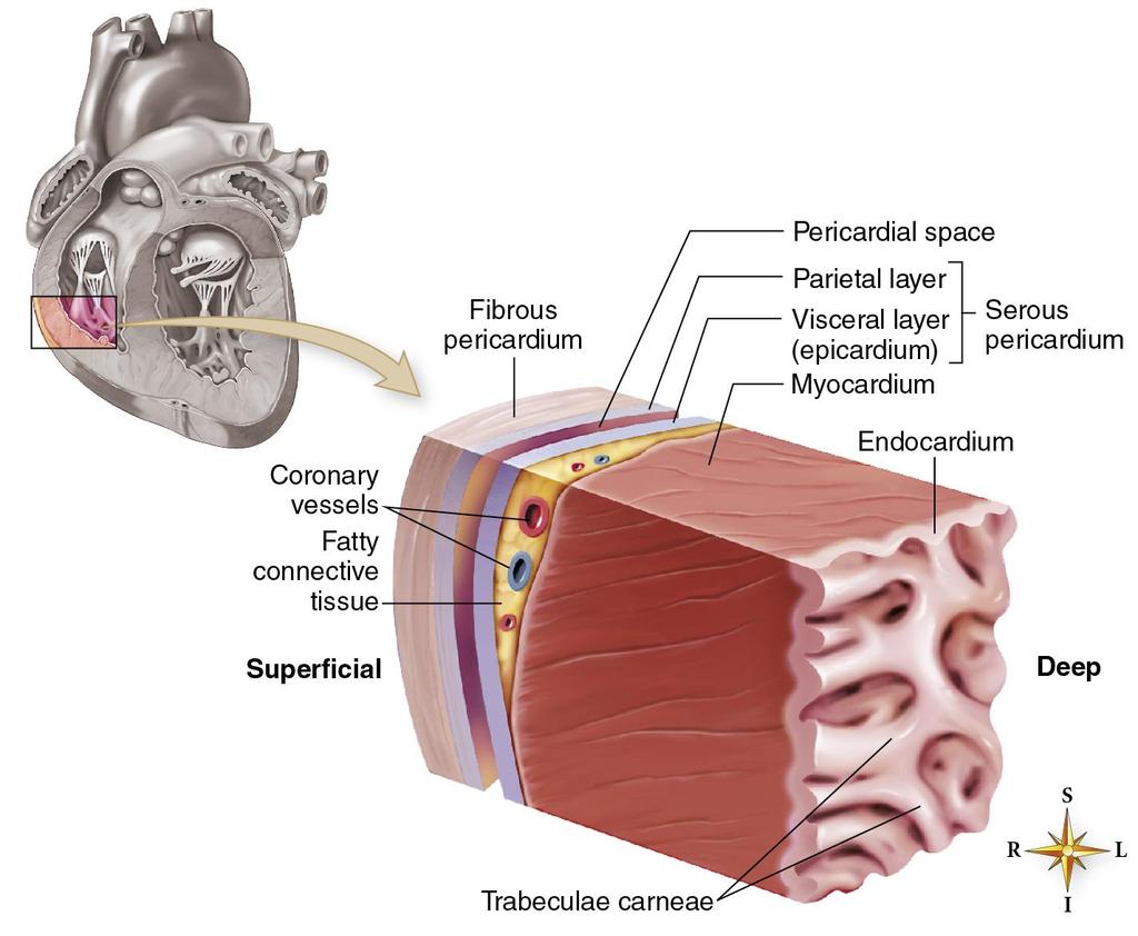
7 Coverings of the Heart Pericardium Parietal pericardium tough outer layer Serous pericardium Pericardial space contains serous fluid 7
8 Layers of the Heart Wall Endocardium Innermost layer Lines heart s inner chambers Continuous with innermost layer of vessels 8
9 Layers of the Heart Wall Myocardium Middle layer Thick, muscular layer Responsible for pumping action 9
10 Layers of the Heart Wall Epicardium Also called visceral layer of serous pericardium External layer of heart Contains blood capillaries, lymph capillaries, nerve fibers, and fat 10
11 11
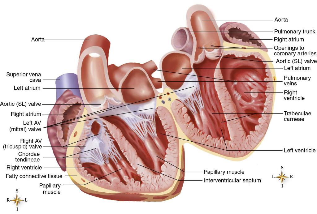
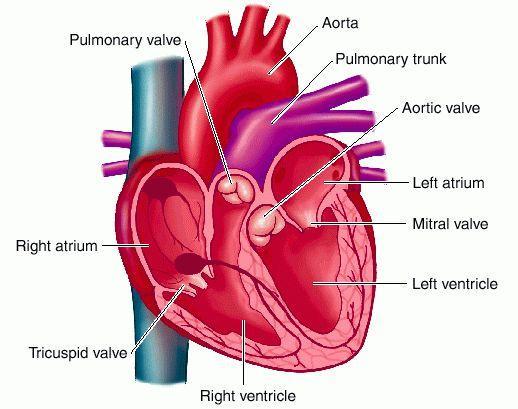
12 Heart Chambers Heart is divided into four chambers Two upper chambers = right and left atria Two lower chambers = right and left ventricles 12
13 13
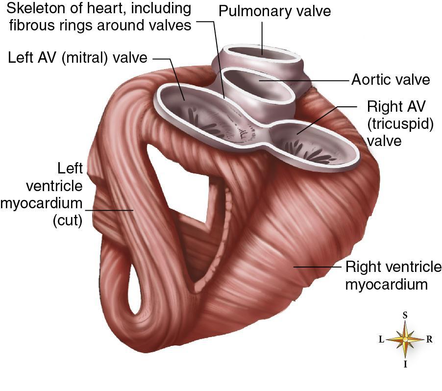
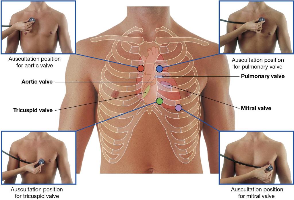
14 Heart Valves Heart contains four valves Two sets of atrioventricular (AV) valves -Leaflet Two sets of semilunar (SL) valves Crescent shaped cusps Function Ensure blood flows in one direction through heart chambers Prevent backflow of blood 14
15 15
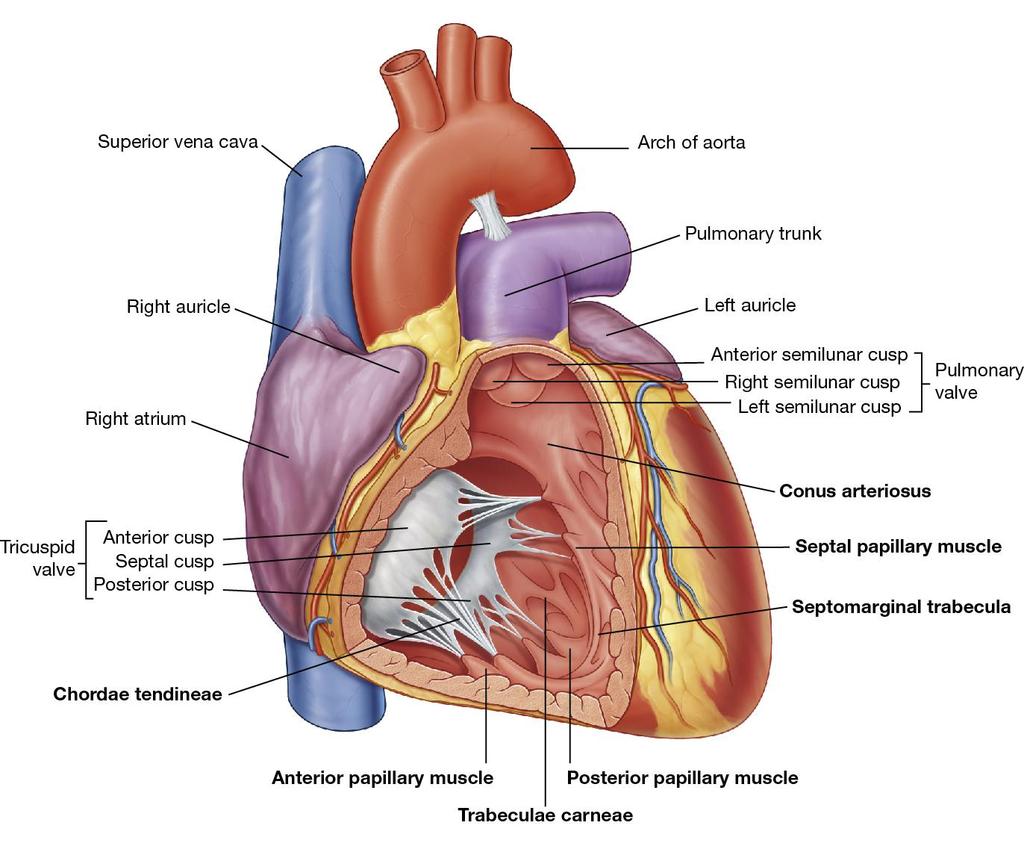
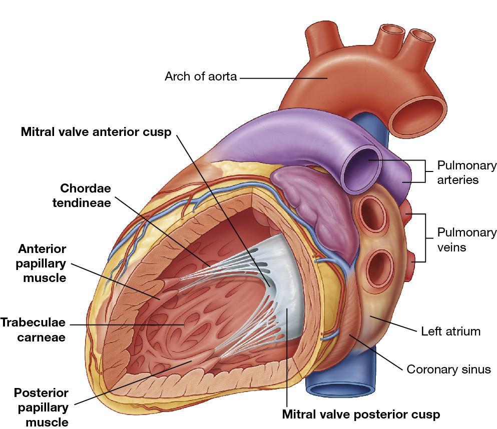
16 Atrioventricular (AV) Valves AV valves separate atria from ventricles Tricuspid valve Lies between right atrium and right ventricle Three separate leaflets Mitral (bicuspid) valve Has only two leaflets Lies between left atrium and left ventricle 16
17 Atrioventricular (AV) Valves Cusps of AV valves are attached to chordae tendineae Heart strings Attach into papillary muscles, which imbed deeply into the endocardium Serve as anchors 17
18 18
19 19
20 Semilunar Valves Prevent backflow of blood from the vessels into the ventricles during diastole Pulmonic valve Aortic valve 20
21 21
22 22
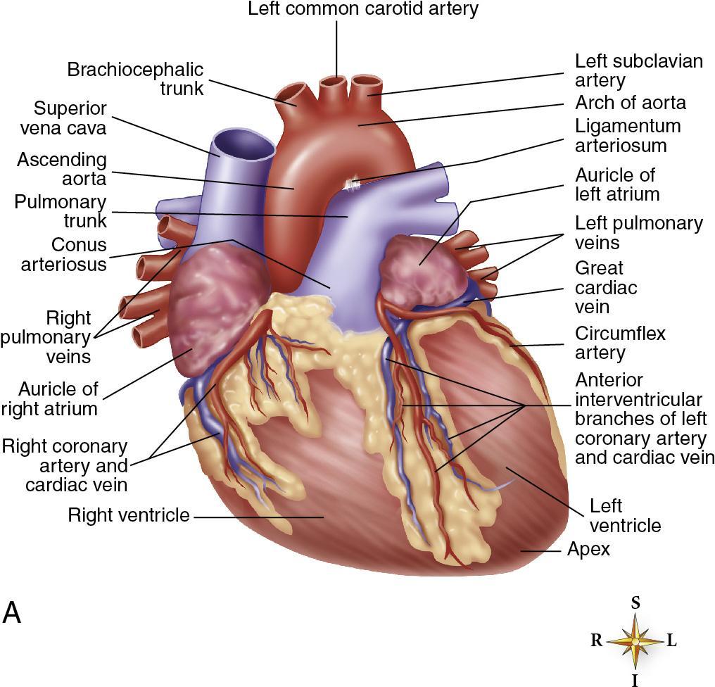
23 Coronary Circulation Coronary arteries Right Left Main Left anterior descending Circumflex Coronary veins Coronary sinus 23
24 24
25 Autonomic Nervous System Sympathetic division fight or flight Parasympathetic division rest and digest 25
26 Sympathetic Stimulation Impulses sent from accelerator center in medulla travel along sympathetic fibers Primary neurotransmitters: norepinephrine, epinephrine 26
27 Sympathetic Receptor Sites Alpha-1 Eyes, blood vessels, bladder, male reproductive organs Alpha-2 Digestive system Peripheral nervous system Beta-1 Heart, kidneys Beta-2 Bronchial smooth muscle Skeletal blood vessels Dopamine Renal, mesenteric, and visceral blood vessels 27
28 Parasympathetic Stimulation Primary neurotransmitter = acetylcholine Main effect = slowing of heart rate 28
29 Baroreceptors Also called pressoreceptors Specialized nerve tissue Found in internal carotid arteries/aortic arch Detect changes in blood pressure 29
30 Chemoreceptors Located in internal carotid arteries and aortic arch Detect and respond to changes in: Oxygen content of blood ph Carbon dioxide tension 30
31 Cardiac Cycle Systole Period during which the chamber is contracting and blood is being ejected Diastole Period of relaxation during which the chamber is filling 31
32 Atrial Systole and Diastole Atrial diastole Blood enters the right atrium Right atrium fills and distends Tricuspid valve opens and the right ventricle fills Same sequence occurs a split second earlier in the left heart Left atrium receives blood from pulmonary veins Mitral valve opens as the left atrium fills Blood flows into the left ventricle Atrial systole Atrial kick 32
33 Ventricular Systole and Diastole Ventricular systole Blood is propelled through the systemic and pulmonary circulation AV valves close Aortic and Pulmonic valves open Ventricular diastole Ventricles begin to passively fill with blood Aortic and Pulmonic valves close AV valves open 33
34 Cardiac Output Cardiac output Volume of blood pumped into the aorta each minute by the heart Stroke volume Amount of blood ejected from a ventricle with each heartbeat 34
35 Cardiac Output Cardiac output (CO) equals stroke volume (SV) multiplied by heart rate (HR) CO = SV HR In a healthy adult, CO at rest is about 5 L/min 35
36 Cardiac Output Affected by change in heart rate OR stroke volume Factors that influence heart rate Factors that affect stroke volume 36
37 Ejection Fraction The percentage of blood pumped out of a ventricle with each contraction Used as a measure of ventricular function A normal ejection fraction is between 50% and 65% 37
38 Signs and Symptoms of Decreased Cardiac Output: Your Thoughts
39 Signs and Symptoms of Decreased Cardiac Output Acute changes in blood pressure Acute changes in mental status Cold, clammy skin Color changes in the skin and mucous membranes Crackles (rales) Dyspnea Dysrhythmias Fatigue Orthopnea Restlessness 39
40 Questions?
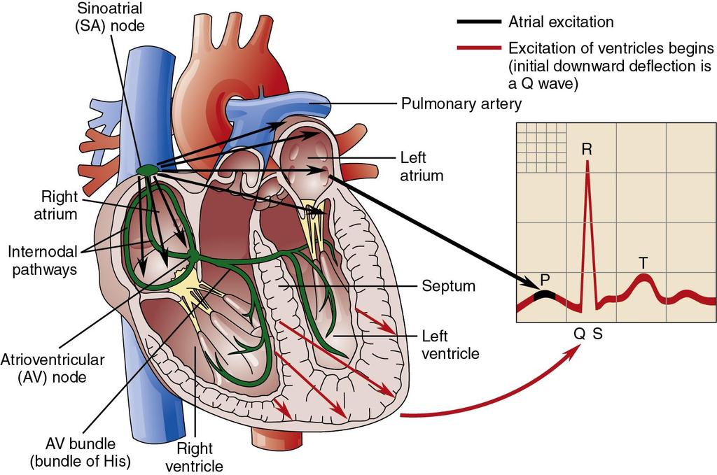
41 The Conduction System
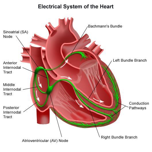
42 The Conduction System Conduction system Specialized electrical (pacemaker) cells Arranged in a system of pathways 42
43 43
 node")
44 The Conduction System Primary pacemaker Sinoatrial (SA) node 44
45 Atria The Conduction System Impulse leaves SA node to Bachmann s Bundle (to the left atrium) to simultaneously depolarize both atria Spreads from cell to cell across atrial muscle 45
46 Some abnormal heart rhythms that originate in the atria result in ineffectual atrial contraction. What effect would this have on atrial kick?
47 The Conduction System Internodal pathways Impulse is spread to AV node via internodal pathways Merge gradually with cells of AV node 47
48 The Conduction System AV node Located in floor of right atrium Supplied by right coronary artery in most people Delays conduction of impulse from atria to the ventricles Allows time for atria to empty into ventricles 48
49 The AV node and AV bundle 49
50 The Conduction System Bundle of His (AV bundle) Connects AV node with bundle branches Pacemaker cells have an intrinsic rate of 40 to 60 bpm Conducts impulse to right and left bundle branches 50
51 The Conduction System Right bundle branch Left bundle branch Divides into three fascicles Anterior fascicle Posterior fascicle Septal fascicle 51

52 The Conduction System Purkinje fibers Receive impulse from bundle branches Relay it to ventricular myocardium Pacemaker cells have an intrinsic rate of 20 to 40 bpm 52
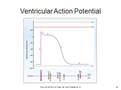
53 Cardiac Action Potential

54 Cardiac Action Potential Electrons Current Polarity Voltage 54
55 Cardiac Action Potential Electrolytes Ions Cell membranes 55
 56")
56 Polarization (Resting) 56

57 Depolarization = Stimulation 57
58 58
59 Refractory Periods Refractoriness The period of recovery that cells need after being discharged before they are able to respond to a stimulus 59

60 Refractory Periods 60

61 Refractory Periods 61
62 The Electrocardiogram (ECG)
63 63
64 The ECG Can provide information about: The orientation of the heart in the chest Conduction disturbances The electrical effects of medications and electrolytes The mass of cardiac muscle The presence of ischemic damage 64
65 The ECG Does not provide information about the mechanical (contractile) condition of the myocardium Evaluated by assessment of pulse and blood pressure 65
66 Electrodes Applied at specific locations on the patient's chest wall and extremities One end of a monitoring cable is attached to the electrode The other end is attached to an ECG machine The cable conducts current back to the cardiac monitor 66
67 EASI Lead Placement 67
68 Bipolar Leads A lead that consists of a positive and negative electrode All ECG leads are technically bipolar leads Leads I, II, and III use two distinct electrodes 68
69 Standard Limb Leads Leads I, II, and III Right arm electrode is always negative Left leg electrode is always positive 69

70 Einthoven s Triangle 70

71 Standard Limb Leads 71
 Lead avr Views the heart from the right shoulder Does not view any wall of the")
72 Augmented Limb Leads Leads avr, avl, avf a = Augmented V = Voltage R = Right arm L = Left arm F = Foot (usually of the left leg) Lead avr Views the heart from the right shoulder Does not view any wall of the heart 72

73 Augmented Limb Leads 73

74 Horizontal Plane Leads Six chest (precordial or V ) leads view the heart in the horizontal plane Chest leads V1 V2 V3 V4 V5 V6 74
75 Chest Leads 75
76 MCL 1 Variation of chest lead V 1 Negative electrode below left clavicle toward left shoulder Positive electrode right of sternum in 4th intercostal space Views ventricular septum 76
77 ECG Paper and Systematic Rhythm Interpretation
78 ECG Paper
79 Waveforms P wave QRS complex T wave U wave PR interval QRS duration QT interval 79
80 ECG Paper ECG paper is graph paper made up of small and larger, heavy-lined squares Smallest squares are 1 mm wide and 1 mm high 5 small squares between the heavier black lines 25 small squares within each large square 80
81 Horizontal Axis = Time Width of each small box = 0.04 second. Width of each large box (5 small boxes) = 0.20 second 81

82 Horizontal Axis = Time 5 large boxes = 1 second. 15 large boxes = 3 seconds. 30 large boxes = 6 seconds. 82
83 Vertical Axis = Voltage/Amplitude Size or amplitude of a waveform is measured in millivolts (voltage) or millimeters (amplitude). 83
84 ECG Paper 84
85 Waveforms Terms Baseline (isoelectric line) Waveform Segment Interval Complex 85
86 P Wave: Atrial Depolarization 86
87 Normal P Wave Characteristics Smooth and rounded No more than 2.5 mm in height No more than 0.11 seconds in duration Positive in leads I, II, avf, and V 2 through V 6 87

88 Abnormal P Waves 88
89 89
 Q waves More than 0.")
90 Q Wave Normal (physiologic) Q waves Less than 0.04 sec Less than 1/3 the height of R wave in that lead Abnormal (pathologic) Q waves More than 0.04 sec More than 1/3 the height of the following R wave in that lead 90
91 R and S Waves R wave The first positive, or upward, deflection following the P wave Always positive S wave A negative waveform following the R wave Always negative R and S waves represent depolarization of the right and left ventricles 91
92 QRS Complex 92
93 Normal QRS Complex Measure the QRS complex with the longest duration and clearest onset and end In adults, the normal QRS duration is 0.11 seconds or less 93
94 QRS Variations 94
95 QRS Variations 95

96 ST Segment Portion of the ECG tracing between QRS complex and T wave Represents early part of repolarization of right and left ventricles 96
97 ST Segment 97
98 ST Segment Elevation 98
99 T Wave Represents ventricular repolarization 99

100 Abnormal T Waves The T wave following an abnormal QRS complex is usually opposite in direction of the QRS 100
101 PR Interval P wave + PR segment = PR interval Normally measures sec 101
102 PR Interval The PR interval reflects: Depolarization of the right and left atria (Pwave) Spread of the impulse through the AV node, AV bundle, right and left bundle branches, and the Purkinje fibers (PR segment) 102
103 Abnormal PR Interval Long PR interval (greater than 0.20 sec) Indicates the impulse was delayed as it passed through the atria, AV node, or AV bundle Short PR interval (less than 0.12 sec) May be seen when the impulse originates in the atria close to the AV node or in the AV bundle 103
104 Portion of the ECG tracing from the beginning of the QRS complex to the end of the T wave Represents total ventricular activity sec QT Interval 104

105 QT Interval = seconds 105
106 QT Rule of Thumb Tachycardia shortens QT intervals Bradycardia prolongs QT Intervals Congenital Prolongation of the QT interval Many medications can prolong the QT Interval Quinolones, amiodarone, anti-psychotics, Zofran, Albuterol, Cipro, Haldol are just a few for example 106
107 R-R Interval Used to determine ventricular rate and regularity 107
108 P-P Interval Used to determine atrial rate and regularity 108
109 Artifact Distortion of an ECG tracing by electrical activity that is noncardiac in origin Can mimic various cardiac dysrhythmias, including ventricular fibrillation Patient evaluation essential before initiating any medical intervention 109
110 Artifact Causes Loose electrodes Broken ECG cables or broken wires Muscle tremor Patient movement External chest compressions 60-cycle interference 110

111 Artifact Loose Electrodes 111

112 Artifact Muscle Tremor 112

113 Artifact 60-Cycle Interference 113
114 Systematic Rhythm Interpretation
115 Systematic Rhythm Interpretation Assess rhythmicity (atrial and ventricular) Assess rate (atrial and ventricular) Identify and examine waveforms Assess intervals (PR, QRS, and QT) and examine ST segments Interpret the rhythm (and assess clinical significance) 115
116 Rhythmicity Ventricular Compare R-R intervals Atrial Compare P-P intervals 116
117 Rhythmicity - Terminology Regular rhythm Essentially regular rhythm Irregular rhythm Regularly irregular rhythm Irregularly irregular rhythm 117

118 Assessing Ventricular Rhythmicity 118
119 Six-Second Method- Can be Used in Regular or Irregular Rhythms Ventricular rate Count the number of complete QRS complexes within a period of 6 sec Multiply that number by 10 to determine the number of QRS complexes in 1 min 119
120 Large Box Method- Only in Regular Rhythms Count the number of large boxes between two consecutive waveforms (R-R interval or P-P interval) and divide into 300 Best used if the rhythm is regular 120

121 Large Box Method 121
122 Sequence Method Only in Regular Rhythms Select an R wave that falls on a dark vertical line Number the next 6 consecutive dark vertical lines as follows: 300, 150, 100, 75, 60, and 50 Note where the next R wave falls in relation to the 6 dark vertical lines already marked this is the heart rate 122
123 Small Box Method Only in Regular Rhythms Count the number of small boxes between two consecutive waveforms (R-R interval or P-P interval) and divide into 1500 Time consuming, but accurate 123
124 Identify and Examine Waveforms Look to see if the normal waveforms (P, Q, R, S, and T) are present P waves Present? Upright? Do they look alike? Is there one P wave before each QRS complex? 124
125 Identify and Examine Waveforms QRS Are QRS complexes present? If so, does a QRS follow each P wave? Do the QRS complexes look alike? T waves Does a T wave follow each QRS complex? Does a P wave follow the T wave? Are the T waves upright and of normal height? 125
126 Assess Intervals and Examine Segments PR interval QRS duration QT interval ST segment 126
127 Interpret the Rhythm Interpret the rhythm Specify site of origin (pacemaker site) of the rhythm (sinus) Specify mechanism (bradycardia) and ventricular rate For example: Sinus bradycardia at 38 beats/min Evaluate patient s clinical presentation to determine how he or she is tolerating the rate and rhythm 127
128 Questions?
Chapter 20: Cardiovascular System: The Heart
 Chapter 20: Cardiovascular System: The Heart I. Functions of the Heart A. List and describe the four functions of the heart: 1. 2. 3. 4. II. Size, Shape, and Location of the Heart A. Size and Shape 1.
Cardiovascular System
 Cardiovascular System The Heart Cardiovascular System The Heart Overview What does the heart do? By timed muscular contractions creates pressure gradients blood moves then from high pressure to low pressure
The Heart. Size, Form, and Location of the Heart. 1. Blunt, rounded point; most inferior part of the heart.
 12 The Heart FOCUS: The heart is composed of cardiac muscle cells, which are elongated, branching cells that appear striated. Cardiac muscle cells behave as a single electrical unit, and the highly coordinated
11/10/2014. Muscular pump Two atria Two ventricles. In mediastinum of thoracic cavity 2/3 of heart's mass lies left of midline of sternum
 It beats over 100,000 times a day to pump over 1,800 gallons of blood per day through over 60,000 miles of blood vessels. During the average lifetime, the heart pumps nearly 3 billion times, delivering
4. The two inferior chambers of the heart are known as the atria. the superior and inferior vena cava, which empty into the left atrium.
 Answer each statement true or false. If the statement is false, change the underlined word to make it true. 1. The heart is located approximately between the second and fifth ribs and posterior to the
The Cardiovascular System
 The Cardiovascular System The Cardiovascular System A closed system of the heart and blood vessels The heart pumps blood Blood vessels allow blood to circulate to all parts of the body The function of
The Cardiovascular System
 Essentials of Human Anatomy & Physiology Elaine N. Marieb Seventh Edition Chapter 11 The Cardiovascular System Slides 11.1 11.19 Lecture Slides in PowerPoint by Jerry L. Cook The Cardiovascular System
the Cardiovascular System I
 the Cardiovascular System I By: Dr. Nabil A Khouri MD, MsC, Ph.D MEDIASTINUM 1. Superior Mediastinum 2. inferior Mediastinum Anterior mediastinum. Middle mediastinum. Posterior mediastinum Anatomy of
The Cardiovascular System
 11 PART A The Cardiovascular System PowerPoint Lecture Slide Presentation by Jerry L. Cook, Sam Houston University ESSENTIALS OF HUMAN ANATOMY & PHYSIOLOGY EIGHTH EDITION ELAINE N. MARIEB The Cardiovascular
Cardiovascular System Notes: Physiology of the Heart
 Cardiovascular System Notes: Physiology of the Heart Interesting Heart Fact Capillaries are so small it takes ten of them to equal the thickness of a human hair. Review What are the 3 parts of the cardiovascular
2. right heart = pulmonary pump takes blood to lungs to pick up oxygen and get rid of carbon dioxide
 A. location in thorax, in inferior mediastinum posterior to sternum medial to lungs superior to diaphragm anterior to vertebrae orientation - oblique apex points down and to the left 2/3 of mass on left
10. Thick deposits of lipids on the walls of blood vessels, called, can lead to serious circulatory issues. A. aneurysm B. atherosclerosis C.
 Heart Student: 1. carry blood away from the heart. A. Arteries B. Veins C. Capillaries 2. What is the leading cause of heart attack and stroke in North America? A. alcohol B. smoking C. arteriosclerosis
10/23/2017. Muscular pump Two atria Two ventricles. In mediastinum of thoracic cavity 2/3 of heart's mass lies left of midline of sternum
 It beats over 100,000 times a day to pump over 1,800 gallons of blood per day through over 60,000 miles of blood vessels. During the average lifetime, the heart pumps nearly 3 billion times, delivering
Chapter 18 - Heart. I. Heart Anatomy: size of your fist; located in mediastinum (medial cavity)
") Chapter 18 - Heart I. Heart Anatomy: size of your fist; located in mediastinum (medial cavity) A. Coverings: heart enclosed in double walled sac called the pericardium 1. Fibrous pericardium: dense connective
Chapter 18 - Heart I. Heart Anatomy: size of your fist; located in mediastinum (medial cavity) A. Coverings: heart enclosed in double walled sac called the pericardium 1. Fibrous pericardium: dense connective
Human Anatomy, First Edition
 Human Anatomy, First Edition McKinley & O'Loughlin Chapter 22 : Heart 1 Functions of the Heart Center of the cardiovascular system, the heart. Connects to blood vessels that transport blood between the
The Heart. Happy Friday! #takeoutyournotes #testnotgradedyet
 The Heart Happy Friday! #takeoutyournotes #testnotgradedyet Introduction Cardiovascular system distributes blood Pump (heart) Distribution areas (capillaries) Heart has 4 compartments 2 receive blood (atria)
THE HEART. A. The Pericardium - a double sac of serous membrane surrounding the heart
 THE HEART I. Size and Location: A. Fist-size weighing less than a pound (250 to 350 grams). B. Located in the mediastinum between the 2 nd rib and the 5 th intercostal space. 1. Tipped to the left, resting
ELECTROCARDIOGRAPHY (ECG)
") ELECTROCARDIOGRAPHY (ECG) The heart is a muscular organ, which pumps blood through the blood vessels of the circulatory system. Blood provides the body with oxygen and nutrients, as well as assists in
ELECTROCARDIOGRAPHY (ECG) The heart is a muscular organ, which pumps blood through the blood vessels of the circulatory system. Blood provides the body with oxygen and nutrients, as well as assists in
Matters of the Heart: Comprehensive Cardiology SARAH BEANLANDS RN BSCN MSC
 Matters of the Heart: Comprehensive Cardiology SARAH BEANLANDS RN BSCN MSC Who am I? Class Outline Gross anatomy of the heart Trip around the heart Micro anatomy: cellular and tissue level Introduction
The Heart. C h a p t e r. PowerPoint Lecture Slides prepared by Jason LaPres Lone Star College - North Harris
 C h a p t e r 20 The Heart PowerPoint Lecture Slides prepared by Jason LaPres Lone Star College - North Harris Copyright 2009 Pearson Education, Inc., publishing as Pearson Benjamin Cummings Introduction
Cardiovascular System
 Cardiovascular System Purpose Transport oxygen and nutrients Take waste products away from tissues & organs Things we learned Blood pressure: the force of blood pushing against the walls of blood vessels
The Cardiovascular System (Heart)
") The Cardiovascular System The Cardiovascular System (Heart) A closed system of the heart and blood vessels The heart pumps blood Blood vessels allow blood to circulate to all parts of the body The function
The Cardiovascular System The Cardiovascular System (Heart) A closed system of the heart and blood vessels The heart pumps blood Blood vessels allow blood to circulate to all parts of the body The function
The cardiovascular system is composed of the heart and blood vessels that carry blood to and from the body s organs. There are 2 major circuits:
 1 The cardiovascular system is composed of the heart and blood vessels that carry blood to and from the body s organs. There are 2 major circuits: pulmonary and systemic. The pulmonary goes out to the
Approximately the size of your fist Location. Pericardial physiology
 Heart Anatomy Approximately the size of your fist Location Superior surface of diaphragm Left of the midline Anterior to the vertebral column, posterior to the sternum Wednesday, March 28, 2012 Muscle
Electrical Conduction
 Sinoatrial (SA) node Electrical Conduction Sets the pace of the heartbeat at 70 bpm AV node (50 bpm) and Purkinje fibers (25 40 bpm) can act as pacemakers under some conditions Internodal pathway from
Chapter 14. The Cardiovascular System
 Chapter 14 The Cardiovascular System Introduction Cardiovascular system - heart, blood and blood vessels Cardiac muscle makes up bulk of heart provides force to pump blood Function - transports blood 2
Figure ) The specific chamber of the heart that is indicated by letter A is called the. Diff: 1 Page Ref: 364
 The specific chamber of the heart that is indicated by letter A is called the. Diff: 1 Page Ref: 364") Essentials of Anatomy and Physiology, 9e (Marieb) Chapter 11 The Cardiovascular System Short Answer Figure 11.1 Using Figure 11.1, identify the following: 1) The Purkinje fibers are indicated by label.
Essentials of Anatomy and Physiology, 9e (Marieb) Chapter 11 The Cardiovascular System Short Answer Figure 11.1 Using Figure 11.1, identify the following: 1) The Purkinje fibers are indicated by label.
ECG. Prepared by: Dr.Fatima Daoud Reference: Guyton and Hall Textbook of Medical Physiology,12 th edition Chapters: 11,12,13
 ECG Prepared by: Dr.Fatima Daoud Reference: Guyton and Hall Textbook of Medical Physiology,12 th edition Chapters: 11,12,13 The Concept When the cardiac impulse passes through the heart, electrical current
CV Anatomy Quiz. Dr Ella Kim Dr Pip Green
 CV Anatomy Quiz Dr Ella Kim Dr Pip Green Q1 The location of the heart is correctly described as A) lateral to the lungs. B) medial to the sternum. C) superior to the diaphragm. D) posterior to the spinal
The Heart and Heart Disease
 The Heart and Heart Disease Illustration of the heart by Leonardo DaVinci heart-surgeon.com/ history.html 2/14/2010 1 I. Location, Size and Position of the Heart A. Triangular organ located 1. of mass
THE CARDIOVASCULAR SYSTEM. Heart 2
 THE CARDIOVASCULAR SYSTEM Heart 2 PROPERTIES OF CARDIAC MUSCLE Cardiac muscle Striated Short Wide Branched Interconnected Skeletal muscle Striated Long Narrow Cylindrical PROPERTIES OF CARDIAC MUSCLE Intercalated
37 1 The Circulatory System
 H T H E E A R T 37 1 The Circulatory System The circulatory system and respiratory system work together to supply cells with the nutrients and oxygen they need to stay alive. a) The respiratory system:
The HEART. What is it???? Pericardium. Heart Facts. This muscle never stops working It works when you are asleep
 This muscle never stops working It works when you are asleep The HEART It works when you eat It really works when you exercise. What is it???? Located between the lungs in the mid thoracic region Apex
The Heart and Cardiovascular System
 The Heart and Cardiovascular System What you will learn The location of the heart 3 layers and covering of the heart Explain the function of the heart as 2 separate pumps Identify the 4 chambers of the
- what other structures, besides the heart, does the mediastinum contain?
 Basic A & P II Dr. L. Bacha Chapter Outline (Martini & Nath 2010) An Introduction to the Cardiovascular System - read the paragraphs under this heading on page 580 The Heart is a Four Chambered Organ describe
CARDIOVASCULAR SYSTEM
 CARDIOVASCULAR SYSTEM Overview Heart and Vessels 2 Major Divisions Pulmonary Circuit Systemic Circuit Closed and Continuous Loop Location Aorta Superior vena cava Right lung Pulmonary trunk Base of heart
Ch 19: Cardiovascular System - The Heart -
 Ch 19: Cardiovascular System - The Heart - Give a detailed description of the superficial and internal anatomy of the heart, including the pericardium, the myocardium, and the cardiac muscle. Trace the
Cardiac Cycle. Each heartbeat is called a cardiac cycle. First the two atria contract at the same time.
 The Heartbeat Cardiac Cycle Each heartbeat is called a cardiac cycle. First the two atria contract at the same time. Next the two ventricles contract at the same time. Then all the chambers relax. http://www.youtube.com/watch?v=frd3k6lkhws
Circulation. Circulation = is a process used for the transport of oxygen, carbon! dioxide, nutrients and wastes through-out the body
 Circulation Circulation = is a process used for the transport of oxygen, carbon! dioxide, nutrients and wastes through-out the body Heart = muscular organ about the size of your fist which pumps blood.
BASIC PRINCIPLES OF ECG INTERPRETATION
 Chapter 1 BASIC PRINCIPLES OF ECG INTERPRETATION Cardiac rhythm analysis may be accomplished informally via cardiac monitoring and more diagnostically via a 12-lead electrocardiogram (ECG). An electrocardiogram
3/26/15 HTEC 91. EKG Sign-in Book. The Cardiac Cycle. Parts of the ECG. Waves. Waves. Review of protocol Review of placement of chest leads (V1, V2)
") EKG Sign-in Book HTEC 91 Review of protocol Review of placement of chest leads (V1, V2) Medical Office Diagnostic Tests Week 2 http://www.cvphysiology.com/arrhythmias/a013c.htm The Cardiac Cycle Represents
EKG Sign-in Book HTEC 91 Review of protocol Review of placement of chest leads (V1, V2) Medical Office Diagnostic Tests Week 2 http://www.cvphysiology.com/arrhythmias/a013c.htm The Cardiac Cycle Represents
Cardiovascular System Notes: Heart Disease & Disorders
 Cardiovascular System Notes: Heart Disease & Disorders Interesting Heart Facts The Electrocardiograph (ECG) was invented in 1902 by Willem Einthoven Dutch Physiologist. This test is still used to evaluate
The Heart. The Heart A muscular double pump. The Pulmonary and Systemic Circuits
 C H A P T E R 19 The Heart The Heart A muscular double pump circuit takes blood to and from the lungs Systemic circuit vessels transport blood to and from body tissues Atria receive blood from the pulmonary
Collin County Community College. ! BIOL Anatomy & Physiology! WEEK 5. The Heart
 Collin County Community College! BIOL. 2402 Anatomy & Physiology! WEEK 5 The Heart 1 (1578-1657) A groundbreaking work in the history of medicine, English physician William Harvey s Anatomical Essay on
Electrocardiogram ECG. Hilal Al Saffar FRCP FACC College of medicine,baghdad University
 Electrocardiogram ECG Hilal Al Saffar FRCP FACC College of medicine,baghdad University Tuesday 29 October 2013 ECG introduction Wednesday 30 October 2013 Abnormal ECG ( ischemia, chamber hypertrophy, heart
The Cardiovascular System. Chapter 15. Cardiovascular System FYI. Cardiology Closed systemof the heart & blood vessels. Functions
 Chapter 15 Cardiovascular System FYI The heart pumps 7,000 liters (4000 gallons) of blood through the body each day The heart contracts 2.5 billion times in an avg. lifetime The heart & all blood vessels
Major Function of the Cardiovascular System. Transportation. Structures of the Cardiovascular System. Heart - muscular pump
 Structures of the Cardiovascular System Heart - muscular pump Blood vessels - network of tubes Blood - liquid transport vehicle brachiocephalic trunk superior vena cava right pulmonary arteries right pulmonary
The Cardiovascular System
 The Cardiovascular System The Manila Times College of Subic Prepared by: Stevens B. Badar, RN, MANc THE HEART Anatomy of the Heart Location and Size approx. the size of a person s fist, hollow and cone-shaped,
Cardiovascular System
 Cardiovascular System I. Structure of the Heart A. Average adult heart is 14 cm long and 9 cm wide. B. Lies in the mediastinum. C. Enclosed in the pericardium. 1. Fibrous pericardium- Outer, tough connective
Basic ECG Interpretation Module Notebook
 Basic ECG Interpretation Module Notebook ECG_Notebook_04.27.05 Page 1 of 142 Basic ECG Interpretation Table of Contents Module Objectives... 3 Module Outline... 6 Lesson I... 6 Lesson II... 8 Lesson III...
The Cardiovascular System
 Essentials of Human Anatomy & Physiology Elaine N. Marieb Slides 11.1 11.19 Seventh Edition Chapter 11 The Cardiovascular System Functions of the Cardiovascular system Function of the heart: to pump blood
Chapter 20 (2) The Heart
 The Heart") Chapter 20 (2) The Heart ----------------------------------------------------------------------------------------------------------------------------------------- Describe the component and function of
Chapter 20 (2) The Heart ----------------------------------------------------------------------------------------------------------------------------------------- Describe the component and function of
THE HEART OBJECTIVES: LOCATION OF THE HEART IN THE THORACIC CAVITY CARDIOVASCULAR SYSTEM
 BIOLOGY II CARDIOVASCULAR SYSTEM ACTIVITY #3 NAME DATE HOUR THE HEART OBJECTIVES: Describe the anatomy of the heart and identify and give the functions of all parts. (pp. 356 363) Trace the flow of blood
Anatomy of the Heart. Figure 20 2c
 Anatomy of the Heart Figure 20 2c Pericardium & Myocardium Remember, the heart sits in it s own cavity, known as the mediastinum. The heart is surrounded by the Pericardium, a double lining of the pericardial
Lab #3: Electrocardiogram (ECG / EKG)
") Lab #3: Electrocardiogram (ECG / EKG) An introduction to the recording and analysis of cardiac activity Introduction The beating of the heart is triggered by an electrical signal from the pacemaker. The
Lab #3: Electrocardiogram (ECG / EKG) An introduction to the recording and analysis of cardiac activity Introduction The beating of the heart is triggered by an electrical signal from the pacemaker. The
Cardiovascular system
 BIO 301 Human Physiology Cardiovascular system The Cardiovascular System: consists of the heart plus all the blood vessels transports blood to all parts of the body in two 'circulations': pulmonary (lungs)
Health Science 20 Circulatory System Notes
 Health Science 20 Circulatory System Notes Functions of the Circulatory System The circulatory system functions mainly as the body s transport system. It transports: o Oxygen o Nutrients o Cell waste o
QUIZ/TEST REVIEW NOTES SECTION 1 CARDIAC MYOCYTE PHYSIOLOGY [CARDIOLOGY]
![QUIZ/TEST REVIEW NOTES SECTION 1 CARDIAC MYOCYTE PHYSIOLOGY [CARDIOLOGY]](/thumbs/96/126998162.jpg "QUIZ/TEST REVIEW NOTES SECTION 1 CARDIAC MYOCYTE PHYSIOLOGY [CARDIOLOGY]") QUIZ/TEST REVIEW NOTES SECTION 1 CARDIAC MYOCYTE PHYSIOLOGY [CARDIOLOGY] Learning Objectives: Describe the ionic basis of action potentials in cardiac contractile and autorhythmic cells Explain the relationship
QUIZ/TEST REVIEW NOTES SECTION 1 CARDIAC MYOCYTE PHYSIOLOGY [CARDIOLOGY] Learning Objectives: Describe the ionic basis of action potentials in cardiac contractile and autorhythmic cells Explain the relationship
Cardiovascular System. I. Structures of the heart A. : Pericardium sack that surrounds the heart
 Cardiovascular System I. Structures of the heart A. : Pericardium sack that surrounds the heart 1. : Pericardial Cavity serous fluid filled space between the heart and the pericardium B. Heart Wall 1.
Practice Exercises for the Cardiovascular System
 Practice Exercises for the Cardiovascular System On the diagram below, color the oxygen-rich blood red and the oxygen-poor blood blue. Label the parts: Continued on the next page... Label the parts on
By the end of this lecture, you will be able to: Understand the 12 lead ECG in relation to the coronary circulation and myocardium Perform an ECG
 By the end of this lecture, you will be able to: Understand the 12 lead ECG in relation to the coronary circulation and myocardium Perform an ECG recording Identify the ECG changes that occur in the presence
The Circulatory System. The Heart, Blood Vessels, Blood Types
 The Circulatory System The Heart, Blood Vessels, Blood Types The Closed Circulatory System Humans have a closed circulatory system, typical of all vertebrates, in which blood is confined to vessels and
The Cardiovascular System: The Heart: Part A
 PowerPoint Lecture Slides prepared by Janice Meeking, Mount Royal College CHAPTER 18 The Cardiovascular System: The Heart: Part A Heart Anatomy Approximately the size of a fist Location In the mediastinum
Chapter 20 (1) The Heart
 The Heart") Chapter 20 (1) The Heart Learning Objectives Describe the location and structure of the heart Describe the path of a drop of blood from the superior vena cava or inferior vena cava through the heart out
Chapter 20 (1) The Heart Learning Objectives Describe the location and structure of the heart Describe the path of a drop of blood from the superior vena cava or inferior vena cava through the heart out
d) Cardiovascular System Higher Human Biology
 Cardiovascular System Higher Human Biology") d) Cardiovascular System Higher Human Biology What can your remember about the heart and blood vessels? What is the Cardiovascular System? The cardiovascular system, also known as the circulatory system,
d) Cardiovascular System Higher Human Biology What can your remember about the heart and blood vessels? What is the Cardiovascular System? The cardiovascular system, also known as the circulatory system,
Principles of Anatomy and Physiology
 Principles of Anatomy and Physiology 14 th Edition CHAPTER 20 The Cardiovascular System: The Heart Introduction The purpose of the chapter is to: 1. Learn about the components of the cardiovascular system
Chapter 13 The Cardiovascular System: Cardiac Function
 Chapter 13 The Cardiovascular System: Cardiac Function Overview of the Cardiovascular System The Path of Blood Flow through the Heart and Vasculature Anatomy of the Heart Electrical Activity of the Heart
Pearson's Comprehensive Medical Assisting Administrative and Clinical Competencies
 Pearson's Comprehensive Medical Assisting Administrative and Clinical Competencies THIRD EDITION CHAPTER 27 The Cardiovascular System Lesson 1: Overview of the Cardiovascular System Lesson Objectives Upon
Principles of Biomedical Systems & Devices. Lecture 8: Cardiovascular Dynamics Dr. Maria Tahamont
 Principles of Biomedical Systems & Devices Lecture 8: Cardiovascular Dynamics Dr. Maria Tahamont Review of Cardiac Anatomy Four chambers Two atria-receive blood from the vena cave and pulmonary veins Two
Cardiac physiology. b. myocardium -- cardiac muscle and fibrous skeleton of heart
 I. Heart anatomy -- general gross. A. Size/orientation - base/apex B. Coverings D. Chambers 1. parietal pericardium 2. visceral pericardium 3. Layers of heart wall a. epicardium Cardiac physiology b. myocardium
Heart. Structure Physiology of blood pressure and heartbeat
 Heart Structure Physiology of blood pressure and heartbeat Location and Anatomy Location and Anatomy Pericardial cavity: surrounds, isolates, and anchors heart Parietal pericardium lined with serous membrane
1. What kind of blood is found in the rt. atrium? (oxygenated or deoxygenated)
") Carl Christennsen, PhD Chap. 19, 20, & 21 - Circulatory System Bio. 2304 Human Anatomy HEART 1. What kind of blood is found in the rt. atrium? (oxygenated or deoxygenated) Where does this blood come from?
Carl Christennsen, PhD Chap. 19, 20, & 21 - Circulatory System Bio. 2304 Human Anatomy HEART 1. What kind of blood is found in the rt. atrium? (oxygenated or deoxygenated) Where does this blood come from?
Large Arteries of Heart
 Cardiovascular System (Part A-2) Module 5 -Chapter 8 Overview Arteries Capillaries Veins Heart Anatomy Conduction System Blood pressure Fetal circulation Susie Turner, M.D. 1/5/13 Large Arteries of Heart
The Cardiovascular System: The Heart
 PowerPoint Lecture Slides prepared by Meg Flemming Austin Community College C H A P T E R 12 The Cardiovascular System: The Heart Chapter 12 Learning Outcomes 12-1 12-2 Describe the anatomy of the heart,
INTRODUCTION TO ECG. Dr. Tamara Alqudah
 INTRODUCTION TO ECG Dr. Tamara Alqudah Excitatory & conductive system of the heart + - The ECG The electrocardiogram, or ECG, is a simple & noninvasive diagnostic test which records the electrical
The Cardiovascular System
 PowerPoint Lecture Slide Presentation by Patty Bostwick-Taylor, Florence-Darlington Technical College The Cardiovascular System 11 PART A The Cardiovascular System A closed system of the heart and blood
Electrocardiography Abnormalities (Arrhythmias) 7. Faisal I. Mohammed, MD, PhD
 7. Faisal I. Mohammed, MD, PhD") Electrocardiography Abnormalities (Arrhythmias) 7 Faisal I. Mohammed, MD, PhD 1 Causes of Cardiac Arrythmias Abnormal rhythmicity of the pacemaker Shift of pacemaker from sinus node Blocks at different
Electrocardiography Abnormalities (Arrhythmias) 7 Faisal I. Mohammed, MD, PhD 1 Causes of Cardiac Arrythmias Abnormal rhythmicity of the pacemaker Shift of pacemaker from sinus node Blocks at different
Heart. Heart 2-Tunica media: middle layer (media ='middle') muscle fibers (smooth or cardiac).
 muscle fibers (smooth or cardiac).") t. innermost lumenal General Circulatory system heart and blood vessels walls have 3 layers (inside to outside) 1-Tunica interna: aka tunica intima layer--lumenal layer epithelium--endothelium simple squamous
t. innermost lumenal General Circulatory system heart and blood vessels walls have 3 layers (inside to outside) 1-Tunica interna: aka tunica intima layer--lumenal layer epithelium--endothelium simple squamous
Unit 6: Circulatory System. 6.2 Heart
 Unit 6: Circulatory System 6.2 Heart Functions of Circulatory System 1. The heart is the pump necessary to circulate blood to all parts of the body 2. Arteries, veins and capillaries are the structures
5- The normal electrocardiogram (ECG)
") 5- The (ECG) Introduction Electrocardiography is a process of recording electrical activities of heart muscle at skin surface. The electrical current spreads into the tissues surrounding the heart, a small
5- The (ECG) Introduction Electrocardiography is a process of recording electrical activities of heart muscle at skin surface. The electrical current spreads into the tissues surrounding the heart, a small
Test Review Circulatory System Chapters
 Test Review Circulatory System Chapters 13-2010 1. The tissue that forms the tight fitting sac around the heart is the a. parietal pericardium c. myocardium b. visceral pericardium d. endocardium 2. Which
AS Level OCR Cardiovascular System
 AS Level OCR Cardiovascular System Learning Objectives The link between the Cardiac Cycle and the Conduction system of the heart. The relationship between Stroke volume, Heart rate and Cardiac Output.
(D) (E) (F) 6. The extrasystolic beat would produce (A) increased pulse pressure because contractility. is increased. increased
 (E) (F) 6. The extrasystolic beat would produce (A) increased pulse pressure because contractility. is increased. increased") Review Test 1. A 53-year-old woman is found, by arteriography, to have 5% narrowing of her left renal artery. What is the expected change in blood flow through the stenotic artery? Decrease to 1 2 Decrease
Review Test 1. A 53-year-old woman is found, by arteriography, to have 5% narrowing of her left renal artery. What is the expected change in blood flow through the stenotic artery? Decrease to 1 2 Decrease
BIO 136 Human Anatomy & Physiology For Non-Majors 11:39 am, Mar 08, 2006
 Jim Swan 1 These slides are from class presentations, reformatted for static viewing. The content contained in these pages is also in the Class Notes pages in a narrative format. Best screen resolution
Introduction to Electrocardiography
 Introduction to Electrocardiography Class Objectives: Introduction to ECG monitoring Discuss principles of interpretation Identify the components and measurements of the ECG ECG analysis ECG Monitoring
- why the T wave is deflected upwards although it's a repolarization wave?
 Cardiac Electrograph: - why the T wave is deflected upwards although it's a repolarization wave? After depolarization the ventricle contracts but since the heart is a volume conductor (3D not 2D), when
Introduction. Circulation
 Introduction Circulation 1- Systemic (general) circulation 2- Pulmonary circulation carries oxygenated blood to all parts of the body carries deoxygenated blood to the lungs From Lt. ventricle aorta From
IB TOPIC 6.2 THE BLOOD SYSTEM
 IB TOPIC 6.2 THE BLOOD SYSTEM THE BLOOD SYSTEM TERMS TO KNOW circulation ventricle artery vein 6.2.U1 - Arteries convey blood at high pressure from the ventricles to the tissues of the body Circulation
THE CARDIOVASCULAR SYSTEM. Part 1
 THE CARDIOVASCULAR SYSTEM Part 1 CARDIOVASCULAR SYSTEM Blood Heart Blood vessels What is the function of this system? What other systems does it affect? CARDIOVASCULAR SYSTEM Functions Transport gases,
CIRCULATORY SYSTEM BLOOD VESSELS
 Name: Block: CIRCULATORY SYSTEM Multicellular organisms (above the level of roundworms) rely on a circulatory system to bring nutrients to, and take wastes away from, cells. In higher organisms such as
Lab 16. The Cardiovascular System Heart and Blood Vessels. Laboratory Objectives
 Lab 16 The Cardiovascular System Heart and Blood Vessels Laboratory Objectives Describe the anatomical structures of the heart to include the pericardium, chambers, valves, and major vessels. Describe
Chapter 22. Lesson /10/2012. Cardiology. Cardiovascular Disease Risk Factors, Heart Anatomy, and Physiology
 1 Chapter 22 Cardiology 2 Lesson 22.1 Cardiovascular Disease Risk Factors, Heart Anatomy, and Physiology 3 1 Learning Objectives Identify risk factors and prevention strategies associated with cardiovascular
THE HEART Dr. Ali Ebneshahidi
 THE HEART Dr. Ali Ebneshahidi Functions is of the heart & blood vessels 1. The heart is an essential pumping organ in the cardiovascular system where the right heart pumps deoxygenated blood (returned
Heart Anatomy. 7/5/02 Stephen G Davenport 1
 Heart Anatomy Copyright 1999, Stephen G. Davenport, No part of this publication may be reproduced, stored in a retrieval system, or transmitted, in any form without prior written permission. 7/5/02 Stephen
Biology 212: Anatomy and Physiology II. Lab #5: Physiology of the Cardiovascular System For Labs Associated With Dr. Thompson s Lectures
 Biology 212: Anatomy and Physiology II Lab #5: Physiology of the Cardiovascular System For Labs Associated With Dr. Thompson s Lectures References: Saladin, KS: Anatomy and Physiology, The Unity of Form
This presentation will deal with the basics of ECG description as well as the physiological basics of
 Snímka 1 Electrocardiography basics This presentation will deal with the basics of ECG description as well as the physiological basics of Snímka 2 Lecture overview 1. Cardiac conduction system functional
Cardiac Conduction System
 Cardiac Conduction System What causes the Heart to Beat? Heart contracts by electrical signals! Cardiac muscle tissue contracts on its own an electrical signal is sent out by the heart so that all cells
12 Lead ECG Skills: Building Confidence for Clinical Practice. Presented By: Cynthia Webner, BSN, RN, CCRN-CMC. Karen Marzlin, BSN, RN,CCRN-CMC
 12 Lead ECG Skills: Building Confidence for Clinical Practice NTI 2009 Preconference Session 803 Presented By: Karen Marzlin, BSN, RN,CCRN-CMC 1 12 Lead ECG Fundamentals: The Starting Place for Linking
IB TOPIC 6.2 THE BLOOD SYSTEM
 IB TOPIC 6.2 THE BLOOD SYSTEM TERMS TO KNOW circulation ventricle artery vein THE BLOOD SYSTEM 6.2.U1 - Arteries convey blood at high pressure from the ventricles to the tissues of the body Circulation