The Heart. PowerPoint Lecture Presentations prepared by Jason LaPres
|
|
|
- Dale Gibbs
- 6 years ago
- Views:
Transcription


1 20 The Heart PowerPoint Lecture Presentations prepared by Jason LaPres Lone Star College North Harris NOTE: Presentations extensively modi6ied for use in MCB 244 & 246 at the University of Illinois by Drs. Kwast & Brown ( )
2 Chapter 20 Learning Objectives 1. Describe the anatomy of the heart and vasculature including major blood vessels, chambers, and valves. 2. Describe excitation-contraction coupling in cardiac tissue including electrical and ionic events. 3. Describe the physical events during the cardiac cycle including atrial and ventricular systole and diastole. 4. Define cardiac output, describe factors that affect stroke volume, heart rate and contractility and how these change during physical activity.
 Figure 20 1 An Overview of the Cardiovascular")
3 Chapter 20 Introduction to Cardiovascular System Organization Pulmonary circuit: right ventricle lungs left atrium Systemic circuit: left ventricle body right atrium Arteries = away from heart Veins = toward heart Capillaries = exchange vessels in between (gases, nutrients, wastes) Figure 20 1 An Overview of the Cardiovascular System.

4 20-1 Heart Anatomy: Thoracic Location Heart is fist sized, < 1 lb., beats, ~100,000 times/ day moving 8000 liters of blood/day Figure 20 2a The Location of the Heart in the Thoracic Cavity Lies left of midline, between 2 nd rib and 5 th intercostal space, posterior to sternum, in the pericardial cavity of the mediastinum (the region between the two pleural cavities, which also contains the great vessels, thymus, esophagus and trachea).
 within the")
5 20-1 Heart Anatomy: Thoracic Location Figure 20 2b The Location of the Heart in the Thoracic Cavity: The heart is surrounded by the pericardial sac, which consists of dense network of collagen fibers that stabilizes the position of the heart (and major vessels) within the mediastinum.
 of pericardial fluid, which acts as a lubricant to reduce friction between the opposing surfaces as the heart beats.")
6 20-1 Heart Anatomy: Pericardium Figure 20 2C The serous membrane, which lines the pericardium, is comprised of the visceral and parietal pericardia. In between lies a small amount (15-50 ml) of pericardial fluid, which acts as a lubricant to reduce friction between the opposing surfaces as the heart beats. Pericarditis = inflammation of pericardium, usually due to infection Cardiac tamponade = buildup of fluid in pericardial space, restricts heart movement
 The 2 ventricles: Inferior, thick-walled Lined with trabeculae carneae (muscular ridges) Left ventricle 3x thicker, 5x more friction than right Lt round; rt.")
7 20-1 Superficial Anatomy of the Heart The 2 atria: Superior, thin-walled Smooth posterior walls internally, pectinate muscles (ridges) anteriorly Each has an expandable flap called an auricle (atrial appendage) The 2 ventricles: Inferior, thick-walled Lined with trabeculae carneae (muscular ridges) Left ventricle 3x thicker, 5x more friction than right Lt round; rt. - crescent Coronary sulcus: divides atria & ventricles Interventricular sulci (anterior & posterior) separate lt. & rt. ventricles Figure 20 3b


8 20-1 Superficial Anatomy of the Heart Figure 20 3a The Superficial Anatomy of the Heart
 Simple squamous")
9 20-1 Anatomy of the Heart Wall NOTE: CT= Connective Tissue Epicardium (thin, outer layer) Visceral pericardium: serous membrane with loose CT Myocardium (thick, middle layer) Concentric layers of cardiac muscle tissue with CT attached to vessels and nerves Figure 20 4 Atrial myocardium wraps around great vessels; ventricular divided in 2 Endocardium (thin, inner layer) Simple squamous epithelium Continuous with blood vessel endothelium


10 20-1 Anatomy of Cardiac Muscle Muscle cells = cardiocytes actin & myosin sliding filaments, but small w/ single nucleus rich in mitochondria Cells connected by intercalated discs = desmosomes + gap junctions Propagate action potential and convey timing & force of contraction Contractions are all or none; longer contraction phase than skeletal muscle Figure 20 5

11 20-1 Comparison of Cardiac and Skeletal Muscle Characteristics
 and lt. atrium to lt.")
12 20-1 Internal Anatomy of the Heart: Septa & Atrioventricular Valves Interatrial septum: separates atria Interventricular septum: ventricles Atrioventricular (AV) valves: separates Connect rt. atrium to rt. ventricle (tricuspid) and lt. atrium to lt. ventricle (bicuspid or mitral) Permit one-way blood flow: atria ventricles Cusps attached to chordae tendineae from papillary muscles on ventricle wall Papillary muscles prevent cusps from swinging into atria; during ventricular contraction pressure closes valves Figure 20 6a&b
13 20-1 Internal Anatomy of the Heart: Right Atrium Superior vena cava Receives blood from head, neck, upper limbs, & chest Inferior vena cava Receives blood from trunk, viscera, and lower limbs Coronary sinus Cardiac veins return blood to coronary sinus, which opens into rt. atrium Foramen ovale Before birth, is an opening through interatrial septum Connects the two atria (25% flow); seals off at birth, forming fossa ovalis Figure 20 6a (Fetuses also have ductus arteriosus: connects pulmonary trunk to aorta [90% lung bypass], closes at birth leaving the ligamentum arteriosum)
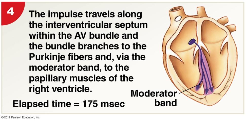
14 20-1 Internal Anatomy of the Heart: Right Ventricle Free edges attach to chordae tendineae from papillary muscles of ventricle Right atrioventricular (AV) Valve (tricuspid) Opening from right atrium to right ventricle Trabeculae carneae Muscular ridges on internal surface of right (and left) ventricle Includes moderator band: ridge contains part of conducting system coordinates contractions of cardiac muscle cells Figure 20 6a
15 20-1 Internal Anatomy of the Heart: Right Ventricle into Pulmonary Trunk Pulmonary Circuit Conus arteriosus (superior end of right ventricle) leads to pulmonary trunk Pulmonary trunk divides into left and right pulmonary arteries Blood flows from right ventricle to pulmonary trunk through pulmonary valve Pulmonary valve has three semilunar cusps Figure 20 6a
16 20-1 Anatomy of the Heart: Left Atrium and Ventricle The Left Atrium Lt. & rt. pulmonary veins deliver oxygenated blood to left atrium Passes to left ventricle through left AV (bicuspid or mitral) valve The Left Ventricle Holds same volume as right but thicker and more powerful muscle No moderator band Systemic Circulation Blood leaves left ventricle through aortic valve into ascending aorta (aortic arch) and becomes descending aorta Figure 20 6a

17 20-1 Internal Gross Anatomy of the Heart Figure 20 6c The Sectional Anatomy of the Heart.
 Left ventricle must produce 4-6x more pressure than right When left ventricle contracts, distance between base & apex decreases as")
18 20-1 Internal Anatomy of the Heart: Comparison of Right & Left Ventricles Right ventricle wall is thinner and develops less pressure than left ventricle (why?--lungs are close to heart and pulmonary vessels are short and wide) Left ventricle must produce 4-6x more pressure than right When left ventricle contracts, distance between base & apex decreases as well as diameter Contraction and bulging of the round left ventricle into the crescent-shaped right ventricle helps eject blood from right ventricle as well (see 20-7b) Figure 20 7 Structural Differences between the Left and Right Ventricles

 Heart Murmur leaky valve (e.g.")
19 20-1 Anatomy of Heart Valves Semilunar Valves Pulmonary and aortic tricuspid valves Prevent backflow from pulmonary trunk and aorta into right & left ventricles, respectively Have no chordae tendineae or muscles Aortic Sinuses Sacs at base of ascending aorta that prevent valve cusps from sticking to aorta Origin of right and left coronary arteries Valvular Heart Disease (VHD) Valve function deteriorates to extent that heart cannot maintain adequate circulation e.g., Rheumatic fever childhood reaction to streptococcal infection, chronic carditis (inflammation of heart) Heart Murmur leaky valve (e.g., mitral valve prolapse cusps don t close properly) Figure 20 8a&b
20 20-1 Connective Tissue and the Cardiac Fibrous Skeleton Connective Tissues Physically support cardiac muscle fibers Distribute forces of contraction Add strength and prevent overexpansion of heart Elastic fibers return heart to original shape after contraction The Cardiac (Fibrous) Skeleton Four bands around heart valves and bases of pulmonary trunk and aorta Stabilize valves Electrically insulate ventricular cells from atrial cells
 originate at aortic sinuses and then branch out Cardiac veins return deoxygenated coronary blood into right atrium High blood pressure & elastic rebound forces blood through coronary arteries")
21 20-1 Coronary Circulation Heart is <1% body mass but requires 5% blood Blood flow to heart may 9x during vigorous activity Coronary Arteries (lt. & rt.) originate at aortic sinuses and then branch out Cardiac veins return deoxygenated coronary blood into right atrium High blood pressure & elastic rebound forces blood through coronary arteries between ventricular contractions Arterial anastomoses = interconnections between arteries (helps stabilize coronary blood flow despite pressure differences between lt. and rt. coronary arteries) Figure 20 9a&b
 & atrioventricular (AV) nodes gives rise to marginal arteries (surface of rt.")
22 20-1 Coronary Circulation Right Coronary Artery -- supplies blood to rt. atrium, portions of both ventricles and cells of sinoatrial (SA) & atrioventricular (AV) nodes gives rise to marginal arteries (surface of rt. Ventricle) Supplies posterior interventricular artery Left Coronary Artery -- supplies blood to lt. atrium, lt. ventricle and interventricular septum gives rise to circumflex artery and anterior interventricular artery Cardiac Veins: small veins drain into great cardiac vein which drains into the coronary sinus and eventually into the rt. atrium (at base of the inferior vena cava) Figure 20 9a&c
 vasodilators (e.")
23 20-1 Coronary Circulation and Disease Coronary Artery Disease (CAD) - partial or complete block of coronary circulation, results in coronary ischemia Can lead to myocardial infarction (heart attack): heart tissue denied oxygen. Common symptom of CAD: angina pectoralis pain in the chest as a result of the ischemia CAD treatments include drugs that block sympathetic stimulation (e.g., propranolol) vasodilators (e.g., nitroglycerin) and calcium channel blockers Figure Plaques can be removed surgically via catheter (laser or roto-rooter ) or via balloon angioplasty; stents (wire mesh) used to keep artery open Coronary bypass surgery - use healthy veins (from legs) to create anatomoses around blockages; most people have 4 major coronary arteries, hence quadruple bypass surgery

24 20-2 The Conducting System Heartbeat single cardiac cycle Two Types of Cardiac Muscle Cells 1. Conducting System - control and coordinate heartbeat 2. Contractile Cells - produce contractions that propel blood 1% of myocardial cells are autorhythmic: depolarize without neural or endocrine stimulation Each heartbeat begins with the action potential generated by pacemaker cells in the sinoatrial (SA) node Depolarization transmitted to other myo-cardiac cells through conduction system Fig The entire heart contracts in series: atria first followed by ventricles Electrocardiogram (ECG or EKG) electrical events in cardiac cycle
25 20-2 The Conducting System Sinoatrial (SA) node: located in wall of right atrium near superior vena cava Atrioventricular (AV) node: located at the junction between atria and ventricles Conducting cells: interconnect the 2 nodes and distribute electrical impulses throughout the myocardium Includes the AV bundle, bundle branches and Purkinje fibers Cells of SA and AV nodes cannot maintain resting membrane potential and spontaneously drift towards threshold potential Called prepotential or pacemaker potential SA node: action potentials/min; AV node: Fig 20 11


26 20-2 Impulse Conduction Through Heart Fig Impulse Conduction
27 20-2 The Conducting System Overall heart rate set by SA node Resting heart rate (sinus rhythm) ~75bpm set by SA node (80-100) + parasympathetic stimulation (Vagus) Max rate 230 bpm set by AV node max; inefficient pumping above 180 bpm Abnormal Pacemaker Function Bradycardia: abnormally slow heart rate (<60 bpm) Tachycardia: abnormally fast heart rate (>100 bpm) Ectopic pacemaker Abnormal cells partially or completely bypass conducting system; disrupts timing of ventricular contractions Allometry fact?: 800 million beats lifespan

28 20-2 Electrocardiogram A recording of electrical events in the heart Obtained by electrodes at specific body locations Abnormal patterns used to diagnose damage P wave = Atria depolarize QRS complex = Ventricles depolarize T wave = Ventricles repolarize Figure Time Intervals Between ECG Waves P R interval From start of atrial depolarization to start of QRS complex Q T interval From ventricular depolarization to ventricular repolarization
29 20-2 ECG Diagnostics P-R longer than ~200ms = damage to AV node or conducting cells Total heart block = damaged AV node No impulses transmitted through, atria and ventricles beat independently (atria fast, ventricles slow) Large QRS = enlarged heart Q-T longer than ~380ms = coronary ischemia or myocardial damage Cardiac arrhythmias = abnormal patterns of cardiac activity Fibrillation = rapid, irregular, out of phase contractions due to activity in areas other than SA node: defibrillation to stop all activity so SA node can resume control

30 20-2 Cardiac Muscle Cell Action Potential Once threshold (~-75mV) reached, AP proceeds rapidly in 3 steps: Figure The Action Potential in Skeletal and Cardiac Muscle
31 20-2 The Cardiac Action Potential Refractory Period Period when AP cannot be elicited with a second stimulus OR requires > than normal stimulus Absolute no AP no matter what the stimulus because Na + channels are either open and/or inactivated Relative can elicit a 2 nd AP with stronger-than-normal stimulus Length of cardiac action potential in ventricular cell ms: 30 times longer than skeletal muscle fiber long refractory period prevents summation and tetany
32 20-2 Role of Calcium Ions and Energetics for Cardiac Contraction 20% of Ca 2+ for contraction enters through slow, voltagesensitive calcium channels in plasma membrane (extracellular sources) Influx triggers release of calcium ion reserves from sarcoplasmic reticulum (SR) As slow calcium channels close, intracellular Ca 2+ is pumped into SR or out of cell Energy for Cardiac Contractions From mitochondrial breakdown of fatty acids and glucose Oxygen from circulating hemoglobin and internal myoglobin

33 20-3 The Cardiac Cycle Fig 20 16: Phases of the Cardiac Cycle Systole = contraction Diastole = relaxation
34 20-3 The Cardiac Cycle 8 Steps in the Cardiac Cycle 1. Atrial systole begins Atrial contraction begins Rt. & lt. AV valves remain open 2. Atria top off ventricles Filling ventricles 3. Atrial systole ends AV valves close Ventricles contain max vol. known as end-diastolic volume (EDV) 4. Ventricular systole begins Isovolumetric ventricular contraction Pressure in ventricles rises with AV valves shut Fig 20 17
35 20-3 The Cardiac Cycle Fig Ventricular ejection Semilunar valves open Blood flows into pulmonary and aortic trunks Stroke volume (SV) = 60% of end-diastolic volume 6. Ventricular pressure falls Semilunar valves close Ventricles contain endsystolic volume (ESV), about 40% of end-diastolic volume 7. Ventricular diastole Ventricular pressure is higher than atrial pressure All heart valves are closed Ventricles relax (isovolumetric relaxation)

36 20-3 The Cardiac Cycle 8. AV valves open & passive atrial filling begins. When ventricular pressure falls below atrial pressure, the AV valves open Blood then flows from the atria into the ventricles while both are in diastole Fig 20 17

37 20-3 The Cardiac Cycle: Sounds Figure Heart Sounds S 1 = lubb AV valves closing at start of ventricular systole S 2 = dubb semilunar valves closing at start of ventricular diastole S 3 = blood flowing into ventricles as AV valves open S 4 = atrial contraction Murmur = sound produced by regurgitation through valve
 am t of blood ejected in single beat (ml/beat) SV = EDV ESV Ejection fraction The percentage of EDV represented by SV Cardiac output (CO) (ml/min)")
38 20-4 Cardiodynamics Cardiodynamics = movement & force generated by cardiac contractions End-diastolic volume (EDV) am t of blood in ventricles at end of diastole End-systolic volume (ESV) am t blood in ventricles at end of systole Stroke volume (SV) am t of blood ejected in single beat (ml/beat) SV = EDV ESV Ejection fraction The percentage of EDV represented by SV Cardiac output (CO) (ml/min) The volume pumped by ventricle in 1 minute CO = HR x SV (heart rate [beats/min] times stroke volume) Fig 20-19

39 20-4 Cardiodynamics Figure Factors Affecting Cardiac Output
40 20-4 Adjusting Cardiodynamics: Innervation Autonomic innervation Both sympathetic (NE) and parasympathetic (ACh) innervation of SA node, AV node and atrial myocardium Sympathetic dominates in ventricles Cardiac centers of medulla oblongata monitor blood pressure (baroreceptors) arterial O 2 and CO 2 levels (chemoreceptors) cardioacceleratory center controls sympathetic neurons (increases heart rate positive chronotropic effect) cardioinhibitory center controls parasympathetic neurons (slows heart rate negative chronotropic effect) Autonomic tone Dual innervation maintains resting tone by releasing ACh (and NE) At rest, parasympathetic tone reduces SA node inherent rate ( bpm) to ~70 bpm

41 20-4 Adjusting Cardiodynamics: Innervation Figure Autonomic Innervation of the Heart
 opens K + channels")
 binds to β-1 receptors")

42 20-4 Adjusting Cardiodynamics: Innervation Effects of Sympathetic & Parasympathetic Stimulation on Pacemaker Cells ACh (parasympathetic stimulation) opens K + channels thereby slowing the rate of spontaneous depolarization and slightly extending the duration of repolarization NE (sympathetic stimulation) binds to β-1 receptors thereby opening Na + - Ca 2+ channels and increasing the rate of depolarization and shortening the duration of repolarization Figure Autonomic Regulation of Pacemaker Function
43 20-4 Adjusting Cardiodynamics: Reflexes, Hormones & Drugs Atrial or Bainbridge Reflex Increased venous return activates stretch receptors in right atrium sympathetic activity HR Hormonal Effects on Heart Rate Epinephrine (E), Norepinephrine (NE) and Thyroid hormone (thyroxine) HR and contractile strength (positive inotropic effect) by acting on SA node β-1 blockers (hypertensive drugs) block E & NE effects Other Heart Rate Effectors Caffeine: rapid depolarization of SA node, HR Nicotine: stimulates sympathetic neurons, HR Changes in K +, Ca 2+, temperature, etc.
44 20-4 Cardiodynamics: Stroke Volume Adjustments End diastolic volume (EDV) is affected by 1. Venous return 2. Filling time EDV preload (amount of ventricular stretch) SV (Frank-Starling Principle) End systolic volume (ESV) is affected by 1. Preload 2. Contractility = force produced during contraction (inotropic) 3. Afterload = tension the ventricle must produce to open the semilunar valve and eject blood ( by any factor that restricts arterial blood flow) ESV SV

45 20-4 Cardiodynamics: Stroke Volume Adjustments Figure 20 23
46 20-4 Cardiodynamics: Contractility Sympathetic stimulation positive inotropic effect NE released by postganglionic fibers of cardiac nerves AND both E & NE released by suprarenal (adrenal) medullae Increases ejection fraction and decreases ESV Parasympathetic activity negative inotropic effect Acetylcholine released by Vagus nerve Hormones can have neg. or pos. inotropic effects Many pharmaceutical drugs mimic E, NE and thyroxine actions β-1 Receptors Mimetics (e.g., isoproterenol, dopamine, dobutamine) β-1 Receptor Blockers (e.g., propranolol, timolol, metoprolol) Others affect Ca 2+ (e.g., digitalis [inhibit SR uptake], nifedipine and verapamil [channel blockers])

47 20-4 Summary of Factors Affecting Cardiac Output Figure A Summary of the Factors Affecting Cardiac Output
The Heart. C h a p t e r. PowerPoint Lecture Slides prepared by Jason LaPres Lone Star College - North Harris
 C h a p t e r 20 The Heart PowerPoint Lecture Slides prepared by Jason LaPres Lone Star College - North Harris Copyright 2009 Pearson Education, Inc., publishing as Pearson Benjamin Cummings Introduction
Ch 19: Cardiovascular System - The Heart -
 Ch 19: Cardiovascular System - The Heart - Give a detailed description of the superficial and internal anatomy of the heart, including the pericardium, the myocardium, and the cardiac muscle. Trace the
Chapter 20: Cardiovascular System: The Heart
 Chapter 20: Cardiovascular System: The Heart I. Functions of the Heart A. List and describe the four functions of the heart: 1. 2. 3. 4. II. Size, Shape, and Location of the Heart A. Size and Shape 1.
Cardiovascular System
 Cardiovascular System The Heart Cardiovascular System The Heart Overview What does the heart do? By timed muscular contractions creates pressure gradients blood moves then from high pressure to low pressure
Chapter 18 - Heart. I. Heart Anatomy: size of your fist; located in mediastinum (medial cavity)
") Chapter 18 - Heart I. Heart Anatomy: size of your fist; located in mediastinum (medial cavity) A. Coverings: heart enclosed in double walled sac called the pericardium 1. Fibrous pericardium: dense connective
Chapter 18 - Heart I. Heart Anatomy: size of your fist; located in mediastinum (medial cavity) A. Coverings: heart enclosed in double walled sac called the pericardium 1. Fibrous pericardium: dense connective
The Cardiovascular System: The Heart
 PowerPoint Lecture Slides prepared by Meg Flemming Austin Community College C H A P T E R 12 The Cardiovascular System: The Heart Chapter 12 Learning Outcomes 12-1 12-2 Describe the anatomy of the heart,
11/10/2014. Muscular pump Two atria Two ventricles. In mediastinum of thoracic cavity 2/3 of heart's mass lies left of midline of sternum
 It beats over 100,000 times a day to pump over 1,800 gallons of blood per day through over 60,000 miles of blood vessels. During the average lifetime, the heart pumps nearly 3 billion times, delivering
The Heart. PowerPoint Lecture Presentations prepared by Jason LaPres. Lone Star College North Harris Pearson Education, Inc.
 20 The Heart PowerPoint Lecture Presentations prepared by Jason LaPres Lone Star College North Harris An Introduction to the Cardiovascular System Learning Outcomes Describe the superficial anatomy of
Approximately the size of your fist Location. Pericardial physiology
 Heart Anatomy Approximately the size of your fist Location Superior surface of diaphragm Left of the midline Anterior to the vertebral column, posterior to the sternum Wednesday, March 28, 2012 Muscle
- what other structures, besides the heart, does the mediastinum contain?
 Basic A & P II Dr. L. Bacha Chapter Outline (Martini & Nath 2010) An Introduction to the Cardiovascular System - read the paragraphs under this heading on page 580 The Heart is a Four Chambered Organ describe
The Heart. The Heart A muscular double pump. The Pulmonary and Systemic Circuits
 C H A P T E R 19 The Heart The Heart A muscular double pump circuit takes blood to and from the lungs Systemic circuit vessels transport blood to and from body tissues Atria receive blood from the pulmonary
10/23/2017. Muscular pump Two atria Two ventricles. In mediastinum of thoracic cavity 2/3 of heart's mass lies left of midline of sternum
 It beats over 100,000 times a day to pump over 1,800 gallons of blood per day through over 60,000 miles of blood vessels. During the average lifetime, the heart pumps nearly 3 billion times, delivering
THE CARDIOVASCULAR SYSTEM. Heart 2
 THE CARDIOVASCULAR SYSTEM Heart 2 PROPERTIES OF CARDIAC MUSCLE Cardiac muscle Striated Short Wide Branched Interconnected Skeletal muscle Striated Long Narrow Cylindrical PROPERTIES OF CARDIAC MUSCLE Intercalated
Human Anatomy, First Edition
 Human Anatomy, First Edition McKinley & O'Loughlin Chapter 22 : Heart 1 Functions of the Heart Center of the cardiovascular system, the heart. Connects to blood vessels that transport blood between the
The Heart. Size, Form, and Location of the Heart. 1. Blunt, rounded point; most inferior part of the heart.
 12 The Heart FOCUS: The heart is composed of cardiac muscle cells, which are elongated, branching cells that appear striated. Cardiac muscle cells behave as a single electrical unit, and the highly coordinated
the Cardiovascular System I
 the Cardiovascular System I By: Dr. Nabil A Khouri MD, MsC, Ph.D MEDIASTINUM 1. Superior Mediastinum 2. inferior Mediastinum Anterior mediastinum. Middle mediastinum. Posterior mediastinum Anatomy of
2. right heart = pulmonary pump takes blood to lungs to pick up oxygen and get rid of carbon dioxide
 A. location in thorax, in inferior mediastinum posterior to sternum medial to lungs superior to diaphragm anterior to vertebrae orientation - oblique apex points down and to the left 2/3 of mass on left
THE HEART. A. The Pericardium - a double sac of serous membrane surrounding the heart
 THE HEART I. Size and Location: A. Fist-size weighing less than a pound (250 to 350 grams). B. Located in the mediastinum between the 2 nd rib and the 5 th intercostal space. 1. Tipped to the left, resting
Chapter 13 The Cardiovascular System: Cardiac Function
 Chapter 13 The Cardiovascular System: Cardiac Function Overview of the Cardiovascular System The Path of Blood Flow through the Heart and Vasculature Anatomy of the Heart Electrical Activity of the Heart
*Generating blood pressure *Routing blood: separates. *Ensuring one-way blood. *Regulating blood supply *Changes in contraction
 *Generating blood pressure *Routing blood: separates pulmonary and systemic circulations *Ensuring one-way blood flow: valves *Regulating blood supply *Changes in contraction rate and force match blood
Anatomy of the Heart. Figure 20 2c
 Anatomy of the Heart Figure 20 2c Pericardium & Myocardium Remember, the heart sits in it s own cavity, known as the mediastinum. The heart is surrounded by the Pericardium, a double lining of the pericardial
Chapter 20 (1) The Heart
 The Heart") Chapter 20 (1) The Heart Learning Objectives Describe the location and structure of the heart Describe the path of a drop of blood from the superior vena cava or inferior vena cava through the heart out
Chapter 20 (1) The Heart Learning Objectives Describe the location and structure of the heart Describe the path of a drop of blood from the superior vena cava or inferior vena cava through the heart out
Collin County Community College. ! BIOL Anatomy & Physiology! WEEK 5. The Heart
 Collin County Community College! BIOL. 2402 Anatomy & Physiology! WEEK 5 The Heart 1 (1578-1657) A groundbreaking work in the history of medicine, English physician William Harvey s Anatomical Essay on
Functions of the Heart
 Cardiovascular System The Heart What is the Cardiovascular System? Blood circulated in Arteries, veins, and capillaries by the Pumping action of the heart Functions of the Heart Generating blood pressure
THE CARDIOVASCULAR SYSTEM. Part 1
 THE CARDIOVASCULAR SYSTEM Part 1 CARDIOVASCULAR SYSTEM Blood Heart Blood vessels What is the function of this system? What other systems does it affect? CARDIOVASCULAR SYSTEM Functions Transport gases,
BIOLOGY 2060 LECTURE NOTES ANATOMY & PHYSIOLOGY II (A. IMHOLTZ) HEART P1 OF 5
 HEART P1 OF 5") BIOLOGY 2060 LECTURE NOTES ANATOMY & PHYSIOLOGY II (A. IMHOLTZ) HEART P1 OF 5 1. Heart Functions a. Generates pressure that propels blood thru blood vessels. (Tissue perfusion.) b. Separates oxygenated
BIOLOGY 2060 LECTURE NOTES ANATOMY & PHYSIOLOGY II (A. IMHOLTZ) HEART P1 OF 5 1. Heart Functions a. Generates pressure that propels blood thru blood vessels. (Tissue perfusion.) b. Separates oxygenated
Chapter 20! The Heart!
 Chapter 20! The Heart! SECTION 20-1! The heart is a four-chambered organ, supplied by the coronary circulation, that pumps oxygen-poor blood to the lungs and oxygen-rich blood to the rest of the body!
Cardiovascular System
 Cardiovascular System Purpose Transport oxygen and nutrients Take waste products away from tissues & organs Things we learned Blood pressure: the force of blood pushing against the walls of blood vessels
Chapter 20 THE CARDIOVASCULAR SYSTEM: THE HEART
 Chapter 20 THE CARDIOVASCULAR SYSTEM: THE HEART INTRODUCTION A. The cardiovascular system consists of the blood, heart, and blood vessels. B. The heart is the pump that circulates the blood through an
Chapter 14. The Cardiovascular System
 Chapter 14 The Cardiovascular System Introduction Cardiovascular system - heart, blood and blood vessels Cardiac muscle makes up bulk of heart provides force to pump blood Function - transports blood 2
The Cardiovascular System. Chapter 15. Cardiovascular System FYI. Cardiology Closed systemof the heart & blood vessels. Functions
 Chapter 15 Cardiovascular System FYI The heart pumps 7,000 liters (4000 gallons) of blood through the body each day The heart contracts 2.5 billion times in an avg. lifetime The heart & all blood vessels
The Cardiovascular System: The Heart: Part A
 PowerPoint Lecture Slides prepared by Janice Meeking, Mount Royal College CHAPTER 18 The Cardiovascular System: The Heart: Part A Heart Anatomy Approximately the size of a fist Location In the mediastinum
BIOLOGY 2060 LECTURE NOTES ANATOMY & PHYSIOLOGY II (A. IMHOLTZ) HEART P1 OF 7
 HEART P1 OF 7") BIOLOGY 2060 LECTURE NOTES ANATOMY & PHYSIOLOGY II (A. IMHOLTZ) HEART P1 OF 7 1. Heart a. Generates the pressure that propels blood thru blood vessels. b. Separates oxygenated and deoxygenated blood separate.
BIOLOGY 2060 LECTURE NOTES ANATOMY & PHYSIOLOGY II (A. IMHOLTZ) HEART P1 OF 7 1. Heart a. Generates the pressure that propels blood thru blood vessels. b. Separates oxygenated and deoxygenated blood separate.
Heart Anatomy. 7/5/02 Stephen G Davenport 1
 Heart Anatomy Copyright 1999, Stephen G. Davenport, No part of this publication may be reproduced, stored in a retrieval system, or transmitted, in any form without prior written permission. 7/5/02 Stephen
The Cardiovascular System Part I: Heart Outline of class lecture After studying part I of this chapter you should be able to:
 The Cardiovascular System Part I: Heart Outline of class lecture After studying part I of this chapter you should be able to: 1. Describe the functions of the heart 2. Describe the location of the heart,
Circulation. Circulation = is a process used for the transport of oxygen, carbon! dioxide, nutrients and wastes through-out the body
 Circulation Circulation = is a process used for the transport of oxygen, carbon! dioxide, nutrients and wastes through-out the body Heart = muscular organ about the size of your fist which pumps blood.
Heart. Structure Physiology of blood pressure and heartbeat
 Heart Structure Physiology of blood pressure and heartbeat Location and Anatomy Location and Anatomy Pericardial cavity: surrounds, isolates, and anchors heart Parietal pericardium lined with serous membrane
Heart Overview. Chapter 18. Heart. Heart. Two Circuits. Organization of the Cardiovascular System. The Heart. Located directly behind sternum
 Heart Overview Chapter 18 The Heart Heart anatomy Cardiac muscle cells Heart chambers, valves and vessels Conducting system EKG Cardiac cycle Contractile and pacemaker cells Cardiodynamics Cardiac disorders
Principles of Anatomy and Physiology
 Principles of Anatomy and Physiology 14 th Edition CHAPTER 20 The Cardiovascular System: The Heart Introduction The purpose of the chapter is to: 1. Learn about the components of the cardiovascular system
The HEART. What is it???? Pericardium. Heart Facts. This muscle never stops working It works when you are asleep
 This muscle never stops working It works when you are asleep The HEART It works when you eat It really works when you exercise. What is it???? Located between the lungs in the mid thoracic region Apex
CV Anatomy Quiz. Dr Ella Kim Dr Pip Green
 CV Anatomy Quiz Dr Ella Kim Dr Pip Green Q1 The location of the heart is correctly described as A) lateral to the lungs. B) medial to the sternum. C) superior to the diaphragm. D) posterior to the spinal
The Heart. Happy Friday! #takeoutyournotes #testnotgradedyet
 The Heart Happy Friday! #takeoutyournotes #testnotgradedyet Introduction Cardiovascular system distributes blood Pump (heart) Distribution areas (capillaries) Heart has 4 compartments 2 receive blood (atria)
The Cardiovascular System
 The Cardiovascular System The Cardiovascular System A closed system of the heart and blood vessels The heart pumps blood Blood vessels allow blood to circulate to all parts of the body The function of
The Cardiovascular System
 Essentials of Human Anatomy & Physiology Elaine N. Marieb Seventh Edition Chapter 11 The Cardiovascular System Slides 11.1 11.19 Lecture Slides in PowerPoint by Jerry L. Cook The Cardiovascular System
10. Thick deposits of lipids on the walls of blood vessels, called, can lead to serious circulatory issues. A. aneurysm B. atherosclerosis C.
 Heart Student: 1. carry blood away from the heart. A. Arteries B. Veins C. Capillaries 2. What is the leading cause of heart attack and stroke in North America? A. alcohol B. smoking C. arteriosclerosis
Blood must move! 4/15/2014. Heart Basics
 What is the CARDIOVASCULAR system? The cardiovascular system carries blood and dissolved substances to and from different places in the body. The Heart has the job of pumping these things around the body.
4. The two inferior chambers of the heart are known as the atria. the superior and inferior vena cava, which empty into the left atrium.
 Answer each statement true or false. If the statement is false, change the underlined word to make it true. 1. The heart is located approximately between the second and fifth ribs and posterior to the
The Circulatory System. The Heart, Blood Vessels, Blood Types
 The Circulatory System The Heart, Blood Vessels, Blood Types The Closed Circulatory System Humans have a closed circulatory system, typical of all vertebrates, in which blood is confined to vessels and
Lab Activity 23. Cardiac Anatomy. Portland Community College BI 232
 Lab Activity 23 Cardiac Anatomy Portland Community College BI 232 Cardiac Muscle Histology Branching cells Intercalated disc: contains many gap junctions connecting the adjacent cell cytoplasm, creates
The Cardiovascular System
 The Cardiovascular System The Manila Times College of Subic Prepared by: Stevens B. Badar, RN, MANc THE HEART Anatomy of the Heart Location and Size approx. the size of a person s fist, hollow and cone-shaped,
Cardiovascular System: The Heart
 Cardiovascular System: The Heart I. Anatomy of the Heart (See lab handout for terms list) A. Describe the size, shape and location of the heart B. Describe the structure and function of the pericardium
Part 1. Copyright 2011 Pearson Education, Inc. Figure Copyright 2011 Pearson Education, Inc.
 PowerPoint Lecture Slides prepared by Leslie Hendon University of Alabama, Birmingham C H A P T E R The Heart 19 Part 1 The Heart A muscular double pump circuit vessels transport blood to and from the
The Cardiovascular System
 Essentials of Human Anatomy & Physiology Elaine N. Marieb Slides 11.1 11.19 Seventh Edition Chapter 11 The Cardiovascular System Functions of the Cardiovascular system Function of the heart: to pump blood
THE HEART OBJECTIVES: LOCATION OF THE HEART IN THE THORACIC CAVITY CARDIOVASCULAR SYSTEM
 BIOLOGY II CARDIOVASCULAR SYSTEM ACTIVITY #3 NAME DATE HOUR THE HEART OBJECTIVES: Describe the anatomy of the heart and identify and give the functions of all parts. (pp. 356 363) Trace the flow of blood
BIOL 4350 Cardiovascular Physiology Dr. Hamilton. Using the figure above, match the following: 1. Purkinje fibers. 2. SA node. 3. AV node.
 BIOL 4350 Cardiovascular Physiology Dr. Hamilton Using the figure above, match the following: 1. Purkinje fibers. 2. SA node. 3. AV node. 1 Using the figure above, match the following: 4. Atrial depolarization.
Cardiovascular System
 Cardiovascular System I. Structure of the Heart A. Average adult heart is 14 cm long and 9 cm wide. B. Lies in the mediastinum. C. Enclosed in the pericardium. 1. Fibrous pericardium- Outer, tough connective
INTRODUCTORY REMARKS:
 INTRODUCTORY REMARKS: The circulatory system provides a way for the blood to be transported throughout the body. This provides nutrients to the cells and allows wastes to be removed. Open vs. Closed Circulatory
Cardiovascular System Notes: Heart Disease & Disorders
 Cardiovascular System Notes: Heart Disease & Disorders Interesting Heart Facts The Electrocardiograph (ECG) was invented in 1902 by Willem Einthoven Dutch Physiologist. This test is still used to evaluate
BIO 136 Human Anatomy & Physiology For Non-Majors 11:39 am, Mar 08, 2006
 Jim Swan 1 These slides are from class presentations, reformatted for static viewing. The content contained in these pages is also in the Class Notes pages in a narrative format. Best screen resolution
Heart. Heart 2-Tunica media: middle layer (media ='middle') muscle fibers (smooth or cardiac).
 muscle fibers (smooth or cardiac).") t. innermost lumenal General Circulatory system heart and blood vessels walls have 3 layers (inside to outside) 1-Tunica interna: aka tunica intima layer--lumenal layer epithelium--endothelium simple squamous
t. innermost lumenal General Circulatory system heart and blood vessels walls have 3 layers (inside to outside) 1-Tunica interna: aka tunica intima layer--lumenal layer epithelium--endothelium simple squamous
CARDIOVASCULAR SYSTEM
 CARDIOVASCULAR SYSTEM Overview Heart and Vessels 2 Major Divisions Pulmonary Circuit Systemic Circuit Closed and Continuous Loop Location Aorta Superior vena cava Right lung Pulmonary trunk Base of heart
The Cardiovascular System. anatom.ua 1
 The Cardiovascular System anatom.ua 1 The Closed Circulatory System Humans have a closed circulatory system, typical of all vertebrates, in which blood is confined to vessels and is distinct from the interstitial
THE HEART Dr. Ali Ebneshahidi
 THE HEART Dr. Ali Ebneshahidi Functions is of the heart & blood vessels 1. The heart is an essential pumping organ in the cardiovascular system where the right heart pumps deoxygenated blood (returned
Cardiovascular system
 BIO 301 Human Physiology Cardiovascular system The Cardiovascular System: consists of the heart plus all the blood vessels transports blood to all parts of the body in two 'circulations': pulmonary (lungs)
human anatomy 2016 lecture thirteen Dr meethak ali ahmed neurosurgeon
 Heart The heart is a hollow muscular organ that is somewhat pyramid shaped and lies within the pericardium in the mediastinum. It is connected at its base to the great blood vessels but otherwise lies
The Cardiovascular System
 Chapter 18 Part A The Cardiovascular System 1/19/16 1 Annie Leibovitz/Contact Press Images Similarities of Cardiac and Skeletal Muscle RMP Ion concentration Deploarization Action Potential Repolarization
The cardiovascular system is composed of the heart and blood vessels that carry blood to and from the body s organs. There are 2 major circuits:
 1 The cardiovascular system is composed of the heart and blood vessels that carry blood to and from the body s organs. There are 2 major circuits: pulmonary and systemic. The pulmonary goes out to the
Electrical Conduction
 Sinoatrial (SA) node Electrical Conduction Sets the pace of the heartbeat at 70 bpm AV node (50 bpm) and Purkinje fibers (25 40 bpm) can act as pacemakers under some conditions Internodal pathway from
Anatomy of the Heart
 Biology 212: Anatomy and Physiology II Anatomy of the Heart References: Saladin, KS: Anatomy and Physiology, The Unity of Form and Function 8 th (2018). Required reading before beginning this lab: Chapter
Figure ) The specific chamber of the heart that is indicated by letter A is called the. Diff: 1 Page Ref: 364
 The specific chamber of the heart that is indicated by letter A is called the. Diff: 1 Page Ref: 364") Essentials of Anatomy and Physiology, 9e (Marieb) Chapter 11 The Cardiovascular System Short Answer Figure 11.1 Using Figure 11.1, identify the following: 1) The Purkinje fibers are indicated by label.
Essentials of Anatomy and Physiology, 9e (Marieb) Chapter 11 The Cardiovascular System Short Answer Figure 11.1 Using Figure 11.1, identify the following: 1) The Purkinje fibers are indicated by label.
Cardiovascular Anatomy Dr. Gary Mumaugh
 Cardiovascular Anatomy Dr. Gary Mumaugh Location of Heart Approximately the size of your fist Location o Superior surface of diaphragm o Left of the midline in mediastinum o Anterior to the vertebral column,
The Cardiovascular System
 PowerPoint Lecture Slide Presentation by Patty Bostwick-Taylor, Florence-Darlington Technical College The Cardiovascular System 11 PART A The Cardiovascular System A closed system of the heart and blood
Principles of Biomedical Systems & Devices. Lecture 8: Cardiovascular Dynamics Dr. Maria Tahamont
 Principles of Biomedical Systems & Devices Lecture 8: Cardiovascular Dynamics Dr. Maria Tahamont Review of Cardiac Anatomy Four chambers Two atria-receive blood from the vena cave and pulmonary veins Two
Unit 6: Circulatory System. 6.2 Heart
 Unit 6: Circulatory System 6.2 Heart Functions of Circulatory System 1. The heart is the pump necessary to circulate blood to all parts of the body 2. Arteries, veins and capillaries are the structures
LECTURE 5. Anatomy of the heart
 LECTURE 5. Anatomy of the heart Main components of the CVS: Heart Blood circulatory system arterial compartment haemomicrocirculatory (=microvascular) compartment venous compartment Lymphatic circulatory
The Cardiovascular System
 11 PART A The Cardiovascular System PowerPoint Lecture Slide Presentation by Jerry L. Cook, Sam Houston University ESSENTIALS OF HUMAN ANATOMY & PHYSIOLOGY EIGHTH EDITION ELAINE N. MARIEB The Cardiovascular
The Cardiovascular System (Heart)
") The Cardiovascular System The Cardiovascular System (Heart) A closed system of the heart and blood vessels The heart pumps blood Blood vessels allow blood to circulate to all parts of the body The function
The Cardiovascular System The Cardiovascular System (Heart) A closed system of the heart and blood vessels The heart pumps blood Blood vessels allow blood to circulate to all parts of the body The function
QUIZ/TEST REVIEW NOTES SECTION 1 CARDIAC MYOCYTE PHYSIOLOGY [CARDIOLOGY]
![QUIZ/TEST REVIEW NOTES SECTION 1 CARDIAC MYOCYTE PHYSIOLOGY [CARDIOLOGY]](/thumbs/96/126998162.jpg "QUIZ/TEST REVIEW NOTES SECTION 1 CARDIAC MYOCYTE PHYSIOLOGY [CARDIOLOGY]") QUIZ/TEST REVIEW NOTES SECTION 1 CARDIAC MYOCYTE PHYSIOLOGY [CARDIOLOGY] Learning Objectives: Describe the ionic basis of action potentials in cardiac contractile and autorhythmic cells Explain the relationship
QUIZ/TEST REVIEW NOTES SECTION 1 CARDIAC MYOCYTE PHYSIOLOGY [CARDIOLOGY] Learning Objectives: Describe the ionic basis of action potentials in cardiac contractile and autorhythmic cells Explain the relationship
Function: Transportation of. Oxygen Nutrients Waste Hormones gases
 Function: Transportation of Oxygen Nutrients Waste Hormones gases Pericardium: double sac of serous membrane filled with fluid (pericardial fluid to be exact) that surrounds the heart. Parietal pericardium:
This lab activity is aligned with Visible Body s A&P app. Learn more at visiblebody.com/professors
 1 This lab activity is aligned with Visible Body s A&P app. Learn more at visiblebody.com/professors 2 PRE-LAB EXERCISES: A. Watch the video 29.1 Heart Overview and make the following observations: 1.
IB TOPIC 6.2 THE BLOOD SYSTEM
 IB TOPIC 6.2 THE BLOOD SYSTEM THE BLOOD SYSTEM TERMS TO KNOW circulation ventricle artery vein 6.2.U1 - Arteries convey blood at high pressure from the ventricles to the tissues of the body Circulation
Using Figure 14.1, match the following: 1) Myelin sheath. 1) 2) Cell body of ANS preganglionic neuron. 2)
 Myelin sheath. 1) 2) Cell body of ANS preganglionic neuron. 2)") Practice Exam 1 AP 2 chapters 14 and 18 Name MATCHING: Match labeled areas with the appropriate terminology from the list below. Figure 14.1 Using Figure 14.1, match the following: 1) Myelin sheath. 1)
Practice Exam 1 AP 2 chapters 14 and 18 Name MATCHING: Match labeled areas with the appropriate terminology from the list below. Figure 14.1 Using Figure 14.1, match the following: 1) Myelin sheath. 1)
The Heart and Heart Disease
 The Heart and Heart Disease Illustration of the heart by Leonardo DaVinci heart-surgeon.com/ history.html 2/14/2010 1 I. Location, Size and Position of the Heart A. Triangular organ located 1. of mass
Cardiovascular System Notes: Physiology of the Heart
 Cardiovascular System Notes: Physiology of the Heart Interesting Heart Fact Capillaries are so small it takes ten of them to equal the thickness of a human hair. Review What are the 3 parts of the cardiovascular
Anatomy & Physiology of Cardiovascular System. Chapter 18 & 19
 Anatomy & Physiology of Cardiovascular System Chapter 18 & 19 Objectives..cont 1. Discuss the physiological stages of cardiac muscle contraction. 2. Trace a typical ECG and label each wave or complex 3.
Pearson's Comprehensive Medical Assisting Administrative and Clinical Competencies
 Pearson's Comprehensive Medical Assisting Administrative and Clinical Competencies THIRD EDITION CHAPTER 27 The Cardiovascular System Lesson 1: Overview of the Cardiovascular System Lesson Objectives Upon
Major Function of the Cardiovascular System. Transportation. Structures of the Cardiovascular System. Heart - muscular pump
 Structures of the Cardiovascular System Heart - muscular pump Blood vessels - network of tubes Blood - liquid transport vehicle brachiocephalic trunk superior vena cava right pulmonary arteries right pulmonary
The Heart and Cardiovascular System
 The Heart and Cardiovascular System What you will learn The location of the heart 3 layers and covering of the heart Explain the function of the heart as 2 separate pumps Identify the 4 chambers of the
37 1 The Circulatory System
 H T H E E A R T 37 1 The Circulatory System The circulatory system and respiratory system work together to supply cells with the nutrients and oxygen they need to stay alive. a) The respiratory system:
Chapter 13. Cardiovascular System
 Chapter 13 Cardiovascular System 1 Introduction A. The cardiovascular system consists of the heart and vessels (arteries, capillaries and veins.) B. A functional cardiovascular system is vital for supplying
Chapter 20 (2) The Heart
 The Heart") Chapter 20 (2) The Heart ----------------------------------------------------------------------------------------------------------------------------------------- Describe the component and function of
Chapter 20 (2) The Heart ----------------------------------------------------------------------------------------------------------------------------------------- Describe the component and function of
Large Arteries of Heart
 Cardiovascular System (Part A-2) Module 5 -Chapter 8 Overview Arteries Capillaries Veins Heart Anatomy Conduction System Blood pressure Fetal circulation Susie Turner, M.D. 1/5/13 Large Arteries of Heart
Chapter 12 Lecture Outline
 Chapter 12 Lecture Outline See separate PowerPoint slides for all figures and tables preinserted into PowerPoint without notes. Copyright The McGraw-Hill Companies, Inc. Permission required for reproduction
Circulatory system. Terminology. Ventricles and resistance. Pressure gradients move blood through the heart and vessels.
 Circulatory system Pressure gradients move blood through the heart and vessels. Pulmonary circulation vs. systemic circulation (to pulmonary circuit) liver head and arms heart aorta diaphragm (from pulmonary
CIRCULATORY SYSTEM BLOOD VESSELS
 Name: Block: CIRCULATORY SYSTEM Multicellular organisms (above the level of roundworms) rely on a circulatory system to bring nutrients to, and take wastes away from, cells. In higher organisms such as
Heart Pump and Cardiac Cycle. Faisal I. Mohammed, MD, PhD
 Heart Pump and Cardiac Cycle Faisal I. Mohammed, MD, PhD 1 Objectives To understand the volume, mechanical, pressure and electrical changes during the cardiac cycle To understand the inter-relationship
UNIT 11: THE CARDIOVASCULAR SYSTEM
 UNIT 11: THE CARDIOVASCULAR SYSTEM Functions of the Heart PUMPS Blood Transports Oxygen and Nutrients Removes Carbon Dioxide and Metabolic Wastes Thermoregulation Immunological Function Clotting Mechanisms
8:49 am, Jan 28, 2008
 Jim Swan 1 These slides are from class presentations, reformatted for static viewing. The content contained in these pages is also in the Class Notes pages in a narrative format. Best screen resolution