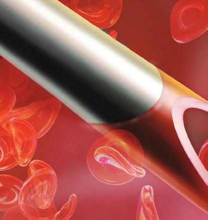
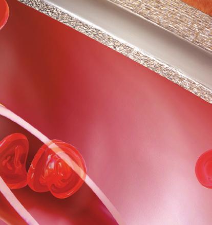
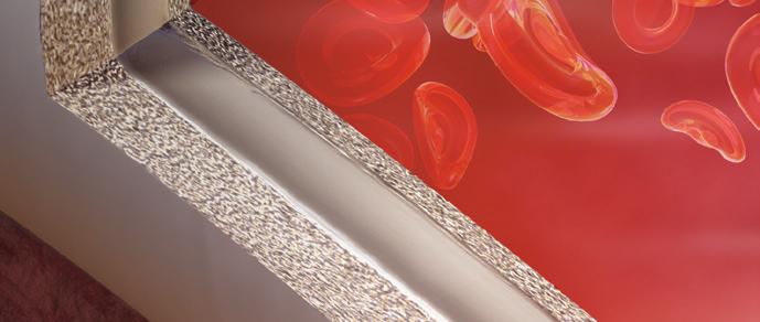
CIRCUIT OPTIMIZING THE VASCULAR ACCESS. Bringing long-term value to AV access creation and intervention. Sponsored by Gore & Associates.
|
|
|
- Barry Shields
- 5 years ago
- Views:
Transcription


1 Supplement to June 2018 OPTIMIZING THE VASCULAR ACCESS CIRCUIT Bringing long-term value to AV access creation and intervention


2 TABLE OF 2 SUPPLEMENT TO ENDOVASCULAR TODAY JUNE 2018 VOL. 17, NO. 6


3 CONTENTS 4 Undersizing to the Outflow Vein Technique and considerations in deployment of the GORE VIABAHN Endoprosthesis at the venous anastomosis of dialysis access arteriovenous grafts. By Daniel V. Patel, MD, FASDIN 9 Stent Grafts: An Important Tool in Today s Dialysis Treatment Algorithm Two dialysis experts discuss their approach to access challenges. With John R. Ross, MD, and Ziv J. Haskal, MD, FSIR, FAHA, FACR 14 GORE VIABAHN Endoprosthesis Flexibility Across the Elbow Maintaining mechanical integrity in this region is critical for optimal outcomes in dialysis access. By Dheeraj Rajan, MD, FRCPC, FSIR, FACR 17 The Pathophysiology of Arteriovenous Graft Thrombosis and Stenosis Understanding the underlying challenges of an optimal access site. With Mitchell Henry, MD 20 Strategies for Approaching Vascular Graft Thrombectomy Insights on the best tactics for declotting arteriovenous grafts. With Scott O. Trerotola, MD VOL. 17, NO. 6 JUNE 2018 SUPPLEMENT TO ENDOVASCULAR TODAY 3
 in dialysis access 1 ; however, AVGs are still essential for dialysis")

4 Undersizing to the Outflow Vein Technique and considerations in deployment of the GORE VIABAHN Endoprosthesis at the venous anastomosis of dialysis access arteriovenous grafts. BY DANIEL V. PATEL, MD, FASDIN In the era of Fistula First, we have seen a dramatic decrease in the prevalence of arteriovenous grafts (AVGs) in dialysis access 1 ; however, AVGs are still essential for dialysis access in patients in whom a fistula is not possible. For this challenging patient population, the use of stent grafts at the venous anastomosis has transformed management of dysfunctional dialysis access grafts. The venous anastomosis has been the Achilles heel of AVGs, where aggressive neointimal hyperplasia leads to stenosis and graft thrombosis. Although primary treatment with angioplasty had been the therapy of choice in the past, more recent studies have shown the superiority of stent graft placement at the venous anastomosis as a more durable treatment choice. 2-4 This article presents a case of venous anastomosis stenosis, with particular attention to practical considerations in deploying the GORE VIABAHN Endoprosthesis at the venous anastomosis of an AVG with a larger-diameter outflow vein. CASE PRESENTATION A 60-year-old man with a history of hypertension resulting in end-stage renal disease presented with decreased clearances at dialysis. He had a left brachial artery to axillary vein 6 mm expanded polytetrafluoroethylene (eptfe) AVG placed 1 year prior to presentation. Six months earlier, endovascular thrombectomy with angioplasty at the venous anastomosis was performed at an outside institution. He presented to our center with a recent history of prolonged bleeding after needle removal at dialysis (> minutes) and elevated venous pressures on dialysis. Upon clinical examination, a pulsatile graft was present. A preprocedural Doppler study suggested a stenosis at the venous anastomosis. Of note, the draining axillary vein proximal to the anastomosis measured 9 to 11 mm in diameter. Figure 1. Left brachial artery to axillary vein AVG. Note the graft in orange and the outflow vein in blue. The arrow demonstrates the site of venous anastomosis stenosis. An angiogram revealed a high-grade stricture at the venous anastomosis of the graft (Figure 1). An 8 mm x 8 cm BARD VACCESS Angioplasty Balloon was used to dilate the venous anastomosis of the graft, with a waist noted at the venous anastomosis before full balloon effacement (Figure 2). After angioplasty of the lesion, ongoing significant recoil remained (Figure 3). Given the data supporting advantages of stent graft placement over balloon angioplasty at the venous Figure 2. Angioplasty performed with an 8 mm x 8 cm balloon at the venous anastomosis. Note the waist seen at the junction of the venous anastomosis and outflow vein prior to full balloon effacement. 4 SUPPLEMENT TO ENDOVASCULAR TODAY JUNE 2018 VOL. 17, NO. 6

. Postprocedure, the patient regained a strong thrill in the access.")

5 A Figure 3. Recoil at the venous anastomosis postangioplasty. anastomosis, as well as with recoil postangioplasty, the decision was made to place a GORE VIABAHN Endoprosthesis at the venous anastomosis stenosis. 4 After dilation with balloon angioplasty, the graft measured approximately 7 mm in diameter, so an 8 mm x 10 cm GORE VIABAHN Endoprosthesis was deployed at the venous anastomosis lesion and further postdilated with the 8 mm balloon (Figure 4). Postprocedure, the patient regained a strong thrill in the access. Venous pressures and clearances improved at dialysis, with bleeding times at < 5 minutes after needle removal. At 6 months postprocedure, the patient has required no further interventions and maintains access use at dialysis. ANGIOPLASTY OF VENOUS ANASTOMOSIS STENOSIS Angioplasty remains as a key component of venous anastomosis lesion management. The challenge of angioplasty here is the need to manage the difference of diameters between the graft and the outflow vein. In our practice, we often see 6 mm straight eptfe used for AV access grafts. The draining outflow vein size can be variable, ranging from 5 to 6 mm to 14 mm or larger; this is contingent on the ability of the vein to further dilate after AVG placement. In general, we tend to see larger-diameter draining veins proximal to the venous anastomosis. Our approach to balloon angioplasty sizing targets matching the diameter of the larger outflow vein. For 6 mm eptfe grafts, we usually treat the venous anastomosis with a 7 to 8 mm percutaneous transluminal angioplasty (PTA) balloon. Although the balloon size is larger than the 6 mm eptfe segment, we have noted no significant eptfe dysfunction with using 8 mm balloons at the 6 mm eptfe graft. This is due to the elastic nature of the eptfe material. When the vein is larger (9 12 mm in diameter), we use 8 to 9 mm PTA balloons. We have limited the use of larger 10 to 12 mm diameter balloons at B C Figure 4. Deployment of an 8 mm x 10 cm GORE VIABAHN Endoprosthesis at the venous anastomosis (A, B). Note the extension of the proximal edge into the draining axillary vein. Representation of the GORE VIABAHN Endoprosthesis (orange) with the proximal edge floating into the larger-caliber outflow vein (blue) (C). 6 mm grafts, with concerns for potential graft trauma with larger-diameter balloons. The general concept is to dilate the site of stenosis as large as possible, without unnecessary trauma to the graft. VOL. 17, NO. 6 JUNE 2018 SUPPLEMENT TO ENDOVASCULAR TODAY 5


6 Figure 5. Depiction of venous anastomosis GORE VIABAHN Endoprosthesis placement at a larger-diameter outflow vein. We also encounter bovine grafts and tapered grafts with venous anastomosis lesions. Although the bovine grafts have the ability to dilate and expand over time, we have been cautious with significantly oversizing PTA balloons at the bovine graft material, with concerns of damage to the bovine graft segment. In these cases, we generally avoid balloon dilation > 2 mm over the graft diameter at the venous anastomosis. When considering 4 to 7 mm tapered AVGs, we generally approach angioplasty with an 8 or 9 mm PTA balloon at the 7 mm venous anastomosis. The known shortfall of venous anastomosis angioplasty is the ability to maintain a durable result. Multiple randomized controlled trials have shown the superiority of venous anastomosis management with stent grafts over balloon angioplasty. Although angioplasty is a necessary step in treating venous anastomosis stenosis, established data now supports further stent graft placement as primary treatment. 2-4 SIZING CONSIDERATIONS WITH VENOUS ANASTOMOSIS STENT GRAFT PLACEMENT Diameter We follow the diameter selection guidelines found in the instructions for use, which recommend oversizing the device 5% to 20% to the size of the target vessel. We generally anchor at least 2 to 4 cm of the GORE VIABAHN Endoprosthesis within the graft and allow the proximal end to freely float in the outflow vein (Figures 5 7). Proper device oversizing and landing zone selection within the graft combine to securely anchor the distal segment of the GORE VIABAHN Endoprosthesis within the graft while maintaining proper flow dynamics. Planning Length and Deployment Position Choice of stent graft length is essential to success in long-term venous anastomosis treatment. The goal is to create an outflow conduit that will decrease future neointimal hyperplasia development. Attention must be paid to the desired location for the proximal edge of the stent graft. Placement of a stent graft at the venous anastomosis creates a new, more proximal (central) venous anastomosis. The proximal edge of the newly placed stent graft becomes the new venous anastomosis of the graft. This may instigate development of upstream neointimal hyperplasia, with the more proximal draining vein now in closer proximity to the high flows from the access. As described in the case, we identify larger-diameter veins proximal to the venous anastomosis. A stent graft serves as a conduit leading from the graft through the site of venous anastomosis stenosis and neointimal hyperplasia to the larger-diameter outflow vein. This allows exclusion Figure 6. Doppler image of the proximal edge of an 8 mm GORE VIABAHN Endoprosthesis floating in a larger-caliber outflow vein. Figure 7. Intravascular ultrasound image of the proximal edge of an 8 mm diameter GORE VIABAHN Endoprosthesis (orange) floating in a 12 mm diameter outflow vein (blue). 6 SUPPLEMENT TO ENDOVASCULAR TODAY JUNE 2018 VOL. 17, NO. 6
.")
. This stenosis was further treated with a second 8 mm x 5 cm GORE VIABAHN Endoprosthesis (D, E).")

 was necessary in this case to create a conduit to the larger axillary vein.")
.")


7 OPTIMIZING THE VASCULAR ACCESS CIRCUIT A B C D E F G Figure 8. Right upper arm brachial-axillary AVG with high-grade venous anastomosis (A). The patient was treated with angioplasty and placement of an 8 mm x 5 cm GORE VIABAHN Endoprosthesis at the venous anastomosis (B). A small venous valve was seen at the time, with no interference with access flow (purple arrow). Four months later, the patient presented with elevated venous pressures. A high-grade stenosis was seen at the site of the venous valve (C). This stenosis was further treated with a second 8 mm x 5 cm GORE VIABAHN Endoprosthesis (D, E). One year later, the graft remained patent with no further intervention necessary (F, G). of the neointimal hyperplasia from the access while providing long-term radial support at the site of stenosis. The overall goal is to create a bypass from the graft to a healthy, suitable vein. A longer-length stent graft (10 cm) was necessary in this case to create a conduit to the larger axillary vein. Venous valves must also be recognized when planning for placement of stent grafts. These valves may develop aggressive stenosis and thrombosis if they are within several centimeters of the proximal edge of a venous anastomosis stent graft (Figure 8). We target placement of the proximal edge of stent grafts through any adjacent venous valves, leading to the larger-diameter draining vein. This excludes the valves from the higher flow through the graft, which minimizes the risk of valve associated venous anastomosis stenosis.5 Of note, in this case, the distal end of the 10 cm long GORE VIABAHN Endoprosthesis extended 5 cm within the graft but was not in the cannulation areas. Stent graft placement in cannulation areas should be avoided as much as possible to decrease risk of infection.6 Wall Apposition Versus Undersizing to the LargerDiameter Outflow Vein Computational fluid dynamic studies have supported the concept that direct venous anastomosis vessel wall apposition can lead to higher shear stresses at the draining outflow vein (Figure 9).7 This high blood flow and shear stress can lead to development of neointimal hyperplasia at the draining vein.8,9 To minimize future issues of edge stenosis and neointimal hyperplasia development at the draining vein, the proximal edge of the stent graft should have minimal venous vessel wall contact. The goal is to deploy the proximal edge of the smaller-diameter stent graft within a larger-diameter outflow vein. This elephant trunk technique allows the GORE VIABAHN Endoprosthesis to lie in the outflow vein without vessel wall apposition. VOL. 17, NO. 6 JUNE 2018 SUPPLEMENT TO ENDOVASCULAR TODAY 7

 and with wall apposition (B).")
8 A B from the stresses of the higher blood flow from the graft, effectively reducing the risk of future stenosis development. The relative undersizing of the proximal portion of the stent graft allows for permissive mobility of the stent graft within the larger-diameter outflow vessel. This undersized proximal part of the stent graft has minimal contact with the vessel wall (Figures 6 and 7). Figure 9. Computational flow dynamic studies of wall shear stress at four representative time points of the cardiac cycle: without wall apposition (A) and with wall apposition (B). Note higher shear stresses (red) at outflow vessel when wall apposition is present. Reproduced from Ross JR. Stent-graft sizing for AV access creation and revision procedures. Endovasc Today. 2014;13(6 suppl):21. TABLE 1. DEVICE APPOSITION RELATIVE TO THE OUTFLOW VEIN 6-Month Outcomes Undersized* Apposed 7 Target lesion primary patency Circuit primary patency 62% 47% 48% 41% Note: Outflow wall apposition is not necessary for quality outcomes. Sixmonth outcomes from the Gore REVISE Clinical Study demonstrate that GORE VIABAHN Endoprosthesis achieves quality outcomes regardless of whether the device is apposed or undersized to the outflow vein. The GORE VIABAHN Endoprosthesis should always be sized 5% to 20% greater than the AVG diameter per the instructions for use. *The difference between the diameter of the vein and device is 1 mm. The difference between the diameter of the vein and device is < 1 mm. Data on file. Courtesy of Gore & Associates. Findings from the Gore REVISE Clinical Study further support the advantages of undersizing the proximal edge of the GORE VIABAHN Endoprosthesis. A > 1 mm difference in vein diameter between the stent graft and the largerdiameter outflow vein resulted in higher 6 month patency (Table 1). In the case presentation, the proximal edge of the GORE VIABAHN Endoprosthesis was placed at the large-caliber draining axillary vein. This bypassed areas of luminal narrowing and stenosis, creating a direct conduit from the graft to the large-caliber outflow vein (Figure 4C). The smaller-diameter outflow vein segments were excluded UNIQUE PROPERTIES OF THE GORE VIABAHN ENDOPROSTHESIS As a flexible device with full end-to-end eptfe lining, the GORE VIABAHN Endoprosthesis is unique in its ability to treat venous anastomosis lesions. The self-expanding support allows for maintained patency at this site of recurrent stenosis. A key feature is the ability of the GORE VIABAHN Endoprosthesis to stay dynamic and flexible within the larger-caliber outflow vein. This flexibility minimizes risks of kinking and access thrombosis as the device follows the natural ability of vessels to bend and contour with the body. In this case, we undersized the diameter of the stent graft to a larger outflow vein, minimizing the risk of future stenosis and helping to maintain long-term access patency. The patient previously had an episode of graft thrombosis but has remained intervention-free over 6 months after stent graft placement. This has been a revolution in managing dysfunctional AVGs, providing for improved access outcomes and quality of care in the end-stage renal disease population. n 1. Lynch JR, Wasse H, Armistead NC, McClellan WM. Achieving the goal of the Fistula First breakthrough initiative for prevalent maintenance hemodialysis patients. Am J Kidney Dis. 2011;57: Haskal ZJ, Trerotola S, Dolmatch B, et al. Stent graft versus balloon angioplasty for failing dialysis-access grafts. N Engl J Med. 2010;362: Haskal ZJ, Saad TF, Hoggard JG, et al. Prospective, randomized, concurrently-controlled study of a stent graft versus balloon angioplasty for treatment of arteriovenous access graft stenosis: 2-year results of the RENOVA study. J Vasc Interv Radiol. 2016;27: Vesely T, DaVanzo W, Behrend T, et al. Balloon angioplasty versus Viabahn stent graft for treatment of failing or thrombosed prosthetic hemodialysis grafts. J Vasc Surg. 2016;64: Patel DV. Case report: endovascular salvage of an abandoned AV graft. Endovasc Today. 2017;16(6 suppl): Kim CY, Guevara CJ, Engstrom BI, et al. Analysis of infection risk following covered stent exclusion of pseudoaneurysms in prosthetic arteriovenous hemodialysis access grafts. J Vasc Interv Radiol. 2012;23: Ross JR. Stent-graft sizing for AV access creation and revision procedures. Endovasc Today. 2014;13(6 suppl): Van Tricht I, De Wachter D, Tordoir J, Verdonck P. Comparison of the hemodynamics in 6 mm and 4-7 mm hemodialysis grafts by means of CFD. J Biomech. 2006;39: VanBavel E. Effects of shear stress on endothelial cells: possible relevance for ultrasound applications. Prog Biophys Mol Biol. 2007;93: Daniel V. Patel, MD, FASDIN Volusia-Flagler Vascular Center Daytona Beach, Florida (386) ; dpatel@vfvascularcenter.com Disclosures: Medical advisory and speaker fees from Gore & Associates. 8 SUPPLEMENT TO ENDOVASCULAR TODAY JUNE 2018 VOL. 17, NO. 6

, but how to provide similarly successful interventions in native fistulae remains a problem.")
9 Stent Grafts: An Important Tool in Today s Dialysis Treatment Algorithm Two dialysis experts discuss their approach to access challenges. WITH JOHN R. ROSS, MD, AND ZIV J. HASKAL, MD, FSIR, FAHA, FACR What are some of the common challenges you face as a physician when attempting to preserve and extend the life of a dialysis circuit? Dr. Haskal: One is naturally having consistent and durable solutions for patients with surgically created permanent accesses. We know what to do with the venous anastomoses of arteriovenous grafts (AVGs), but how to provide similarly successful interventions in native fistulae remains a problem. We cannot support a care model wherein patients consistently return for autoreinterventions when global periods of reimbursement expire we have to be better. Another challenge is the persistent extended use of tunneled catheters in patients. Working to drive down the national use of catheters in patients awaiting access creation will translate to fewer infections, central vein stenoses, and the prototypical 25-year-old dialysis patient with end-stage central vein occlusions we all see. Finally, we ve got to get in line and practice by evidence where evidence exists. Dr. Ross: Graft-vein anastomosis is a recurrent problem, and balloon angioplasty alone has not been rewarding in this particular area. We are able to extend the life of that graft on the outflow side tremendously using the GORE VIABAHN Endoprosthesis. We also use the device in other areas, such as across the elbow where nothing else has shown the durability that we see with the GORE VIABAHN Endoprosthesis. For those patients with reoccurring thrombosis, how have stent grafts like the GORE VIABAHN Endoprosthesis improved outcomes as a treatment option? Dr. Haskal: We now have multiple prospective controlled trials proving the therapeutic and economic value of prompt use of expanded polytetrafluoroethylene (eptfe) stent grafts in stenotic venous anastomoses. I m proud to have designed and published first and subsequent work in this, so I m a true believer. My randomized controlled trials did not include placing stent grafts in thrombosed grafts because we didn t want to muddy the early data. The Gore REVISE trial included such patients and convincingly showed that a GORE VIABAHN Endoprosthesis provides a better outcome over angioplasty when treating a thrombosed graft, not just a stenotic one (Table 1). So, I use stent grafts in just this way. Dr. Ross: There are three reasons that grafts thrombose, and they should be thought of as an inflow failure, an outflow failure, or a conduit failure. Assuming we don t have an inflow failure or a conduit failure (other than thrombosis), and it's only related to the outflow, then in our experience, the GORE VIABAHN Endoprosthesis seems to do an exceedingly good job in maintaining patency for a much longer period of time than simple balloon angioplasties and certainly better than bare-metal stents (BMSs). The data have shown stent graft improvements over percutaneous transluminal angioplasty (PTA), but what are your thoughts on drugcoated balloons (DCBs) as a treatment option in dialysis access? Dr. Ross: Currently, we do not have convincing data on the treatment of graft-vein anastomosis associated with DCBs. Dr. Haskal: I m excited by the prospects of DCBs in AV access. I m happy that we ll see multiple prospective trials appear with different balloons and drug doses. Replicative studies are essential. Personally, I do believe there is a real signal for drug effect in this application of DCBs. I use them, but it s the early days for this technology. Lead papers need to appear in full publication, subanalyses thereafter, and more real-world experience. Regarding DCBs, what are your thoughts about data that have shown decreasing levels of improvement compared to traditional angioplasty over time? VOL. 17, NO. 6 JUNE 2018 SUPPLEMENT TO ENDOVASCULAR TODAY 9
10 TABLE 1. ADDITIONAL MEASURES OF EFFECTIVENESS AT 6 MONTHS TREND TOWARD IMPROVED PATENCY OF STENT GRAFTS Prior interventions to target lesion Target Lesion Primary Patency Circuit Primary Patency Stent grafts, % Balloon angioplasty, % Stent grafts, % Balloon angioplasty, % (36) 43.9 (45) 48.7 (36) 35.0 (45) (64) 29.2 (59) 40.7 (64) 25.2 (59) Stenotic vs thrombosed Stenotic 64.6 (61) 45.8 (64) 49.7 (61) 35.9 (64) Thrombosed 36.1 (39) 23.5 (40) 34.2 (39) 21.8 (40) Antecubital fossa Crossed 72.4 (17) N/A 67.3 (17) N/A Did not 49.2 (83) N/A 39.0 (83) N/A Abbreviations: N/A, not applicable. Percentages cited are the percentage of subjects out of the data available. Parentheses represent number of subjects available at 6-month time point. Reprinted from Journal of Vascular Surgery, 64/5, Vesely T, DaVanzo W, Behrend T, et al., Balloon angioplasty versus Viabahn stent graft for treatment of failing or thrombosed prosthetic hemodialysis grafts, e1., Copyright (2016), with permission from Elsevier. Dr. Ross: DCBs have only completed one clinical trial. There seems to be a positive signal that there may be a future for DCBs in this area, but we need much larger studies to quantify what DCBs will do at 12 or 24 months. There seems to be some improvement over standard angioplasty balloons, but compared to stent grafts (and that s where the comparison should be made), the benefit of DCBs is not definitive. The clinical trial on DCBs, of course, did not use them with graft-vein anastomosis, only in fistulas. The graft-vein anastomosis may be a different pathological process, and as of now, we just don t have any randomized controlled trial data. We do have some individual data from sites around the country that show much better results at the graft-vein anastomosis in thrombosed grafts assuming we can say that the cause of the graft thrombosis was related to the outflow stenosis. If someone misses a problem with the inflow or doesn t realize that there is a thrombogenic graft and is treating the outflow, it reclots, but it has very little to do with the outflow. If we know that it s a stenosis at the graft-vein anastamosis that is causing the thrombosis, then certainly the GORE VIABAHN Endoprosthesis is enhancing patency and lengthening the period of time before we have a dysfunctional graft again (Table 1). 1 Dr. Haskal: Here are some of the fundamental things to keep in mind: the reliable binary effect of the eptfe stent graft in the AVG will never be matched by a DCB. We are not converting an end-to-side anastomosis to a laminar end-end one. We do not get the antirecoil effect of the stent graft when a balloon is used, drug coated or not. That means that the DCB effect will always be weaker and noisier, and may differ across anatomic areas. This means that we have to set appropriate expectations for DCBs. Significant improvement by DCBs over conventional balloons may be less dramatic, but still be enough on arteriovenous fistulae that stent graft may have little or no role. There is no reason to believe that the anatomic responses to PTA and drugs will be similar in a cephalic arch as adjacent to the arterial anastomosis. It s too early to tease apart these essential details. Patience and enthusiasm please, but neither blinded and unbridled, nor negative and dismissive. Why would one not consider a BMS in the case of thrombosis? Dr. Ross: BMSs have a tendency to get inside stent stenosis. It is a very aggressive stenosis that takes place. BMSs in veins, at least in AV access, seems to have gone by the wayside now. Stent grafts are going to be a way to actually prevent the recurrent stenosis. With BMSs, we are trading one complication for another complication, and the complication with a BMS may be greater than the complication than had we simply used PTA. The better outcome is going to be a covered stent. Dr. Haskal: I ve given a popular lecture entitled, Requiem for the Bare Stent in Hemodialysis Access. Thrombosis does not make a special case wherein a BMS might still be preferable. Consider that there are no modern controlled trials of BMSs against balloon angioplasty in the AV access circuit, nor will there be. 10 SUPPLEMENT TO ENDOVASCULAR TODAY JUNE 2018 VOL. 17, NO. 6
11 There is no reason to hitch one s product horse in a multimillion dollar regulatory study that will fail to show BMS advantage. The places for BMSs are few: central veins where a stent is required and a stent graft is problematic because of positioning challenges, jailing of traversed veins, or other technical aspects. Describe the importance of device flexibility in situations where stent grafts are being used. Dr. Ross: It is not infrequent that we place these devices across joints. We have a tremendous amount of flexibility with the GORE VIABAHN Endoprosthesis we ve used it across the elbow joint and certainly in the axilla, and I ve never seen a fracture of a stent graft lattice in that environment at all. I think that has to do with a protracted patency. Dr. Haskal: We need to be careful and specific when discussing device flexibility. The finger bending test we all do with a device handed to us is not a real test of performance. That caveat aside, I absolutely believe that flexibility matters, for example, at the antecubital fossa. All experienced operators have witnessed it. A flexible stent graft has unique benefit there, and I choose devices specifically for that flexibility when placing stent grafts there. How do longer lesion lengths affect your decision when it comes to appropriate treatment? Dr. Haskal: Stent grafts have allowed me to ignore lesion length. Access stent graft trials have not shown any patency impact with use of longer devices. This is entirely unsurprising, as the length of a surgical access graft does not directly impact patency the quality of the downstream veins is much more important. This is the same in endovascular interventions. Twenty centimeter long venous stenoses or occlusions can be lined with eptfe stent grafts and have truly long-term patencies, as long as the inflow and outflow veins (ie, the takeoff and landing strips) are of good caliber and free of stenoses. I ve been practicing this approach for more than 8 years. Dr. Ross: The longer the lesion, the greater the chance of recurrence. Generally speaking, when we see these long lesions, we re looking at what I call the adequate outflow vein, which may be 10 or 12 cm away from a graft vein anastomosis. Typically on long lesions, angioplasty does very poorly, so we need another option. The GORE VIABAHN Endoprosthesis allows us, particularly with these long lesions, to reach far up. How and where do you incorporate devices like the GORE VIABAHN Endoprosthesis into your treatment algorithms? Dr. Ross: If we have a stenotic or occlusive lesion and we have difficulty in getting across with the wire, I like to put a stent graft there because the next time I may not be able to get a wire across and we may lose the access. Difficult wire passage certainly indicates to me that a GORE VIABAHN Endoprosthesis would have its place there. If we have a long lesion, I think the GORE VIABAHN Endoprosthesis is going to be the first time around. If we have a stenosis that occurs early on after an access has been placed, I tend to use GORE VIABAHN Endoprostheses earlier, because the outcomes seem to be much better than just simple angioplasties. Any time that we are performing angioplasty, we always have a GORE VIABAHN Endoprosthesis in the room, just in case. Dr. Haskal: Stenotic and thrombosed AVG anastomoses get stent grafts at initial meeting, unless (rarely) anatomically suitable. I do not delay or deny a patient the proven benefit of longer-term, better patency and the fewer reinterventions that we can guarantee with a stent graft. Cephalic arches move to stent grafts only after repeated angioplasties; I don t hesitate to place flexible stent grafts thereafter. In-stent stenosis in BMSs is treated with stent grafts. Degenerating or unresponsive areas of native fistulae can benefit from stent grafts, if balloon intervals are unsuitably short and surgical alternatives are undesirable. Central vein lesions that require a stent are treated with stent grafts, unless sizes and anatomy aren t suitable. What is the importance of stent grafts like the GORE VIABAHN Endoprosthesis as it relates to the changing health care environment? Dr. Haskal: Here s some perspective: before stent grafts, angioplasty and other intervention patencies were described to 6 month endpoints. After stent graft trials, the endpoints have all changed to 2 year outcomes. We ve long suspected the economic benefit of early stent graft use. I wasn t able to publish that data with the first eptfe stent graft trial even though our Monte Carlo simulations suggested that 10 years ago. But there are such data in favor of the GORE VIABAHN Endoprosthesis, and soon, for the BARD FLAIR Endovascular Stent Graft or BARD FLUENCY PLUS Endovascular Stent Graft. As physicians, we need to be sensitive to these data and act responsibility for cost control and better patient outcomes. When these go hand in hand, as with these devices in these applications, that s a great place to be. VOL. 17, NO. 6 JUNE 2018 SUPPLEMENT TO ENDOVASCULAR TODAY 11

12 Dr. Ross: We re looking at the time interval where we have a dysfunctional access, and even though the GORE VIABAHN Endoprosthesis may be fairly expensive up front, if we do not have to do another procedure within a short period of time, it actually becomes quite economical. It s almost like a functional time ratio. If we can prevent a $10,000 or $15,000 procedure per year, hypothetically, then the GORE VIABAHN Endoprosthesis becomes very economical if it will buy us that particular amount of functional access over time. In patients who have had critical access dysfunctions, the GORE VIABAHN Endoprosthesis is an integral part of the practice. has been able to get us time such that we hopefully will have the patients to get fewer and fewer procedures done. And in the future when there is probably going to be some component of capitation, the total amount of money that s going to be utilized, particularly for access, then indeed we can show, again, back to the same thing, that if the outcomes are very predictable as a function of time, then again I think this is going to be one of the most important things that we can look at in the future, it s going to be very important. What future applications do you see for such devices in dialysis access? Dr. Ross: We ve been using the GORE VIABAHN Endoprosthesis for very difficult access since its launch around I would love to see larger diameter GORE VIABAHN Endoprosthesis studied for performance in the central venous system. I would love to see a study on very large graft-vein anastomosis. A very important question that we need to answer is how cost will be a predictor of which device we re using. If we re getting the appropriate amount of time from the device, it may not be as expensive as it seems upfront when you look at that ratio. n Recommended Reading Aitken E, Thomson P, Bainbridge L, et al. A randomized controlled trial and cost-effectiveness analysis of early cannulation arteriovenous grafts versus tunneled central venous catheters in patients requiring urgent vascular access for hemodialysis. J Vasc Surg. 2017;65: Bittl JA, Cohen DJ, Seek MM, Feldman RL. Economic analysis of angiography and preemptive angioplasty to prevent hemodialysis-access thrombosis. Catheter Cardiovasc Interv. 2010;75: Dolmatch B, Hogan A, Ferko N. An economic analysis of stent grafts for treatment of vascular access stenosis: point-ofcare and Medicare perspectives in the United States [published online April 26, 2018]. J Vasc Interv Radiol. Falk A, Maya ID, Yevzlin AS; RESCUE Investigators. A prospective, randomized study of an expanded polytetrafluoroethylene stent graft versus balloon angioplasty for in-stent restenosis in arteriovenous grafts and fistulae: two-year results of the RESCUE study. J Vasc Interv Radiol. 2016;27: Haskal ZJ, Trerotola S, Dolmatch B, et al. Stent graft versus balloon angioplasty for failing dialysis-access grafts. N Engl J Med. 2010;362: Haskal ZJ, Saad TF, Hoggard JG, et al. Prospective, randomized, concurrently-controlled study of a stent graft versus balloon angioplasty for treatment of arteriovenous access graft stenosis: 2-year results of the RENOVA study. J Vasc Interv Radiol. 2016;27: e3. Haskal ZJ. Time to purposefully plan ahead: a call for quality in research. J Vasc Interv Radiol. 2016;27: Mohr BA, Sheen A, Rodriguez A, Vesely T. Economic evaluation of the Viabahn stent-graft vs. angioplasty for \ hemodialysis graft stenosis: evidence from the REVISE clinical trial. J Vasc Interv Radiol. 2015;26: S14. United States Renal Data System USRDS annual data report: Epidemiology of kidney disease in the United States. National Institutes of Health, National Institute of Diabetes and Digestive and Kidney Diseases; Bethesda, MD; Accessed May 14, Vesely T, Siegel JB. Use of the peripheral cutting balloon to treat hemodialysis-related stenoses. J Vasc Interv Radiol. 2005;16: Vesely, T, DaVanzo W, Behrend, T, et al. Balloon angioplasty versus Viabahn stent graft for treatment of failing or thrombosed prosthetic hemodialysis grafts. J Vasc Surg. 2016;64: e1. John R. Ross, MD Medical Director Dialysis Access Institute Orangeburg, South Carolina jrrsurgery@aol.com Disclosures: Consultant to Gore & Associates. Ziv J. Haskal, MD, FSIR, FAHA, FACR Professor of Interventional Radiology University of Virginia Charlottesville, Virginia Disclosures: Consultant to and received research support from Gore & Associates and Bard Peripheral Vascular/ Becton, Dickinson and Company. 12 SUPPLEMENT TO ENDOVASCULAR TODAY JUNE 2018 VOL. 17, NO. 6

 to Revise AV Grafts in Hemodialysis (REVISE). ClinicalTrials.gov. Bethesda, MD: National Library of Medicine; 2014.")
13 Proven success in the most challenging AV Access cases. 83% Secondary Patency Across the Elbow at 2 Years 1 The GORE VIABAHN Endoprosthesis is the choice for a wide range of complex peripheral disease cases, including complex AV Access (early PTA failures, thrombosis, and points of flexion). See the proof at goremedical.com/avaccess 1. W. L. Gore & Associates, Inc. GORE VIABAHN Endoprosthesis Versus Percutaneous Transluminal Angioplasty (PTA) to Revise AV Grafts in Hemodialysis (REVISE). ClinicalTrials.gov. Bethesda, MD: National Library of Medicine; Published August 15, Updated October 14, Accessed April 1, NLM Identifier: NCT Angiogram image courtesy of William DaVanzo, MD. Please see accompanying prescribing information in this journal. Products listed may not be available in all markets. GORE, VIABAHN, and designs are trademarks of W.L. Gore & Associates W. L. Gore & Associates, Inc. AX0559-EN1 MAY 2018
14 GORE VIABAHN Endoprosthesis Flexibility Across the Elbow Maintaining mechanical integrity in this region is critical for optimal outcomes in dialysis access. BY DHEERAJ RAJAN, MD, FRCPC, FSIR, FACR Hemodialysis prosthetic grafts provide a relatively stable conduit for hemodialysis; however, they are prone to dysfunction and eventual failure primarily due to intimal hyperplasia at the venous anastomosis leading to venous stenosis, disruption in flow, and eventual thrombosis. Primary intervention-free patency from creation of prosthetic arteriovenous grafts (AVGs) at 1 year is 40%. 1 Mainstay therapy in the past has been plain old balloon angioplasty (POBA). With repeated POBA interventions, cumulative patency rises modestly to 50% for AVGs; however, patency is limited, and multiple reinterventions are required to maintain functionality of the access over time. WHY INTERVENE? The Kidney Disease Outcomes Quality Initiative indicates that a 50% primary patency at 6 months is expected following POBA; this consensus statement was based on retrospective data. 2 Despite this, four randomized prospective studies indicate that primary patency of POBA, at least within prosthetic grafts at the venous anastomosis, is actually much poorer. In studies by Vesely and Haskal et al, 6-month primary patency ranged from 23% to 40%. 3-6 Given the relatively poor patency of POBA and the associated attendant costs, multiple devices have been investigated to improve patency and capitalize on the opportunity of improving patency of dialysis accesses, which cost the global economy billions of dollars annually. WHY STENT GRAFTS OVER POBA? Compared to all other previous devices, stent grafts work simply because they exclude areas of pathology (eg, intimal hyperplasia), whereas previous devices only partially treated intimal hyperplasia without fully treating or excluding it (eg, cutting balloons and cryoplasty). The covering consists of a relatively inert polymeric covering (expanded polytetrafluoroethylene [eptfe]), which acts as a barrier to migration of smooth muscle cells and the diseased and/or thrombogenic wall from luminal blood flow. 7 This exclusion is achieved without trauma, whereas surgical manipulation of the vessels triggers a hypoxic event leading to intimal hyperplasia and future stenosis. 8 Although there are multiple peripheral stent grafts available on the market, some of which have been studied in multicenter randomized prospective studies within dialysis grafts, the GORE VIABAHN Endoprosthesis device was the only device studied across the elbow and in patients who present with thrombosed AVGs. 6 Loop AVGs within the forearm are more common than other locations of dialysis grafts; they frequently traverse the elbow joint and patients often present with thrombosed grafts for intervention. The REVISE trial using the GORE VIABAHN Endoprosthesis reflected this real-life clinical practice. 6 The GORE VIABAHN Endoprosthesis is composed of reinforced eptfe attached to an external nitinol stent structure and a heparin-bonded surface. The single helically wound nitinol wire has no longitudinal connections, contributing to the flexibility and mechanical integrity for which the device has become known. Importantly, this external nitinol support does not extend beyond the eptfe. CASE 1 A 75-year-old man had undergone six prior POBA interventions within 1 year and presented for a declotting procedure of his right forearm loop graft. The only lesion identified within the access circuit was a venous anastomotic stenosis, where the AVG was anastomosed to the basilic vein at the elbow joint. The graft was sized to 6 mm at the venous anastomosis. The stenosis was 3 to 4 cm in length (Figure 1A). Given the multiple previously failed POBA events within a year and the patient s presented thrombosed access, we elected to place a stent graft. The GORE VIABAHN Endoprosthesis was selected based on proven patency efficacy across the elbow joint. We avoided using the BARD FLAIR Endovascular Stent Graft or BARD FLUENCY PLUS Endovascular Stent Graft given their relative rigidity and no patency evidence for placement across a joint space. The diameter of the GORE VIABAHN Endoprosthesis should be at least 5% to 20% 14 SUPPLEMENT TO ENDOVASCULAR TODAY JUNE 2018 VOL. 17, NO. 6
.")
. No repeat interventions were required within a year following placement of the stent graft.")


. DISCUSSION The GORE VIABAHN Endoprosthesis is highly flexible and kink resistant.")
15 A B Figure 1. Venous anastomotic stenosis extending across elbow joint (A). An 8 x 100 mm GORE VIABAHN Endoprosthesis was deployed and following postdilation, there was a satisfactory angiographic result (B). larger than the diameter of the graft and there should be at least 1 cm of coverage before and after the stenotic vein segment for secure placement. Given the case criteria, an 8 mm diameter x 100 mm length device was chosen and deployed (Figure 1B). No repeat interventions were required within a year following placement of the stent graft. CASE 2 A 67-year-old man had a 2-year-old 6 mm loop graft within the left arm that had clotted twice within the previous 2 months and had two prior POBA interventions for venous anastomotic stenosis over a 4-month period prior to that. He now presented with a clotted graft for the third time. The only offending lesion was found at the venous anastomosis to the basilic vein, which had undergone angioplasty four times with an 8 mm balloon (Figure 2A). After declotting of the graft with POBA resulted in a poor angiographic result, an 8 mm x 50 mm GORE VIABAHN Endoprosthesis was placed across the stenosis, which was selected (based upon the measured size of the post-poba size of the distal landing location) and placed across the elbow joint (Figure 2B). Three months later, the AVG continued to function with no additional interventions (Figure 2C). DISCUSSION The GORE VIABAHN Endoprosthesis is highly flexible and kink resistant. The release/deployment mechanism of the device allows for more accurate placement and less of the forward jumping that is seen with other deployment mechanisms. Specifically, the release mechanism is a pull chord that releases the device from the delivery shaft. Other devices employ a pin-and-pull mechanism that results in a more variable position of deployment, often with forward jumping of the stent graft. In terms of sizing the device and diameter selection, the GORE VIABAHN Endoprosthesis is sized to the venous outflow end of the graft to ensure sufficient A B C Figure 2. Venous stenosis at the end of the AVG across the elbow joint after suboptimal POBA. Note the venous collateral overlying the radial head, indicating resistance to antegrade flow (A). Device with flow through it with the arm straight (B). Flow across the GORE VIABAHN Endoprosthesis with the elbow flexed and the device bent more than 90 with no inhibition of flow (C). wall apposition for anchoring and reducing the risk of migration. Recommended oversizing is 5% to 20%. Wall apposition to the vein at the outflow of the GORE VOL. 17, NO. 6 JUNE 2018 SUPPLEMENT TO ENDOVASCULAR TODAY 15

16 VIABAHN Endoprosthesis is not required, and the device can be smaller in diameter than the outflow vein. The REVISE study was a randomized prospective study that compared treatment of venous anastomotic stenosis of AVGs with POBA versus placement of the GORE VIABAHN Endoprosthesis. As previously mentioned, the study allowed inclusion of patients with stenosis across the elbow joint and patients presenting with thrombosed accesses. Overall, the 6-month target lesion primary patency was significantly better with the GORE VIABAHN Endoprothesis at 52% versus 34% for POBA. 6 There were also multiple other notable results within this study. First, 44% of patients presented with thrombosed accesses and 17% (n = 22) had devices placed across the elbow. Second, despite patients presenting with thrombosed accesses, the GORE VIABAHN Endoprosthesis had superior target lesion patency over POBA (36% vs 24%), and third, no devices placed across the elbow fractured up to 24 months with improved patency at 6 months (72% vs 49%). There are anecdotal concerns regarding stent grafts for AVG anastomotic stenosis. With the introduction of drug-coated balloons to dialysis interventions, it is important to note that most studies have been performed in arteriovenous fistulas rather than AVGs. Also, initial results suggest that although target lesion patency is better than POBA, it is still inferior to the outcomes of stent graft studies. On the surgical side, there is the consideration of loss of venous capital when a GORE VIABAHN Endoprosthesis is placed. If sizing criteria are followed and the device does not extend 1 to 2 cm beyond the stenosis into normal vein, then no potential usable vein is lost, as postsurgical scarring often extends this same length along the vein, making it more difficult if not possible to use later. Other concerns include the cost of device to overall cost of maintaining the access without using a GORE VIABAHN Endoprosthesis. In a presented abstract, cost of access maintenance over 24 months, particularly if the access was thrombosed, was significantly lower with use of the GORE VIABAHN Endoprosthesis. 9 CONCLUSION In summary, I prefer the GORE VIABAHN Endoprosthesis for interventions within AVGs due to its flexibility, kink resistance, ability to use across the elbow, and proven efficacy in the treatment of thrombosed AVGs. In addition, visibility and ease of deployment with minimal if no deployment migration are added advantages of this device compared to other available stent grafts in dysfunctional dialysis accesses. n 1. Huber TS, Carter JW, Carter RL, Seeger JM. Patency of autogenous and polytetrafluoroethylene upper extremity arteriovenous hemodialysis accesses: a systematic review. J Vasc Surg. 2003;38: Vascular Access 2006 Work Group. Clinical practice guidelines for vascular access. Am J Kidney Dis. 2006;48:S176-S Haskal ZJ, Trerotola S, Dolmatch B, et al. Stent graft versus balloon angioplasty for failing dialysis-access grafts. N Engl J Med. 2010;362: Vesely TM. Use of stent grafts to repair hemodialysis graft-related pseudoaneurysms. J Vasc Interv Radiol. 2005;16: Haskal ZJ, Saad TF, Hoggard JG, et al. Prospective, randomized, concurrently-controlled study of a stent graft versus balloon angioplasty for treatment of arteriovenous access graft stenosis: 2-year results of the RENOVA study. J Vasc Interv Radiol. 2016;27: e3. 6. Vesely T, DaVanzo W, Behrend T, et al. Balloon angioplasty versus Viabahn stent graft for treatment of failing or thrombosed prosthetic hemodialysis grafts. J Vasc Surg. 2016;64: e1. 7. Sick PB, Brosteanu O, Niebauer J, et al. Neointima formation after stent implantation in an experimental model of restenosis: polytetrafluoroethylene-covered versus uncovered stainless steel stents. Heart Dis. 2002;4: Roy-Chaudhury P, Sukhatme VP, Cheung AK. Hemodialysis vascular access dysfunction: a cellular and molecular viewpoint. J Am Soc Nephrol. 2006;17: Mohr BA, Sheen A, Rodriguez A, Vesely T. Economic evaluation of the Viabahn stent-graft vs. angioplasty for hemodialysis graft stenosis: evidence from the revise clinical trial. J Vasc Interv Radiol. 2015;26:S14. Dheeraj Rajan, MD, FRCPC, FSIR, FACR Head and Professor, Division of Vascular & Interventional Radiology Department of Medical Imaging University of Toronto University Health Network Toronto, Ontario dheeraj.rajan@uhn.ca Disclosures: Consultant to Becton, Dickinson and Company and Gore & Associates. 16 SUPPLEMENT TO ENDOVASCULAR TODAY JUNE 2018 VOL. 17, NO. 6
 require adequate arterial inflow. Is this the same for arteriovenous grafts (AVGs)? AVGs also need adequate arterial inflow.")
17 The Pathophysiology of Arteriovenous Graft Thrombosis and Stenosis Understanding the underlying challenges of an optimal access site. WITH MITCHELL HENRY, MD Successful arteriovenous fistulas (AVFs) require adequate arterial inflow. Is this the same for arteriovenous grafts (AVGs)? AVGs also need adequate arterial inflow. Arterial narrowing and calcification are relatively common in patients with chronic kidney disease, especially those with diabetes and hypertension. 1 Preoperative arterial evaluation, including size and flow, is an important tool to help choose optimal inflow. When in doubt about the size of the artery for the graft anastomosis, what are the options? How small of an artery might you consider trying for a graft anastomosis? In general, preoperative arterial diameter 1.5 mm is adequate for an AVF, but one would ideally prefer 2 mm. As important as size, a soft, compliant artery is necessary for dilation and increased flow over time. A noncompliant atherosclerotic artery is not able to dilate to allow the fistula to mature over time to deliver proper inflow. A technically adept surgeon can perform an AVG anastomosis with a 2 mm artery, but again, ideally it would be 2.5 mm. The same issue in arterial quality is important when choosing an artery for the graft inflow. In addition to adequate arterial inflow, a compliant vein is needed in order to have a successful AVF. How does the quality of the vein affect the function of a vascular graft? What is the ideal size of a vein for the graft anastomosis? A venous diameter of 2.5 mm at the anastomosis in the hands of a skilled surgeon is desired for fistula or graft creation. Venous size and quality should be assessed both preoperatively and in the operating room at the time of fistula or graft creation. One not only needs to consider the quality of the vein at or near the anastomosis, but the entire outflow tract all the way to the right atrium. In addition, the size and angle of the anastomosis may be important in optimizing flow. A group from Belgium has done an elegant computerized study investigating the impact of anastomotic size and the angle of placement of the vein on flow in an AVF. 2 Although these findings were in reference to an AVF, in practice, one would strive to keep the angle as acute as possible with either the vein (AVF) or graft (AVG) anastomosis. My goal is to create an anastomotic length of approximately 1.5 times the diameter of the artery. The opening in the artery, when placing a graft, should match the graft opening created by the angle fashioned, usually not as acute of an angle as previously mentioned. Without sufficient blood flow through an AVF or AVG, the hemodialysis treatment may be compromised and the fistula or graft may thrombose. How do you verify which vessels to use for fistula creation or vascular graft implantation? Evaluation of the patient for a hemodialysis fistula or graft includes the patient's medical history, a thorough physical examination, and preoperative vascular mapping with duplex ultrasound. If any uncertainty remains, we do more extensive arterial and/or venous evaluations with the help of our radiology colleagues. This is especially important for individuals who have had previous attempts at creating peripheral access or central venous manipulations. How is blood flow through the graft assessed in the operating room and during follow-up appointments? Historically, the fistula is evaluated by palpation following reperfusion. A palpable thrill provides feedback that there is good flow through the vein, and gentle obstruction of the outflow vein results in a palpable pulse. Absence of a palpable thrill may indicate a downstream obstruction. Duplex ultrasound to assess the blood flow through superficial vascular grafts or fistulas is relatively easy and is a useful complement to physical exam. In grafts, a stenosis may be present in a significant number of patients with normal findings on simple clinical evaluation. VOL. 17, NO. 6 JUNE 2018 SUPPLEMENT TO ENDOVASCULAR TODAY 17


 dialysis grafts. 4 Why is the graft thrombosis rate so high?")
18 Studies have also shown that when access flow is measured repeatedly, the trends of decreasing flow add predictive power for the detection of access stenosis or thrombosis. 3 Speaking of blood flow, what is the ideal blood flow range for an AVG? A good general rule is that a graft should have at least 600 ml/min of flow to provide adequate dialysis and ongoing patency. Grafts that fall below that number have a high incidence of subsequent dysfunction or thrombosis. A similar flow is desirable in AVFs, although chronically developed fistulas may still function optimally and remain patent, even if they fall below that number. Figure 1 gives some general characteristics that result in the different scenarios. The literature states that graft thrombosis is the cause of 80% of all vascular access dysfunction in polytetrafluoroethylene (PTFE) dialysis grafts. 4 Why is the graft thrombosis rate so high? First, it is important to remember that cardiovascular disease is the leading cause of death in patients undergoing hemodialysis. A history of myocardial infarction or heart failure leads to poor cardiac output, low heart rate, and low systolic blood pressure (< 100 mm Hg), which may result in insufficient blood flow, ultimately affecting AVF or AVG patency. In addition, the graft vein interface is fraught A with changes in normal physiology including changes in shear stress, compliance mismatching, and perianastomotic vibration. These all contribute to changes in the venous outflow, leading to neointimal hyperplasia, which may significantly alter access flow, resulting in dysfunction or thrombosis. How important is blood pressure? Hypertension is the single most important predictor of coronary artery disease in uremic patients, even more so than cigarette smoking and hypertriglyceridemia. 5 However, even hypertensive patients can develop a systolic pressure < 100 mm Hg, which may lead to graft thrombosis. Blood pressure may be viewed as a surrogate of cardiac output. Low blood pressure may be a reflection of an abnormal pump. In addition, there are numerous causes of low blood pressure, but some that may be more likely with a hemodialysis patient include hypotension during the dialysis session or a decrease in blood volume due to dehydration. The incidence of intradialytic hypotension is the most common complication of outpatient hemodialysis; however, it is variable among centers depending on the patient, dialysis prescription, and center factors. 6 Dehydration may occur due to medications such as diuretics. Hypotension can be caused by other drugs that treat hypertension, heart medications such as beta blockers, drugs for Parkinson's disease, tricyclic antidepressants, narcotics, and alcohol. Even over-the-counter drugs may cause low blood pressure when taken in combination with high blood pressure medication. B Figure 1. Access flow level guidelines for fistulas in adult patients (A). PTFE graft access flow level guidelines in adult patients (B). Graphics used with permission of Transonic Systems Inc. Are there coagulation issues that may lead to AVF or AVG thrombosis? Hemodialysis may activate platelets by adherence to the extracorporeal circuit. Proteinuria in the nephrotic range may also increase coagulation. Abnormal physiologic states that are increased in hemodialysis patients, which activate platelets or are responsible for a thrombotic tendency, include serum fibrinogen, anticardiolipin and/ or lupus antibodies, von Willebrand factor, and factor VIII procoagulant. Historically, many studies have tried unsuccessfully to link abnormal coagulation parameters to graft thrombosis. However, O Shea et al 18 SUPPLEMENT TO ENDOVASCULAR TODAY JUNE 2018 VOL. 17, NO. 6
19 studied 31 patients who presented as repeat clotters and found that 90% of the patients had an elevated factor VIII level, 62% had elevated fibrinogen, and 42% had elevated C-reactive protein. 7 Even so, searching for coagulation disorders can be a resource intensive and disappointing endeavor. In > 90% of thrombosed grafts, the underlying pathology is venous neointimal hyperplasia at the venous anastomotic site or in the proximal vein. 3 Recently, there have been reports of growing evidence of arterial inflow stenosis. What are your thoughts? We do have a reasonable understanding of the pathology and the pathogenesis of venous neointimal hyperplasia and vascular stenosis in the setting of dialysis access dysfunction. 4 We also now know that chronic kidney disease promotes the development of arterial wall lesions by modifications of calcium and phosphorus metabolism, chronic inflammation, volume overload, hypertension, and dialysis stress. Khan and Vesely noted that in patients with PTFE AVGs, 28% of patients studied demonstrated significant arterial inflow abnormalities. 8 When doing evaluations for graft or fistula dysfunction it is very important to study both arterial inflow and venous outflow. As we often say, the clinician needs to consider the entire vascular access circuit from the aortic valve to the right atrium. The presence of a stenotic or calcified artery may jeopardize the surgeon s attempt to create a successful AVF or AVG and, importantly, may contribute to dysfunction of an established access. n 1. Asif A, Ravani P, Roy-Chaudhury P, et al. Vascular mapping techniques: advantages and disadvantages. J Nephrol. 2007;20: Van Canneyt K, Pourchez T, Eloot S, et al. Hemodynamic impact of anastomotic size and angle in side-to-side arteriovenous fistulae: a computer analysis. J Vasc Access. 2010;11: National Kidney Foundation Kidney Disease Outcomes Quality Initiative. Clinical practice guidelines and clinical practice recommendations, 2006 updates section: clinical practice guidelines for vascular access. org/professionals/kdoqi/guideline_uphd_pd_va/va_guide4.htm. Accessed February 11, Roy-Chaudhury P, Kelly BS, Melhem M, et al. Vascular access in hemodialysis: issues, management, and emerging concepts. Cardiol Clin. 2005;23: Ritz E, Koch M. Morbidity and mortality due to hypertension in patients with renal failure. Am J Kidney Dis. 1993;21(supplement 2): Davenport A. Intradialytic complications during hemodialysis. Hemodial Int. 2006;10: O'Shea SI, Lawson JH, Reddan D, et al. Hypercoagulable states and antithrombotic strategies in recurrent vascular access site thrombosis. J Vasc Surg. 2003;38: Khan FA, Vesely TM. Arterial problems associated with dysfunctional hemodialysis grafts: evaluation of patients at high risk for arterial disease. J Vasc Interv Radiol. 2002;13: Mitchell Henry, MD Professor of Surgery Division of Transplantation Department of Surgery The Ohio State University Wexner Medical Center Columbus, Ohio Mitchell.Henry@osumc.edu Disclosures: None. VOL. 17, NO. 6 JUNE 2018 SUPPLEMENT TO ENDOVASCULAR TODAY 19
, frequently due to intimal hyperplasia at the venous anastomosis or in the outflow vein.")
20 Strategies for Approaching Vascular Graft Thrombectomy Insights on the best tactics for declotting arteriovenous grafts. WITH SCOTT O. TREROTOLA, MD Thrombosis can be a common event with arteriovenous grafts (AVGs), frequently due to intimal hyperplasia at the venous anastomosis or in the outflow vein. Are there advantages to declotting an AVG relatively soon after a thrombotic event? Within a 2 or 3 day period, there is no advantage at all. There is no urgency in terms of success rates. Of note, there are reports of the grafts being declotted much later (eg, weeks to months). After a week or so, it becomes more difficult to declot a graft, especially if there is any clot within the venous outflow, because the longer that clot is in contact with the vein wall, the greater the adherence of the clot. If the clot is limited to the graft, it is not usually a problem even after a week or more. Does it have to be declotted the exact day the thrombosis occurs? No, that s not the case. There is no difference in outcomes between declotting it that day and declotting it 48 to 72 hours later. It does become an issue later. After 1 to 2 weeks, the success rate goes down, but it goes down incrementally maybe from the high 90s to the mid 80s. Are there differences in how you address a fresh thrombus versus an old thrombus? For the most part, you re treating fresh clot because you usually declot grafts within a couple days from when it clotted. It doesn t come up all that often, but sometimes you treat an older clot that is wall adherent. The older the clot, the more difficult it may be to treat. You are then more likely to need to use wall-contact mechanical thrombectomy devices if you are not a physician who uses them anyway. You may have more difficulty clearing that clot, so you may have to use stent grafts, which is something I avoid if possible in declots. How often do you see an arterial plug during a thrombectomy procedure? What is the best technique for removing an arterial plug percutaneously? In graft thrombectomy, you always see arterial plugs. Some physicians mistakenly dilate the arterial anastomosis thinking there s a stenosis there, when it s almost always the arterial plug. It s there 100% of the time. In my practice, I use the TELEFLEX ARROW-TREROTOLA Percutaneous Thrombectomy Device (PTD) to treat the arterial plug. Most people who aren t using the TELEFLEX ARROW-TREROTOLA PTD use an EDWARDS LIFESCIENCES FOGARTY Balloon Catheter or an angioplasty balloon, but I don t recommend using the latter because you want a compliant balloon, not a noncompliant balloon, to dislodge that plug and avoid injury to the artery. Caution should be used when dislodging the plug at the arterial anastomosis to minimize the risk of arterial embolization. What tips can you offer in order to minimize possible arterial embolization? There is a great deal of misconception regarding the genesis of arterial emboli (AE) during declots. First of all, AE are common (~5%) in all declots and if asymptomatic, they are often clinically inconsequential; they are also easily treated with backbleeding and other techniques. That said, AE occur far more commonly due to the operator pressurizing a graft (eg, flushing, contrast injection) that is not yet fully declotted than by dislodgement of the arterial plug during removal. If you re careful with injection, AE will be very uncommon in your practice. Regarding dislodging the plug, we've seen that the TELEFLEX ARROW-TREROTOLA PTD had fewer AE than an EDWARDS LIFESCIENCES FOGARTY Balloon Catheter, but both have low AE rates. Again, it s not the plug removal that causes most AE. For the over-the-wire (OTW) mechanical thrombectomy devices, what cautions should be used when declotting vascular grafts? Some physicians think OTW devices, of which there are only a few, may decrease AE but there s no proof of this. In general, for graft thrombectomy, OTW devices are not necessary except in rare situations. For 20 SUPPLEMENT TO ENDOVASCULAR TODAY JUNE 2018 VOL. 17, NO. 6
21 fistula declotting, they are more often needed. OTW versus non-otw is more often than not an operator preference. There are times when it is difficult to remove all of the thrombus from the wall of the AVG, and on an angiogram, there is residual clot. Why might this be, and what techniques do you employ to completely clear out the residual clot? There are different types of residual clot, but all need the same treatment: wall contact. You often find areas of what I call cannulation site lesions, because they re not really aneurysms and they re not pseudoaneurysms; it s a graft, but some people call them aneurysms or aneurysmal dilatation of a graft. You get laminar clot buildup along the wall that is layers upon layers of mature, organized clot it s not fresh clot. You can also get this type of clot lining older grafts even without dilatation of the graft. If you are declotting old thrombus, the best way to manage it is mechanical thrombectomy, specifically wall-contact mechanical thrombectomy devices. Often, you need to apply external pressure with your hand or an ultrasound probe to bring the clot into contact with the device. Non wall-contact devices that use suction or related principles will not work on this type of clot. Sometimes it s impossible to get the graft to become patent, and the AVG keeps reclotting. Why do you think this might be, and what do you suggest in this situation? I think the most common reason for that is an underlying infection within the graft. When I start to see this, I first make sure that my anticoagulation is good and I m not missing something, such as stenosis or residual clot at my sheath entry sites. However, if you see a patient in whom you get good flow and then it just clots in front of your eyes, my purely anecdotal experience has been that when those patients are sent to the operating room for thrombectomy, infection is found. I think that s a common and underrecognized cause for that problem. I also think that there are times when you fall behind in the patient s anticoagulation, and this causes rethrombosis; if it happens, you want to check the activated clotting time (ACT) and make sure that the patient s heparin levels are adequate. Different patients need different doses of heparin. Also, make sure that the sheaths are not crossed and they are not creating some sort of an artificial obstruction to flow by the various access devices. In the clot management device market, what devices are used most often for thrombectomy of AVGs? What are the features and benefits of each device? Are there any limitations of each device? I believe the ARGON CLEANER XT Rotational Thrombectomy System, the TELEFLEX ARROW- TREROTOLA PTD, and the BOSTON SCIENTIFIC ANGIOJET Thrombectomy System are currently used for dialysis grafts in the United States, but it is hard to keep up with this area so there may be more. The TELEFLEX ARROW-TREROTOLA PTD and the BOSTON SCIENTIFIC ANGIOJET Thrombectomy System were both studied in randomized trials leading to their approvals in the 1990s, and both have multiple subsequent published studies supporting their use in this area. 1-3 The big difference between the BOSTON SCIENTIFIC ANGIOJET Thrombectomy System and the others is that the BOSTON SCIENTIFIC ANGIOJET Thrombectomy System is not a wall-contact device. Non-wall-contact devices have great difficulty clearing wall-adherent material and can t be used to treat the arterial plug. Are there times when a lytic agent is required? What are the benefits and drawbacks of thrombolysis? Some physicians prefer to use a lytic agent such as in the "Lyse and Wait" technique, but if you re using a mechanical thrombectomy device, you may never need to use a lytic agent, as it just increases the cost and prolongs the hemostasis time. There s an excellent randomized trial by Vogel et al who compared the TELEFLEX ARROW-TREROTOLA PTD to the "Lyse and Wait" technique with tissue plasminogen activator (tpa) and found that there was no difference in immediate outcomes, but importantly no difference in the in-room procedure time. 4 Physicians think they are reducing the room time using lyse and wait, but they re actually not according to this randomized trial. So in fact, the time taken to do the lytic injection beforehand prolongs the overall procedure compared to using the TELEFLEX ARROW-TREROTOLA PTD. The only time we ever use a lytic agent is when a patient has extensive central venous thrombosis. One of my partners uses the BOSTON SCIENTIFIC ANGIOJET Thrombectomy System in this setting. I prefer to place an infusion catheter and lyse them overnight with tpa. This is very effective. In terms of drawbacks, using a lytic agent prolongs hemostasis and has rare risks of bleeding complications. In my view, if you don t need to use a lytic agent to get the job done with a mechanical device, why take on those risks? VOL. 17, NO. 6 JUNE 2018 SUPPLEMENT TO ENDOVASCULAR TODAY 21

22 If the thrombectomy fragment basket or device wire amplitude is 9 mm, how does this function in a 6 mm or 4 to 7 mm vascular graft lumen? The 9 mm is the maximum diameter of the present TELEFLEX ARROW-TREROTOLA PTD. The device is selfadjusting to the size of the graft that s the beauty of it. Is there a difference between a mechanical thrombectomy device with a rotator drive unit that is 3,000 RPM versus 4,000 RPM? That hasn t been studied in a head-to-head fashion, so I don t think anyone knows the answer. I think it is highly unlikely. As noted, there is copious evidence including a randomized controlled trial for 3,000 RPM, but little or nothing is published for 4,000 RPM. Although rare, mechanical thrombectomy devices have been known to damage the luminal wall of an arteriovenous fistula or AVG. What advice do you have to prevent this? I m not sure that is really true. I don t know of any published paper showing that, and in fact the opposite has been repeatedly shown. Mechanical thrombectomy devices are extraordinarily safe, and the TELEFLEX ARROW- TREROTOLA PTD has been found to be less injurious than an EDWARDS LIFESCIENCES FOGARTY Balloon Catheter. 5 Obviously, misuse of any device can result in trauma to a vessel or a graft. Regarding the TELEFLEX ARROW-TREROTOLA PTD, it is meant to be used in a pullback fashion. Using the device in a back-and-forth or toothbrush fashion to address refractory clot can result in the tip becoming caught in the vessel or graft wall. n 1. Trerotola SO, Vesely TM, Lund GB, et al. Treatment of thrombosed hemodialysis access grafts: Arrow-Trerotola percutaneous thrombolytic device versus pulse-spray thrombolysis. Radiology. 1998;206: Lazzaro CR, Trerotola SO, Shah H, et al. Modified use of the Arrow-Trerotola percutaneous thrombolytic device for the treatment of thrombosed hemodialysis access grafts. J Vasc Interv Radiol. 1999;10: Yang CC, Yang CW, Wen SC, Wu CC. Comparisons of clinical outcomes for thrombectomy devices with different mechanisms in hemodialysis arteriovenous fistulas. Catheter Cardiovasc Interv. 2012;80: Vogel PM, Bansal V, Marshall MW. Thrombosed hemodialysis grafts: lyse and wait with tissue plasminogen activator or urokinase compared to mechanical thrombolysis with the Arrow-Trerotola percutaneous thrombolytic device. J Vasc Interv Radiol. 2001;12: Lajvardi A, Trerotola SO, Strandberg JD, et al. Evaluation of venous injury caused by a percutaneous mechanical thrombolytic device. Cardiovasc Intervent Radiol. 1995;18: Scott O. Trerotola, MD Stanley Baum Professor of Radiology Professor of Radiology in Surgery Vice Chair for Quality Associate Chair and Chief, Interventional Radiology Perelman School of Medicine of the University of Pennsylvania Philadelphia, Pennsylvania streroto@uphs.upenn.edu Disclosures: Consultant to BD Interventional, Teleflex, Cook Medical, Gore & Associates, B. Braun Interventional Systems, Inc., Medcomp; receives royalties from Cook Medical and Teleflex. 22 SUPPLEMENT TO ENDOVASCULAR TODAY JUNE 2018 VOL. 17, NO. 6
23 GORE VIABAHN Endoprosthesis INDICATIONS FOR USE IN THE U.S.: The GORE VIABAHN Endoprosthesis is indicated for improving blood flow in patients with symptomatic peripheral arterial disease in superficial femoral artery de novo and restenotic lesions up to 270 mm in length with reference vessel diameters ranging from mm, in superficial femoral artery in-stent restenotic lesions up to 270 mm in length with reference vessel diameters ranging from mm, and in iliac artery lesions up to 80 mm in length with reference vessel diameters ranging from mm. The GORE VIABAHN Endoprosthesis is also indicated for the treatment of stenosis or thrombotic occlusion at the venous anastomosis of synthetic arteriovenous (AV) access grafts. CONTRAINDICATIONS: The GORE VIABAHN Endoprosthesis with Heparin Bioactive Surface is contraindicated for noncompliant lesions where full expansion of an angioplasty balloon catheter was not achieved during pre-dilatation, or where lesions cannot be dilated sufficiently to allow passage of the delivery system. Do not use the GORE VIABAHN Endoprosthesis with Heparin Bioactive Surface in patients with known hypersensitivity to heparin, including those patients who have had a previous incidence of Heparin- Induced Thrombocytopenia (HIT) type II. Refer to Instructions for Use at goremedical.com for a complete description of all warnings, precautions, and adverse events. INDICATIONS FOR USE IN EU UNDER CE MARK: The GORE VIABAHN Endoprosthesis is a flexible, self-expanding endoluminal prosthesis for endovascular grafting of peripheral arteries. The GORE VIABAHN Endoprosthesis is also indicated for improving blood flow in symptomatic obstructions of peripheral veins. CONTRAINDICATIONS: Non-compliant lesions where full expansion of an angioplasty balloon catheter was not achieved during pre-dilatation, or where lesions cannot be dilated sufficiently to allow passage of the delivery system. Do not use the GORE VIABAHN Endoprosthesis with PROPATEN Bioactive Surface in patients with known hypersensitivity to heparin, including those patients who have had a previous incidence of Heparin-Induced Thrombocytopenia (HIT) type II. Refer to Instructions for Use at goremedical.com for a complete description of all warnings, precautions, and adverse events. Gore products referenced within are used within their FDA approved/cleared indications. Gore does not have knowledge of the indications and FDA approval/clearance status of non-gore products. Gore makes no representations as to the surgical techniques, medical conditions or other factors that may be described in this article. The reader is advised to contact the manufacturer for current and accurate information.


")


24 Early cannulation Immediate benefits for hemodialysis care GORE ACUSEAL Vascular Graft: The leading 1 on-label early cannulation graft Enables cannulation within 24 hours Allows for earlier removal or avoidance of a central venous catheter (CVC) Reduces the 49% higher risk of fatal infections due to CVC complications2 Lowers the costs of treating CVC sepsis by earlier removal of a CVC 3 Learn more at goremedical.com/avaccess W. L. Gore & Associates, Inc. I Flagstaff, AZ I goremedical.com 1. Marketrack: PriceTrack Database. Burlington, MA: Decision Resources Group; Accessed August 14, Ravani P, Palmer SC, Oliver MJ, et al. Associations between hemodialysis access type and clinical outcomes: a systematic review. Journal of the American Society of Nephrology 2013;24(3): Mohr BA, Trovillion PJ. Economic value of preventing central venous catheter sepsis infections with early cannulation arteriovenous grafts (ECAVGS) compared to non-ecavgs. Presented at the ISPOR 20TH Annual International Meeting; May 16-20, 2015; Philadelphia, PA. Value in Health 2015;18(3):A42. Products listed may not be available in all markets. GORE, ACUSEAL, and designs are trademarks of W. L. Gore & Associates W. L. Gore & Associates, Inc. AX0214-EN1 MAY 2018 All other trademarks are the property of their respective owners.
Regardless of whether you are a vascular surgeon,
 C A S E R E P O R T The Versatility of the GORE VIABAHN Endoprosthesis Several case reports highlighting its unique design and why it is a valuable tool for the interventionist. BY PETER WAYNE, MD Regardless
COVERA Vascular Covered Stents in the Management of Dysfunctional AV Access
 COVERA Vascular Covered Stents in the Management of Dysfunctional AV Access Bart L. Dolmatch, M.D., FSIR Palo Alto Medical Foundation Mountain View, CA USA This presentation is being made on behalf of
All arteriovenous access circuits, whether native vein
 Summary of the Gore REVISE Clinical Study This nationwide study reports on the safety and effectiveness of the GORE VIHN Endoprosthesis for the treatment of stenoses and thrombotic occlusions involving
Introduction to the Native Arteriovenous Fistula: A primer for medical students and radiology residents
 Introduction to the Native Arteriovenous Fistula: A primer for medical students and radiology residents Jesus Contreras, D.O. PGY-4 John Yasmer, D.O. Department of Radiology No Disclosures Objectives Introduce
Prospective, randomized controlled study of paclitaxel-coated versus plain balloon angioplasty for the treatment of failing dialysis access
 Prospective, randomized controlled study of paclitaxel-coated versus plain balloon angioplasty for the treatment of failing dialysis access Disclosure Speaker name:... I have the following potential conflicts
Proven Performance Through Innovative Design *
 Proven Performance Through Innovative Design * Deliver Our Next Generation AV Covered Stent Results The COVERA Vascular Covered Stent builds upon proven technologies from the category leader in AV Access.
Lutonix AV Clinical Trial
 Long Term Effects of LUTONIX 035 DCB Catheter Interim 24 Month Results Scott O. Trerotola, MD, for the Lutonix AV Investigators Stanley Baum Professor of Radiology Professor of Radiology in Surgery Associate
ASDIN 9th Annual Scientific Meeting
 Stent Graft Placement is Best for Cephalic Arch and Central Vein Stenosis Dheeraj K. Rajan MD, FRCPC, FSIR Head - Division of Vascular and Interventional Radiology University of Toronto University Health
IN.PACT AV Access IDE Study Full Baseline Data. Robert Lookstein, MD MHCDL New York, NY On Behalf of the IN.PACT AV ACCESS Investigators
 IN.PACT AV Access IDE Study Full Baseline Data Robert Lookstein, MD MHCDL New York, NY On Behalf of the IN.PACT AV ACCESS Investigators Disclosures Speaker name: Robert Lookstein, MD... I have the following
LUTONIX AV Clinical Trial
 LUTONIX AV Clinical Trial A Prospective, Global, Multicenter, Randomized, Controlled Study Comparing LUTONIX 035 AV Drug Coated Balloon PTA Catheter vs. Standard Balloon PTA Catheter for the Treatment
The Role of LUTONIX 035 DCB in AV Fistula Dysfunction Management in our Practice
 The Role of LUTONIX 035 DCB in AV Fistula Dysfunction Management in our Practice Dr Kate Steiner Consultant Interventional Radiologist East and North Hertfordshire NHS Trust Disclosure Speaker name: Dr
COVERA Vascular Covered Stents Innovation in AV Access
 IL0118 Rev.0 1 COVERA Vascular Covered Stents Innovation in AV Access Dheeraj Rajan, MD, FRCPC, FSIR Head and Associate Professor, Division of Vascular & Interventional Radiology Department of Medical
Angioplasty remains the most common method
 When AVF Angioplasty Fails Defining procedural success and overcoming common problems. By Thomas M. Vesely, MD Angioplasty remains the most common method of treating obstructive vascular stenoses associated
Few occlusive problems in arteriovenous access
 Treatment of VG Venous nastomotic Stenosis Clinical indicators and treatment techniques for this common cause of arteriovenous graft failure. y art L. Dolmatch, MD Few occlusive problems in arteriovenous
Technical Aspects for Treating AV Dialysis Fistulae with the IN.PACT DCB. Andrew Holden Auckland Hospital Auckland, New Zealand
 Technical Aspects for Treating AV Dialysis Fistulae with the IN.PACT DCB Andrew Holden Auckland Hospital Auckland, New Zealand LINC 2017 26 th January 2017 Disclosure Speaker name: Andrew Holden I have
Role of Covered Stents and Drug Eluting Balloons In Dialysis Access
 Role of Covered Stents and Drug Eluting Balloons In Dialysis Access Joseph Habib, MD, FACS Assistant Professor Department of Surgery Division of Vascular and Endovascular Surgery University of Florida
The HeRO Graft. Shawn M. Gage, PA Division of Vascular Surgery Duke University Medical Center
 The HeRO Graft Shawn M. Gage, PA Division of Vascular Surgery Duke University Medical Center Faculty Disclosure I disclose the following financial relationships: CryoLife/Hemosphere, Inc. & W.L. Gore and
Jimmy Wei Hwa Tan, Surg, MD
 Jimmy Wei Hwa Tan, Surg, MD Director, Department of Surgery Chief, Department of Cardiovascular Surgery Tainan An-Nan Municipal Hospital, China Medical University, Taiwan Disclosure I have the following
Disclosures. Consultant/Independent Contractor: B Braun, Teleflex, MedComp, Cook, Bard, WL Gore Royalty: Cook, Teleflex
 Disclosures Consultant/Independent Contractor: B Braun, Teleflex, MedComp, Cook, Bard, WL Gore Royalty: Cook, Teleflex DCB in Hemodialysis Access Scott Trerotola, MD 2016: 50 th birthday of the AVF History
Cephalic Arch Stenosis: A unique entity
 Cephalic Arch Stenosis: A unique entity Arif Asif, M.D. Professor of Medicine Director, Interventional Nephrology University of Miami, Miami, FL PUMP Kian K, Asif A: Semin Dial 2008 Prevalence of CAS 39%
Lutonix in AV fistula and Early look AV IDE trial data
 Lutonix in AV fistula and Early look AV IDE trial data Jackie P Ho Dept of Cardiac, Thoracic & Vascular Surgery National University of Singapore NUHS, Singapore Lutonix in AV fistula and final AV IDE trial
Lutonix AV Clinical Trial
 Long Term Effects of LUTONIX 035 DCB Catheter Interim 24 Month Results Ta-Wei Su MD Vascular surgery CGMH Taiwan Disclosure I have the following potential conflicts of interest to report: Consulting Employment
The recent United States Renal Data System
 Maintenance of Hemodialysis ccess With Stents and Covered Stents Hemodialysis access maintenance advances beyond angioplasty. Y RT L. DOLMTCH, MD The recent United States Renal Data System report 1 shows
Salvaging prosthetic dialysis fistulas with stents: Forearm versus upper arm grafts
 From the Society for Clinical Vascular Surgery Salvaging prosthetic dialysis fistulas with stents: Forearm versus upper arm grafts Stephen Kolakowski, Jr, MD, Matthew J. Dougherty, MD, and Keith D. Calligaro,
*PTA Failure: Recoil >30% stenosis or more than 2 PTA s within 3 mo. Bart Dolmatch, MD
 The views presented reflect those of the author/presenter and do not necessarily reflect those of ASDIN nor serve as an endorsement of safety, efficacy or applicability of said procedure. the Endovascular
AV Access Technology and Innovation DEVICES CHANGING HOW WE THINK ABOUT VASCULAR ACCESS
 AV Access Technology and Innovation DEVICES CHANGING HOW WE THINK ABOUT VASCULAR ACCESS Vascular Access New Technology What problem are you trying to solve for? Most common problem is inadequate outflow
Lutonix AV Clinical Trial
 Lutonix AV Clinical Trial Long Term Effects of LUTONIX 035 DCB Catheter 18 month Interim Results Scott O. Trerotola, MD Stanley Baum Professor of Radiology Professor of Surgery Associate Chair and Chief,
CATHETER REDUCTION. Angelo N. Makris, M.D. Medical Director Chicago Access Care
 CATHETER REDUCTION Angelo N. Makris, M.D. Medical Director Chicago Access Care Objectives Discuss tools/techniques proven to improve AVF rates & decrease catheter rates Implement a change process in your
First experience with DCB for treatment of dialysis access stenosis The Greek experience
 First experience with DCB for treatment of dialysis access stenosis The Greek experience D Karnabatidis Department of Interventional Radiology Patras University Hospital Patras, Greece Background Vessel
Bare Metal Stents vs Stent Grafts
 Bare Metal Stents vs Stent Grafts ASDIN 12th Annual Scientific Meeting Phoenix, AZ, February 20, 2016 Dirk Hentschel, MD Director, Interventional Nephrology Brigham and Women s Hospital Disclosure Consultant:
COVERA covered stent to treat stenosis in arteriovenous fistula: 6-month results from the prospective, multi-center, randomized AVeNEW study
 COVERA covered stent to treat stenosis in arteriovenous fistula: 6-month results from the prospective, multi-center, randomized AVeNEW study Panagiotis Kitrou MD, MSc, PhD, EBIR Consultant Interventional
What s on the Horizon in Dialysis Access? Libby Watch, MD, FACS Miami Cardiac & Vascular Institute
 What s on the Horizon in Dialysis Access? Libby Watch, MD, FACS Miami Cardiac & Vascular Institute Disclosures No relevant disclosures Employee Advanced Access Care Dialysis Work 5 operating surgeons 3
Interventions for AV-Shunt stenosis: What works best PTA, Stent or DCB?
 Interventions for AV-Shunt stenosis: What works best PTA, Stent or DCB? Martin Forlee Vascular Surgeon Cape Town Disclosure Speaker name: Martin Forlee I have the following potential conflicts of interest
Juxta-anastomotic stenoses: angioplasty or surgery (or when/why should we wait)?
?") Juxta-anastomotic stenoses: angioplasty or surgery (or when/why should we wait)? Richard Shoenfeld MD, FSIR, FAHA The Access Center at West Orange West Orange, New Jersey USA SEDAV 2015 Madrid November
Juxta-anastomotic stenoses: angioplasty or surgery (or when/why should we wait)? Richard Shoenfeld MD, FSIR, FAHA The Access Center at West Orange West Orange, New Jersey USA SEDAV 2015 Madrid November
Current Status of DCB Experience with Non- Femoropopliteal Applications (Dialysis, Tibial, Venous)
") Current Status of DCB Experience with Non- Femoropopliteal Applications (Dialysis, Tibial, Venous) Saher Sabri, MD University of Virginia Health System Charlottesville, Virginia Dialysis vascular access
Current Status of DCB Experience with Non- Femoropopliteal Applications (Dialysis, Tibial, Venous) Saher Sabri, MD University of Virginia Health System Charlottesville, Virginia Dialysis vascular access
4/29/2012. Management of Central Vein Stenoses. Central Venous Stenoses and Occlusions
 Central Venous Stenoses and Occlusions Management of Central Vein Stenoses Robert K. Kerlan Jr. M.D. Professor of Clinical Radiology and Surgery University of California San Francisco Key Questions What
Recurrent lesions in AV access & Initial DCB experience in India
 Recurrent lesions in AV access & Initial DCB experience in India Dr. Virender K Sheorain Consultant Interventional Radiologist Medanta-The Medicity Hospital Gurgaon, INDIA Disclosure Speaker name:... I
Surgical Options in Thrombectomy for Non-Surgeons
 Surgical Options in Thrombectomy for Non-Surgeons Shouwen Wang, MD, PhD, FASDIN AKDHC Ambulatory Surgery Center Arizona Kidney Disease and Hypertension Center Phoenix, Arizona Disclosure No relevant financial
ENDOVASCULAR TREATMENT OF MALFUNCTIONING HAEMODIALYSIS GRAFTS/FISTULAS. Dr R Uberoi Interventional Radiologist John Radcliffe hospital Oxford.
 ENDOVASCULAR TREATMENT OF MALFUNCTIONING HAEMODIALYSIS GRAFTS/FISTULAS Dr R Uberoi Interventional Radiologist John Radcliffe hospital Oxford. Stenosis Stenosis Native fistula/grafts. Radio-cephalic 55-75%
Which Stent Is Best for Various Femoropopliteal Anatomy? 2018 Pacific Northwest Endovascular Conference June 15-26, 2018 Seattle, WA
 Which Stent Is Best for Various Femoropopliteal Anatomy? 2018 Pacific Northwest Endovascular Conference June 15-26, 2018 Seattle, WA Brian DeRubertis, MD, FACS Associate Professor of Surgery UCLA Division
Accurate Vessel Sizing Drives Clinical Results. IVUS In the Periphery
 Accurate Vessel Sizing Drives Clinical Results IVUS In the Periphery Discussion Iida O, et. al. Study Efficacy of Intravascular Ultrasound in Femoropopliteal Stenting for Peripheral Artery Disease With
Case #1. Case #1- Possible codes. Unraveling the -59 modifier. Principles of Interventional. CASE 1: Simple angioplasty
 Unraveling the -59 modifier Principles of Interventional Coding Donald Schon, MD, FACP Debra Lawson, CPC, PCS Distinct or independent from other services performed on the same day Normally not reported
The Art of Angioplasty
 The Art of Angioplasty Achieving and Defining Success Thomas M. Vesely, MD Saint Louis, Missouri Dr. Vesely is a consultant for: W.L. Gore & Associates Lutonix Imaging the Vascular Access Circuit A well
Guidelines for Arteriovenous Access Intervention, Management and Abandonment, and for Removal of Pre- Study Dialysis Catheter
 Guidelines for Arteriovenous Access Intervention, Management and Abandonment, and for Removal of Pre- Study Dialysis Catheter The guidelines for arteriovenous access intervention, management and abandonment,
Why Can't I Cannulate This Fistula? Fistula Immaturity: The Simple But Critical Steps for a Functioning (Mature) AVF
 AVF") Why Can t I Cannulate This Access? Steven J. Bander M.D. Adjunct Professor of Nephrology St. Louis University Director, Vascular Access Center, St. Luke s Hospital Saint Louis, MO Why Can't I Cannulate
Why Can t I Cannulate This Access? Steven J. Bander M.D. Adjunct Professor of Nephrology St. Louis University Director, Vascular Access Center, St. Luke s Hospital Saint Louis, MO Why Can't I Cannulate
FLEXIBLE, BALOON EXPANDABLE
 EARLY RESULTS OF A CLINICAL TRIAL OF FLEXIBLE, BALOON EXPANDABLE COVERED STENT GRAFT IN ILIAC OCCLUSIVE DISEASE Chris LeCroy Coastal Vascular and Interventional Pensacola, Florida Clinical Trial WL GORE
ASDIN 7th Annual Scientific Meeting DISCLOSURES TECHNICAL CONSIDERATIONS TECHNICAL CONSIDERATIONS UTILITY OF ULTRASOUND IN EVALUATING ACCESS
 DISCLOSURES UTILITY OF ULTRASOUND IN EVALUATING ACCESS DYSFUNCTION None Vandana Dua Niyyar, MD Assistant Professor of Medicine, Division of Nephrology, Emory University UTILITY OF ULTRASOUND IN ACCESS
Percutaneous transluminal angioplasty in the treatment of stenosis of hemodialysis arteriovenous fistulae: our experience
 Percutaneous transluminal angioplasty in the treatment of stenosis of hemodialysis arteriovenous fistulae: our experience Poster No.: C-3355 Congress: ECR 2010 Type: Scientific Exhibit Topic: Interventional
Since the initiation of hemodialysis, multiple inventions
 Special Feature Stent Graft for Nephrologists: Concerns and Consensus Loay Salman and Arif Asif Division of Nephrology, Section of Interventional Nephrology, University of Miami Miller School of Medicine,
Konstantinos Katsanos, MSc, MD, PhD, EBIR
 Konstantinos Katsanos, MSc, MD, PhD, EBIR Interventional Radiologist Patras University Hospital, Rion, Greece & Guy s and St. Thomas Hospitals, London, UK Honoraria from MEDTRONIC, BOSTON SCI Research
ASDIN 7th Annual Scientific Meeting
 Partial aneurysmectomy is effective in managing aneurysm-associated complications and preserving dialysis arteriovenous fistulae Ammar Almehmi, MD, MPH Shouwen Wang, MD, PhD University of Arizona and AKDHC-ASC
2006 NKF-DOQI Guidelines Preferred Vascular Access Order 1. Radiocephalic (wrist) fistula 2. Brachiocephalic (elbow) fistula 3. Basilic vein transposi
 fistula 2. Brachiocephalic (elbow) fistula 3. Basilic vein transposi") Stage 5 Chronic Kidney Disease Assessing the Results of AV Access: Realistic Outcomes in 2009 Sean P. Roddy, MD Albany, NY Defined as a GFR
Stage 5 Chronic Kidney Disease Assessing the Results of AV Access: Realistic Outcomes in 2009 Sean P. Roddy, MD Albany, NY Defined as a GFR
Supera for the Juxta-anastomotic AVF Stenosis
 Supera for the Juxta-anastomotic AVF Stenosis Dr Shannon D. Thomas Vascular Access Surgeon Conjoint Senior Lecturer University of New South Wales Sydney, Australia Disclosure Speaker name: Dr S. D. Thomas...
Case Endovascular management of non maturing dyalisis vascular access
 Case 10238 Endovascular management of non maturing dyalisis vascular access Guedes Pinto 1, Erique; Madeira 2, Célia; Sousa 3, Marta; Penha 1, Diana; Rosa 1, Luís; Germano 1, Ana; Baptista 1, Manuela 1
HD Scanning: Velocities and Volume Flow
 HD Scanning: Velocities and Volume Flow Non-Invasive Lab Symposium West Orange, NJ April 27, 2018 Volume Flow Cindy Sturt, MD, FACS, RVT 500,000 Americans on dialysis 20-25% annual mortality 65% 5 year
Clinical Study Endovascular Stent Placement for Hemodialysis Arteriovenous Access Stenosis
 International Vascular Medicine Volume 215, Article ID 97122, 7 pages http://dx.doi.org/1.1155/215/97122 Clinical Study Endovascular Stent Placement for Hemodialysis Arteriovenous Access Stenosis Brendon
DCB in the treatment of av-accessa single center prospective study
 DCB in the treatment of av-accessa single center prospective study Dr. med. Tobias Steinke Schön Klinik Fachzentrum Gefäßchirurgie, Düsseldorf Disclosure Speaker name: Dr. med. Tobias Steinke I have the
Expanding to every demand: The GORE VIABAHN VBX Stent Graft
 Expanding to every demand: The GORE VIABAHN VBX Stent Graft GORE, VIABAHN, and designs are trademarks of W. L. Gore & Associates. 2017 W. L. Gore & Associates, Inc. Program Faculty Martin Austermann, MD
Christian Wissgott MD, PhD Assistant Director, Radiology Westküstenkliniken Heide
 2-Year Results Of The Tack Optimized Balloon Angioplasty (TOBA) Trial For Fem- Pop Lesions Demonstrates Safety and Efficacy Of The Tack Endovascular System In Repairing Focal Post-PTA Dissections Christian
Thrombectomy, open, arteriovenous fistula without revision, autogenous or nonautogenous dialysis graft (separate procedure)
") Dialysis Vascular Access Coverage, Coding and Reimbursement Overview Hospital Outpatient 2019 Edition All Reimbursement Amounts are Listed at ational Unadjusted Medicare Rates and Do ot Include the 2%
Dialysis Vascular Access Coverage, Coding and Reimbursement Overview Hospital Outpatient 2019 Edition All Reimbursement Amounts are Listed at ational Unadjusted Medicare Rates and Do ot Include the 2%
IVUS Guided Case Review Case Performed by Frank R. Arko III, MD Charlotte, NC
 IVUS Guided Case Review Case Performed by Frank R. Arko III, MD Charlotte, NC The opinions and clinical experiences presented herein are for informational purposes only. Dr. Arko is a paid consultant for
Postoperative AV Fistula Evaluation. Postoperative examination protocol. Postoperative AVF Protocol. Hemodialysis Access Surveillance
 Hemodialysis Access Surveillance Postoperative AV Fistula Evaluation Failure of maturation Stenosis Perigraft mass/fluid collection Joseph L. Mills, Sr., M.D. Professor of Surgery Chief, Division of Vascular
UC SF. End Stage Renal Disease. National Kidney Foundation Dialysis Outcomes Quality Initiative (K/DOQI) BUT-- No Cephalic Vein What s Next
 BUT-- No Cephalic Vein What s Next") No Cephalic Vein What s Next End Stage Renal Disease Charles Eichler MD Clinical Professor of Surgery Division of Vascular Surgery UCSF April 5, 2014 Population of patients with ESRD growing rapidly -
No Cephalic Vein What s Next End Stage Renal Disease Charles Eichler MD Clinical Professor of Surgery Division of Vascular Surgery UCSF April 5, 2014 Population of patients with ESRD growing rapidly -
IN ARTERIOVENOUS FISTULA FAILURE
 DRUG ELUTING BALLOON ANGIOPLASTY IN ARTERIOVENOUS FISTULA FAILURE Nicola Troisi, MD GUIDELINES GUIDELINES VAS 2007 GUIDELINES VAS 2007 GUIDELINES VAS 2007 GUIDELINES VAS 2007 2007!!!!!!!!!! GUIDELINES
Disclosures. In the DCB Era, How Do I Choose To Use a Stent? When to Stent and What Devices to Use in the SFA
 In the DCB Era, How Do I Choose To Use a Stent? When to Stent and What Devices to Use in the SFA Disclosures No financial disclosures. Cameron M. Akbari, MD, MBA, FACS Site Director, Vascular Surgery Medstar
AV ACESS COMPLICATIONS. Ass. Prof. Dr. Habas
 AV ACESS COMPLICATIONS Ass. Prof. Dr. Habas COMPLICATION AVF IS CONSIDERED A MINOR PROCEDURE INCIDENCE OF COMPLICATION- 20-27% MANY A COMPLICATION LEADS TO FAILURE OF FISTULA LOSS OF SITE AND VEIN FOR
Pitfalls in pushing fistulas ----
 Pitfalls in pushing fistulas ---- An argument for more grafts Marc Webb, MD, FACS Michigan Vascular Access, PC March 27 th, 2009 Vascular Access for Hemodialysis ------- Basic facts - the need for Access
Intervention for Lower Extremity PAD: When, why and what?! Robert F Cuff, MD FACS RVT RPVI
 Intervention for Lower Extremity PAD: When, why and what?! Robert F Cuff, MD FACS RVT RPVI 1 Disclosures I have no financial disclosures related to this talk Objectives 1. Discuss indications for intervention
Access More Patients. Customize Each Seal.
 Access More. Customize Each Seal. The Least Invasive Path Towards Proven Patency ULTRA LOW PROFILE TO EASE ADVANCEMENT The flexible, ultra-low 12F ID Ovation ix delivery system enables you to navigate
Thoughtful vs. Dogmatic
 Debate on Balloon Assisted Maturation The Biology is Wrong: BAM Won t Work BAM Dr. Rapp Debate BIVF Dr. Lawson Jeffrey H. Lawson, M.D., Ph.D. Departments of Surgery and Pathology Duke University Medical
UPDATE IN VASCULAR ACCESS Mercedeh Kiaii MD FRCPC Rick Luscombe RN BSN CNeph(C) Elizabeth Lee MD FRCPC
 Elizabeth Lee MD FRCPC") UPDATE IN VASCULAR ACCESS Mercedeh Kiaii MD FRCPC Rick Luscombe RN BSN CNeph(C) Elizabeth Lee MD FRCPC Background Endovascular AVF Outline Data from FLEX and NEAT study SPH data Clinical experience Cannulation
UPDATE IN VASCULAR ACCESS Mercedeh Kiaii MD FRCPC Rick Luscombe RN BSN CNeph(C) Elizabeth Lee MD FRCPC Background Endovascular AVF Outline Data from FLEX and NEAT study SPH data Clinical experience Cannulation
Autogenous arteriovenous fistula for hemodialysis complicated with a giant venous aneurysm
 ISPUB.COM The Internet Journal of Thoracic and Cardiovascular Surgery Volume 12 Number 2 Autogenous arteriovenous fistula for hemodialysis complicated with a giant venous aneurysm K Ergüne?, U Yetkin,
Steal Syndrome: The Role of the Vascular Lab
 Steal Syndrome: The Role of the Vascular Lab Eighth Overlook Noninvasive Vascular Lab Symposium Larry A. Scher, M.D. Professor of Surgery Division of Vascular Surgery Montefiore Medical Center Albert Einstein
Superficialización de la vena basílica. Pierre BOURQUELOT, Paris
 Superficialización de la vena basílica. Pierre BOURQUELOT, Paris 1 Basilic Vein Superficialization. Pierre BOURQUELOT, Paris 2 (Upper arm) Basilic Vein 3 Technique 2-stage Basilic Vein Tunnel-Superficialization
Vasile Goldiş Western University of Arad Faculty of Medicine, Pharmacy and Dental Medicine, Arad, Romania
 ENDOVASCULAR TREATMENT FOR VASCULAR GRAFT RESTENOSIS Bogdan Totolici 1, Francisca Blanca Călinescu 1*, Ionel Droc 2, Carmen Neamţu 1 1 Vasile Goldiş Western University of Arad Faculty of Medicine, Pharmacy
Anatomical challenges in EVAR
 Anatomical challenges in EVAR M.H. EL DESSOKI, MD,FRCS PROFESSOR OF VASCULAR SURGERY CAIRO UNIVERSITY Disclosure Speaker name:... I have the following potential conflicts of interest to report: Consulting
Robert W. Fincher, DO The Ritz-Carlton, Dove Mountain Marana, Arizona February 7th, 2015
 Robert W. Fincher, DO The Ritz-Carlton, Dove Mountain Marana, Arizona February 7th, 2015 Disclosure I have nothing to disclose Randomized Controlled Studies In SFA Technology: What s The Best Tool For
John E. Campbell, MD Assistant Professor of Surgery and Medicine Department of Vascular Surgery West Virginia University, Charleston Division
 John E. Campbell, MD Assistant Professor of Surgery and Medicine Department of Vascular Surgery West Virginia University, Charleston Division John Campbell, MD For the 12 months preceding this CME activity,
Percutaneous and Surgical Treatments
 AVG Pseudoaneurysms Percutaneous and Surgical Treatments Marius Florescu M.D. Feb. 2015 ASDIN DEFINITION AVG Pseudoaneurysm Formation Incidence AVG Pseudoaneurysm incidence rate ranges between 2% and 10%
During the 1980s and early 1990s, the arteriovenous
 Balloon-Assisted Maturation of Arteriovenous Fistulas The important role that endovascular techniques play in helping the dialysis community meet their goals. BY GREGG MILLER, MD, AND ALEX FRIEDMAN, BA
Percutaneous AV Fistula Creation. Ellipsys EndoAVF System
 Percutaneous AV Fistula Creation Ellipsys EndoAVF System Presented by Forest Rawls Jr CHT,CCHT-A,FNKF No Disclosures Various Access Types Some old Some new Scribner Shunt OUR OLD DEPENDABLE FRIEND
Ultrasound and the dialysis patient
 Ultrasound and the dialysis patient Poster No.: C-1765 Congress: ECR 2011 Type: Educational Exhibit Authors: T. M. O. Couto, H. Matos, Â. Moreira, A. Estevao ; vila conde/ 1 2 2 2 1 2, Coimbra/ Keywords:
Introduction What Causes Peripheral Vascular Disease? How Do Doctors Treat Peripheral Vascular Disease?... 9
 Patient Information Table of Contents Introduction... 3 What is Peripheral Vascular Disease?... 5 What Are Some of the Symptoms of Peripheral Vascular Disease?... 7 What Causes Peripheral Vascular Disease?...
Inflow stenosis in arteriovenous fistulas and grafts: A multicenter, prospective study
 Kidney International, Vol. 67 (2005), pp. 1986 1992 Inflow stenosis in arteriovenous fistulas and grafts: A multicenter, prospective study ARIFASIF, FLORIN N. GADALEAN, DONNA MERRILL, GAUTAM CHERLA, CRISTIAN
RESULTS OF VIABAHN IN AORTO ILIAC OCCLUSION
 RESULTS OF VIABAHN IN AORTO ILIAC OCCLUSION JORGE FERNÁNDEZ NOYA ANGIOLOGY AND VASCULAR SURGERY DEPARTMENT UNIVERSITY CLINICAL HOSPITAL SANTIAGO DE COMPOSTELA ALTERNATIVAS ENDOVASCULAR TERAPÉUTICAS TREATMENT
Mechanical thrombectomy in acute thrombosis of dialysis fistulas: a multi-center study
 Mechanical thrombectomy in acute thrombosis of dialysis fistulas: a multi-center study Clément Marcelin 1 Y Le Bras 1, MD, F. Petitpierre 1, MD, N. Grenier 1, MD PHD, J V D Berg MD PHD 3, B Huasen 2, MD
A Practical Review of the Use of Stents for the Maintenance of Hemodialysis Access
 How I Do It 217 A Practical Review of the Use of Stents for the Maintenance of Hemodialysis Access Michael Ginsburg, MD 1 Jonathan M. Lorenz, MD 2 Sean P. Zivin, MD 3 Steven Zangan, MD 2 Don Martinez,
Sid Bhende MD Sentara Vascular Specialists April 28 th Dialysis Access Review: Understanding the Access Options our Patients Face
 Sid Bhende MD Sentara Vascular Specialists April 28 th 2018 Dialysis Access Review: Understanding the Access Options our Patients Face Disclosures Dialysis Background Why is it important? Outline National
MIHÁLY TAPOLYAI, MD, FASN, FACP Associate Professor, Louisiana State University; Shreveport, Louisiana, USA Associate Professor; University of Hawai
 MIHÁLY TAPOLYAI, MD, FASN, FACP Associate Professor, Louisiana State University; Shreveport, Louisiana, USA Associate Professor; University of Hawai i Postgraduate Training Program at Chubu Hospital, Okinawa,
Ultrasound as a Tool for Preoperative Planning, Monitoring, and Interventions in Dialysis Arteriovenous Access
 Vascular and Interventional Radiology Review Shenoy and Darcy Ultrasound in Dialysis Interventions Vascular and Interventional Radiology Review FOUS ON: Surendra Shenoy 1 Michael Darcy 2 Shenoy S, Darcy
PATIENTS WITH CLI THE THREE YEARS OUTCOME OF ENDOLUMINAL BYPASS FOR PATIENTS WITH CLI
 JORGE FERNÁNDEZ NOYA ANGIOLOGY AND VASCULAR SURGERY DEPARTMENT UNIVERSITARY CLINICAL HOSPITAL SANTIAGO DE COMPOSTELA THE THREE YEARS OUTCOME OF ENDOLUMINAL BYPASS FOR Disclosure Speaker name: Jorge Fernández
Interventional Treatment for Complete Occlusion of Arteriovenous Shunt: Our Experience in 39 cases
 Chin J Radiol 2003; 28: 137-142 137 Interventional Treatment for Complete Occlusion of Arteriovenous Shunt: Our Experience in 39 cases SHE-MENG CHENG SUK-PING NG FEI-SHIH YANG SHIN-LIN SHIH Department
KDOQI Guidelines. Overview. Predicting Successful Fistula Maturation Warren Gasper MD UCSF Vascular Surgery Fellow 2011 UCSF Vascular Symposium
 DISCLOSURES: NONE Predicting Successful Fistula Maturation Warren Gasper MD UCSF Vascular Surgery Fellow 2011 UCSF Vascular Symposium KDOQI Guidelines AV fistulas have better outcomes than grafts or catheters
AMMAR SERAWAN, MD. Ain Wzain Hospital. April 21, 2012 Vascular Access Study Workshop
 AMMAR SERAWAN, MD Ain Wzain Hospital April 21, 2012 Vascular Access Study Workshop 1 Inspection for signs of: - infection (redness, discharge, edema) -aneurysms (may be cannulated using the lateral side
Zenith Renu AAA Converter Graft. Device Description Planning and Sizing Deployment Sequence Patient Follow-Up
 Zenith Renu AAA Converter Graft Device Description Planning and Sizing Deployment Sequence Patient Follow-Up Device description: Device indications The Zenith Renu AAA Converter Graft with Z-Trak Introduction
Talent Abdominal Stent Graft
 Talent Abdominal with THE Xcelerant Hydro Delivery System Expanding the Indications for EVAR Treat More Patients Short Necks The Talent Abdominal is the only FDA-approved device for proximal aortic neck
Access Preservation: Recurrent Central Venous Stenosis, Pacemaker Wires and other Nightmares. Who am I? Disclosures
 Access Preservation: Recurrent Central Venous Stenosis, Pacemaker Wires and other Nightmares Jason Burgess, MD RVT Surgical Specialists of Charlotte CMC-Mercy Charlotte, NC Disclosures Gore Vascular- Consultant
Turbo-Power. Laser atherectomy catheter. The standard. for ISR
 Turbo-Power Laser atherectomy catheter The standard for ISR Vaporize the ISR challenge In-stent restenosis (ISR) Chance of recurring 7 115,000 + /year (U.S.) 1-6 Repeated narrowing of the arteries after
Selection of Permanent Hemodialysis Vascular Access
 Selection of Permanent Hemodialysis Vascular Access TABLE OF CONTENTS 1.0 Scope...1 2.0 Recommendations & Rationale... 2 3.0 References... 3 4.0 Sponsors... 9 5.0 Effective Date... 10 Appendix 1: Key Elements