Prevalence of left ventricular hypertrophy in peripheral arterial disease and its relation to blood pressure
|
|
|
- Aubrey Cameron
- 5 years ago
- Views:
Transcription
1 University of Dundee DOCTOR OF MEDICINE Prevalence of left ventricular hypertrophy in peripheral arterial disease and its relation to blood pressure Wright, Gary Allan Award date: 2014 Awarding institution: University of Dundee Link to publication General rights Copyright and moral rights for the publications made accessible in the public portal are retained by the authors and/or other copyright owners and it is a condition of accessing publications that users recognise and abide by the legal requirements associated with these rights. Users may download and print one copy of any publication from the public portal for the purpose of private study or research. You may not further distribute the material or use it for any profit-making activity or commercial gain You may freely distribute the URL identifying the publication in the public portal Take down policy If you believe that this document breaches copyright please contact us providing details, and we will remove access to the work immediately and investigate your claim. Download date: 07. Oct. 2018
2 PREVALENCE OF LEFT VENTRICULAR HYPERTROPHY IN PERIPHERAL ARTERIAL DISEASE AND ITS RELATION TO BLOOD PRESSURE Dr Gary Allan Wright MD Thesis University of Dundee 2014
3 1 CHAPTER1 Contents INTRODUCTION 1.1 PeripheralArterialDisease DefinitionandPrevalence PathogenesisandRiskFactorsforPAD PrognosisofPAD(MortalityandMorbidity) ManagementofPeripheralArterialDisease BloodPressureOptimization AntiGPlateletTherapy LipidLoweringTherapy SmokingCessation ExerciseTherapy Summary 1.2 PathogenesisofLeftVentricularHypertrophy Pathophysiology SignalsforCardiacGrowth LVHandPressureandVolumeOverload PressureOverload VolumeOverload HypertrophyandConnectiveTissue
4 2 1.3 EchocardiogramandLeftVentricularHypertrophy LVMassMeasurements ImagingModeandAcquisition DefiningEndocardialBorders CalculatingLVMass LVMassFormulas IndexingforthePatientsBodySize DeterminingReferenceValuesforLVH GeometricPatternsofLVH 1.4 BloodPressureandLeftVentricularHypertrophy 1.5 PrognosisandLeftVentricularHypertrophy LVHandCardiovascularMorbidity ElectrocardiographicLVHandMorbidity EchocardiographicLVHandMorbidity SummaryofLVHandCardiovascularMorbidity LVHandMortality ElectrocardiographicLVHandMortality EchocardiographicLVHandMortality SummaryofLVHandMortality LVHGeometryandPrognosis PrognosisandLVHSummary ConsequencesofLVH LVH,AtrialFibrillationandStroke LVH,CoronaryFlowReserveandVentricular Arrhythmias LimitationsofLVHStudies
5 3 1.6 RegressionofLeftVentricularHypertrophy RegressionofLVHandPrognosis 1.7LVHRegressionandPharmacology CHAPTER2 MATERIALSANDMETHODS 2.1StudyPopulationandSampleSizeEstimates 2.2Echocardiography 2.2.1LeftVentricularHypertrophyAssessment 2.2.2LeftVentricularSystolicFunctionAssessment 2.3BloodPressureAssessment 2.3.1OfficeBloodPressure 2.3.2AmbulatoryBloodPressure(24Hour) 2.4BloodSamples 2.5Statistics CHAPTER3 RESULTS 3.1EchocardiographicLeftVentricularHypertrophyindexedto BodySurfaceArea 3.2EchocardiographicLeftVentricularHypertrophyindexedto Height 2.7
6 4 3.3AmbulatoryBloodPressure(24Hour) 3.3.1LeftVentricularHypertrophyindexedtoBodySurfaceArea 3.3.2LeftVentricularHypertrophyindexedtoHeight Severity of Peripheral Arterial Disease, History of Atrial FibrillationandMyocardialInfarctioninPatientsWithandWithout LeftVentricularHypertrophy CHAPTER4 DISCUSSIONANDFUTUREWORK REFERENCES APPENDIX
7 5 ABBREVIATIONS AII ABPM ASE BNP BSA BMI BP CABG CAD ECG LV LVH LVM LVMI LVEF IVS LVIDD MI PCI PWT RWT Angiotensin II Ambulatory Blood Pressure Monitoring American Society of Echocardiography B-type natriuretic peptide Body surface area Body mass index Blood pressure Coronary artery bypass graft Coronary artery disease Electrocardiogram Left ventricular Left ventricular hypertrophy Left ventricular mass Left ventricular mass index Left ventricular ejection fraction Interventricular septal wall thickness Left ventricular internal diameter during diastole Myocardial infarction Percutaneous coronary intervention Posterior wall thickness Relative wall thickness
8 6 ACKNOWLEDGMENTS I thank the Chief Scientist Office (Scotland) for financial support. Sample size estimates were performed by Dr Simon Ogston, Senior Lecturer in Statistics, Department of Epidemiology, Ninewells Hospital. Statistical analysis of the Study data was performed with the help of Dr Donald Ang, Research Fellow, Department of Clinical Pharmacology, Ninewells Hospital Declaration I herby declare that: a) I gained ethical approval for the study from the Tayside Committee on Medical Ethics. b) I recruited all the patients in the Study and performed all the investigations including the echocardiolgram, physical examination, questionnaire and 24 hour blood pressure monitoring. c) I am the author of this Thesis d) All references have been consulted by me e) The work of which the thesis is a record, has been done by me f) This Thesis has not previously been accepted for a higher degree Gary A Wright Date
9 7 Abstract Objectives: To assess the prevalence of left ventricular hypertrophy (LVH) in patients with newly diagnosed peripheral arterial disease (PAD). Methods: Consecutive patients referred for the first time for assessment of PAD with a history of intermittent claudication and ankle brachial pressure of index of 0.9 were recruited. All subjects underwent a full echocardiogram, office blood pressure and 24 hour ambulatory blood pressure monitoring. Results: Out of 350 subjects screened, left ventricular mass measurements were available on 227 (65%). The prevalence of LVH indexed to body surface area was 50%. In a multiple regression model the factors independently related to LVH were age, sex and history of diabetes. There was no relation between presence of LVH and 24 hour blood pressure. Conclusion: LVH is prevalent in patients with PAD and is not associated with 24 hour blood pressure.
10 8 PapersarisingfromthisResearch WrightGA,AndDS,StonebridgePA,BelchJJ,StruthersAD. Leftventricularhypertrophyispresentinonehalfofnewly disagnosesperipheralarterialdiseasepatients.journal(of( Hypertension,(2007;(25(2):463=469( AngDS,FaheyTP,WrightGA,StruthersAD.Developmentand validationofaclinicalscoretoidentifyechocardiographicleft ventricularhypertrophyinpatientswithcardiovascular disease.american(journal(of(hypertension.(2008;21(9):1011= 1017
11 9 HYPOTHESIS Peripheral Arterial Disease (PAD) is a common condition, affecting approximately 3 to 25% of the population and this group of patients are known to be at high risk of premature death (1-6). It is generally thought that this is exclusively due to coincidental coronary artery disease, which leads to cardiac death due to ischaemic events. While this is likely to be a major factor, left ventricular hypertrophy (LVH) is potentially a second major contributor to cardiac death in these patients but little attention is routinely paid to this added risk factor (7). LVH is an important predictor of cardiovascular risk and studies in the comparable vascular disease of angina, suggest that left ventricular hypertrophy poses a bigger risk of cardiac death than multivessel coronary disease with a relative risk of 2.4 for LVH and 1.6 for multivessel coronary artery disease (CAD) (7). LVH is known from all population studies to be a strong independent risk factor for cardiovascular death but what we do not yet know is how common LVH is in PAD patients (8-12). To have a significant impact on a condition, the risk factor should be common. There are several reasons to believe that LVH might be common in PAD paients. Firstly, by its nature, PAD is associated with stiff arteries, which increases left ventricular (LV) afterload and in theory promotes LVH.
12 10 Secondly, in patients of similar high cardiovascular risk, data shows that LVH is remarkably common. Data from this institution (University of Dundee) demonstrated that 52% of angina patients, 42% of type 2 diabetics, 25% in a random group of stroke survivors and 25% in a random group of patients attending a geriatric day hospital have evidence of LVH (13, 14). Thirdly, PAD patients have an increased incidence of renovascular disease (overt and subclinical) which increases Blood Pressure (BP) and thereby promotes LVH. Fourthly, often a vascular surgeon is the only specialist to see a PAD patient and they may understandably focus more on surgical possibilities than on the pharmacological management of these patients BP or secondary cardiovascular protection. If LVH does turn out to be common in PAD patients, then detecting and treating it could be a major new way to reduce cardiac deaths in this group of with high mortality and morbidity. This possibility arises because LVH regression has been shown to strongly improve prognosis, irrespective of BP changes (15). Indeed, it appears that full LVH regression returns risk to that of someone with no LVH (12, 16). In this study I set out to assess how LVH is prevalent in PAD patients and to elucidate its relation to blood pressure.
13 11 CHAPTER1 INTRODUCTION
14 PERIPHERAL ARTERIAL DISEASE DEFINITION AND PREVALENCE The American Heart Association/ American College of Cardiology (AHA/ACC) 2005 Practice guidelines for the management of patients with peripheral arterial disease state: The term peripheral arterial disease includes a diverse group of disorders that lead to progressive stenosis or occlusion, or aneurysmal dilation, of the aorta and its non-coronary branch arteries, including the carotid, upper extremity, visceral, and lower extremity arterial branches. (17) Historically, the term peripheral vascular disease has been most used to describe the noncardiac diseases that affect the circulation as a whole. This term encompasses numerous pathophysiological syndromes that affect the arterial, venous, and lymphatic circulations. It therefore includes all vascular diseases that alter organ perfusion. Arterial diseases include those disorders that cause either fixed obstruction or abnormal vascular reactivity of the arterial supply to a given tissue. This can lead to impaired blood delivery and produce ischemia. PAD is very common in the western world and a recent systematic review of the literature demonstrated that contrary to common belief, it is just as common in low and middle income countries than in the more affluent parts of the world (6). Estimates of the prevalence of peripheral arterial disease vary widely, from 3% to 57%, depending on how the disease is identified and
15 13 the populations studied (2, 5, 6, 18-34). PAD is just as common in females as it is in males; indeed it is more prevalent in females in some age groups compared to their male counterparts. In both sexes the prevalence increases from 2.7% in those aged 25 to 29 years to 24% in those aged above 95 years in the same proportion in high income countries (HIC) (6). In low and middleincome (LMIC) countries the prevalence increases from 1.2% in males and 4% in females at age 25 to 29 years to 21.5% in males and 18.6% in females at age over 95 years. The prevalence of PAD in females from age 25years to 64 years is approximately double that of their male counterparts in low and middle-income countries. Rates of newly diagnosed disease are in the range of 7% to 13% per year (28). The proportion of people with PAD has increased by up to 50% worldwide, with most of this increase occurring in low and middle-income countries (6). PAD can be progressive, with about a third of patients reporting worsening symptoms that require surgical interventions over 5 to 10 years (17) PATHOGENESIS AND RISK FACTORS FOR PAD PAD typically refers to the atherosclerotic process that involves the lower extremities, commonly the aorta and ilio-femoral arteries. Atherosclerosis is a complex process involving vascular remodeling, inflammation, oxidative stress, thrombosis, platelet aggregation, lipid abnormalities and endothelial disturbance. Similar to the analogous process of coronary atherosclerosis, this process involves a number of stages including lesion initiation with endothelial dysfunction, fatty streak development, fibroproliferative
16 14 atheroma, lipid rich core with fibrous cap and intimal hyperplasia. This causes remodeling and intrusion into the lumen of the artery causing claudication (angina of the leg muscles). Claudication symptoms occur due to mismatch between oxygen requirements and delivery to the skeletal muscles of the lower limbs. This process is usually insidious but the presence and severity of symptoms can be related to other factors. These factors include endothelial function, collateral blood supply, oxygen delivery, muscle mechanics and energy metabolism and patient comorbidities. Acute occlusions occur when there is disruption of the fibrous cap, resulting in exposure of the necrotic lipid rich core and subendothelial tissue resulting in thrombus formation and acute ischaemia. PAD is a marker for generalized systemic atherosclerosis and it has typical cardiovascular risk factors such as advanced age, cigarette smoking, diabetes mellitus, hypercholesteroaemia, and hypertension (6). In addition to age with an odds ratio of 1.39, several risk factors showed a consistently significant association with PAD in both HIC and LMIC. Smoking was the strongest risk factor with an OR of 2.1 for all countries, and diabetes a close second with an OR of Almost every study on PAD has reported an important link with hypertension, with 50% to 92% of subjects having a history of arterial hypertension (5, 27). The Framingham study demonstrated a 2.5 to 4-fold increase in the risk of developing PAD in subjects with hypertension (35). As well as the traditional cardiovascular risk factors, other factors have been demonstrated to be important in PAD such as race and ethnicity, genetics and
17 15 abnormal hip to waist ratios (Table 1.1). There are well-recognized differences in cardiovascular disease in different ethnic groups. Although South Asians living in the UK have a worse risk factor profile and greater risk of coronary artery disease than the Caucasian population, they have a lower prevalence of PAD and lower limb amputation rates (36, 37). Caucasians have a higher incidence of abdominal aortic aneurysm. The reason for this discrepancy is not clear and it is not know whether PAD disease distribution is due to genetic or environmental differences. It is known that South Asians have a higher mortality from CAD than the Caucasian population and it may be that South Asians don t live long enough to develop symptomatic PAD; given that PAD is strongly associated with age (37). To date, no major gene has been discovered for PAD, although observational studies suggest an increased rate amongst healthy relatives of patients with claudication (38). Table1.1 Risk Factors for Peripheral Arterial Disease Traditional Advanced Age Smoking Diabetes & Impaired Glucose Tolerance Hypertension Dyslipidaemia Non-Traditional Race/Ethnicity Genetics Inflammatory Markers (CRP, Fibrinogen, IL-6) Hypercoagulable States Abnormal hip to waist ratio Homocysteine Chronic Kidney Disease Metabolic Syndrome
18 PROGNOSISOFPAD(MORTALITYANDMORBIDITY) Compared to the large publicity and public health initiatives on myocardial infarctions (MIs) and strokes, public recognition of the risks, symptoms, morbidity and mortality associated with PAD has been largely neglected. A number of studies suggest that risk factor management is treated less aggressively in those patients with PAD as opposed to those with CHD (5, 39). The prevailing evidence shows that PAD is a worldwide disease and its prevalence has increases by almost 50% over the last 10 years. This represents a significant public health problem, given the increased mortality and morbidity associated with PAD (40, 41). PAD is strongly associated with age and has the same traditional cardiovascular risk factors as MI and stroke. The prevalence of coronary heart disease in patients with PAD is between 14% and 90%, depending on the population screened and tests used (2, 30, 42-44). The prevalence of cerebrovascular disease varies just as wildly. Depending on how it is quantified it ranges from less than 20% in those diagnosed from symptoms to 80% in those with a stenosis of a major head and neck artery of >30% (3, 4, 21, 22, 27, 34, 45-47). A large retrospective study performed in Canada over a 10-year period from 1985 to 1995, demonstrated that the annual mortality was higher among patients with symptomatic PAD (8.2%) than those with a prior myocardial infarction (6.3%) (28). The investigators compared outcomes of the PAD
19 17 cohort (16,440 patients) to reference populations with a first diagnosis of myocardial infarction (15,590 patients) and stroke (18,704 patients). 10% of the PAD population suffered a subsequent stroke, a further 10% had an MI and almost half were dead (49%) within the mean follow-up of 5.9 years. This compares to a mortality rate of 40% in the MI group and 49% in the stroke group. Broadly similar findings have also been reported in previous studies involving patients with PAD symptoms (32, 46, 48-52). This compares impressively with a recent meta analysis of four large cardiovascular trials in post MI and heart failure patients (CAPRICORN, VALIANT, EPHESUS and OPTIMAAL) (53). In this meta analysis 28,771 patients were analyzed with a PAD prevalence of 8.2%. Over a mean follow up of 2.7 years 18.8% of these patients died and 52.3% experienced a composite of cardiovascular death or hospitalization. In patients with PAD the adjusted HR for heart failure hospitalization was 1.37 and all cause mortality of 1.26 (53). The model controlled for the following covariates: age, gender, race, systolic blood pressure, BMI, smoking history, Killip class, history of diabetes, hypertension, angina, MI, Atrial fibrillation, dyslipidaemia, renal insufficiency, heart failure, chronic obstructive pulmonary (COPD) and stroke. Even adjusting for all these factors, PAD was strongly associated in a multivariate model, with all cardiovascular morbidity including, CV hospitalizations, heart failure, MI, and all composite endpoints with an adjusted HR of between 1.17 and The strongest association was with subsequent MI. Surprisingly; the only CV endpoint that was not statistically increased in a multivariate model was that of stroke, although it was in a univariate model. These figures are consistent with other studies, including
20 18 the Global Registry of Acute Coronary Events (GRACE), where the prevalence of PAD was 9.7% in 41,108 participants with acute coronary syndrome (54). The increase in cardiovascular risk in those with PAD was dependent on the number of additional risk factors at the time of diagnosis, illustrating how risk factors are additive, resulting in a 'cumulative' cardiovascular risk that parallels cardiac disease. The risk of myocardial infarction was significantly increased in those aged over 65 years, especially with concomitant angina, diabetes mellitus, heart failure, and hypertension. Having PAD confers high cardiovascular risk, which is comparable to populations of patients with prior MI and stroke, and this message needs to be emphasized. Importantly, the majority of PAD subjects remain asymptomatic but constitute a high-risk population. Of concern, risk factor reduction in those with a diagnosis of PAD is less frequently applied in comparison to those with cardiac disease. Thus, PAD patients may have had a less intensive risk reduction, leading to an increased cardiovascular event rate. PAD commonly presents in secondary care to vascular surgeons in the UK and the onus is on them to initiate full cardiovascular risk prevention therapies, such as smoking cessation and the aggressive treatment of hypertension and lipids. However, many PAD patients also have cardiac problems, and may attend hypertension and lipid clinics. Perhaps the time has come to organise vascular clinics jointly run by vascular surgeons and cardiovascular physicians, as one possible solution. The approach to the PAD patient should not simply be surgical but should include intense focus on
21 19 aggressive risk factor identification and modification (5, 17, 31, 51, 55, 56). As in cardiac disease, blockade of the renin-angiotensin-aldosterone system (RAAS) may offer another therapeutic target. For example, in the Heart Outcomes Prevention Evaluation (HOPE) study, 44% of the study population (patients at high risk of cardiovascular events including patients with cardiovascular disease, diabetes and age >55 years) had PAD and an impressive reduction in cardiovascular events was seen in the ACE inhibitor (Ramipril) group. This reduction was independent of blood pressure reduction (57). In summary, there is now growing evidence that PAD patients are a high-risk group, although still relatively under detected and under treated. This is despite the fact that they have an increased mortality rate comparable to those with pre-existing or established cardiovascular disease (myocardial infarction, stroke) MANAGEMENT OF PERIPHERAL ARTERIAL DISEASE The importance of risk factor management in patients with PAD is less well appreciated compared to those patients with coronary artery or cerebrovascular disease and is treated less aggressively. The Scottish Intercollegiate Guidelines Network (SIGN) and National Institute for Health and Clinical Excellence (NICE) guidelines mandate that patients with PAD should be seen in a cardiovascular clinic and have a cardiovascular risk factor assessment.
22 20 The overall management in this group of patients involves reducing cardiovascular events through risk factor optimization, preventing progression of PAD and limb loss and improving symptoms and general wellbeing thorough counseling, pharmacological medication, exercise programs and surgery or vascular endovascular intervention where required Blood Pressure Optimization Hypertension is an established risk factor for mortality and morbidity and that adequate treatment reduces cardiovascular risk in the population. Blood pressure control in PAD patients is supported by the Heart Outcome Prevention Evaluation study (57). The ACE inhibitor Rampril resulted in a 20% relative risk reduction in MI, Stroke and vascular death. Beta blocker therapy is advocated in patients with coronary disease and there is no evidence of worsening of claudication in patients with PAD, despite previous historical concerns regarding this side effect (58) Anti-platelet Therapy The antiplatelet agent Aspirin has been strongly advocated in all guidelines. Aspirin may reduce the occurrence of acute cardiovascular events by decreasing the risk of thrombus formation. A meta analysis performed by the Antithrombotic Trialists Collaboration demonstrated a significant benefit from daily anti-platelet agent use in patients at high cardiovascular risk. In a subgroup analysis of PAD patients, there was a statistically significant 23% reduction in major vascular events in the group taking anti-platelet therapy (59). A recent randomized controlled trial assessing the efficacy of low dose
23 21 aspirin in patients with PAD demonstrated a 26% reduction in vascular events compared to patients not taking aspirin (60). In a subgroup analysis of the CHARISMA trial, dual anti-platelet therapy with the combination of aspirin and clopidogrel versus aspirin alone showed no benefit in the composite end point of MI, stroke and CV events (61). Therefore, single agent anti-platelet agents are recommended for prevention of CV events in patients with PAD Lipid Lowering Therapy The use of lipid lowering therapy and in particular, HMG-CoA reductase inhibitors (Statins) such as Simvastatin and Atorvastatin, has been associated with a significant improvement in outcome in primary and secondary cardiovascular patients. Less is known about the benefit of these agents in patients with peripheral arterial disease compared to the extensively studied group of patients with cardiovascular and cerebrovascular disease. A subgroup analysis of PAD patients in the Heart Protection Study demonstrated a 22% relative risk reduction in the rate of first major vascular events in the group taking Simvastatin, regardless of baseline LDL level (62). Moreover, there is evidence that these agents improve symptoms of claudication and increase walking distance (63). Therefore, lipid lowering with a statin is recommended for patients with PAD and a total cholesterol of >3.5mmol/l Smoking Cessation Smoking is one of the most potent risk factors for the development of PAD. It has a 2 to 6-fold increase in the risk of developing PAD with a dose-response
24 22 effect (64). Patients who smoke also have a worse outcome, with higher rates of critical limb ischaemia and amputation. Smoking cessation improves survival and improves exercise capacity; this is evident even after 1-year of abstinence (65). Smoking cessation is therefore at the foundation of risk reeducation in this group of patients Exercise Therapy Patients with symptomatic PAD have reduced exercise capacity and quality of life. Supervised exercise programs have consistently been shown to improve walking performance, claudication severity and quality of life. A Cochrane review and meta analysis of trials comparing supervised and unsupervised exercise therapy in patients with symptomatic PAD showed that supervised exercise produced significant improvements in walking distance of between 60% to 337%, compared to unsupervised exercise (66). Therefore, in all guidelines, patients with PAD and intermittent claudication should be encouraged to partake in supervised exercise.
25 Summary PAD is an under diagnosed and undertreated condition and is associated with a profound increase in mortality and morbidity. Claudication is a marker of systemic atherosclerosis which mandates aggressive risk factor management and intensive medical therapy. This is not always achieved in the primary or secondary care setting. The therapeutic interventions that should be implemented include: Antihypertensive Therapy Lipid Lowering Therapy Antiplatelet Agents Smoking cessation Supervised Exercise Therapy These measures not only improve symptoms and quality of life but also significantly reduce cardiovascular events.
26 PATHOGENESIS OF LEFT VENTRICULAR HYPERTROPHY PATHOPHYSIOLOGY Left Ventricular Hypertrophy (LVH) is thought to be a structural adaptation of the heart, at least in part, as a compensatory mechanism for increased blood pressure and wall stress (i.e. increased mechanical load such as increased blood pressure and aortic stenosis) (67). The heart is able to compensate for haemodynamic burden by: 1) Using the Frank-Starling mechanism to increase contractility 2) Employing hormonal mechanisms to increase contractility 3) Increasing muscle mass to bear the extra load The first mechanism is limited physiologically and the second has been shown to be deleterious chronically, such as in heart failure. Increasing cardiac muscle mass has a key compensatory role in haemodynamic overload. This increase in mass is due to hypertrophy (increase in size) rather than hyperplasia (increase in number) of cardiac myocytes. Cardiac muscle hypertrophy is the chronic adaptation of the left ventricle to increased cardiac load. Increased wall stress provides a stimulus for increased mrna
27 25 transcription of myocyte proteins. Studies performed on the pathways that can lead to the increased protein synthesis that causes LVH have provided evidence that LVH may occur in the absence of recognisable changes in cardiac loading conditions (68-70). It is believed that mechanical signals initiate a cascade of biological events leading to cardiac growth and hypertrophy. Hypertrophy is usually accompanied by complex changes in gene expression. These changes include the re-expression of immature foetal cardiac genes, variable expression of genes that modify intracellular ion homeostasis and important parasympathetic and sympathetic receptors are down regulated (e.g. down regulation of α 1 - adrenergic receptors, M2 muscarinic receptors and increase in ratio of angiotensin-ii AT2 to AT1 receptor subtypes). However, the long-term implications of these changes in gene expression are still unclear in vivo (68-71). After birth, the myocytes lose their ability to proliferate (increase their numbers). Subsequent growth therefore occurs as a result of enlargement of preexisting myocyte cells. Myocardial gene expression is up regulated in the initiation of ventricular hypertrophy, although the precipitating factors involved in this up regulation are not well understood. A number of molecular risk factors for hypertrophy including modulation of the renin-aldosteoneangiotensin system (RAAS), growth factors, natriuretic peptides, endothelin, and the cardiac myosin heavy chain genes have been reported (68-71).
28 26 Hypertension has long been implicated as one of the most important underlying causes of LV hypertrophy. Other factors implicated include: obesity, age, dietary sodium intake, volume load, diabetes, arterial hypertrophy and stiffening (pressure overload), insulin resistance, and neurohumoral factors (e.g., adrenergic factors and the reninangiotensin-aldosterone system). Many of these factors differ across ethnic groups, and may partially account for the observed differences in LV hypertrophy across populations. Given the number of potential determinants of LV hypertrophy, there are likely to be several genes acting independently or synergistically to increase risk for LV hypertrophy SIGNALS FOR CARDIAC GROWTH The search for a chemical signal that serves as a master switch for cardiac growth has so far proved fruitless. Multiple kinases that are implicated in hypertrophy are located within the extracellular matrix (ECM) of cardiac myocytes. It appears that angiotensin II, via the AT1 receptor, plays a crucial role in the induction of hypertrophy as this hormone can induce the molecular events of early cardiac growth in mammals (68, 69, 71). However, the
29 27 observation that pressure overload produces cardiac hypertrophy in AT1 receptor knock-out mice suggests that there are other important mechanisms. The pathways through which different stimuli increase LV mass are unknown but likely involve expression of numerous genes and cardiac transcription of neorohormones including angiotensin-ii, aldosterone and endothelin (72, 73). Neurohormones have both haemodynamic activity (e.g. increase blood pressure, reduce sodium excretion) and are direct growth factors. Studies suggest that the process of hypertrophy begins at the time of increased mechanical strain and progresses through its compensatory stage to a pathological stage leading to an increase in LV mass, which is deleterious. It is clear that the signals involved in the initiation and perpetuation of ventricular hypertrophy are complex and not well understood. It is beyond the scope of this thesis to discuss in detail, the molecular and genetic basis for left ventricular hypertrophy.
30 LVH AND PRESSURE AND VOLUME OVERLOAD PRESSURE OVERLOAD In response to pressure overload in conditions such as aortic stenosis or peripheral arterial hypertension, the parallel addition of sarcomeres causes an increase in myocyte width (not number), which in turn increases wall thickness. This remodeling results in concentric hypertrophy (increase in ratio of wall thickness/chamber dimension). A principle of physics is that the tension on the wall of a sphere is the product of the pressure times the radius of the chamber and the tension is inversely related to the thickness of the wall. According to the law of LaPlace, the load on the myocardium is given as follows: Pressure x Radius Wall Thickness Therefore, an increase in pressure can be offset by an increase in wall thickness.
31 VOLUME OVERLOAD Volume overload in conditions such as chronic aortic valve regurgitation, mitral valve regurgitation, anemia or renal failure, creates myocyte lengthening by sarcomere replication in series and therefore an increase in ventricular volume (74, 75). This pattern of eccentric hypertrophy (cavity dilatation with a decrease in ratio of wall thickness/chamber dimension) is also initially compensatory. The heart can meet the demand to sustain a high stroke volume. However, chronic hypertrophy may be deleterious because it increases the risk of developing heart failure and consequently premature death (76) HYPERTROPHY AND CONNECTIVE TISSUE For myocyte growth to support an increased mechanical load, it must be accompanied by increases in the surrounding extra cellular matrix architecture of connective tissue and ground substance, as well as the capillary and nerve networks. The connective tissue itself is primarily composed of collagen with smaller amounts of elastin, laminin, and fibronectin. Although collagen types I, III, and V are found in the myocardium, type I comprises 85% of the total collagen in this location. The complex collagen weave provides a mechanism for translating individual myocyte force generation into ventricular contraction, it controls the development of cardiac interstitial edema, and it is responsible for much of the ventricle s passive diastolic stiffness. Autopsy
32 30 and biopsy studies of patients with severe pressure overload in the form of chronic hypertension or aortic stenosis frequently show changes in collagen architecture. This is manifest as significant increases in the percentage of fibrosis occupying the myocardium (77, 78). Therefore, ventricular hypertrophy is as a consequence of both myocyte growth and increase in extra cellular matrix. This is initially compensatory but then leads to deleterious effects physiologically.
33 ECHOCARDIOGRAM AND LEFT VENTRICULAR HYPERTROPHY LV Mass Measurements In clinical practice, LV chamber dimensions are used to derive measures of LV systolic function, whereas in epidemiological studies and treatment trials, one of the largest applications of echocardiography has been the estimation of LV mass in populations and its change with antihypertensive therapy. All LV mass algorithms, whether using M-mode, 2D, or 3D echocardiographic measurements are based on subtraction of the LV cavity volume from the volume enclosed by the LV epicardium (outer surface) to obtain LV muscle volume. This volume is then converted to mass by multiplying by myocardial density. Following this principle, several methodologies have been used to calculate left ventricular mass and to define hypertrophy. Each method has its own flaws and strengths at each step, all of which results in a wide range of values.
34 32 Critical Steps in Determining and Interpreting Left Ventricular Hypertrophy using Echocardiography 1. Imaging Mode and Acquisition 2. Estimating Left Ventricular Volume 3. Defining Border Limits 4. Calculating Mass LV Mass Formulas 5. Indexing for Body Size 6. Determining Cut-off Points a. Using a reference sample (normality/statistical criteria) b. Using prognostic data (driven by clinical endpoint) 7. Evaluation of Left Ventricular Structure The most significant echocardiographic limitation is related to inadequate image quality. Population-based studies using standard trans-thoracic echocardiography are not able to obtain complete images in almost a quarter of screened patients mainly due to inadequate acoustic windows and poor endocardial definition (79) Imaging Mode and Acquisition LV mass can be calculated using M-mode and 2D imaging, although M-mode imaging allows better endocardial border definition as it has greater resolution due to higher frame-rate (80). To date, most LV mass calculations in studies and guidelines have been made using 2D targeted M-mode measurements.
35 Defining Endocardial Borders Ultrasound signals are enhanced where surfaces change density and this allows definition of edges. The exclusion or inclusion of these signals from myocardial interfaces can cause significant discrepancies in the overall measurements. Initial M-mode standards recommended measurement from the leading edge (nearest the echo probe) to trailing edge (edge further from echo probe) in the septum and from leading edge to leading edge of the posterior wall. The Penn Convention Criteria excludes echos from the wall boundary edges. This approach underestimated LV mass when compared to the most accepted border definition criteria of the American and European Societies of Echocardiography criteria, which measure leading edge to leading edge (Figure 1.1) (79).
 (79) The Standard convention used the leading edge to trailing edge in the septum and leading edge to leading edge in the posterior wall.")
36 34 Figure 1.1 Echocardiographic Endocardial Definition of Left Ventricular Mass Formulas. (Image from Foppa et al 2005) (79) The Standard convention used the leading edge to trailing edge in the septum and leading edge to leading edge in the posterior wall. The Penn formula excludes all echoes from all walls and uses the trailing edge to leading edge in the septum and posterior walls The ASE criteria use leading edge to leading edge in all walls ASE American Society of Echocardiography LVDd Left Ventricular Dimensions in diastole
37 Calculating LV Mass LV Mass Formulas Formulas to estimate LV mass are variations on the same mathematical principle. Where LVIDD, PWTD and IVSD are Left Ventricular Internal Diameter in Diastole, Posterior Wall Thickness in Diastole and Septal Wall Thickness in Diastole, respectively. Original calculation from Troy were the first to be recommended (81): TROY FORMULA: LV Mass = 1.05 ((LVIDD + PWTD + IVSD) 3 (LVIDD) 3 )g DEVEREUX FORMULA 1: Devereux used a different modified equation based on narcolepsy findings of 34 patients, using the Penn Conversion as the border definition criteria (82): LV Mass (Penn) = 1.04 ((LVIDD + PWTD + IVSD) 3 (LVIDD) 3 ) 13.6g. DEVEREUX FORMULA 2: Devereux then proposed a new adjusted equation, validated on necropsy findings of 52 individuals, using the ASE convention on border definition (83): LV Mass (ASE) = 0.8(1.04 ((LVIDD + PWTD + IVSD) 3 (LVIDD) 3 )) +0.6g
38 36 The American and European Societies of Echocardiography both recommend Devereux Formula 2 for estimation of LV mass as it is validated by necropsy (r = 0.90, P < 0.001) (17). This is the reason I have used this formula to calculate LV mass in this study and I have excluded patients with LV systolic dysfunction as this formula is appropriate for evaluating patients without major distortions of LV geometry (e.g. patients with previous myocardial infarctions and poor LV systolic function) Indexing for the Patients Body Size Both body size and body habitus are associated with LV dimensions and mass. Several indices for body size correlation have been proposed such as height, body surface area and body mass index. Different criteria for body size adjustment and their cut off values result in different prevalence of LVH in population studies. The body surface area correction reduces variability due to body size and gender, but this index underestimates LV mass in overweight and obese individuals (84, 85). The ability to detect LV hypertrophy related to obesity is enhanced by indexing LV mass for the power of its growth relation with height (height 2.7 ). This is derived from regression models in normal samples from De Simone and appears to offer the most accurate estimation of LV hypertrophy particularly in obese individuals (86). Other studies have found LVH indexed to Height 2.7 to be a better predictor of cardiovascular events than LVH indexed using body surface area (87-89).
39 Determining Reference Values for LVH LV mass, as with most biological variables, are in a Gaussian distribution. The determination of abnormal values is a source of controversy that includes differences in LVH formulas, sex, ethnicity, body size etc as explained previously. In this study LVH was defined as LV mass index greater than 110g/m 2 for females and 134g/m 2 for males using standard criteria (85). LV mass indexed to Height 2.7 greater than 50g/m 2.7 in males and greater than 47g/m 2.7 in females was defined as LVH. These represent the upper limits of the normal sex-specific 95% confidence intervals (CIs) of a reference population of 137 men and 91 women aged 18 to 73 years (84, 86) Geometric Patterns of LVH Calculation of Relative Wall Thickness (RWT) by the formula: 2 X PWTD/ LVIDD permits categorization of an increase in LV mass as either concentric (RWT>0.45) or eccentric (RWT<0.45) hypertrophy and allows identification of concentric remodeling (normal LV mass with increased RWT).
40 38 Numerous studies have established that the left ventricle adapts to stress, such as hypertension, by developing a variety of geometric patterns of which four have been defined (Figure 1.2): 1. Normal geometry (Normal LVMI & RWT) 2. Concentric remodeling (Normal LVMI & Increased RWT) 3. Eccentric LVH (Increased LVMI & Normal RWT) 4. Concentric LVH (Increased LVMI & RWT). The traditional hypertensive pattern of concentric LVH has been observed in only a minority (6%-24%) of asymptomatic hypertensive patients in most studies (90-95). In these studies a larger proportion of hypertensive patients had, eccentric LVH, concentric remodeling, or normal LV geometry than concentric LVH. In a prospectively planned echocardiographic sub-study involving 960 patients of the Losartan Intervention For Endpoint reduction in Hypertension (LIFE) study, 45.6% of patients had Eccentric Hypertrophy compared to 23.9% who had Concentric Hypertrophy, 19.1% who had normal geometry and 10.5% who had Concentric Remodeling (91). The average age of this population was 65 years with just over half being male. In one of the largest studies to date investigating the prevalence of LVH in the general population, Milani and colleagues assessed echocardiograms from 35,602 participants from a clinical echocardiographic database in New Orleans (96). These participants had no evidence of aortic stenosis and had a normal left ventricular ejection fraction. The average age of this population
41 39 was 60 years and 47% were male. There was no information in the paper about the prevalence of hypertension in this group. The most common pattern was of normal geometry (54%) followed by concentric remodeling (35%), concentric hypertrophy (6%) and eccentric hypertrophy (5%). Krumholz and colleagues studied the echocardiographic pattern of LV remodeling in 3,216 participants of the Framingham population who had adequate quality echocardiograms and found different relative prevalences to that of Milani, despite both populations being similar (97). The average age of the Framingham cohort was 55years for male and 57 years for female subjects with 43.5% of the population being male. In this population 76% of males and 72% of females had normal geometry followed by 8% of males and 12% of females having eccentric hypertrophy. There were 8% of males and females with concentric remodeling and concentric hypertrophy. The main difference in the two groups was that there was more concentric remodeling in Milanis paper and more normal geometry in the Framingham population. The prevalence of concentric hypertrophy and eccentric hypertrophy were similar in both groups of between 5% and 8%. Of this cohort; 23% of females and 18% of males were taking antihypertensive medication. Therefore, in a general population, the majority of people have normal geometry and the three patterns of abnormal geometry are equally distributed.
42 40
43 41 In a large cohort study of 988 patients undergoing coronary angiography for investigation of coronary artery disease, there was a very large prevalence of concentric hypertrophy in those with (25%) and without (30%) coronary artery disease (98). Of those without coronary artery disease 25% had normal geometry, 33% had concentric remodeling and 17% had eccentric hypertrophy. In patients with confirmed coronary artery disease (at least one artery with >70% narrowing) 31% had normal geometry, 19% had concentric remodeling and 20% had eccentric hypertrophy. The average age of this population was 55 years with 36% of those without coronary artery disease and 55% with coronary artery disease being male. There was 83% prevalence of hypertension in both groups. In a study of 165 patients with untreated hypertension, abnormal LV geometry was found in 48%. This comprised 13% with concentric remodeling, 27% with eccentric LVH, and 8% with concentric LVH (90). In another series of 271 untreated hypertensive patients, abnormal LV geometry was found in 35%, including 20% with concentric remodeling, 9% with eccentric LVH, and 6% with concentric LVH (94). Therefore, in a population of patients with hypertension, abnormal geometry is very prevalent, generally more than 30% of patients. The distribution of geometry is heterogeneous in this group in the different studies. In a case control study involving patients with first diagnosed stroke and age, sex and ethnic race matched controls; Di Tullio and colleagues examined the prevalence of abnormal LV geometry and prognosis (99). Stroke patients had
44 42 a significantly higher proportion of concentric hypertrophy (13% versus 6%) and eccentric hypertrophy (33% versus 20%) than controls. Normal LV pattern was significantly more frequent in controls compared to stroke patients (65% versus 43%). There was no statistical difference in amount of concentric remodeling between the groups, with 11% in stroke patients and 9% in controls. Patients who have suffered ischaemic strokes have similar prevalence of LVH to that of patients with hypertension.
45 BLOOD PRESSURE AND LEFT VENTRICULAR HYPERTROPHY There is an important misconception that patients with LVH are always hypertensive. In Framingham, LVH occurred in 28% of women over 60 years with a systolic blood pressure within the normal range (BP mmHg) (10). The Strong Heart study looked at the relationship of echocardiographically determined LV mass to demographic variables, blood pressure and cardiac function in 1,935 Native American Indians. This large study showed that half of the variability in LV mass was unexplained. Of the proportion that could be explained, there were six independent predictors of LV mass; and systolic blood pressure was only the third most important (100). In a study of diabetic patients in Ninewells Hospital, Dundee, blood pressure could not accurately identify those patients that had LVH. Twenty Six percent (26%) of diabetics who had normotensive office blood pressures, on or off antihypertensive medication (SBP<140mmHg) had echocardiographic LVH (101). In 7,924 adults in the second National Health and Nutrition Examination Survey (NHANES II) population there was evidence of LVH in 6.4% of those without hypertension (8). The lack of correlation between blood pressure and LVH seen in these studies is likely to be mirrored by my population of PAD patients in this study. The important point here is that the prevailing level of blood pressure is in general, a poor predictor of the presence of LVH and probably an even poorer predictor in PAD patients. This will be investigated in my study.
46 44 The existence of normotensive LVH does illustrate that all cut-offs are artificial to some extent and that all parameters are really in a continuous distribution. In clinical practice, so-called "normotensive" patients with LVH exist and they deserve better control of their risk factors. In routine clinical practice, doctors act more in response to cut-offs and targets than respond to continuous distributions; this is understandable but potentially puts patients at danger if risk factors are not modified. Another related issue is that normotensive LVH patients are probably a heterogeneous group, which includes normotensive LVH individuals with BPs in the upper normal range and whose "normal BPs" should perhaps be reclassified as high. There are also groups of patients with normal clinic blood pressure but hypertensive out of the office setting, or those patients whose blood pressure does not follow the circadian rhythm and drop overnight. There are a proportion of hypertensive patients whose LV masses have not regressed into the normal range but whose BPs have fallen into the normal range, who also may benefit from more intensive risk factor modification. Indeed, in a study of 2,051 people in a general Italian population aged 25 to 74years, 20% of successfully treated hypertensive individuals using 24 hour Blood Pressure monitoring and 5% of normotensive individuals had LVH (102). In this large population study LVH was more common in patients with poorly controlled hypertension (33%) compared to those that had good blood pressure control (20%) suggesting regression of LVH with good anti-hypertensive treatment.
47 45 What is clear is that hypertension is a risk factor for LVH but not everyone with hypertension develops LVH and conversely not everyone with LVH has hypertension. There is a significant population with normal blood pressure but have LVH evident and are at risk of complications from this. This study will shed light on the association of blood pressure and LVH in a population of patients with newly diagnosed PAD.
48 PROGNOSIS AND LEFT VENTRICULAR HYPERTROPHY It is well established that LVH determined by ECG, Echocardiography or MRI is a powerful risk factor for cardiovascular morbidity and mortality with relative risks of between 1.5 to 4.0 for cardiovascular morbidity and between 1.5 and 8.0 for all-cause mortality (8, 9, 11-13, 15, 88, 89, 91, 95, 97, 99, ). Starting as an adaptive mechanism to compensate for increased cardiac workload, LVH can contribute to increased rates of cardiovascular events through its deleterious effects on ventricular function, coronary circulation, and arrhythmogenesis. The prevalence of LVH is dictated somewhat by age, increasing from 6% in Framingham subjects under age 30 years to 43% in those aged 70 years or older, this remains true even after multivariate analysis (10). LVH prevalence also varies with severity of hypertension, ranging from less than 10% in people with normal blood pressure to >40% in patients with hypertension (8, 10, 33, 67, 90, 92, 100, 102, 118, ). After adjustment for confounding factors left ventricular hypertrophy remains an important independent risk factor for morbidity and mortality LVH and Cardiovascular Morbidity Electrocardiographic LVH and Morbidity The difficulty interpreting studies using ECG LVH criteria is that they vary between studies and include the Perugia score, the Minnesota Code, voltage
49 47 criteria (Sokolow-Lyon index, Cornell voltage, Romhilt-Estes point score) and other study-specific criteria using QRS voltage, ST segment, and T-wave changes. Studies using ECG criteria reveal that LVH is associated with a 1.6- to 4.0-fold higher risk of future cardiovascular morbid events (114, ). The populations in these studies were heterogeneous and included those with acute myocardial infarction, hypertension, elderly subjects (over the age of 70 years), and the Framingham population. Most studies had a predominantly male population. LVH was more prevalent in male compared to female patients. The prevalence of ECG LVH ranged from 6% to 35% with the lowest prevalence in the Framingham group and the highest in male patients in an outpatient hypertension clinic using the Sokolow-Lyon criteria (139). Follow-up times in these studies ranged from 1 to 10 years. In most of these studies LVH was associated with increased cardiovascular morbidity in the form of myocardial infarction, angina, stroke, transient ischaemic attack, symptomatic aorto-illiac occlusive disease, peripheral thromboembolism and congestive cardiac failure even after multivariate analysis. Echocardiography is more sensitive and specific for diagnosing LVH and so I have presented only limited data on ECG diagnosis of LVH and concentrated on presenting data in the next section on Echo LVH.
50 Echocardiographic LVH and Morbidity The prevalence of LVH using echocardiographic criteria varies in all studies due to differing LVH criteria and, similar to those studies using ECG criteria, different heterogeneous population studied; but ranges from 10% to over 70% (7, 11, 12, 16, 89, 91-93, 95, 97, 99, 103, 104, 106, 107, 110, 115, 116, 122, 124, 126, ). The majority of these studies were on Caucasian populations with hypertension or recent history of myocardial infarction. Other groups studied were dialysis patients, elderly patients, Afro-American populations, the Framingham population, inner city hospital populations, the LIFE Study population and patients presenting with first stroke. All these studies demonstrate that LVH is exceedingly common in different populations of patients with cardiovascular risk factors and disease. These studies also reveal that LVH is consistently and powerfully associated with cardiovascular morbidity with a 1.5 to 3.5 fold higher risk of future cardiovascular morbid events even after adjustment for cardiovascular risk factors such as age, sex, cardiovascular disease, cholesterol, smoking, hypertension, diabetes, BMI and LV systolic function. End points defining cardiovascular disease varied among studies but included cerebrovascular thrombotic events (stroke or TIA), myocardial infarction, heart failure and angina. LV mass was indexed to body surface area or height with different
51 49 cutoff values for LVMI in each study. Follow-up periods in these studies ranged from 1 to 10 years. Even in a relatively low risk group of individuals LVH is a powerful independent prognosticator for future cardiovascular morbidity. In one of the largest studies to date looking at the prognostic implications of LVH, Levy and colleagues demonstrated a relative risk of cardiovascular disease (in the form of coronary heart disease, congestive heart failure, stroke or TIA and intermittent claudication) of 1.49 for each increment of 50g per metre in LV mass corrected for height in men and a relative risk of 1.57 in women (11). These relative risks were based on multivariate analyses correcting for age, diastolic blood pressure, pulse pressure, antihypertensive treatment, cholesterol, smoking, diabetes, BMI and ECG evidence of LVH. This was during a 4-year follow-up of 3,220 subjects enrolled in the Framingham Heart Study who were free of clinically apparent cardiovascular disease and had echocardiographically determined LV mass. 43.5% of this population were male and the average age was 54.5 years for males and 56.9 years for females. 15.5% of males and 21% of females in this cohort had echocardiographically determined LVH. So, even in a low risk general population LVH is seen to be very prevalent and an important pointer to future cardiovascular events. In my PAD population, I expect the prevalence of LVH to be higher and therefore a very important and potentially reversible prognostic marker. Another study with a relatively low risk population demonstrated a similar risk
52 50 of stroke in people with LVH compared to those without. The study population consisted of 1,792 participants of the Atherosclerosis Risk in Communities (ARIC) study which was a prospective, population based investigation in the United States (147). The unadjusted relative risk of ischaemic stroke was 1.36 for every 10g/m 2.7 increment. In a multivariate analysis adjusting for age, sex, hypertension, systolic blood pressure, smoking diabetes mellitus, cholesterol, BMI, coronary artery disease, congestive heart failure, left ventricular ejection fraction and left atrial size the relative risk was The average age of this population was 58.8 years with 35.7% men. In this mainly African American cohort with a median follow up of 8.8 years there was a high prevalence of LVH in those who developed a stroke (62.2%) compared to those with no stroke (38.6%) (147). It is important to point out that in this population based study; LVH was a significant independent predictor of ischaemic stroke, even taking into account traditional risk factors. In a population based case-control study comparing 394 mostly Hispanic patients with first ischaemic stroke with 413 age, sex and ethnic race matched, stroke free controls, LVH was associated with a 2.5 fold increase in stroke risk after adjustment for other risk factors (99). Concentric LVH carried the greatest stroke risk with a relative risk of 3.5. The average age of this population was 68 years with 46% males, 17.2% white, 28.2% black and 54.6% Hispanic. Few studies are available on a mixed race population, especially representing the Hispanic community. This study demonstrated that
53 51 LVH was independently associated with the risk of stroke in all ethnic groups with an adjusted odds ratio of 4.6 for whites, 2.1 for blacks and 2.3 for Hispanic individuals. The mechanism of ischaemic stroke is not clear. LVH may cause cerebrovascular events due to its relation to left atrial enlargement and atrial fibrillation; which are known risk factors for stroke and TIA ( ).. This will be discussed in a future section of this thesis Summary of LVH and Cardiovascular Morbidity There is a strong and reliable relationship between the presence of either ECG or Echocardiographic LVH and cardiovascular morbidity in the form of angina, myocardial infarction, stroke, transient ischaemic attacks, peripheral embolism, symptomatic aorto-iliac disease and heart failure. The overall weighted adjusted relative risk of cardiovascular morbidity in ECG and echocardiographic LVH studies combined is approximately 2.0. This is true for all ethnic races studied.
54 LVH AND MORTALITY Electrocardiographic LVH and Mortality ECG is the first line method for assessing for LVH due to its availability, cost and ease of use. In a recent review of 26 studies investigating the utility of ECG LVH as a prognostic marker for mortality the prevalence of ECG LVH varied from 0.6% to 40%, with an average prevalence of 18% in the pooled population (157). This extensive review included 40,444 individuals with hypertension and LVH was defined by 15 different criteria, demonstrating the heterogeneous nature of defining ECG LVH. Vakili published a review of ten studies evaluating a total of 38,262 individuals from diverse populations with cardiovascular risk factors or disease, and demonstrated the risk of all-cause mortality was 1.5- to 6.8 fold higher among those with ECG evidence of LVH at baseline (12). Mean age was greater than 48 years with the prevalence of LVH ranging from 1%, in patients undergoing angiography to 44% in patients with hypertension. The ECG LVH criteria varied between studies and included the Romhilt-Estes, Minnesota Code, and other study-specific criteria using QRS voltage, ST segment, and T-wave changes. Mean follow-ups were between 4 years to 10 years. The highest RR (6.8) was observed among patients with LVH seen in the emergency department with angina; however, this figure was not adjusted for other risk factors. In the two studies reporting outcomes by sex, LVH conferred a higher risk in women in one study (141) and a slightly lower risk in another (139), compared with men (2.4 vs 2.0 and
55 vs 2.2, respectively). ECG evidence of LVH is a useful and powerful test for risk stratification regardless of the patient population or ECG criteria used. Each ECG criteria has its own sensitivity and specificity, which is inferior to that of echocardiography. These are the reasons I have concentrated on echocardiographically assessed LVH in my study Echocardiographic LVH and Mortality The prevalence of Echocardiographically determined LVH in studies looking at mortality, as with morbidity, varies substantially depending on the criteria used and the population studied. In a review of 30 studies, including 37,700 untreated and treated hypertensive patients, prevalence varies from 10% up to 77% (158). Eccentric LVH was more frequent than concentric LVH (20.3% versus 14.8% respectively). As with ECG diagnosis of LVH, the studies using echocardiography are also heterogeneous with 23 different criteria being used. This emphasizes the fact that there is weak consensus about the most accurate method and values used to assess the presence of LVH and different formulas used in different populations of subjects. This limits somewhat the accurate assessment of risk of LVH in defined populations. Nevertheless, there is no doubt that LVH is prevalent is prognostically devastating. In a review of seven heterogeneous prospective studies evaluating a total of 5,478 patients, the relative risk for all-cause mortality associated with baseline LVH ranged from 1.0 to 8.0 (12). These studies included low risk patients such
56 54 as the Framingham population and high-risk populations, such as those with end stage renal disease, diabetes, ischaemic heart disease and hypertension. The prevalence of LVH ranged from 16% among Framingham men to 74% in patients with end-stage renal disease (11, 159). Follow-up ranged from 2 to 12 years, mean age was greater than 50 years and the majority of patients were men. LVM indexed for body surface area was used in most studies to define LVH; however, one study used LVM indexed for height and another used a combination of wall thickness and ejection fraction (11). All studies except one, reported outcomes after adjustment for baseline risk characteristics. Studies that reported end points by sex found a higher RR in women (2.0 to 4.3) with baseline LVH than their male counterparts (1.5 to 2.0). The one study that did not find any increased risk associated with baseline LVH was conducted in patients with end-stage renal disease, 79% of whom had CAD, peripheral arterial disease, or arrhythmia and had an overall median survival of 50 months (159). This is likely to be because this population has a very poor prognosis anyway because of severe concomitant cardiovascular disease and the presence of LVH in this group has minimal extra effect.
57 Summary of LVH and Mortality There is a consistently powerful relationship between the presence of either ECG or Echocardiographic LVH and all-cause mortality. Excluding the study of high risk dialysis patients (159), the overall weighted adjusted RR of allcause mortality associated with LVH, in ECG and echocardiographic LVH studies combined, was 2.5 in the review by Vakili and colleagues (12). The mean overall risk of cardiovascular morbidity, in the form of angina, MI, stroke, peripheral arterial disease in this study is 2.3. The overall risk of morbid or mortal events is roughly similar for ECG and Echo LVH but echocardiography has a better sensitivity and specificity at diagnosing this condition. The prevalence of baseline LVH in studies using echocardiography was between 16% and 74%, compared to between 1% and 44% in studies using ECG criteria(12). In this paper they estimate that, if the prevalence of LVH was 20% in a population and the adjusted RR is 2.0, then almost 20% of cardiovascular events would be associated with LVH. Left ventricular hypertrophy has not been studied in patients with peripheral arterial disease but is likely to be far more prevalent than 20%, given its risk factors and therefore confer a significant risk of cardiovascular morbidity and mortality.
58 LVH GEOMETRY AND PROGNOSIS We have seen that LVH is prevalent in many populations, especially those with cardiovascular risk factors or disease and is a significant risk factor for morbidity and mortality. Geometric LV pattern may also be important and contain additional prognostic information. In a study of 988 consecutive patients undergoing coronary catheterization, the risk of all-cause mortality per 100 patient-years for patients with normal, concentric remodeling, eccentric and concentric LVH was 1.5%, 1.7%, 2.8%, and 4.2% respectively, in patients without coronary disease. Mean follow-up was 9 years. In patients with proven coronary artery disease the risk was almost doubled at 2.7%, 3.6%, 5.4%, and 7.6% respectively (Figure 1.3) (110). In this cohort, concentric LVH conferred a significantly increased relative risk of all cause mortality (2.21) and cardiovascular mortality (2.97). Eccentric LVH conferred a moderate risk of all cause mortality and significant risk of cardiovascular mortality (RR 1.33 and 2.87 respectively) (98). In an observational study of 280 patients with essential hypertension and no prior cardiac disease, the 10-year incidence of cardiovascular events and death was worst in patients with concentric hypertrophy (31%, 21%) compared to those with eccentric hypertrophy (23%, 10%), concentric remodeling (15%, 3%) and normal geometry (11%, 0%) (92). Verdecchia and colleagues demonstrated in a study of 694 hypertensive patients with normal LV mass (<125g/m 2 ) the risk of cardiovascular events was statistically higher in the group with concentric remodeling compared to the
 in patients undergoing coronary catheterization.")
59 57 patients with normal geometry (relative risk 2.56) over a mean follow-up of 7.7 years (116). Figure 1.3 Risk of All Cause Mortality in Patients with and without Coronary Artery Disease (CAD) in patients undergoing coronary catheterization. Data from Ghali et al (1998) (98)
60 58 Data from the Framingham Heart Study using 3,209 patients, who were 40 years old and free from cardiovascular disease with adequate follow up and good quality echocardiogram, showed that the cardiovascular event rates in men and women were highest in the concentric hypertrophy group and lowest on the normal geometry group. This was even after adjustment for traditional cardiovascular risk factors (97). This study showed a relative risk of 2.1 for all cause mortality in men and a RR of 1.6 in women with concentric hypertrophy (97). This study demonstrated that subjects with concentric hypertrophy had the worst prognosis followed by those with eccentric hypertrophy, concentric remodeling and normal geometry. It is important to emphasize that this was in a population who were free of cardiovascular disease at baseline. Taken together, these studies illustrate that LV geometry stratifies risk of adverse outcomes and is independent of BP and other conventional risk factors. More recently, LV geometry was also demonstrated as a predictor of cardiovascular risk during antihypertensive drug therapy in the LIFE Echocardiography Sub-study (91). Out of the 9,193 patients in the parent LIFE Study, 960 subjects were prospectively assessed with annual echocardiographic follow-up. Among the 960 patients, analysis was undertaken in the 937 patients with measurable LV dimensions on the baseline echocardiogram. The study demonstrated at baseline that among hypertensive patients with ECG signs of LVH, the most common LV geometry was eccentric LVH (47%), followed by concentric LVH (24%),
61 59 normal (19%) and concentric remodelling (10%). However, LV geometry changed significantly during follow-up as a result of aggressive antihypertensive treatment. By the end of the follow-up period of 4.8 years, the proportions with each of these LV geometries were 30%, 5%, 63%, and 3%, respectively (P< 0.001). Baseline LV geometry did not predict outcome in the LIFE substudy but in multivariate Cox regression analysis, each abnormal LV pattern after treatment independently predicted risk of the composite endpoint compared with normal LV geometry (P<0.05) (Figure 1.4).
62 60 Figure 1.4 Influence of LV Geometry on Cardiovascular Risk after Treatment in the Losartan Intervention for Endpoint Reduction in Hypertension (LIFE) Echocardiography Study. P<0.05 No patients with concentric remodeling at final study experienced myocardial infarction Modified from Gerdts et al 2008 (91)
63 61 When the individual components of the composite endpoint were considered separately in similar multivariate models: Concentric remodeling was associated with increased risk of cardiovascular death (HR 7.85) and stroke (HR 3.02), Eccentric hypertrophy with increased risk of cardiovascular death (HR 3.24) and MI (HR 2.56), and Concentric hypertrophy with increased risk of MI (HR 4). This analysis demonstrates that evaluation of LV geometry after treatment adds prognostic information to clinical evaluation and assessment of LVH in patients with hypertension, with concentric remodeling showing the highest risk of cardiovascular death. A population based case-control study with 394 patients with first ischemic stroke and 413 age, sex and ethnically matched controls demonstrated concentric hypertrophy carried the greatest stroke risk with an adjusted OR of 3.5. This was followed by eccentric hypertrophy (OR 2.4) and concentric remodeling (OR 1.7) (99). This is in contrast to the LIFE substudy population. Analysis using 5,888 participants of the Cardiovascular Health Study (CHS), which is a longitudinal, multicenter cohort study of elderly men and women (>65 years old) found that eccentric and concentric LVH increased the risk of heart failure (Adjusted HR 2.95 and 3.32 respectively) and coronary heart
64 62 disease (Adjusted HR 2.02 and 1.61 respectively) (109). No LV geometric pattern increased the risk of stroke in this elderly general population. Additional data will be needed to determine whether the adverse implications of specific LV geometric patterns are consistent across populations and remain significant in analyses adjusting for co-variates. Overall, it appears that concentric hypertrophy consistently confers the worst prognosis in most of the studies with information on geometric patterns. In the LIFE substudy by Gerdts and colleagues, concentric remodeling conferred the worst prognosis in terms of cardiovascular death (91). A possible explanation for this is that this abnormal geometric pattern is more difficult to reverse with pharmacological therapy, thereby increasing the risk. In comparison, concentric hypertrophy regresses more easily with anti-hypertensive therapy and therefore may improve prognosis. I will discuss this in more detail in the later section on LVH regression.
65 PROGNOSIS AND LVH SUMMARY The relationship between the presence of either ECG or Echocardiographic LVH and subsequent cardiovascular and all-cause mortality is impressively strong. This is consistent for all populations studied. Women with LVH appear to be at equal, if not higher risk, of adverse outcomes compared to their male counterparts. The average weight adjusted RR of cardiovascular morbidity is 2.0 for females versus 2.4 for males. With a RR of all-cause mortality being 2.3 for females versus 1.9 for males. On a population basis, the clinical significance of the association between LVH and cardiovascular events depends, in part, on the prevalence of LVH. The prevalence of baseline LVH is higher in studies using anatomically validated echocardiographic methods than in studies using electrocardiographic criteria (16%-74% vs 1%-44%, respectively). This suggests that the population burden of cardiovascular events associated with LVH is extremely considerable. For instance, with a population prevalence of LVH of 20% and an adjusted relative risk of 2.0, almost 20% of cardiovascular events would be associated with LVH (12). If LVH is more prevalent than 20% in PAD patients then the amount of cardiovascular events is likely to be even higher in this group. One of the most comprehensive studies looking at adverse cardiovascular outcomes was that of Liao et al (1995). In addition to standard adjusted risk calculations, LVH was shown to have an even worse outcome (RR 2.4) when
66 64 compared to multivessel coronary artery disease (RR 2.0) and reduced ejection fraction (RR 1.6) (7). All these data suggested that LVH is likely to be far more prevalent in PAD patients and the morbidity and mortality burden of LVH is expected to be considerable CONSEQUENCES OF LVH It has been consistently shown that LVH is strongly associated with the risk of cardiovascular morbidity, in the form of stroke, MI, heart failure, arrhythmia and angina. It is also strongly associated with all cause and cardiovascular mortality. It starts off as a compensatory mechanism and then becomes a powerful and independent risk factor for morbidity and mortality. In this section we will discuss the possible physiological reasons for these consequences LVH, Atrial Fibrillation and Stroke The mechanism of ischaemic stroke is not clear but LVH may predispose individuals due to its relation to left atrial enlargement. Left atrial dilatation is a known risk factor for thromoembolism ( ). A separate mechanism is that both left atrial dilatation and LVH are risk factors for atrial fibrillation (AF). Atrial fibrillation is the most common arrhythmia in the general population and its prevalence increases with age ( ). AF, itself, is a strong risk factor for ischaemic stoke, primarily because it causes left atrial appendage thrombus formation (154, 155, ). The mechanisms underlying the increased thrombogenic risk of AF are complex and remain
67 65 only partially elucidated. In the Framingham population, AF increased the risk of stroke by almost five fold (168). Suggested mechanisms of the increased stroke risk include stasis, procoaguable state with platelet and clotting factor abnormalities and endothelial dysfunction and inflammation (164). Abnormalities in the Renin-Angiotensin-Aldosterone-System (RAAS) are important in numerous cardiovascular diseases. Atrial tissue has the ability to produce these hormones, primarily Angiotensin II, which is up regulated in AF (169, 170). Angiotensin II is proinflammatory, pro-apoptotic and pro-thrombotic. Modulation of the RAAS with either ACE-inhibitors, Angiotensin Receptor blockers or Aldosteone has been proven to be prognostically beneficial in a number of diseases including hypertension, LVH and heart failure. Importantly, one can reduce the incidence of atrial fibrillation and decrease left atrial size by treating hypertension with commonly prescribed medication such as angiotensin converting enzyme inhibitors (ACE-I) and angiotensin receptor blockers (ARB) (166, 167). Various substudies of the LIFE study have demonstrated a strong relationship between LVH and left atrial size and new onset AF. In 941 participants of the LIFE echo substudy (mean age 66 years), left atrial diameter was measured at baseline (171). Enlarged left atrial diameter was present in 56% of women and 38% of men and was strongly associated with LVH (OR 2.46) and AF (7.18) (171). In logistic regression analysis, LA enlargement was related to LVH and in particular eccentric geometry. This is surprising, as it appears from previous LIFE studies that concentric hypertrophy carries the strongest risk for cerebrovascular events and not eccentric hypertrophy (171).
68 66 In various LIFE substudies the use of the angiotensin receptor blocker Losartan significantly reduces the incidence of AF by 33% and reduced stroke by an impressive 55% (163, 167, ). Losartan reduced left atrial diameter by 19mm compared to just 6mm in the Atenolol arm (174). This was despite similar reductions in blood pressure between the two treatment groups (Atenolol versus Losartan). Thus, RAAS modulation has beneficial effects on both atrial fibrillation and stroke reduction in patients with LVH, perhaps by its beneficial effect on LA size LVH, Coronary Flow Reserve and Ventricular Arrhythmias A study from the Framingham population demonstrated that LVH was associated with a relative risk of sudden cardiac death of 1.53 over a 14-year follow-up period (148). Importantly, regression of LVH in the LIFE study, during a mean follow-up period of 4.8 years was associated with a significant reduction in sudden cardiac death (HR 0.7) (175). Sudden cardiac death is likely related to increased ventricular arrhythmias in LVH patients. The possible mechanisms by which LVH causes ventricular arrhythmia are not well understood and are numerous. Some mechanisms may be that LVH prolongs the action potential in cardiac myocytes and this predisposes them to early afterdopolarisations and increased triggered activity (176, 177). It also increases refractoriness within the LV and both these mechanisms have been
69 67 associated with increased risk of polymorphic ventricular tachycardia and ventricular fibrillation ( ). A hallmark of severe chronic LVH is fibrosis and collagen deposition ( ). Fibrosis and collagen deposition is akin to scar, which is a potent risk factor for ventricular arrhythmia (184). LVH reduces coronary flow reserve and increases myocardial oxygen requirements (185, 186). Coronary flow reserve is a measure of the ability of the coronary vasculature to increase its blood flow to compensate for states of relative ischaemia. Coronary reserve has been defined as the ratio of coronary resistance under control conditions (rest) and of coronary resistance after maximal coronary vasodilatation (using vasodilatory drugs in the cardiac laboratory, such as dipyridamole). This imbalance may predispose to cardiac ischaemia, arrhythmia and sudden death ( ). Structural and functional alterations of the coronary circulation have been reported in LVH. In people with LVH there is a lower ratio of subendocardial capillaries to LV mass and therefore when vascular growth does not match myocyte growth there is a reduction in coronary flow reserve. This picture is worsened if there is medial hypertrophy of the blood vessel wall giving rise to luminal narrowing and a further reduction in oxygen supply. The reduction in coronary flow reserve in hypertrophic hearts is caused by both a concomitant increase in resting myocardial blood flow, due to higher work load and oxygen consumption, and a reduction in hyperaemic response to endothelial dependant and independent stressors( ). Increased myocardial and extravascular compressive forces contribute mechanically to myocardial blood flow reduction in LVH. The subendocardium is underperfused during systole and it must therefore
70 68 compensate during diastole. Elevated end diastolic pressure in LVH can reduce subendocardial perfusion, particularly during physical or pharmacological stress, causing signs and symptoms of ischemia in the absence of significant epicardial coronary artery lesions (192). Moreover, the risk of ischemia is higher in dilated hearts (193). Impaired coronary flow reserve is associated with both abnormal left ventricular relaxation and increased left ventricular filling pressure (194). Coronary flow reserve is negatively associated with LV mass (194). LVH and its effect on cardiac microcirculation and deleterious effect on the interstitium with increase in fibrosis can initiate and maintain a reduction in myocardial flow reserve. This is turn, can result in, not only depression of systolic function but also impairment of LV dispensability and increase in myocardial filling pressures (190, 191, 194, 195). In a substudy of The Losartan Intervention for Endpoint Reduction in Hypertension (LIFE) study, LVH was associated with impaired LV diastolic filling in a multiple regression model (195). A reduction in coronary flow reserve in patients with LVH initiates a process of reduced microvascular perfusion paralleling increased metabolic demand secondary to increased LV mass and pressure overload. The reduction in microvascular perfusion is also further reduced by the increased LV filling pressure and delay in LV relaxation found in patients with LVH (196). This is independent of levels of blood pressure. The beta-blocker, Nebivolol, improves LV filling pressure as well as coronary flow reserve (196).
71 69 There may also be asymptomatic coronary artery disease and narrowing in patients with LVH as they have similar risk factors. This will create more ischaemia and risk of arrhythmias and sudden death. In a recent study of 317 patients with ischaemic cardiomyopathy (mean age 65 years) and implantable cardioverter defibrillators (ICD), ECG evidence of LVH was the only significant predictor of VT and VF in univariate analysis (105). In a multivariate analysis model it was a significant predictor of arrhythmia and death in this cohort. The unique advantage of this study is that they were able to detect arrhythmia easily due to the fact that the patients had an ICD and could therefore detect all significant arrhythmias. In another LIFE substudy of 1,326 subjects, Losartan significantly reduced the risk of sudden cardiac death compared to Atenolol with a HR of 0.49 (197). Losartan may have this beneficial effect by regressing LVH, reducing myocardial fibrosis and reducing cardiac arrhythmias. In summary, LVH leads to worse outcome, in terms of morbidity and mortality, secondary to reduced coronary flow reserve, increased LV filling pressure, reduced systolic and diastolic LV function and increased risk of myocardial infarction, stroke, atrial fibrillation, ventricular arrhythmias and heart failure. Numerous studies, including the numerous LIFE sub-studies, have proven this. Importantly, regressing LVH, and especially using medications that target the RAAS can attenuate much of these events.
72 LIMITATIONS OF LVH STUDIES ECG and echocardiographic criteria used to define LVH in published studies are not uniform and vary substantially. These differences have a potential impact on the prevalence of LVH, introducing a bias toward lower prevalence of LVH with more stringent ECG LVH criteria or higher LV Mass cutoff values for defining LVH and vice versa. Also, end points except all-cause mortality vary among studies. However, the risk of cardiovascular events associated with LVH is significantly increased irrespective of the prevalence of LVH or the end point used, suggesting that interstudy differences in LVH criteria or end point did not have a substantial effect on the overall evidence of a strong association between LVH and adverse outcomes. Taking all this into consideration, an important strength of most published studies is that they derived the estimates of risk in multivariate analyses that took potential confounding effects of conventional risk factors into account. This unequivocally confirms the strong association between LVH and adverse outcome. Given the higher sensitivity of the echocardiogram for detecting LVH and the adverse prognostic implications associated with LVH, it would be desirable to reach a consensus with regard to a uniform definition of echocardiographic LVH. This would allow more direct comparison of studies and make possible pooling of such data in the form of a true meta-analysis. The classic indexation of LV mass for body surface area is useful for detecting
73 71 hypertension-associated LVH but tends to mask that associated with obesity. In contrast, indexation of LV mass to body height (height 2.7 ) appears to detect obesity-independent and obesity-related LVH equally well.
74 REGRESSION OF LEFT VENTRICUALR HYPERTROPHY A fundamentally important issue with regard to the clinical significance of LVH is the degree to which LVH regression is associated with improvement in prognosis. Individuals in whom ECG or echocardiographic LVH regressed have significantly lower rates of subsequent cardiovascular events than those in whom LVH persisted or developed. LVH regression reduces events by up to 75% (12, 112, 114, 118, 123, 129, 175, 198, 199). Verdecchia and colleagues showed in a meta-analysis in 2003 that LVH regression lead to a significant reduction in cardiovascular events (OR 0.41)(200). A metaanalysis published 7 years later looking at the prognostic relevance of echocardiographically determined LVH regression in hypertensive patients confirmed that regression of LVH was associated with a significant reduction of cardiovascular events (118). In this rigorous analysis, only studies with multivariate analyses and adjusted hazard ratios, examining echocardiographic LVH regression and prognosis in hypertension were included. Five studies were identified, including a total of 3,149 patients with a follow-up of between 3 and 9 years. Overall, there was an adjusted HR of 0.54 for either LVH regression or persistent normal LV geometry versus LVH development or persistence. This contemporary meta-analysis demonstrates that LVH regression is associated with a significant improvement in cardiovascular outcome in patients with hypertension.
75 73 Important insights regarding the physiology of regression of hypertrophy in patients with hemodynamic overload can be drawn from a number of studies on patients before and after Aortic valve replacement and septal myectomy for patients with hypertrophic obstructive cardiomyopathy ( ). Normalisation of systolic load through aortic valve replacement causes a rapid reduction in myocyte hypertrophy and LV mass (35% reduction) within a few weeks after valve replacement. In this early phase there is rapid regression of myocyte hypertrophy but little change in collagen and matrix. During continued reduction of load many months to a few years after valve replacement, regression of interstitial fibrosis and further regression of LVH occurs, resulting in near-normalisation of both muscle mass and fibrous tissue. In these studies, the increased biomechanical loads were abruptly reduced by mechanical valve replacement in the absence of pharmacological interventions. Dahlof and colleagues performed a meta-analysis of 109 studies involving 2357 hypertensive patients on the effects of antihypertensive treatment on LVH (204). In this analysis, ACE inhibitors reduced LV mass by 15%. Lesser reductions were achieved with diuretics (11%), ß-blockers (8%), and calcium-channel blockers (8.5%). Overall, LV mass was reduced by 11.9%, which is less than the magnitude of regression observed after valve replacement.
76 74 The lower magnitude of regression observed in pharmacological trials in hypertensive patients is likely related to an incomplete reduction of hypertension itself. Nevertheless, regression of LVH by pharmacological means reduces morbidity and mortality over and above reduction in blood pressure REGRESSION OF LVH AND PROGNOSIS A double-blinded, randomized, parallel-group substudy of the LIFE (Losartan Intervention For Endpoint Reduction) study demonstrated that regression of electrocardiographic LVH by Cornell product and/or Sokolow-Lyon voltage criteria during antihypertensive therapy was associated with a lower likelihood of CV morbidity and mortality (129). This was independent of treatment modality and of reductions in BP. In contrast, persistence or development of electrocardiographic LVH by these criteria was associated with increased risk of CV morbidity and mortality. These findings support the value of electrocardiographic LVH criteria for assessing CV risk over time in patients with hypertension and suggest that antihypertensive therapy targeted at regression or prevention of electrocardiographic LVH by these criteria may improve prognosis.
77 75 Data from the Heart Outcomes Prevention Evaluation (HOPE) trial provided strong evidence supporting the hypothesis that regression of ECG-LVH improves prognosis (57). The combined end point of either regression of ECG-LVH or prevention of progression to ECG-LVH in response to a Ramipril-based therapy was associated with reduced risk of death, MI, stroke, and congestive heart failure. However, the usefulness of these findings is limited by the low prevalence of ECG-LVH in the HOPE trial (7.1%), the absence of adjustment for other clinical variables in outcome analyses, and by the absence of specific data addressing the value of changes in Sokolow-Lyon voltage for predicting outcome. A substudy of LIFE by Okin et al (2004) supports the importance of serial measurement of ECG LVH during antihypertensive treatment for risk stratification(129). They found that significantly lower values of both Cornell product and Sokolow-Lyon voltage were associated with 14.5% to 16.6% reductions in the incidence of major CV morbidity and mortality over 4.8 years of follow-up, independent of primary study assignment to Losartan or Atenolol, baseline Framingham risk score, and of baseline and in-treatment levels of BP. Intriguingly, a simultaneous 1-Standard Deviation decrease in both Cornell product and Sokolow-Lyon voltage was associated with a 29% reduction in the composite end point of CV morbidity and mortality (38% decrease risk of CV mortality, 18.9% lower risk of MI and a 26.8% reduced risk of stroke).
78 76 The strong, independent relation between lower values of electrocardiographic LVH and reduced rates of CV events in this study is paralleled by findings from the echocardiographic substudies of LIFE, which provide evidence of a similarly powerful association of changing left ventricular mass with CV morbidity and mortality (112, 123, 129, 175, 199). Lower values of left ventricular mass were associated with a 22% lower rate of composite end points, a 38% reduction in CV mortality, a 24% reduction in stroke, and a 15% lower rate of MI. Lack of regression of LVH in these studies also portends a poorer prognosis. Taken together, these electrocardiographic and echocardiographic findings demonstrate that the strong association between serial assessments of LVH and CV outcomes is independent of the method used to serially assess the degree of hypertrophy. What is clear is that regression of LVH improves patients prognosis drastically and either development of LVH or lack of regression is an omen for a dismal prognosis.
79 LVH REGRESSION AND PHARMACOLOGY For many years, clinicians have been interested in finding the best drugs to regress LVH or improve other surrogate markers of hypertensive target organ damage (such as microproteinuria or endothelial dysfunction). One metaanalysis which included only double-blind, randomized, controlled clinical studies with parallel-group design (39 trials) found that more LVH regression occurred with greater blood pressure reduction and a longer duration of therapy (205). Specifically, LVH regression occurred in 13% of patients treated with the ACE inhibitors, 9% treated with calcium channel blockers, 6% treated with ß-blockers, and 7% treated with diuretics, suggesting that overall, the ACE inhibitors (or ARBs) are probably the best drugs for LVH regression. The LVH regression substudy from the Heart Outcomes Prevention Evaluation (HOPE) trial, which studied a broad range of patients with normal or controlled blood pressure, showed that ECG-LVH was regressed in 46.1% of patients treated with Ramipril compared with 38.6% of those treated with placebo (198). Importantly, this effect was independent of hypertension or blood pressure reduction. Furthermore, LVH prevention/regression using Ramipril resulted in a reduction of the primary outcomes (cardiovascular death, myocardial infarction, or stroke) and the prevention of congestive heart failure. This study represents one of the first trials that convincingly shows
80 78 that LVH regression really does matter, even in patients without hypertension. Unfortunately, the study depends on ECG criteria of LVH, and it is recognized that although the ECG is a useful screening tool for LVH, it has a relatively low specificity and sensitivity compared to echocardiography. The use of ACE inhibitors is well-established for the treatment of hypertension, heart failure, left ventricular systolic dysfunction, and perhaps for all patients with myocardial infarction, diabetic nephropathy and diabetic retinopathy. Data from the Perindopril Protection Against Recurrent Stroke Study (PROGRESS) also point toward the beneficial effects of the ACE inhibitor, Perindopril, in patients after stroke. Therefore, it is possible that ACE inhibitors may be the best drugs for regressing LVH and result in corresponding improvements in prognosis through their benefits in concomitant comorbidity, including underlying left ventricular dysfunction, diabetes, renal dysfunction, cerebrovascular disease. The pathophysiology of cardiovascular disease and in particular LVH may help us understand why ACE inhibitors provide therapeutic hope to the many patients with cardiovascular disease and LVH. The most obvious system influenced by ACE inhibitors is the renin-angiotensin-aldosterone system. Because angiotensin and aldosterone have deleterious effects on myocardial and vascular remodeling, the beneficial effects of these drugs may come through the blockade of these neurohormones, resulting in an antihypertrophic effect on the myocardium and an antiproliferative effect on smooth muscles.
81 79 Furthermore, despite blood vessels being exposed to higher pressures, the complications of hypertension (that is, myocardial infarction and stroke) are paradoxically thrombotic rather than hemorrhagic. As mentioned previously, abnormalities of haemostasis, platelets, and endothelial dysfunction are present in hypertension and LVH, contributing to a prothrombotic state. Hypertension and other vascular diseases are associated with endothelial dysfunction. ACE inhibitors have been shown to improve endothelial function. There is also some evidence that the ACE inhibitors may also improve insulin resistance. Nevertheless, unanswered questions remain. For example, the Afro-Caribbean ethnic groups are at particular risk of developing LVH, and it remains uncertain whether LVH regression in this group would translate into a prognostic benefit. Ethnic differences in cardiovascular disease are increasingly important considerations, and in the United Kingdom, blacks are at high risk of hypertension and hypertension-related complications such as stroke and renal damage, whereas south Asians are at a high risk of coronary artery disease. Indeed, the recent analysis from the Studies of Left Ventricular Dysfunction (SOLVD) investigators suggests that black patients with left ventricular dysfunction do not respond well to ACE inhibitors, especially if they have a past history of hypertension. Limited data on LVH regression and other hypertensive target organ damage are available in south Asians, although it is perceived that they respond to antihypertensive drugs similarly to whites.
82 80 In this study I set out to assess how prevalent LVH is in PAD patients and to elucidate its relation to blood pressure. If it is indeed prevalent then its burden on morbidity and mortality may potentially be significant in this group of patients. Targeting the RAAS may be a powerful tool to improve the outcome for these patients.
83 81 CHAPTER 2 MATERIALS AND METHODS
84 Study Population and Sample Size Estimates I gained Ethical Approval from the Tayside Committee on Medical Research Ethics and the study was undertaken in accordance with their standards. Three hundred and fifty patients who were referred as outpatients for the first time because of PAD were consecutively recruited at Ninewells Hospital, Dundee, between March 2003 and June Six hundred and thirty one patients were approached by letter until 350 agreed to participate in the study. Sample size estimates were performed by Dr Simon Ogston (Senior Lecturer in Statistics, Department of Epidemiology, Ninewells Hospital). Sample size calculations are based on expecting the prevalence of LVH in PAD patients to be 30%. This figure is based on the prevalence of LVH in analogous populations to be approximately 35%. Studies from this institution (University of Dundee) demonstrated that 52% of angina patients, 42% of type 2 diabetics, 25% in a random group of stroke survivors and 25% in a random group of patients attending a geriatric day hospital have evidence of LVH (13, 14, 206). This means that a sample size of 323 PAD patients would be required to give a precision (standard error) of 2.5% in assessing the expected prevalence i.e. this would give a two sided confidence interval of 95%. The standard error of a prevalence estimate in a population is: (P(1-P)/n) where n=total number of subjects being studied, P= expected prevalence of an abnormality expressed as a decimal (e.g. if the prevalence is 30% then P=0.3).
85 83 i.e. if we expect the prevalence of LVH to be 30% and we study 323, then the standard error will be: (0.3(1-0.3)/323) = = Expressed as a percentage, this gives a standard error of 2.5%. The inclusion criteria were a history of intermittent claudication and the presence of a low ( 0.9) ankle brachial pressure index. The exclusion criteria included: (1) Patients with valvular, pericardial or congenital heart disease (2) Patients with an impaired left ventricular systolic function on echocardiography (EF<45%). (3) Patients with ABPI <0.5 Subjects were approached by letter from a database detailing new referrals to the Vascular Laboratory in Ninewells Hospital, Dundee (Sample letter in Appendix). All subjects who volunteered for the study attended a single clinic visit in the hospital and underwent the following: routine history, clinical examination, electrocardiography, routine blood tests, transthoracic echocardiography and 24 hour ambulatory Blood Pressure monitoring. (Consent form and reporting form in Appendix). Hypertension on office BP
86 84 was taken as a BP of >140/90 mmhg as in the Joint British Societies (JB2) guidelines (2005). 2.2 ECHOCARDIOGRAPHY Transthoracic echocardiography was performed by a single operator (Dr Gary Wright) using a Hewlett Packard Sonos 5500 (Andover, MA, USA) Echocardiographic machine. The scan was performed with the patient lying in the left lateral position at approximately 45 o Left ventricular hypertrophy assessment Patients were studied with two-dimensional guided M-mode echocardiography in standard views. All measurements were made according to the American Society of Echocardiography (ASE) recommendation at end diastole, taken as the onset of QRS complex. The leading edge to leading edge convention was used to measure interventricular septal (IVS) thickness, left ventricular internal diameter (LVIDD) and left ventricular posterior wall thickness (PWT). Measurements were made over at least 3 separate cardiac cycles and the average taken. Left ventricular mass (LVM) was calculated according to the formula of Devereux et al (ASE left ventricular mass) +0.6 (83)
87 85 and indexed to body surface area (BSA) to give a left ventricular mass index (LVMI). Left ventricular hypertrophy was defined as LVMI greater than 110g/m 2 in females and greater than 134 g/m 2 in males. Left ventricular mass was also indexed to height 2.7 and LVH was defined as LVMI greater than 47g/m 2.7 in females and greater than 50g/m 2.7 in males. LVMI was not calculated in cases in which either poor image quality or inadequate image alignment prevented accurate M-mode measurements from being made. Left ventricular geometry was classified as normal, concentric remodeling, eccentric left ventricular hypertrophy or concentric left ventricular hypertrophy, based on left ventricular mass and relative wall thickness (RWT). RWT was calculated as follows: 2 x LV posterior wall thickness/ LV end diastolic dimension X 100%. A value of > 45% was defined as abnormal. Normal LV geometry was defined as normal left ventricular mass and normal RWT Concentric remodeling defined as normal left ventricular mass and increased RWT Eccentric LVH defined as increased left ventricular mass and normal RWT
88 86 Concentric LVH defined as increased left ventricular mass and increased RWT Left ventricular systolic function assessment Quantitative assessment of left ventricular systolic function was made using the modified biplane Simpson s method to calculate a left ventricular ejection fraction. Three measurements from successive cardiac cycles were made in the two chamber and four chamber views. 2.3 BLOOD PRESSURE ASSESSMENT Office Blood Pressure Blood pressure was taken in the seated position, on the non-dominant arm (brachial artery) using a calibrated OMRON sphygmomanometer (Omron Healthcare Europe, Hoofddorp, Netherlands) after a period of 15 minutes rest using an appropriate size cuff. The average of 3 consecutive measurements was used Ambulatory Blood Pressure (24 Hour) Ambulatory BP monitoring was performed with Meditech ABPM-04 (Meditech Ltt, Budapest, Hungary) recorders on a day of typical activity in 131 patients fitted to the non-dominant arm. Those who underwent the test
89 87 were randomly chosen due to the availability of the machines, with a bias toward those patients with echocardiographic LVH. Ambulatory BP readings were obtained at 15 min intervals from 8 am to 10 pm (daytime period) and 30 min intervals from 10 pm to 8 am (night time period). The following ambulatory BP parameters were evaluated: average daytime, average night time and 24 hour systolic and diastolic BP. Subjects included in the study analysis had recordings of good technical quality (at least 70% of valid readings). 2.4 BLOOD SAMPLES Blood samples were taken from an antecubital vein and sent for analysis to Ninewells Hospital Biochemistry laboratory for total cholesterol, HDL cholesterol, serum creatinine (5ml of clotted blood). Four milliliters of whole blood EDTA samples were taken for near patient BNP testing using the Biosite Triage BNP fluorescence immunoassay test kit (Biosite Inc, San Diego, CA, USA). Normal ranges for blood tests: Test Male Range Female Range Creatinine µmol/L 44-80µmol/L Total Cholesterol <5.0 mmol/l <5.0mmol/L HDL Cholesterol >1.0 mmol/l >1.2 mmol/l BNP <100 pg/ml <100pg/ml
90 STATISTICS Values are quoted as means and 95% confidence intervals. A minimum of three measurements were used to calculate the mean for each parameter. Comparisons of continuous variables, such as blood pressure, between groups were performed using the one-way ANOVA test. The one-way ANOVA test is useful when the variables are not in a normal distribution and can tolerate data that is not in a typical bell shaped curve. Comparisons between categorical variables, such as presence or absence of LVH, were performed using the chi-square test. Multivariate forward regression analysis was performed to establish which variables were independently related to left ventricular mass. All statistical analyses were performed using SPSS for Windows version A value of p<0.05 was considered to be statistically significant. See section 2.1 Study Population for a more in-depth explanation of sample size calculations and rationale. Sample size calculations were performed by Dr Simon Ogston (Senior Lecturer in Statistics, Department of Epidemiology, Ninewells Hospital). Sample size calculations are based on expecting the prevalence of LVH in PAD patients to be 30%. This figure is based on the prevalence of LVH in analogous populations to be approximately 35% as stated above.
91 89 CHAPTER 3 RESULTS
92 90 The following consort diagram shows a flowchart of recruitment with six hundred and thirty one patients being approached resulting in three hundred and fifty participants in the study. Analysis was carried out in two hundred and twenty seven patients as LV mass was available in two hundred and forty one. Fourteen patients were excluded because of an LV ejection fraction of less that forty five percent.
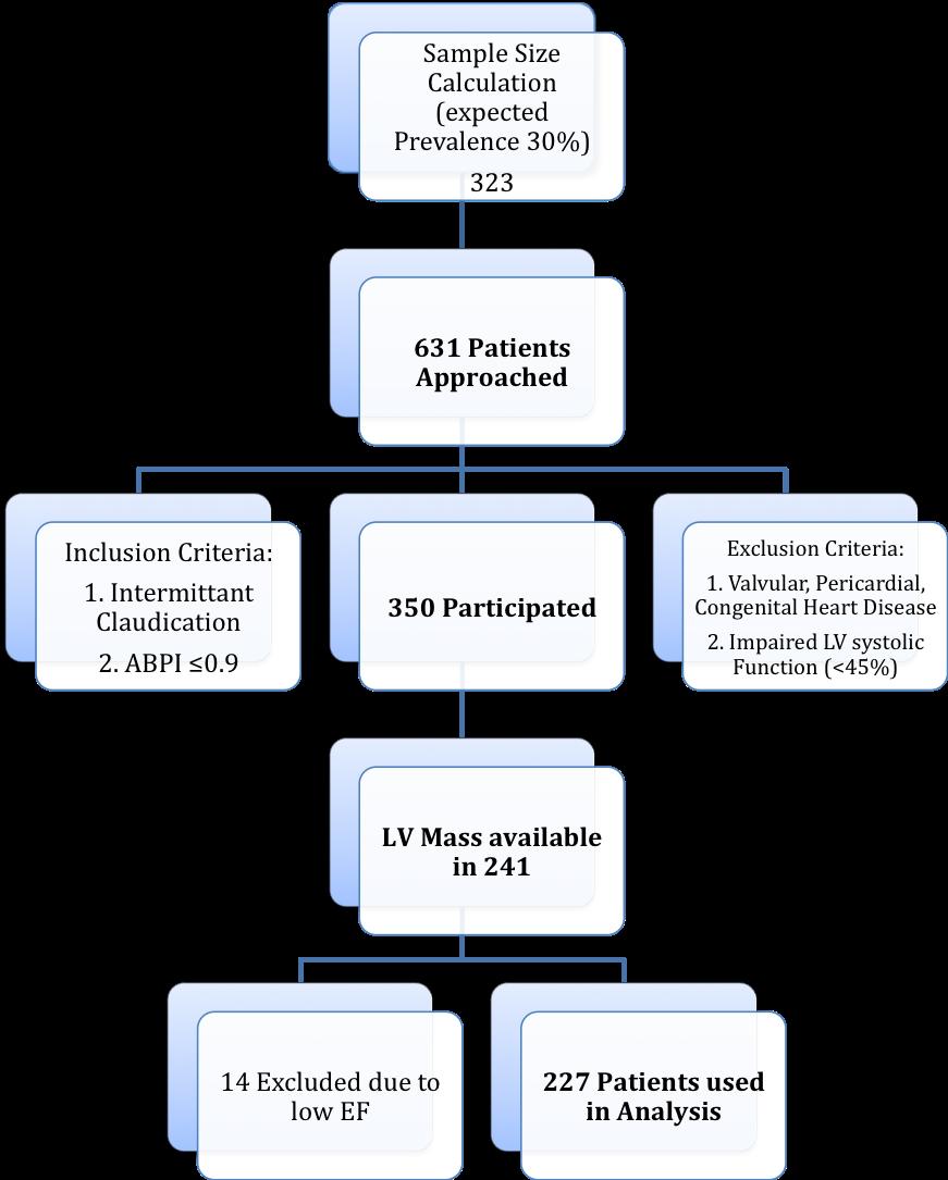
93 91
94 ECHOCARDIOGRAPHIC LEFT VENTRICULAR HYPERTROPHY (INDEXED TO BODY SURFACE AREA) Patient characteristics of the 350 patients recruited are listed in Table 3.1. M- mode measurement of LV mass was obtainable in 241 patients (69%). Those in whom M-mode LV mass could not be assessed did not differ significantly in any other parameters from those in whom an adequate M-mode measurement was obtained (Table 3.2). Out of the 241 patients with M-mode LV mass, 14 patients were excluded due to low ejection fraction. Of the remaining 227 patients with preserved LV systolic function, the proportion of patients with LVH was 50% (114/227) when left ventricular mass was indexed to body surface area (BSA). Table 3.3 shows the characteristics of patients with and without left ventricular hypertrophy. The data was normally distributed, with similar mean, median and modes, with a typical bell shaped curve for all the continuous variables. Multiple regression analysis was performed to include all possible determinants of left ventricular mass index: age, sex, body mass index (BMI), history of hypertension, previous myocardial infarction (MI), diabetes mellitus, ischaemic heart disease, systolic blood pressure, creatinine and total cholesterol. This model explained only 11.4% of the variation in LVMI, and the factors independently related to LVMI were age (standardised β= 0.208, p=0.002), sex (standardised β= 0.215, p=0.001) and diabetes (standardised β= 0.169, p=0.009). Notably systolic BP was not an independent predictor. Patients with LVH were significantly more likely to be prescribed a beta-blocker (27% vs. 12%, p=
95 ), ACE inhibitor (48% vs. 24%, p<0.001) and nitrate (18% vs. 5%, p=0.006). 35% of the studied patients (79/227) had a normal LV geometry calculated according to body surface area (Figure 3.1). The association between history of hypertension, clinic BP and the presence/ absence of LVH is shown in Figure % of the study population had a history of hypertension (n=145). In this group of patients, 13% were normotensive and had LVH. In the group of patients without a history of hypertension (n=82), 17% were normotensive and had evidence of LVH. Overall, 14% of the entire study population had echo LVH despite a normal clinic BP (Figure 3.3). The equivalent data using a lower office BP cutoff (<130/80) are shown for completeness (Figure 3.4).
96 ECHOCARDIOGRAPHIC LEFT VENTRICULAR HYPERTROPHY (INDEXED TO HEIGHT 2.7 ) The proportion of patients with LVH was 72% (163/227) when left ventricular mass was indexed to height 2.7. Table 3.4 shows the characteristics of patients with and without left ventricular hypertrophy. Multiple regression analysis was performed to include all possible determinants of left ventricular mass index: age, sex, body mass index (BMI), history of hypertension, previous myocardial infarction (MI), diabetes mellitus, ischaemic heart disease, systolic blood pressure, creatinine and total cholesterol. This model explained only 15.2% of the variation in LVMI, and the factors independently related to LVMI were age (standardised β=0.227, p=<0.001), BMI (standardised β= 0.274, p<0.001) and diabetes (standardised β= 0.141, p=0.029). Again systolic BP was not an independent predictor. Patients with LVH were significantly more likely to be prescribed a beta-blocker (24% vs. 8%, p= 0.005), ACE inhibitor (41% vs. 22%, p=0.008), statin (76% vs. 61%, p=0.032), diuretic (44% vs. 28%, p=0.035) and nitrate (16% vs. 0%, p=0.001). Concentric LVH was the most common geometry pattern (38%, 86/227) with 23% (52/227) of patients having normal geometry (Figure 3.5). The association between history of hypertension, clinic BP and the presence/ absence of LVH is shown in Figure 3.6. In the group of patients without a history of hypertension (n=82), 33% were normotensive and had evidence of LVH. Overall, 24% of the entire study population (n=227) had echo LVH
97 95 despite a normal clinic BP (Figure 3.7). The equivalent data using a lower office BP cutoff (<130/80) are shown for completeness (Figures 3.8). Even using this lower cutoff, 16% of LVH patients had a normal BP, irrespective of which LV mass criteria are applied.
98 AMBULATORY BLOOD PRESSURE (24 HOUR) LVH indexed to Body Surface Area 24 hour ambulatory blood pressure readings were carried out in 64% of the LVH patients (73/114) and 51% of the non-lvh patients (58/113). Those who underwent this test were randomly chosen purely due to the availability of spare 24 hour Blood pressure monitoring equipment, albeit with a bias towards performing this test more frequently in patients with LVH than those without LVH because the LVH patients were obviously of more interest here. Mean ambulatory systolic blood pressure was significantly lower in the non- LVH group compared to the LVH group (Table 3.5). There was no significant difference in the mean diastolic blood pressure reading in both groups. The percentage of patients with a normal ambulatory BP reading was not significantly different in both the LVH and non-lvh group. 40% of the patients in the LVH group had a normal 24-hour ABPM reading. The data for the number of patients in each group in Figure 3.9 were adjusted to represent the whole PAD population rather than just the subset who received ABPM, taking into account the bias toward measuring ABPM in those with LVH. Approximately every 1 in 2 patients in this study had a normal ABPM reading. In this normotensive group, 43% of the patients had LVH. In fact, a rough summary of the data in Figure 3.9 is that half of the patients had LVH and a somewhat different half had a high BP. PAD patients
99 97 fall into four equal groups: one quarter had LVH and a high BP, another two quarters had either LVH or a high BP and another quarter had neither LVH nor a high BP LVH indexed to Height 2.7 Mean ambulatory systolic blood pressure was significantly lower in the non- LVH group compared to the LVH group (Table 3.6). There was no significant difference in the mean diastolic blood pressure reading in both groups. The percentage of patients with a normal 24-hour BP reading was not significantly different in both the LVH and non-lvh group. 41% of the patients in the LVH group had a normal 24-hour ABPM reading. The data for the number of patients in each group in Figure 3.10 were adjusted to represent the whole PAD population rather than just the subset who received ABPM, taking into account the bias toward measuring ABPM in those with LVH. Approximately every 1 in 2 patients in this study had a normal ABPM reading. In this normotensive group, 63% of the patients had LVH.
100 SEVERITY OF PERIPHERAL ARTERIAL DISEASE, HISTORY OF ATRIAL FIBRILLATION AND MYOCARDIAL INFARCTION IN PATIENTS WITH AND WITHOUT LEFT VENTRICULAR HYPERTROPHY There was no difference in the severity of PAD, in terms of ABPI, in patients with or without LVH (Table 3.3 and 3.4). There was a mean ABPI of 0.68 in patients with LVH according to BSA compared to 0.69 in patients without LVH. The ABPI was the same (0.69) in patients with and without LVH according to height 2.7. Out of two hundred and twenty seven patients studied, there were fourteen (6%) who had atrial fibrillation (AF) on a 12-lead ECG performed during the assessment (Table 3.7). The patients with AF had a higher mean LVMI of 189.2g/m 2 compared to 130.7g/m 2 in those in sinus rhythm at the time of assessment (p<0.001). Despite a higher mean mass, there was no statistical difference in percentage of patients with LVH between those patients with or without AF (64% V 49%, p=0.279). Unsurprisingly, patients with AF had a larger left atrial diameter (4.08cm V 3.34cm, p<0.001), smaller LV ejection fraction (58% V 67%, p=0.009) and had a significantly higher BNP (195pg/ml V 75pg/ml, p<0.001). Patients who were in AF were less likely to have a smoking history (64% V 90%, p=0.003) compared to those in sinus
101 99 rhythm. All other parameters were similar and in particular, there was no difference in either clinic blood pressure or history of hypertension between those in AF or sinus rhythm. Patients with a history of myocardial infarction were more likely to have LVH than those without (69% V 47%, p=0.024), although their mean LV mass was statistically similar (150.5g/m 2 V 131.6g/m 2, p=0.059) compared to patients with no history of prior MI (Table 3.8). Patients with a history of MI were more likely to be male (81% V 59%, p=0.015) and have a lower total cholesterol (4.16mmol/L V 4.60mmol/L, P=0.004). This is likely to be because more patients with a prior MI were prescribed a Statin (91% V 69%, P=0.01). There was no difference in office systolic blood pressure, but patients with prior MI had a lower diastolic blood pressure (72mmHg V 78mmHg, p=0.002). This is likely to be because patients with a prior MI are more likely to be prescribed anti-hypertensive agents such as beta blockers (38% V 17%, P<0.001), ACE inhibitors (52% V 33%, p=0.046) and calcium channel receptor antagonists (53% V 35%, p=0.048). Importantly, there was no difference in ejection fraction (65% V 67%, p=0.493) between those with and without a history of MI.
102 100 Table 3.1: Patient characteristics (mean +/-SD) of the 350 subjects studied Variable Mean (SD) Males (%) 65.1 Age (years) 68.9 (9.5) BMI (kg/m2) 28.3 (4.6) Office systolic BP (mmhg) 147 (21) Office diastolic (mmhg) 77 (11) Creatinine (μmol/l) 107 (50) Total Cholesterol (mmol/l) 4.57 (0.95) HDL Cholesterol (mmol/l) 1.42 (0.46) History of hypertension (%) 61.4 History of Type II Diabetes (%) 18.3 Current smokers (%) 39.1 Ex-smokers (%) 50.5 History of ischaemic heart disease (%) 32.9 Previous MI (%) 16.9 LVMI Males (61.7) Females 123 (38.7) Left Ventricular Ejection Fraction (%) 64.6 (12.1)
103 101 Treated with Aspirin 70 Statin 71.7 Beta blockers 18 ACE inhibitors 36.8 Angiotensin receptor blocker (ARB) 9.7 Clopidogrel 8 Calcium Antagonist 38.6 Nicorandil 4 Nitrate 13.4 Diuretic 40.9 BMI Body Mass Index BP Blood Pressure MI Myocardial Infarction LVMI Left Ventricular Mass Index ACE Angiotensin Converting Enzyme
104 102 Table 3.2: Patient characteristics classified according to presence or absence of M-mode LV mass measurements being obtainable. Variable LVM available No LVM available P mean (SD) mean (SD) value (n=241) (n=109) Males (%) Age (years) 68 (10) 70 (9) BMI (kg/m 2 ) 28.2 (4.4) 28.4 (4.8) Clinic systolic BP (mmhg) 147 (21) 147 (23) Clinic diastolic BP (mmhg) 77 (11) 77 (11) Creatinine (μmol/l) 107 (57) 108 (31) Total Cholesterol (mmol/l) 4.52 (0.96) 4.67 (0.94) Ex/current smoker (%) History of hypertension (%) D.M. (%) History of MI (%) BMI Body Mass Index D.M. Diabetes Mellitus MI Myocardial Infarction
105 103 Table 3.3: Patient characteristics classified according to presence or absence of left ventricular hypertrophy indexed to body surface area. Variable LVH No LVH P value mean (n=114) mean (n=113) Males % Age (years) <0.001 BMI (kg/m2) ABPI Clinic systolic BP (mmhg) Clinic diastolic BP (mmhg) History of hypertension (%) History of type II Diabetes (%) Previous MI (%) IHD (%) Current smokers (%) BNP (pg/ml) Creatinine (μmol/l) Total Cholesterol (mmol/l) HDL Cholesterol (moml/l) LVMI (g/m2) Males <0.001 Females <0.001 LV ejection fraction (%)
106 104 IVS (cm) <0.001 LVID (cm) <0.001 PWT (cm) <0.001 Left atrial diameter (cm) BMI Body Mass Index MI Myocardial Infarction IHD Ischaemic Heart Disease BNP - B-Type Natriuretic Peptide LVMI - Left Ventricular Mass Index LV Left Ventricle IVS Interventricular Septum LVID Left Ventricular Internal Dimension PWT Posterior Wall Thickness
107 105 Table 3.4: Patient characteristics classified according to presence or absence of left ventricular hypertrophy indexed to height 2.7. Variable LVH No LVH P value mean (n=163) mean (n=64) Males % (n) Age (years) <0.001 BMI (kg/m2) <0.001 ABPI Clinic systolic BP (mmhg) Clinic diastolic BP (mmhg) History of hypertension (%) History of type II Diabetes (%) Previous MI (%) IHD (%) <0.001 Current smokers (%) BNP (pg/ml) Creatinine (umol/l) Total Cholesterol (mmol/l) HDL Cholesterol (moml/l) LVMI (height 2.7) Males <0.001 Females <0.001 LV ejection fraction (%)
108 106 IVS (cm) <0.001 LVID (cm) <0.001 PWT (cm) <0.001 Left atrial diameter (cm) BMI Body Mass Index MI Myocardial Infarction IHD Ischaemic Heart Disease BNP - B-Type Natriuretic Peptide LVMI - Left Ventricular Mass Index LV Left Ventricle IVS Interventricular Septum LVID Left Ventricular Internal Dimension PWT Posterior Wall Thickness
109 107 Table 3.5: 24 hour ambulatory blood pressure results classified according to presence or absence of LVH (indexed to body surface area) Variable LVH mean No LVH mean P value (n=73) (n=58) 24 hour systolic (mmhg) hour diastolic (mmhg) DT systolic (mmhg) DT diastolic (mmhg) NT systolic (mmhg) NT diastolic (mmhg) Normal 24 hour BP ( 130/80) (%) Normal DT reading ( 135/85) (%) Normal NT reading ( 120/70) (%) Abnormal 24 hour BP (>135/85) (%) Abnormal DT reading (>140/90) (%) Abnormal NT reading (>125/75) (%) DT Day Time NT Night Time
110 108 Table 3.6: 24 hour ambulatory blood pressure results classified according to presence or absence of LVH (indexed to height 2.7) Variable LVH mean No LVH mean P value (n=101) (n=30) 24 hour systolic (mmhg) hour diastolic (mmhg) DT systolic (mmhg) DT diastolic (mmhg) NT systolic (mmhg) NT diastolic (mmhg) Normal 24 hour BP ( 130/80) (%) Normal DT reading ( 135/85) (%) Normal NT reading ( 120/70) (%) Abnormal 24 hour BP (>135/85) (%) Abnormal DT reading (>140/90) (%) Abnormal NT reading (>125/75) (%) DT Day Time NT Night Time
111 109 Table 3.7: Patient characteristics classified according to presence or absence of Atrial Fibrillation (AF) on 12 lead ECG Variable AF No AF P value mean (n=14) mean (n=213) Males % Age (years) <0.001 BMI (kg/m2) ABPI Clinic systolic BP (mmhg) Clinic diastolic BP (mmhg) History of hypertension (%) History of type II Diabetes (%) Previous MI (%) IHD (%) Smokers (current or ex) (%) BNP (pg/ml) <0.001 Creatinine (μmol/l) Total Cholesterol (mmol/l) HDL Cholesterol (mmol/l) LVMI (g/m 2 ) <0.001 LV ejection fraction (%) LVH (%) LVID (cm)
112 110 PWT (cm) Left atrial diameter (cm) <0.001
113 111 Table 3.8: Patient characteristics classified according to History of prior Myocardial Infarction (MI) Variable MI No MI P value mean (n=32) mean (n=195) Males % Age (years) BMI (kg/m2) ABPI Clinic systolic BP (mmhg) Clinic diastolic BP (mmhg) History of hypertension (%) History of type II Diabetes (%) Angina (%) Smokers (current or ex) (%) BNP (pg/ml) Creatinine (μmol/l) Total Cholesterol (mmol/l) HDL Cholesterol (moml/l) LVMI (g/m 2 ) LV ejection fraction (%) LVH (%) LVID (cm) <0.001 PWT (cm)
114 112 Left atrial diameter (cm)

115 113 Figure 3.1: Left ventricular geometry in the studied population, using left ventricular mass indexed to body surface area
116 114 Figure 3.2: Association between history of hypertension, clinic BP (normal clinic BP <140/90) and presence or absence of LVH (indexed to BSA) in this study population A History of Hypertension, High Clinic BP, LVH 28% B History of Hypertension, High Clinic BP, No LVH 21% C History of Hypertension, Normal Clinic BP, LVH 8% D History of Hypertension, Normal Clinic BP, No LVH 7% E No History of Hypertension, High Clinic BP, LVH 9% F No History of Hypertension, High Clinic BP, No LVH 9% G No History of Hypertension, Normal Clinic BP, LVH 6% H No History of Hypertension, Normal Clinic BP, No LVH 12%
117 115 Figure 3.3 Association with Clinic Blood Pressure and presence or absence of LVH (indexed to BSA) in the study population.
118 116 Figure 3.4: Association between history of hypertension, clinic BP (normal clinic BP <130/80) and presence or absence of LVH (indexed to BSA) in this study population A History of Hypertension, High Clinic BP, LVH 31% B History of Hypertension, High Clinic BP, No LVH 26% C History of Hypertension, Normal Clinic BP, LVH 4% D History of Hypertension, Normal Clinic BP, No LVH 3% E No History of Hypertension, High Clinic BP, LVH 11% F No History of Hypertension, High Clinic BP, No LVH 15% G No History of Hypertension, Normal Clinic BP, LVH 4% H No History of Hypertension, Normal Clinic BP, No LVH 6%

119 117 Figure 3.5: Left ventricular geometry in the studied population, using left ventricular mass indexed to height 2.7
120 118 Figure 3.6: Association between history of hypertension, clinic BP (normal clinic BP <140/90) and presence or absence of LVH (indexed to height 2.7) in this study population A History of Hypertension, High Clinic BP, LVH 36% B History of Hypertension, High Clinic BP, No LVH 12% C History of Hypertension, Normal Clinic BP, LVH 12% D History of Hypertension, Normal Clinic BP, No LVH 4% E No History of Hypertension, High Clinic BP, LVH 12% F No History of Hypertension, High Clinic BP, No LVH 5% G No History of Hypertension, Normal Clinic BP, LVH 12% H No History of Hypertension, Normal Clinic BP, No LVH 7%
121 119 Figure 3.7 Association with Clinic Blood Pressure and presence or absence of LVH (indexed to Height 2.7) in the study population.
122 120 Figure 3.8: Association between history of hypertension, clinic BP (normal clinic BP<130/80) and presence or absence of LVH (indexed to height 2.7) in this study population A History of Hypertension, High Clinic BP, LVH 43% B History of Hypertension, High Clinic BP, No LVH 14% C History of Hypertension, Normal Clinic BP, LVH 5% D History of Hypertension, Normal Clinic BP, No LVH 2% E No History of Hypertension, High Clinic BP, LVH 17% F No History of Hypertension, High Clinic BP, No LVH 9% G No History of Hypertension, Normal Clinic BP, LVH 7% H No History of Hypertension, Normal Clinic BP, No LVH 3%

123 121 Figure 3.9: Association between ambulatory BP reading and the presence or absence of LVH (indexed to BSA) in the study population.

124 122 Figure 3.10: Association between ambulatory BP reading and the presence or absence of LVH (indexed to Height 2.7) in the study population.
Introduction. Risk factors of PVD 5/8/2017
 PATHOPHYSIOLOGY AND CLINICAL FEATURES OF PERIPHERAL VASCULAR DISEASE Dr. Muhamad Zabidi Ahmad Radiologist and Section Chief, Radiology, Oncology and Nuclear Medicine Section, Advanced Medical and Dental
Left ventricular hypertrophy: why does it happen?
 Nephrol Dial Transplant (2003) 18 [Suppl 8]: viii2 viii6 DOI: 10.1093/ndt/gfg1083 Left ventricular hypertrophy: why does it happen? Gerard M. London Department of Nephrology and Dialysis, Manhes Hospital,
NATIONAL INSTITUTE FOR HEALTH AND CLINICAL EXCELLENCE
 NATIONAL INSTITUTE FOR HEALTH AND CLINICAL EXCELLENCE QUALITY AND OUTCOMES FRAMEWORK (QOF) INDICATOR DEVELOPMENT PROGRAMME Briefing paper QOF indicator area: Peripheral arterial disease Potential output:
Evaluation of Left Ventricular Function and Hypertrophy Gerard P. Aurigemma MD
 Evaluation of Left Ventricular Function and Hypertrophy Gerard P. Aurigemma MD Board Review Course 2017 43 year old health assistant Severe resistant HTN LT BSA 2 Height 64 1 Here is the M mode echocardiogram
Controversies in Cardiac Pharmacology
 Controversies in Cardiac Pharmacology Thomas D. Conley, MD FACC FSCAI Disclosures I have no relevant relationships with commercial interests to disclose. 1 Doc, do I really need to take all these medicines?
Circulation. Blood Pressure and Antihypertensive Medications. Venous Return. Arterial flow. Regulation of Cardiac Output.
 Circulation Blood Pressure and Antihypertensive Medications Two systems Pulmonary (low pressure) Systemic (high pressure) Aorta 120 mmhg Large arteries 110 mmhg Arterioles 40 mmhg Arteriolar capillaries
Review of Cardiac Imaging Modalities in the Renal Patient. George Youssef
 Review of Cardiac Imaging Modalities in the Renal Patient George Youssef ECHO Left ventricular hypertrophy (LVH) assessment Diastolic dysfunction Stress ECHO Cardiac CT angiography Echocardiography - positives
In Vivo Animal Models of Heart Disease. Why Animal Models of Disease? Timothy A Hacker, PhD Department of Medicine University of Wisconsin-Madison
 In Vivo Animal Models of Heart Disease Timothy A Hacker, PhD Department of Medicine University of Wisconsin-Madison Why Animal Models of Disease? Heart Failure (HF) Leading cause of morbidity and mortality
Martin G. Keane, MD, FASE Temple University School of Medicine
 Martin G. Keane, MD, FASE Temple University School of Medicine Measurement of end-diastolic LV internal diameter (LVIDd) made by properly-oriented M-Mode techniques in the Parasternal Long Axis View (PLAX):
10/8/2018. Lecture 9. Cardiovascular Health. Lecture Heart 2. Cardiovascular Health 3. Stroke 4. Contributing Factor
 Lecture 9 Cardiovascular Health 1 Lecture 9 1. Heart 2. Cardiovascular Health 3. Stroke 4. Contributing Factor 1 The Heart Muscular Pump The Heart Receives blood low pressure then increases the pressure
Outline. Pathophysiology: Heart Failure. Heart Failure. Heart Failure: Definitions. Etiologies. Etiologies
 Outline Pathophysiology: Mat Maurer, MD Irving Assistant Professor of Medicine Definitions and Classifications Epidemiology Muscle and Chamber Function Pathophysiology : Definitions An inability of the
Relationship between body mass index, coronary disease extension and clinical outcomes in patients with acute coronary syndrome
 Relationship between body mass index, coronary disease extension and clinical outcomes in patients with acute coronary syndrome Helder Dores, Luís Bronze Carvalho, Ingrid Rosário, Sílvio Leal, Maria João
Cardiovascular Disorders Lecture 3 Coronar Artery Diseases
 Cardiovascular Disorders Lecture 3 Coronar Artery Diseases By Prof. El Sayed Abdel Fattah Eid Lecturer of Internal Medicine Delta University Coronary Heart Diseases It is the leading cause of death in
Lecture 8 Cardiovascular Health Lecture 8 1. Introduction 2. Cardiovascular Health 3. Stroke 4. Contributing Factors
 Lecture 8 Cardiovascular Health 1 Lecture 8 1. Introduction 2. Cardiovascular Health 3. Stroke 4. Contributing Factors 1 Human Health: What s Killing Us? Health in America Health is the U.S Average life
1 Functions of endothelial cells include all the following EXCEPT. 2 Response to vascular injury is characterised by
 airns ase Hospital mergency epartment Part 1 FM MQs 1 Functions of endothelial cells include all the following XPT Formation of von-willebrand factor Formation of collagen and proteoglycans Formation of
Pathophysiology: Heart Failure
 Pathophysiology: Heart Failure Mat Maurer, MD Irving Assistant Professor of Medicine Outline Definitions and Classifications Epidemiology Muscle and Chamber Function Pathophysiology Heart Failure: Definitions
Assessing Cardiac Risk in Noncardiac Surgery. Murali Sivarajan, M.D. Professor University of Washington Seattle, Washington
 Assessing Cardiac Risk in Noncardiac Surgery Murali Sivarajan, M.D. Professor University of Washington Seattle, Washington Disclosure None. I have no conflicts of interest, financial or otherwise. CME
Ischemic Heart Disease
 Ischemic Heart Disease Definition: Ischemic heart disease (IHD) is a condition in which there is an inadequate supply of blood and oxygen to a portion of the myocardium; it typically occurs when there
Diastolic Heart Failure. Edwin Tulloch-Reid MBBS FACC Consultant Cardiologist Heart Institute of the Caribbean December 2012
 Diastolic Heart Failure Edwin Tulloch-Reid MBBS FACC Consultant Cardiologist Heart Institute of the Caribbean December 2012 Disclosures Have spoken for Merck, Sharpe and Dohme Sat on a physician advisory
Heart Failure in Women: Dr Goh Ping Ping Cardiologist Asian Heart & Vascular Centre
 Heart Failure in Women: More than EF? Dr Goh Ping Ping Cardiologist Asian Heart & Vascular Centre Overview Review pathophysiology as it relates to diagnosis and management Rational approach to workup:
Supplementary Online Content
 Supplementary Online Content Inohara T, Manandhar P, Kosinski A, et al. Association of renin-angiotensin inhibitor treatment with mortality and heart failure readmission in patients with transcatheter
Ischemic heart disease
 Ischemic heart disease Introduction In > 90% of cases: the cause is: reduced coronary blood flow secondary to: obstructive atherosclerotic vascular disease so most of the time it is called: coronary artery
LV geometric and functional changes in VHD: How to assess? Mi-Seung Shin M.D., Ph.D. Gachon University Gil Hospital
 LV geometric and functional changes in VHD: How to assess? Mi-Seung Shin M.D., Ph.D. Gachon University Gil Hospital LV inflow across MV LV LV outflow across AV LV LV geometric changes Pressure overload
Cardiovascular Disorders. Heart Disorders. Diagnostic Tests for CV Function. Bio 375. Pathophysiology
 Cardiovascular Disorders Bio 375 Pathophysiology Heart Disorders Heart disease is ranked as a major cause of death in the U.S. Common heart diseases include: Congenital heart defects Hypertensive heart
Hypertension in Aortic Valve Disease
 Hypertension in Aortic Valve Disease Hanna M. Nosseir MRCP, FRCP Head of Cardiology department Galaa Military Medical Complex Aortic stenosis: Introduction Arterial hypertension and aortic stenosis are
PRELIMINARY STUDIES OF LEFT VENTRICULAR WALL THICKNESS AND MASS OF NORMOTENSIVE AND HYPERTENSIVE SUBJECTS USING M-MODE ECHOCARDIOGRAPHY
 Malaysian Journal of Medical Sciences, Vol. 9, No. 1, January 22 (28-33) ORIGINAL ARTICLE PRELIMINARY STUDIES OF LEFT VENTRICULAR WALL THICKNESS AND MASS OF NORMOTENSIVE AND HYPERTENSIVE SUBJECTS USING
Coronary artery disease (CAD) risk factors
 risk factors") Background Coronary artery disease (CAD) risk factors CAD Risk factors Hypertension Insulin resistance /diabetes Dyslipidemia Smoking /Obesity Male gender/ Old age Atherosclerosis Arterial stiffness precedes
Background Coronary artery disease (CAD) risk factors CAD Risk factors Hypertension Insulin resistance /diabetes Dyslipidemia Smoking /Obesity Male gender/ Old age Atherosclerosis Arterial stiffness precedes
During exercise the heart rate is 190 bpm and the stroke volume is 115 ml/beat. What is the cardiac output?
 The Cardiovascular System Part III: Heart Outline of class lecture After studying part I of this chapter you should be able to: 1. Be able to calculate cardiac output (CO) be able to define heart rate
 Five chapters 1. What is CVD prevention 2. Why is CVD prevention needed 3. Who needs CVD prevention 4. How is CVD prevention applied 5. Where should CVD prevention be offered Shorter, more adapted to clinical
2013 ACC/AHA Guidelines on the Assessment of Atherosclerotic Cardiovascular Risk: Overview and Commentary
 2013 ACC/AHA Guidelines on the Assessment of Atherosclerotic Cardiovascular Risk: Overview and Commentary The Johns Hopkins Ciccarone Center for the Prevention of Heart Disease Becky McKibben, MPH; Seth
Cardiovascular Disease in CKD. Parham Eftekhari, D.O., M.Sc. Assistant Clinical Professor Medicine NSUCOM / Broward General Medical Center
 Cardiovascular Disease in CKD Parham Eftekhari, D.O., M.Sc. Assistant Clinical Professor Medicine NSUCOM / Broward General Medical Center Objectives Describe prevalence for cardiovascular disease in CKD
Copyright 2011, 2007 by Mosby, Inc., an affiliate of Elsevier Inc. Normal Cardiac Anatomy
 Mosby,, an affiliate of Elsevier Normal Cardiac Anatomy Impaired cardiac pumping Results in vasoconstriction & fluid retention Characterized by ventricular dysfunction, reduced exercise tolerance, diminished
Structure and organization of blood vessels
 The cardiovascular system Structure of the heart The cardiac cycle Structure and organization of blood vessels What is the cardiovascular system? The heart is a double pump heart arteries arterioles veins
Index of subjects. effect on ventricular tachycardia 30 treatment with 101, 116 boosterpump 80 Brockenbrough phenomenon 55, 125
 145 Index of subjects A accessory pathways 3 amiodarone 4, 5, 6, 23, 30, 97, 102 angina pectoris 4, 24, 1l0, 137, 139, 140 angulation, of cavity 73, 74 aorta aortic flow velocity 2 aortic insufficiency
Heart Failure. Cardiac Anatomy. Functions of the Heart. Cardiac Cycle/Hemodynamics. Determinants of Cardiac Output. Cardiac Output
 Cardiac Anatomy Heart Failure Professor Qing ZHANG Department of Cardiology, West China Hospital www.blaufuss.org Cardiac Cycle/Hemodynamics Functions of the Heart Essential functions of the heart to cover
Wellness: Concepts and Applications 8 th Edition Anspaugh, Hamrick, Rosato
 Wellness: Concepts and Applications 8 th Edition Anspaugh, Hamrick, Rosato Preventing Cardiovascular Disease Chapter 2 Cardiovascular Disease the leading cause of death in the U.S. 35.3% of all deaths
Clinical Features and Subtypes of Ischemic Stroke Associated with Peripheral Arterial Disease
 Cronicon OPEN ACCESS EC NEUROLOGY Research Article Clinical Features and Subtypes of Ischemic Stroke Associated with Peripheral Arterial Disease Jin Ok Kim, Hyung-IL Kim, Jae Guk Kim, Hanna Choi, Sung-Yeon
Identification of patients with heart failure and PREserved systolic Function : an Epidemiologic Regional study
 Identification of patients with heart failure and PREserved systolic Function : an Epidemiologic Regional study Dr. Antonio Magaña M.D. (on behalf I-PREFER investigators group) Stockholm, Sweden, August
Heart Failure (HF) Treatment
 Treatment") Heart Failure (HF) Treatment Heart Failure (HF) Complex, progressive disorder. The heart is unable to pump sufficient blood to meet the needs of the body. Its cardinal symptoms are dyspnea, fatigue, and
Heart Failure (HF) Treatment Heart Failure (HF) Complex, progressive disorder. The heart is unable to pump sufficient blood to meet the needs of the body. Its cardinal symptoms are dyspnea, fatigue, and
4. Which survey program does your facility use to get your program designated by the state?
 STEMI SURVEY Please complete one survey for each TCD designation you have in your facility. There would be a maximum of three surveys completed if your facility was designated as a trauma, stroke and STEMI
Cardiovascular Nursing Practice: A Comprehensive Resource Manual and Study Guide for Clinical Nurses 2 nd Edition
 Cardiovascular Nursing Practice: A Comprehensive Resource Manual and Study Guide for Clinical Nurses 2 nd Edition Table of Contents Volume 1 Chapter 1: Cardiovascular Anatomy and Physiology Basic Cardiac
DIASTOLIC HEART FAILURE
 DIASTOLIC HEART FAILURE M Mohsen Ibrahim, MD Alexandria, Proposed Criteria for Diastolic Heart Failure ESC Working Group (EHJ 1998) CHF signs/symptoms EF 45% Hemodynamic or echo evidence of diastolic dysfunction
4/7/ The stats on heart disease. + Deaths & Age-Adjusted Death Rates for
 + Update on Lipid Management Stacey Gardiner, MD Assistant Professor Division of Cardiovascular Medicine Medical College of Wisconsin + The stats on heart disease Over the past 10 years for which statistics
HEART FAILURE. Study day November 2018 Sarah Briggs
 HEART FAILURE Study day November 2018 Sarah Briggs Overview and Introduction This course is an introduction and overview of heart failure. Normal heart function and basic pathophysiology of heart failure
Cardiac Drugs: Chapter 9 Worksheet Cardiac Agents. 1. drugs affect the rate of the heart and can either increase its rate or decrease its rate.
 Complete the following. 1. drugs affect the rate of the heart and can either increase its rate or decrease its rate. 2. drugs affect the force of contraction and can be either positive or negative. 3.
Quality Payment Program: Cardiology Specialty Measure Set
 Quality Payment Program: Cardiology Specialty Set Title Number CMS Reporting Method(s) Heart Failure (HF): Angiotensin- Converting Enzyme (ACE) Inhibitor or Angiotensin Receptor Blocker (ARB) Therapy for
APPENDIX F: CASE REPORT FORM
 APPENDIX F: CASE REPORT FORM Instruction: Complete this form to notify all ACS admissions at your centre to National Cardiovascular Disease Registry. Where check boxes are provided, check ( ) one or more
Quality Payment Program: Cardiology Specialty Measure Set
 Measure Title * Reportable via PINNACLE α Reportable via Diabetes Collaborative CQMC v1.0 Measure High Priority Measure Cross Cutting Measure Heart Failure (HF): Angiotensin- Converting Enzyme (ACE) Inhibitor
Chapter 4: Cardiovascular Disease in Patients with CKD
 Chapter 4: Cardiovascular Disease in Patients with CKD The prevalence of cardiovascular disease (CVD) was 65.8% among patients aged 66 and older who had chronic kidney disease (CKD), compared to 31.9%
Vascular disease. Structural evaluation of vascular disease. Goo-Yeong Cho, MD, PhD Seoul National University Bundang Hospital
 Vascular disease. Structural evaluation of vascular disease Goo-Yeong Cho, MD, PhD Seoul National University Bundang Hospital resistance vessels : arteries
DIFFERENTE RELAZIONE TRA VALORI PRESSORI E MASSA VENTRICOLARE SX NEI DUE SESSI IN PAZIENTI IPERTESI.
 DIFFERENTE RELAZIONE TRA VALORI PRESSORI E MASSA VENTRICOLARE SX NEI DUE SESSI IN PAZIENTI IPERTESI. Franco Cipollini, Carlo Porta, Enrica Arcangeli, Carla Breschi, & Giuseppe Seghieri Azienda USL 3, Ambulatorio
Cardiovascular System Notes: Heart Disease & Disorders
 Cardiovascular System Notes: Heart Disease & Disorders Interesting Heart Facts The Electrocardiograph (ECG) was invented in 1902 by Willem Einthoven Dutch Physiologist. This test is still used to evaluate
Autonomic nervous system, inflammation and preclinical carotid atherosclerosis in depressed subjects with coronary risk factors
 Autonomic nervous system, inflammation and preclinical carotid atherosclerosis in depressed subjects with coronary risk factors Carmine Pizzi 1 ; Lamberto Manzoli 2, Stefano Mancini 3 ; Gigliola Bedetti
Objectives. Systolic Heart Failure: Definitions. Heart Failure: Historical Perspective 2/7/2009
 Objectives Diastolic Heart Failure and Indications for Echocardiography in the Asian Population Damon M. Kwan, MD UCSF Asian Heart & Vascular Symposium 02.07.09 Define diastolic heart failure and differentiate
Listing Form: Heart or Cardiovascular Impairments. Medical Provider:
 Listing Form: Heart or Cardiovascular Impairments Medical Provider: Printed Name Signature Patient Name: Patient DOB: Patient SS#: Date: Dear Provider: Please indicate whether your patient s condition
Acute Myocardial Infarction
 Acute Myocardial Infarction Hafeza Shaikh, DO, FACC, RPVI Lourdes Cardiology Services Asst.Program Director, Cardiology Fellowship Associate Professor, ROWAN-SOM Acute Myocardial Infarction Definition:
DECLARATION OF CONFLICT OF INTEREST
 DECLARATION OF CONFLICT OF INTEREST Is there a mortality risk associated with aspirin use in heart failure? Results from a large community based cohort Margaret Bermingham, Mary-Kate Shanahan, Saki Miwa,
The Failing Heart in Primary Care
 The Failing Heart in Primary Care Hamid Ikram How fares the Heart Failure Epidemic? 4357 patients, 57% women, mean age 74 years HFSA 2010 Practice Guideline (3.1) Heart Failure Prevention A careful and
Repeat ischaemic heart disease audit of primary care patients ( ): Comparisons by age, sex and ethnic group
: Comparisons by age, sex and ethnic group") Repeat ischaemic heart disease audit of primary care patients (2002-2003): Comparisons by age, sex and ethnic group Baseline-repeat ischaemic heart disease audit of primary care patients: a comparison
Repeat ischaemic heart disease audit of primary care patients (2002-2003): Comparisons by age, sex and ethnic group Baseline-repeat ischaemic heart disease audit of primary care patients: a comparison
Peripheral Arterial Disease (PAD): Presentation, Diagnosis, and Treatment
: Presentation, Diagnosis, and Treatment") Peripheral Arterial Disease (PAD): Presentation, Diagnosis, and Treatment Prepared and Presented by Jon Manocchio, Pharm D Blanchard Valley Hospital October 2011 Introduction PAD is a condition that is
Peripheral Arterial Disease (PAD): Presentation, Diagnosis, and Treatment Prepared and Presented by Jon Manocchio, Pharm D Blanchard Valley Hospital October 2011 Introduction PAD is a condition that is
Skin supplied by T1-4 (medial upper arm and neck) T5-9- epigastrium Visceral afferents from skin and heart are the same dorsal root ganglio
 T5-9- epigastrium Visceral afferents from skin and heart are the same dorsal root ganglio") Cardio 2 ECG... 3 Cardiac Remodelling... 11 Valvular Diseases... 13 Hypertension... 18 Aortic Coarctation... 24 Erythropoiesis... 27 Haemostasis... 30 Anaemia... 36 Atherosclerosis... 44 Angina... 48 Myocardial
Cardio 2 ECG... 3 Cardiac Remodelling... 11 Valvular Diseases... 13 Hypertension... 18 Aortic Coarctation... 24 Erythropoiesis... 27 Haemostasis... 30 Anaemia... 36 Atherosclerosis... 44 Angina... 48 Myocardial
ARIC HEART FAILURE HOSPITAL RECORD ABSTRACTION FORM. General Instructions: ID NUMBER: FORM NAME: H F A DATE: 10/13/2017 VERSION: CONTACT YEAR NUMBER:
 ARIC HEART FAILURE HOSPITAL RECORD ABSTRACTION FORM General Instructions: The Heart Failure Hospital Record Abstraction Form is completed for all heart failure-eligible cohort hospitalizations. Refer to
Results of Ischemic Heart Disease
 Ischemic Heart Disease: Angina and Myocardial Infarction Ischemic heart disease; syndromes causing an imbalance between myocardial oxygen demand and supply (inadequate myocardial blood flow) related to
C1: Medical Standards for Safety Critical Workers with Cardiovascular Disorders
 C1: Medical Standards for Safety Critical Workers with Cardiovascular Disorders GENERAL ISSUES REGARDING MEDICAL FITNESS-FOR-DUTY 1. These medical standards apply to Union Pacific Railroad (UPRR) employees
Term-End Examination December, 2009 MCC-006 : CARDIOVASCULAR EPIDEMIOLOGY
 MCC-006 POST GRADUATE DIPLOMA IN CLINICAL CARDIOLOGY (PGDCC) 00269 Term-End Examination December, 2009 MCC-006 : CARDIOVASCULAR EPIDEMIOLOGY Time : 2 hours Maximum Marks : 60 Note : There will be multiple
Beta-blockers in Patients with Mid-range Left Ventricular Ejection Fraction after AMI Improved Clinical Outcomes
 Beta-blockers in Patients with Mid-range Left Ventricular Ejection Fraction after AMI Improved Clinical Outcomes Seung-Jae Joo and other KAMIR-NIH investigators Department of Cardiology, Jeju National
Acute Myocardial Infarction. Willis E. Godin D.O., FACC
 Acute Myocardial Infarction Willis E. Godin D.O., FACC Acute Myocardial Infarction Definition: Decreased delivery of oxygen and nutrients to the myocardium Myocardial tissue necrosis causing irreparable
Cardiac Pathophysiology
 Cardiac Pathophysiology Evaluation Components Medical history Physical examination Routine laboratory tests Optional tests Medical History Duration and classification of hypertension. Patient history of
Complications of Diabetes mellitus. Dr Bill Young 16 March 2015
 Complications of Diabetes mellitus Dr Bill Young 16 March 2015 Complications of diabetes Multi-organ involvement 2 The extent of diabetes complications At diagnosis as many as 50% of patients may have
The Cardiovascular System Part I: Heart Outline of class lecture After studying part I of this chapter you should be able to:
 The Cardiovascular System Part I: Heart Outline of class lecture After studying part I of this chapter you should be able to: 1. Describe the functions of the heart 2. Describe the location of the heart,
THE HEART AND HYPERTENSION. Philippe Gosse Hypertension Unit University Hospital Bordeaux
 THE HEART AND HYPERTENSION Philippe Gosse Hypertension Unit University Hospital Bordeaux INCREASED LVM Cardiomyocytes hypertrophy is a response to pressure overload This response is influenced by many
C-Reactive Protein and Your Heart
 C-Reactive Protein and Your Heart By: James L. Holly, MD Inflammation is the process by which the body responds to injury. Laboratory evidence and findings at autopsy studies suggest that the inflammatory
In the name of GOD. Animal models of cardiovascular diseases: myocardial infarction & hypertension
 In the name of GOD Animal models of cardiovascular diseases: myocardial infarction & hypertension 44 Presentation outline: Cardiovascular diseases Acute myocardial infarction Animal models for myocardial
LXIV: DRUGS: 4. RAS BLOCKADE
 LXIV: DRUGS: 4. RAS BLOCKADE ACE Inhibitors Components of RAS Actions of Angiotensin i II Indications for ACEIs Contraindications RAS blockade in hypertension RAS blockade in CAD RAS blockade in HF Limitations
HYPERTENSION AND HEART FAILURE
 HYPERTENSION AND HEART FAILURE Kenya Cardiac Society Symposium Feb 2017 Dr Jeilan Mohamed No conflict of interests . Geoffrey, 45 yr old hypertensive office worker male from Nairobi, has just watched his
SUPPLEMENTAL MATERIAL
 SUPPLEMENTAL MATERIAL Table S1: Number and percentage of patients by age category Distribution of age Age
The importance of follow-up after a cardiac event: CARDIAC REHABILITATION. Dr. Guy Letcher
 The importance of follow-up after a cardiac event: CARDIAC REHABILITATION Dr. Guy Letcher The National Medicare Experience Mortality After Angioplasty 225,915 patients Mortality After Bypass Surgery 357,885
Arteriosclerosis & Atherosclerosis
 Arteriosclerosis & Atherosclerosis Arteriosclerosis = hardening of arteries = arterial wall thickening + loss of elasticity 3 types: -Arteriolosclerosis -Monckeberg medial sclerosis -Atherosclerosis Arteriosclerosis,
Topic Page: congestive heart failure
 Topic Page: congestive heart failure Definition: congestive heart f ailure from Merriam-Webster's Collegiate(R) Dictionary (1930) : heart failure in which the heart is unable to maintain an adequate circulation
HEART FAILURE. Study day November 2017 Sarah Briggs and Janet Laing
 HEART FAILURE Study day November 2017 Sarah Briggs and Janet Laing Overview and Introduction This course is an introduction and overview of heart failure. Normal heart function and basic pathophysiology
Detailed Order Request Checklists for Cardiology
 Next Generation Solutions Detailed Order Request Checklists for Cardiology 8600 West Bryn Mawr Avenue South Tower Suite 800 Chicago, IL 60631 www.aimspecialtyhealth.com Appropriate.Safe.Affordable 2018
Egyptian Hypertension Guidelines
 Egyptian Hypertension Guidelines 2014 Egyptian Hypertension Guidelines Dalia R. ElRemissy, MD Lecturer of Cardiovascular Medicine Cairo University Why Egyptian Guidelines? Guidelines developed for rich
Declaration of conflict of interest. None to declare
 Declaration of conflict of interest None to declare Risk management of coronary artery disease Arrhythmias and diabetes Hercules Mavrakis Cardiology Department Heraklion University Hospital Crete, Greece
The ESC Registry on Chronic Ischemic Coronary Disease
 EURObservational Research Programme The ESC Registry on Chronic Ischemic Coronary Disease Prof. Fausto J. Pinto, FESC, FACC, FASE, FSCAI Immediate Past-President, ESC University Hospital Sta Maria University
Left atrial function. Aliakbar Arvandi MD
 In the clinic Left atrial function Abstract The left atrium (LA) is a left posterior cardiac chamber which is located adjacent to the esophagus. It is separated from the right atrium by the inter-atrial
CARDIOVASCULAR RISK FACTORS & TARGET ORGAN DAMAGE IN GREEK HYPERTENSIVES
 CARDIOVASCULAR RISK FACTORS & TARGET ORGAN DAMAGE IN GREEK HYPERTENSIVES C. Liakos, 1 G. Vyssoulis, 1 E. Karpanou, 2 S-M. Kyvelou, 1 V. Tzamou, 1 A. Michaelides, 1 A. Triantafyllou, 1 P. Spanos, 1 C. Stefanadis
Η σημασία της αρτηριακής σκληρίας στην εκτίμηση της διαστολικής δυσλειτουργίας στην υπέρταση. Θεραπευτικές παρεμβάσεις
 Η σημασία της αρτηριακής σκληρίας στην εκτίμηση της διαστολικής δυσλειτουργίας στην υπέρταση. Θεραπευτικές παρεμβάσεις Ελένη Τριανταφυλλίδη Επιμελήτρια Α Β Πανεπιστημιακή Καρδιολογική Κλινική Αττικό Νοσοκομείο
Management of Stable Ischemic Heart Disease. Vinay Madan MD February 10, 2018
 Management of Stable Ischemic Heart Disease Vinay Madan MD February 10, 2018 1 Disclosure No financial disclosure. 2 Overview of SIHD Diagnosis Outline of talk Functional vs. Anatomic assessment Management
The Struggle to Manage Stroke, Aneurysm and PAD
 The Struggle to Manage Stroke, Aneurysm and PAD In this article, Dr. Salvian examines the management of peripheral arterial disease, aortic aneurysmal disease and cerebrovascular disease from symptomatology
Antihypertensive Trial Design ALLHAT
 1 U.S. Department of Health and Human Services Major Outcomes in High Risk Hypertensive Patients Randomized to Angiotensin-Converting Enzyme Inhibitor or Calcium Channel Blocker vs Diuretic National Institutes
Mechanisms of False Positive Exercise Electrocardiography: Is False Positive Test Truly False?
 Mechanisms of False Positive Exercise Electrocardiography: Is False Positive Test Truly False? Masaki Izumo a, Kengo Suzuki b, Hidekazu Kikuchi b, Seisyo Kou b, Keisuke Kida b, Yu Eguchi b, Nobuyuki Azuma
HTA ET DIALYSE DR ALAIN GUERIN
 HTA ET DIALYSE DR ALAIN GUERIN Cardiovascular Disease Mortality General Population vs ESRD Dialysis Patients 100 Annual CVD Mortality (%) 10 1 0.1 0.01 0.001 25-34 35-44 45-54 55-64 66-74 75-84 >85 Age
SECONDARY HYPERTENSION
 HYPERTENSION Hypertension is the clinical term used to describe a high blood pressure of 140/90 mmhg or higher (National Institute of Health 1997). It is such a health risk the World Health Organisation
Stress echo workshop STRESSORS
 Stress echo workshop STRESSORS Adham Ahmed, MD Lecturer of Cardiology, Ain Shams Indications of Stress Echo CAD Diagnosis Prognosticat ion 1 Physiologic Basis 1930s: Tennant and Wiggers Relationship between
Heart disease remains the leading cause of morbidity and mortality in industrialized nations. It accounts for nearly 40% of all deaths in the United
 Heart disease remains the leading cause of morbidity and mortality in industrialized nations. It accounts for nearly 40% of all deaths in the United States, totaling about 750,000 individuals annually
Therapeutic Targets and Interventions
 Therapeutic Targets and Interventions Ali Valika, MD, FACC Advanced Heart Failure and Pulmonary Hypertension Advocate Medical Group Midwest Heart Foundation Disclosures: 1. Novartis: Speaker Honorarium
Summary of Research and Writing Activities In Cardiovascular Disease
 Summary of Research and Writing Activities In Cardiovascular Disease Carole Alison Chrvala, PhD 919.545.2149 (Work) 919.951.5230 (Mobile) cchrvala@centurylink.net www.healthmattersmedwriting.com 1 Manuscripts
Quality Measures MIPS CV Specific
 Quality Measures MIPS CV Specific MEASURE NAME Atrial Fibrillation and Atrial Flutter: Chronic Anticoagulation Therapy CAHPS for MIPS Clinician/Group Survey Cardiac Rehabilitation Patient Referral from
This clinical study synopsis is provided in line with Boehringer Ingelheim s Policy on Transparency and Publication of Clinical Study Data.
 abcd Clinical Study Synopsis for Public Disclosure This clinical study synopsis is provided in line with s Policy on Transparency and Publication of Clinical Study Data. The synopsis which is part of the
ATHEROSCLEROSIS. Secondary changes are found in other coats of the vessel wall.
 ATHEROSCLEROSIS Atherosclerosis Atherosclerosis is a disease process affecting the intima of the aorta and large and medium arteries, taking the form of focal thickening or plaques of fibrous tissue and