Disturbances of Circulation. Histopathology Lab #2 (Web)
|
|
|
- Christiana Bennett
- 5 years ago
- Views:
Transcription
 Paul Hanna")
1 Disturbances of Circulation Histopathology Lab #2 (Web) Paul Hanna Winter 2015


2 Slide #96 History: pig was fine in the morning & found dead in the afternoon there was ~100 mls of clear fluid in the pericardial sac patchy red areas evident on the epicardium & throughout the ventricular myocardium the lungs were red, heavy and wet
3 Questions from the history? What is the term used to indicate a clear fluid (ie non-inflammatory transudate) within the pericardial sac? Hydropericardium What is indicated by the lungs being red, heavy and wet? Pulmonary congestion and edema


4 Appearance of heart Note the ecchymotic to suffusive hemorrhages on the epicardial surface of the heart. Also note on the cut surface of the ventricle, the multifocal to coalescing hemorrhage within the myocardium.

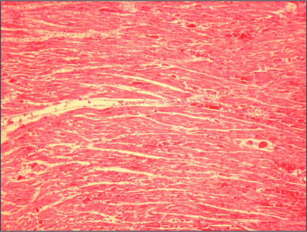
5 Slide #96 Low power magnification is not very exciting. You might be able to determine that this is heart and that there are hypereosinophilic areas.
6 Slide #96

7 Slide #96 At medium magnification you can start to appreciate the congestion, hemorrhage and slight separation of the myofibers by edema.

8 Slide #96 At medium magnification you can start to appreciate the congestion, hemorrhage and slight separation of the myofibers by edema. Also note apparent fibrin thrombi within some of the small vessels
9 At high magnification you can clearly see the blood outside the capillaries (ie hemorrhage) and the swelling, acidophilia, loss of striation and karyolysis of the cardialc myofibers (ie necrosis).

10 PAS stain note fibrin thrombi within capillaries
 the interstitium is mildly edematous fibrin thrombi are present within many capillaries (seen best with PAS")
11 Description: multiple areas of congestion & hemorrhage are scattered throughout the myocardium in some areas cardiac myofibres show karyolytic / pyknotic / karyorrhectic nuclei and have hypereosinophilic cytoplasm (ie indicative of coagulative necrosis) the interstitium is mildly edematous fibrin thrombi are present within many capillaries (seen best with PAS stain)

12 Slide #96 Morph Dx: 1) Myocardial congestion & hemorrhage, multifocal to coalescing, severe with microvascular thrombosis 2) Myocardial degeneration and necrosis, multifocal, acute, severe Comment: the findings in this case are characteristic of Mulberry Heart Disease (note, similar gross lesions can be seen with certain bacterial infections, esp Strep. suis infection) MHD is associated with a deficiency of Vit E (and sometimes selenium) some other diseases in swine associated with deficiencies of Vit E & Selenium, and can occur separately or in conjunction with MHD, include: Nutritional myopathy (white muscle disease) Hepatosis dietetica (see massive hepatic necrosis) Nutritional fat necrosis (aka steatitis or yellow fat disease ) This is a Mulberry!
13
 of the abdominal")
14 Slide #39 History: a 4-year-old Standardbred horse with a history of going around the race track twice and then losing control of its hind limbs hind limbs were cold to the touch & weak pulse felt in femoral artery at necropsy a thrombus / thromboembolus was present at the bifurcation ( quadrifurcation ) of the abdominal aorta

 Branches of the")
15 Fig (Dyce) Branches of the abdominal aorta, horse; 1, Aorta; 10, external iliac a.; 11, internal iliac a.; Fig (Evans & de Lahunta) Branches of the abdominal aorta, ventral aspect, dog.


16 Aortic-iliac thrombosis is occasionally seen in horses & causes exercise intolerance & hind-leg lamness. The underlying cause is usually not identified, but speculation about strongyle-related thromboemboli or hypercoagulability syndromes associated with sepsis / endotoxemia have been suggested.


17 You can see why they are called saddle thrombi Saddle thrombi are also occasional seen in dogs, esp those with hypercoagulability states (eg loss of antithrombin 3 with glomerular disease) Saddle thromboemboli are most often seen in cats which have cardiomyopathy with dilation and turbulence in the left atrium (ie thrombi form in the left atrium and then travel / impact at the bifrucation of the abdominal aorta)
 at the bottom segment of the")
18 Slide #39 note thrombus obstructing most of the lumen of this large artery; also note crescent shaped area of palor (ie area of organization) at the bottom segment of the lumen

19 note area of organization with a few recanalized vessels note thrombus composed of fibrin and entrapped cells & basophilic debris

20 note area of organization with a few recanalized vessels

21 Slide #39 at this magnification you can see that the area of organization is composed of fibrous connective tissue with a few newly formed recanalized vessels
 and the bright yellow pigment (ie hematoidin which is from bilirubin")
22 Slide #39 at this magnification you are starting to see the macrophages that contain pigments from erythrocyte breakdown; the dark brown granular pigment (ie hemosiderin which is from iron storage) and the bright yellow pigment (ie hematoidin which is from bilirubin accumulation)
 and the bright yellow pigment (ie hematoidin which is from bilirubin")
23 Slide #39 at this magnification you are starting to see the macrophages that contain pigments from erythrocyte breakdown; the dark brown granular pigment (ie hemosiderin which is from iron storage) and the bright yellow pigment (ie hematoidin which is from bilirubin accumulation)

24 Slide #39 At this magnification you can clearly see the macrophages with the hemosiderin & hematoidin embedded in abundant fibrous connective tissue
25 Slide #39 Description: this is a section of a large, muscular artery. the lumen is nearly completely occluded by a thrombus. at one margin there is a crescent shaped area of organization where there has been removal of the thrombotic material (as evidenced by macrophages containing hemosiderin and hematoidin pigment, derived from phagocytosed rbc s that were trapped in the thrombus) and replacement with fibrous connective tissue. note the recanalized areas within the area of organization. Morphologic Diagnosis: Arterial organizing thrombus, chronic, severe Comment: the clinical signs can be accounted for by ischemia to the rear legs resulting from the thrombus, ie cold temperature, lack of arterial pulse & muscle weakness. there is not complete ischemia and infarction of the hindlimbs because of collateral circulation and this also accounts for the loss of function only showing up during exercise
26

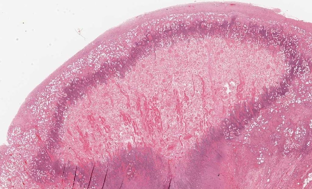
27 Slide #91 Clinical History: a necropsy was performed on an aged cat. the gross pathological changes included: widespread subcutaneous edema, ascites, hydrothorax, multiple, pale, wedge-shaped lesions in kidneys Well demarcated pale wedge shaped lesion in renal cortex; with base of the wedge near the capsule and apex near cortico-medullary junction.


28 Normal kidney, gross sagittal section, cat (above left) and normal histology of the kidney


29 Well demarcated pale wedge shaped lesion in renal cortex; with base of the wedge near the capsule and apex near corticomedullary junction.
30
 of the original cells. Note surrounding layer of inflammatory cell debris.")
31 Slide #91 note within the affected area the basic architectural arrangement of the glomeruli and tubules is apparent however the cells resemble eosinophilic shadow (ghostlike remnant) of the original cells. Note surrounding layer of inflammatory cell debris.
 of the original cells Note surrounding layer of inflammatory cell debris")
32 note within the affected area the basic architectural arrangement of the glomeruli and tubules is apparent however the cells resemble eosinophilic shadows (ghost-like remnants) of the original cells Note surrounding layer of inflammatory cell debris
 of the original cells. Most nuclei are inapparent (ie karyolysis) Note surrounding layer of inflammatory cell debris.")
33 note within the affected area the basic architectural arrangement of the glomeruli and tubules is apparent however the cells resemble eosinophilic shadows (ghost-like remnants) of the original cells. Most nuclei are inapparent (ie karyolysis) Note surrounding layer of inflammatory cell debris.
 of the original cells.")
34 note within the affected area the basic architectural arrangement of the glomeruli and tubules is apparent however the cells resemble eosinophilic shadows (ghost-like remnants) of the original cells. Most nuclei are inapparent (ie karyolysis)
35 Slide #91 Description: on low-power this section of kidney contains an irregular, wedge-shaped pale eosinophilic area which has a basophilic border. the apex of this triangular area is within the medulla, while the base is approximately 1-2 mm from the capsular surface. the inner material is composed of ghost-like remnants of the renal parenchyma (coagulation necrosis of tubules, glomeruli, etc) and the whole area is surrounded by a thick layer of inflammatory cell debris. there is an increase of fibrous connective tissue and some inflammatory cells within the interstitium of the remainder of the renal cortex (pre-existing nephritis). Morphologic Diagnosis: Renal infarct


36 Slide #91 Comment: an interlobar artery was obstructed by a thrombus or thromboembolus resulting in ischemia to the renal parenchyma and subsequently coagulation necrosis.
37
Cellular Pathology. Histopathology Lab #2 (web) Paul Hanna Jan 2018
 Paul Hanna Jan 2018") Cellular Pathology Histopathology Lab #2 (web) Paul Hanna Jan 2018 Slide #91 Clinical History: a necropsy was performed on an aged cat the gross pathological changes included: widespread subcutaneous edema
Cellular Pathology Histopathology Lab #2 (web) Paul Hanna Jan 2018 Slide #91 Clinical History: a necropsy was performed on an aged cat the gross pathological changes included: widespread subcutaneous edema
WSC , Conference 9, Case 1. Tissue from a nyala.
 WSC 2009-2010, Conference 9, Case 1. Tissue from a nyala. MICROSCOPIC DESCRIPTION: Heart, atrium (1 pt.): Approximately 40% of the atrial myocardium is replaced by areas of fibrous connective tissue (1
Ischaemia It means local anemia, it is characterized by a decrease amount of blood in an organ or region. Causes of Ischemia: *1.
 المرحلة الثالثة م. هالة عباس ناجي Ischaemia It means local anemia, it is characterized by a decrease amount of blood in an organ or region. Causes of Ischemia: *1.External pressure upon an artery e.g:
Disturbances of Circulation, Lab 1: Edema and Congestion/Hyperemia. Shannon Martinson, Feb
 Disturbances of Circulation, Lab 1: Edema and Congestion/Hyperemia Shannon Martinson, Feb 2017 http://people.upei.ca/smartinson/ Case #1 Signalment and History: 6-month old feeder lamb found dead on pasture
General Pathology. Hemorrhage (Web)
") General Pathology Hemorrhage (Web) Paul Hanna Feb 2015 Hemorrhage escape of blood from the cardiovascular system may be external or internal Hemorrhage Causes Trauma Sepsis, viruses or toxins Coagulation
General Pathology Hemorrhage (Web) Paul Hanna Feb 2015 Hemorrhage escape of blood from the cardiovascular system may be external or internal Hemorrhage Causes Trauma Sepsis, viruses or toxins Coagulation
Hemodynamic Disorders, Thrombosis, and Shock. Richard A. McPherson, M.D.
 Hemodynamic Disorders, Thrombosis, and Shock Richard A. McPherson, M.D. Edema The accumulation of abnormal amounts of fluid in intercellular spaces of body cavities. Inflammation and release of mediators
VPM Pigment and other tissue deposits. Shannon Martinson
 VPM 152 - Pigment and other tissue deposits Shannon Martinson http://people.upei.ca/smartinson/ Case 1 Signalment: 2 month old heifer beef calf Clinical History: Lateral recumbency for 4 days Tachycardia,
ECVP/ESVP Summer School in Veterinary Pathology Summer School 2015 Histology Case 5 DOG HD: Kidney.
 Case 5 DOG HD: Kidney. 100% of mid to deep renal cortex is characterized by coagulative necrosis/infarction, linear widespread haemorrhages and multifocal vasculitis with thrombosis. Throughout the section
5 DISTURBANCES IN CIRCULATION. Congestion / Hyperemia Haemorrhage Thrombosis Embolism Ischemia Infarction Oedema Shock Sludged blood Model Questions
 5 DISTURBANCES IN CIRCULATION Congestion / Hyperemia Haemorrhage Thrombosis Embolism Ischemia Infarction Oedema Shock Sludged blood Model Questions CONGESTION/ HYPEREMIA Hyperemia is increased amount of
Cellular Pathology Gross Pathology Laboratory 2 Cell Injury. VPM 152: General Pathology Instructor: Chelsea Martin Winter 2016
 Cellular Pathology Gross Pathology Laboratory 2 Cell Injury VPM 152: General Pathology Instructor: Chelsea Martin Winter 2016 Gross Specimens The following slides consist of images from the specimens presented
VPM Pigment and other tissue deposits. Shannon Martinson
 VPM 152 - Pigment and other tissue deposits Shannon Martinson http://people.upei.ca/smartinson/ Case 1: Signalment: 2 month old heifer beef calf Clinical History: Lateral recumbency for 4 days. Tachycardia,
PATHOLOGY Intracellular Degeneration LAB 1
 PATHOLOGY Intracellular Degeneration LAB 1 Cellular swelling Liver Organ :- Liver Lesion :- 1. Narrowing of hepatic sinusoids due to the swelling of hepatocyte. 2. The cytoplasm of affected hepatocyte
HISTOPATHOLOGY. Shannon Martinson
 HISTOPATHOLOGY Shannon Martinson March 2013 Case #1 History: 8 year old beagle Neck pain for the past couple of weeks Paresis, followed by paralysis developed over the past few days Gross Description courtesy
MORPHOLOGIC DIAGNOSIS: Liver: Hepatitis, necrotizing, multifocal to coalescing, severe, with numerous trichomonads. (3 pt)
") Case 1. Tissue from a pelican. MICROSCOPIC DESCRIPTION: Liver: Approximately 80% (1 pt) of the liver is replaced by multifocal to coalescing areas of coagulative and lytic necrosis. Centrally, within these
Case 1. Tissue from a pelican. MICROSCOPIC DESCRIPTION: Liver: Approximately 80% (1 pt) of the liver is replaced by multifocal to coalescing areas of coagulative and lytic necrosis. Centrally, within these
Fourth Practical Pathology. Circulatory disturbances
 Fourth Practical Pathology Circulatory disturbances 12.12.2018 1 Organ: Lung (40X, low power) 1) The blood capillaries within the alveolar septa are engorged with blood 2) Pinkish proteinaceous fluid,
Disturbance of Circulation Hemodynamic Disorder
 Disturbance of Circulation Hemodynamic Disorder 2/17/2017 By Dr. Hemn Hassan Othman PhD, Pathology Fall 2016 1 Thrombosis Definition: Thrombosis is the formation of solid or semisolid blood clot within
AP2 Lab 1 - Blood & Heart
 AP2 Lab 1 - Blood & Heart Project 1 - Formed Elements Identification & Recognition See fig. 17.10 and Table 17.2. Instructor may also provide other images. Note: See Fig. 17.11 All formed elements are
2015 Descriptive Vet Path Course. Histo Exam #3 KEY
 2015 Descriptive Vet Path Course Histo Exam #3 KEY Test 3, Slide 1 Tissue from a guinea pig. MORPHOLOGIC DIAGNOSIS: Heart: Multifocally and randomly (1 pt), within the left and right ventricular myocardium
Inflammation Laboratory 2. Shannon Martinson: VPM 152: March 2012
 Inflammation Laboratory 2 Shannon Martinson: http://people.upei.ca/smartinson VPM 152: March 2012 Reminder - Creating a Morphologic Diagnosis for Inflammatory Lesions Organ and Process Exudate Distribution
Circulatory Disturbances 5: Thrombosis, Embolism, Infarction, Shock
 Circulatory Disturbances 5: Thrombosis, Embolism, Infarction, Shock Shannon Martinson, Feb 2016 http://people.upei.ca/smartinson/ VPM 152 General Pathology Thrombosis, Embolism, Infarction, Shock Learning
Blood and Heart. Student Learning Objectives:
 Blood and Heart Student Learning Objectives: Identify the major components of the blood. Identify the primary structures associated with the heart Follow the blood through the path of the circulation.
Avian Pathology. Bacterial diseases: histo slides. ECVP-ESVP Summer School 2012 Frédérique NGUYEN
 Avian Pathology Bacterial diseases: histo slides ECVP-ESVP Summer School 2012 Frédérique NGUYEN Bacterial diseases: histo slides B1. Turkey. Organs? Morphologic diagnosis? Special procedure? B2. Hen. Organ?
Histopathology: Cell necrosis and cytoplasmic accumulations
 Histopathology: Cell necrosis and cytoplasmic accumulations These presentations are to help you identify basic histopathological features. They do not contain the additional factual information that you
Histopathology: healing
 Histopathology: healing These presentations are to help you identify, and to test yourself on identifying, basic histopathological features. They do not contain the additional factual information that
CARDIOVASCULAR PATHOLOGY LABORATORY CASES AND SLIDES. VPM Lisa Miller CARDIOVASCULAR LAB
 CARDIOVASCULAR PATHOLOGY LABORATORY CASES AND SLIDES VPM 222 2009 Lisa Miller CARDIOVASCULAR LAB Case 1 Heart - Echoplane section from a 7-month-old Labrador retriever Give two morphologic diagnoses 1.
Pathophysiology. Tutorial 3 Hemodynamic Disorders
 Pathophysiology Tutorial 3 Hemodynamic Disorders ILOs Recall different causes of thrombosis. Explain different types of embolism and their predisposing factors. Differentiate between hemorrhage types.
Histopathology: Vascular pathology
 Histopathology: Vascular pathology These presentations are to help you identify basic histopathological features. They do not contain the additional factual information that you need to learn about these
THROMBOSIS. Dr. Nisreen Abu Shahin Assistant Professor of Pathology Pathology Department University of Jordan
 THROMBOSIS Dr. Nisreen Abu Shahin Assistant Professor of Pathology Pathology Department University of Jordan NORMAL BLOOD VESSEL HISTOLOGY THROMBOSIS Pathogenesis (called Virchow's triad): 1. Endothelial*
HUMAN HEART. Learn the following structures on the heart models.
 HUMAN HEART Learn the following structures on the heart models. The human heart has four chambers that consist of the right atrium, left atrium, right ventricle, and left ventricle. The atria are smaller
Describing and interpreting gross lesions. Prepared for VPM 4600, May 2018; Shannon Martinson
 Describing and interpreting gross lesions Prepared for VPM 4600, May 2018; Shannon Martinson How to Describe (and Interpret) Lesions Step 1 Step 2 Step 3 Step 4 Look at the specimen: Is it normal or abnormal
Cellular Injury. Intracellular degeneration. By Dr. Hemn Hassan Othman PhD, Pathology Fall /20/2018 1
 Cellular Injury Intracellular degeneration By Dr. Hemn Hassan Othman PhD, Pathology Fall 2018 10/20/2018 1 Types of cell injury Cell injury is divided into: 1. Reversible cell injury 2. Irreversible cell
Urinary system. Urinary system
 INTRODUCTION. Several organs system Produce urine and excrete it from the body Maintenance of homeostasis. Components. two kidneys, produce urine; two ureters, carry urine to single urinary bladder for
Necrosis is death of cells and tissues in the living animal. Focal/ Multifocal necrosis- terms used for one
 Necrosis Necrosis Necrosis is death of cells and tissues in the living animal. Focal/ Multifocal necrosis- terms used for one or more, small, clearly defined areas of necrosis. Diffuse necrosis- term used
Histopathology: Glomerulonephritis and other renal pathology
 Histopathology: Glomerulonephritis and other renal pathology These presentations are to help you identify basic histopathological features. They do not contain the additional factual information that you
SESSION 1: GENERAL (BASIC) PATHOLOGY CONCEPTS Thursday, October 16, :30am - 11:30am FACULTY COPY
 PATHOLOGY CONCEPTS Thursday, October 16, :30am - 11:30am FACULTY COPY") SESSION 1: GENERAL (BASIC) PATHOLOGY CONCEPTS Thursday, October 16, 2008 9:30am - 11:30am FACULTY COPY GOAL: Describe the basic morphologic (structural) changes which occur in various pathologic conditions.
SESSION 1: GENERAL (BASIC) PATHOLOGY CONCEPTS Thursday, October 16, 2008 9:30am - 11:30am FACULTY COPY GOAL: Describe the basic morphologic (structural) changes which occur in various pathologic conditions.
Cardiac Ischemia (is-kē-mē-uh)
") Chapter 21 Cardiac Ischemia (is-kē-mē-uh) By: Alejandra & Lindsay I. Cardiac Ischemia =the most common cause of death in Western Culture ~35% of deaths. -Suddenly from acute coronary occlusion or fibrillation
Chapter 21 Cardiac Ischemia (is-kē-mē-uh) By: Alejandra & Lindsay I. Cardiac Ischemia =the most common cause of death in Western Culture ~35% of deaths. -Suddenly from acute coronary occlusion or fibrillation
ATHEROSCLEROSIS. Secondary changes are found in other coats of the vessel wall.
 ATHEROSCLEROSIS Atherosclerosis Atherosclerosis is a disease process affecting the intima of the aorta and large and medium arteries, taking the form of focal thickening or plaques of fibrous tissue and
HEMODYNAMIC DISORDERS
 HEMODYNAMIC DISORDERS Normal fluid homeostasis requires vessel wall integrity as well as maintenance of intravascular pressure and osmolarity within certain physiologic ranges. Increases in vascular volume
Inflammation Laboratory 1
 Inflammation Laboratory 1 Lab1 Emphasis: The exudates of acute inflammation Descriptions Morphologic Diagnoses Shannon Martinson: http://people.upei.ca/smartinson VPM 152: February 2012 Describing Lesions
Shock, Hemorrhage and Thrombosis
 Shock, Hemorrhage and Thrombosis 1 Shock Systemic hypoperfusion due to: Reduction in cardiac output Reduction in effective circulating blood volume Hypotension Impaired tissue perfusion Cellular hypoxia
Alfonso López. Cardiac Hypertrophy and Dilation
 Cardiac Hypertrophy and Dilation Alfonso López Professor of Anatomic Pathology Dept. Pathology and Microbiology Atlantic Veterinary College University of Prince Edward Island Canada Jan 23, 2012 Compensatory
HYPEREMIA AND CONGESTION
 HYPEREMIA AND CONGESTION Learning Objectives Define congestion and hyperemia Differentiate between the two with regard to: Mechanisms / underlying causes Appearance (gross and histologic) Effects Differentiate
Inflammation Laboratory 1
 Inflammation Laboratory 1 Lab1 Emphasis: The exudates of acute inflammation Descriptions Morphologic Diagnoses Shannon Martinson: http://people.upei.ca/smartinson VPM 152: March 2013 Describing Lesions
Disorders of Cell Growth & Neoplasia. Histopathology Lab
 Disorders of Cell Growth & Neoplasia Histopathology Lab Paul Hanna April 2010 Case #84 Clinical History: 5 yr-old, West Highland White terrier. skin mass from axillary region. has been present for the
Chapter 14. Circulatory System Images. VT-122 Anatomy & Physiology II
 Chapter 14 Circulatory System Images VT-122 Anatomy & Physiology II The mediastinum Dog heart Dog heart Cat heart Dog heart ultrasound Can see pericardium as distinct bright line Pericardial effusion Fluid
Chapter 23. The Nephron. (functional unit of the kidney
 Chapter 23 The Nephron (functional unit of the kidney Renal capsule The Nephron Renal cortex Nephron Collecting duct Efferent arteriole Afferent arteriole (a) Renal corpuscle: Glomerular capsule Glomerulus
1- Thromboembolism. 2- fat embolism. 3- air embolism. 4- amniotic fluid embolism.
 Embolism Definition:- An embolus is a detached intravascular solid, liquid or gaseous mass that is carried by blood to sites distant from its point of origin. After traveling via the blood, the embolus
The Circulatory System. The Heart, Blood Vessels, Blood Types
 The Circulatory System The Heart, Blood Vessels, Blood Types The Closed Circulatory System Humans have a closed circulatory system, typical of all vertebrates, in which blood is confined to vessels and
BLOOD RUNS THROUGH YOUR BODY
 BLOOD RUNS THROUGH YOUR BODY WORKSHEET A Your heart and blood vessels make up your blood system. At the centre of your blood system is your heart. Its job is to pump the blood around your body. The rest
Cellular responses to stress
 Cellular responses to stress (Adaptations, injury and death) (2 of 5) Most injurious stimuli are grouped into: Oxygen deprivation Chemical agents Infectious agents Immunologic reactions Genetic factors
Chapter 12 Cardiovascular System
 Chapter 12 Cardiovascular System Cardiovascular System Includes Heart and Blood Vessels Transports, nutrients and wastes to and from the tissues 1 The Blood Vessels Three Types of Blood Vessels Arteries:
LABORATORY EXERCISES FOR THE URINARY SYSTEM
 LABORATORY EXERCISES FOR THE URINARY SYSTEM cortex Medulla DEMO SLIDE BOX 172 (450-E001-H-76). Kidney, horse. the inner medulla medullary rays, Uriniferous tubules expand both the cortex and medulla corticomedullary
SESSION IV: MECHANISMS OF HUMAN DISEASE: LABORATORY SESSIONS PULMONARY PATHOLOGY I. December 5, 2012
 SESSION IV: MECHANISMS OF HUMAN DISEASE: LABORATORY SESSIONS PULMONARY PATHOLOGY I December 5, 2012 FACULTY COPY GOAL: Describe the basic morphologic and pathophysiologic changes in various conditions
Levels of Organization. Chapter 19 6/11/2012. Homeostasis & Organization of the animal body. 4 Primary Tissues
 Levels of Organization Chapter 19 Homeostasis & Organization of the animal body Chemical Cellular Tissue Organs System Level Organismic 1-2 4 Primary Tissues 1. Epithelial Tissue: covers surfaces lines
19. RENAL PHYSIOLOGY ROLE OF THE URINARY SYSTEM THE URINARY SYSTEM. Components and function. V BS 122 Physiology II 151 Class of 2011
 19. RENAL PHYSIOLOGY THE URINARY SYSTEM Components and function The urinary system is composed of two kidneys, the functionally filtering apparatus, which connect through two tubular structures called
Pathophysiology of Cardiovascular System. Dr. Hemn Hassan Othman, PhD
 Pathophysiology of Cardiovascular System Dr. Hemn Hassan Othman, PhD hemn.othman@univsul.edu.iq What is the circulatory system? The circulatory system carries blood and dissolved substances to and from
Cell injury, adaptation and death. Unite one Second Lab.
 Cell injury, adaptation and death Unite one Second Lab. The two lung abscesses seen here are examples of liquefactive necrosis in which there is a liquid center in an area of tissue injury. One abscess
Histopathology Description:
 2013-2-1 CANINE HEART Ahmed M. Abubakar BOVINE PATHOLOGY CONTRIBUTING INSTITUTION : The Royal Veterinary college, Dept. of Pathology and Biology Signalment: 11-month-old male Border Collie dog (Canis familiaris)
THE URINARY SYSTEM. The cases we will cover are:
 THE URINARY SYSTEM The focus of this week s lab will be pathology of the urinary system. Diseases of the kidney can be broken down into diseases that affect the glomeruli, tubules, interstitium, and blood
CELL INJURY AND CELL DEATH
 CELL INJURY AND CELL DEATH INTRODUCTION Cell Injury is a result of the sequence of events that occur if the limits of the adaptive capability of cells are exceeded or there is no adaptive response is possible,
THE URINARY SYSTEM. The cases we will cover are:
 THE URINARY SYSTEM The focus of this week s lab will be pathology of the urinary system. Diseases of the kidney can be broken down into diseases that affect the glomeruli, tubules, interstitium, and blood
Lymph I: The Peripheral Lymph System
 Lymph I: The Peripheral Lymph System Peripheral = Secondary Primary Immune Organs = bone marrow, thymus Site of maturation of cells of the immune system Secondary Immune Organs = Nodes, MALT, spleen Filter
Pathology of Hypertension
 2016-03-07 Pathology of Hypertension Honghe Zhang honghezhang@zju.edu.cn Tel:88208199 Department of Pathology ❶ Genetic predisposition ❷ Dietary factors ❸ Environmental factors ❹ Others Definition and
Cardiovascular System
 System examination The initial examination of the cardiovascular system should be done in-situ on opening the thoracic cavity, to evaluate abnormalities of size and position. The four major areas for gross
Read Chapters 21 & 22, McKinley et al
 ACTIVITY 9: BLOOD AND HEART OBJECTIVES: 1) How to get ready: Read Chapters 21 & 22, McKinley et al., Human Anatomy, 5e. All text references are for this textbook. Read dissection instructions BEFORE YOU
Aortic Thromboembolism
 Aortic Thromboembolism (Blood Clots in the Aorta) Basics OVERVIEW Aortic refers to the aorta, the main artery of the body; thromboembolism is blockage of blood flow secondary to the presence of a blood
Quantification of Coronary Arterial Narrowing at Necropsy in Acute Transmural Myocardial Infarction
 Quantification of Coronary Arterial Narrowing at Necropsy in Acute Transmural Myocardial Infarction Analysis and Comparison of Findings in 27 Patients and 22 Controls WILLIAM C. ROBERTS, M.D., AND ANCIL
Chapter 14. The Cardiovascular System
 Chapter 14 The Cardiovascular System Introduction Cardiovascular system - heart, blood and blood vessels Cardiac muscle makes up bulk of heart provides force to pump blood Function - transports blood 2
Lab 1 Blood Composition and formed elements
 Lab 1 Blood Composition and formed elements Plasma 55% of whole blood 90% water 8% proteins from liver 2% misc. Nutrients: AA, glucose, lipids vitamins, minerals Wastes: urea, uric acid, creatine, ammonium
The Cardiovascular System. Chapter 15. Cardiovascular System FYI. Cardiology Closed systemof the heart & blood vessels. Functions
 Chapter 15 Cardiovascular System FYI The heart pumps 7,000 liters (4000 gallons) of blood through the body each day The heart contracts 2.5 billion times in an avg. lifetime The heart & all blood vessels
EXPERIMENTAL THERMAL BURNS I. A study of the immediate and delayed histopathological changes of the skin.
 EXPERIMENTAL THERMAL BURNS I A study of the immediate and delayed histopathological changes of the skin. RJ Brennan, M.D. and B. Rovatti M.D. The purpose of this study was to determine the progressive
An aneurysm is a localized abnormal dilation of a blood vessel or the heart Types: 1-"true" aneurysm it involves all three layers of the arterial
 An aneurysm is a localized abnormal dilation of a blood vessel or the heart Types: 1-"true" aneurysm it involves all three layers of the arterial wall (intima, media, and adventitia) or the attenuated
The Cardiovascular System home study course
 The Cardiovascular System home study course harmony house holistic therapy treatment centre and training academy www.harmony-house.org 1 Copyright 2010 by Mark and Katy Rogers All rights reserved. No part
Interpretation of Renal Transplant Biopsy. Arthur H. Cohen Wake Forest University School of Medicine Winston-Salem, North Carolina USA
 Interpretation of Renal Transplant Biopsy Arthur H. Cohen Wake Forest University School of Medicine Winston-Salem, North Carolina USA Renal Transplant Biopsies Tissue Processing Ideal world process as
Kidney Structure. Renal Lobe = renal pyramid & overlying cortex. Renal Lobule = medullary ray & surrounding cortical labryinth.
 Kidney Structure Capsule Hilum ureter renal pelvis major and minor calyxes renal and vein segmental arteries interlobar arteries arcuate arteries interlobular arteries Medulla renal pyramids cortical/renal
General Anatomy of Urinary System
 General Anatomy of Urinary System URINARY SYSTEM ORGANS Kidneys (2) Ureters (2) Urinary bladder Urethra KIDNEY FUNCTIONS Control blood volume and composition KIDNEY FUNCTIONS Filter blood plasma, eliminate
The Cardiovascular System Part I: Heart Outline of class lecture After studying part I of this chapter you should be able to:
 The Cardiovascular System Part I: Heart Outline of class lecture After studying part I of this chapter you should be able to: 1. Describe the functions of the heart 2. Describe the location of the heart,
Cardiovascular Pathology Lab. Shannon Martinson,
 Cardiovascular Pathology Lab Shannon Martinson, 2017 http://people.upei.ca/smartinson/ Case 1 Signalment: 10 year old MC DSH Cat History Heart murmur detected on PE recommended cardiac US Blood work was
Lung diseases of Vascular Origin. By: Shefaa Qa qqa
 Lung diseases of Vascular Origin By: Shefaa Qa qqa Pulmonary Hypertension Pulmonary hypertension is defined as a mean pulmonary artery pressure greater than or equal to 25 mm Hg at rest. Based on underlying
Figure 10.1A Transparency Master 79
 Brain Carotid arteries Jugular vein Right front leg Lungs (inflated) Cranial Right atrium To left front leg Left subclavian Bronchus capillaries Brachiocephalic vein Left atrium Dorsal aorta Right ventricle
2.01 Remember the structures of the circulatory system
 2.01 Remember the structures of the circulatory system Essential questions What are the structures of blood? What are the structures of the circulatory system? circulatory system 2 Structures of the circulatory
Section 5.1 The heart and heart disease
 Section 5.1 The heart and heart disease Mammals are too large to rely on diffusion. They need a circulatory system to move substances around the body. Blood moves down pressure gradients, from high to
Acute arterial embolism
 Acute arterial embolism Definition Thrombus come from heart or blood vessel or other embolus such as tumor,air gas or fat flow with blood stream and occlude distal limb or visceral arteries which causes
Right-Sided Congestive Heart Failure Basics
 Right-Sided Congestive Heart Failure Basics OVERVIEW Failure of the right side of the heart to pump blood at a sufficient rate to meet the needs of the body or to prevent blood from pooling within the
Heart Dissection. 5. Locate the tip of the heart or the apex. Only the left ventricle extends all the way to the apex.
 Heart Dissection Page 1 of 6 Background: The heart is a four-chambered, hollow organ composed primarily of cardiac muscle tissue. It is located in the center of the chest in between the lungs. It is the
The Endocrine System Pituitary
 The Endocrine System Pituitary Look at your slide of the human pituitary with your naked eye. You should see a cellular region and a more fibrous region. Then view each region with your microscope under
Read Me. covering the Heart Anatomy. Labs. textbook. use. car: you
 Heart Anatomy Lab Pre-Lab Exercises Read Me These exercises should be done before coming to lab, after watching the videos covering the Heart Anatomy Labs. Answer the questions in this guide using the
Circulatory Disturbances 1: Introduction and Edema
 Circulatory Disturbances 1: Introduction and Edema Shannon Martinson, January 2016 http://people.upei.ca/smartinson/ VPM 152 General Pathology INTRODUCTION NORMAL CIRCULATORY SYSTEM Important concepts
Urinary System Laboratory
 Urinary System Laboratory 1 Adrenal gland Organs of The Urinary System Renal artery and vein Kidney Ureter Urinary bladder Figure 26.1 2 Urethra Functions of the urinary system organs: Urethra expels urine
2. right heart = pulmonary pump takes blood to lungs to pick up oxygen and get rid of carbon dioxide
 A. location in thorax, in inferior mediastinum posterior to sternum medial to lungs superior to diaphragm anterior to vertebrae orientation - oblique apex points down and to the left 2/3 of mass on left
Urinary system. Urinary system
 Distal convoluted tubule (DCT) Highly coiled, ~ 5 mm in length Last part of the nephron. Wall; simple cuboidal epithelium Less metabolically active than the PCT no brush border light eosinophilic cytoplasm
EDEMA. Learning Objectives
 EDEMA Learning Objectives Define edema Recognize and be able to describe the gross and microscopic appearance of edema Know the four pathophysiological mechanisms by which edema develops Understand the
Cardiovascular System
 Cardiovascular System angio BELLWORK Day One: Define using technology hemo/hema cardio Medical Therapeutics Standards 11) Outline the gross normal structure and function of all body systems and summarize
Blood Functions. Blood and the Cardiovascular System. Blood. Plasma. Erythrocytes (RBCs) Erythrocytes (RBCs) 4/7/2017
 Erythrocytes (RBCs) 4/7/2017") Blood Functions Blood and the Cardiovascular System Distribution Delivery of oxygen and nutrients to all body cells; Transport of wastes to lungs and excretory organs; Transport of hormones Regulation
Blood Functions Blood and the Cardiovascular System Distribution Delivery of oxygen and nutrients to all body cells; Transport of wastes to lungs and excretory organs; Transport of hormones Regulation
Scrub In: Red blood cells are called: Which component of blood is necessary for the initiation of the blood clotting process:
 Scrub In: Red blood cells are called: a. erythrocytes b. leukocytes c. melanocytes d. thrombocytes Which component of blood is necessary for the initiation of the blood clotting process: a. erythrocytes
Thrombosis and emboli. Peter Nagy
 Thrombosis and emboli Peter Nagy A thrombus is any solid object developing from the blood in vivo within the vascular system or heart. Thrombosis is hemostasis in the wrong place. Major components, forms:
AP2 Lab 3 Coronary Vessels, Valves, Sounds, and Dissection
 AP2 Lab 3 Coronary Vessels, Valves, Sounds, and Dissection Project 1 - BLOOD Supply to the Myocardium (Figs. 18.5 &18.10) The myocardium is not nourished by the blood while it is being pumped through the
The Heart. The Heart A muscular double pump. The Pulmonary and Systemic Circuits
 C H A P T E R 19 The Heart The Heart A muscular double pump circuit takes blood to and from the lungs Systemic circuit vessels transport blood to and from body tissues Atria receive blood from the pulmonary
Hemodynamic Disorders Thrombosis and Shock. 1. Interstitial, between the cells, but outside of the vascular system. - water making up the blood and
 Hemodynamic Disorders Thrombosis and Shock I. Body water, where is it and what keeps it there? A. Intracellular B. Extracellular (intercellular) 1. Interstitial, between the cells, but outside of the vascular