HEMODYNAMIC DISORDERS. J v = ([Pc Pi] σ[πc πi])
|
|
|
- Pierce Skinner
- 5 years ago
- Views:
Transcription
1 HEMODYNAMIC DISORDERS J v = ([Pc Pi] σ[πc πi])
2 Hemodynamic Disorders Thromboembolic Disease Shock
3 Overview Edema Hyperemia Congestion Hemorrhage Hemostasis Thrombosis Embolism Infarction Shock
4 EDEMA ONLY 4 POSSIBILITIES!!! Increased Hydrostatic Pressure Reduced Oncotic Pressure Lymphatic Obstruction Sodium/Water Retention
5 WATER 60% of body 2/3 of body water is INTRA-cellular The rest is INTERSTITIAL Only 5% is INTRA-vascular EDEMA is SHIFT to the INTERSTITIAL SPACE HYDRO- -THORAX, -PERICARDIUM, -PERICARDIUM EFFUSIONS, ASCITES, ANASARCA
6 INCREASED HYDROSTATIC PRESSURE Impaired venous return Congestive heart failure Constrictive pericarditis Ascites (liver cirrhosis) Venous obstruction or compression Thrombosis External pressure (e.g., mass) Lower extremity inactivity with prolonged dependency Arteriolar dilation Heat Neurohumoral dysregulation
7 REDUCED PLASMA ONCOTIC PRESSURE (HYPOPROTEINEMIA) Protein-losing glomerulopathies (nephrotic syndrome) Liver cirrhosis (ascites) Malnutrition Protein-losing gastroenteropathy
8 LYMPHATIC OBSTRUCTION (LYMPHEDEMA) Inflammatory Neoplastic Postsurgical Postirradiation
9 Na+ RETENTION Excessive salt intake with renal insufficiency Increased tubular reabsorption of sodium Renal hypoperfusion Increased renin-angiotensin-aldosterone secretion
10 INFLAMMATION Acute inflammation Chronic inflammation Angiogenesis
11 Jv = ([Pc Pi] σ[πc πi])
12 CHF EDEMA INCREASED VENOUS PRESSURE DUE TO FAILURE DECREASED RENAL PERFUSION, triggering of RENIN- ANGIOTENSION-ALDOSTERONE complex, resulting ultimately in SODIUM RETENTION
13 HEPATIC ASCITES PORTAL HYPERTENSION HYPOALBUMINEMIA
14 ASCITES
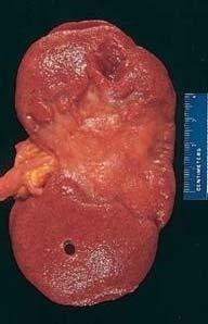
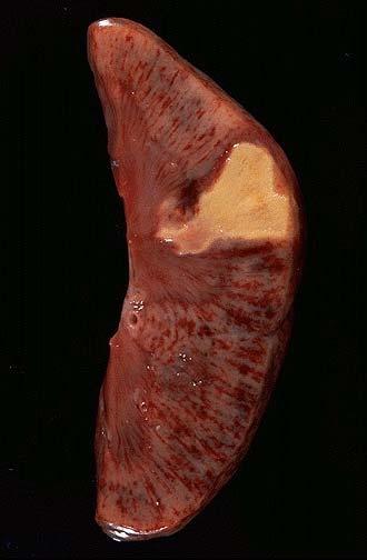
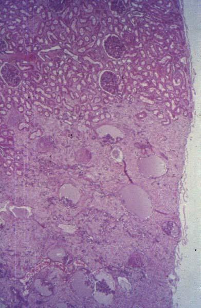
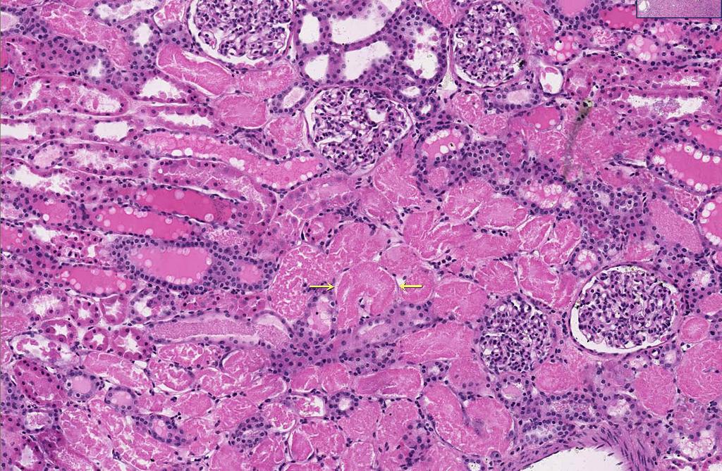
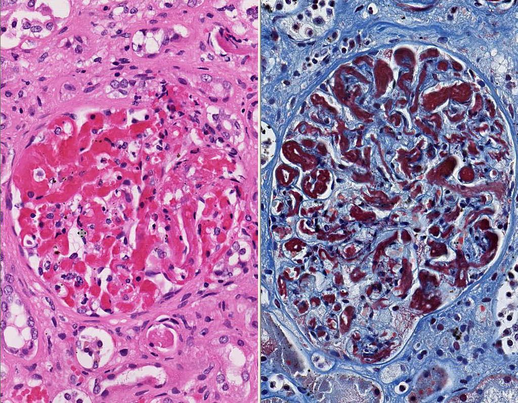
15 RENAL EDEMA SODIUM RETENTION PROTEIN LOSING GLOMERULOPATHIES (NEPHROTIC SYNDROME)
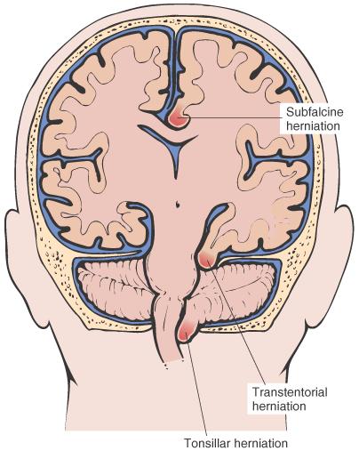
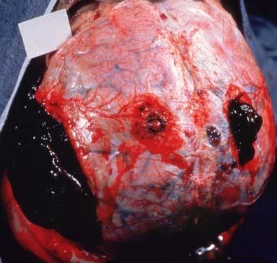
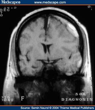
 HERNIATION of cerebellar tonsils HERNIATION of hippocampal uncus over tentorium HERNIATION,")
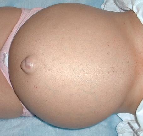
16 EDEMA SUBCUTANEOUS ( PITTING ) DEPENDENT ANASARCA LEFT vs RIGHT HEART PERIORBITAL (RENAL) PULMONARY CEREBRAL (closed cavity, no expansion) HERNIATION of cerebellar tonsils HERNIATION of hippocampal uncus over tentorium HERNIATION, subfalcine

17 Pitting Edema
18 Transudate vs Exudate Transudate results from disturbance of Starling forces specific gravity < protein content < 3 g/dl Exudate results from damage to the capillary wall specific gravity > protein content > 3 g/dl
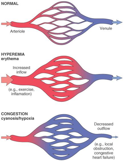
19 HYPEREMIA/(CONGESTION)
20 HYPEREMIA Active Process CONGESTION Passive Process Acute or Chronic
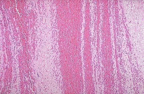
21 LUNG CONGESTION ACUTE CHRONIC LIVER ACUTE CHRONIC CEREBRAL

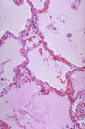
22 ACUTE PASSIVE HYPEREMIA/CONGESTION, LUNG

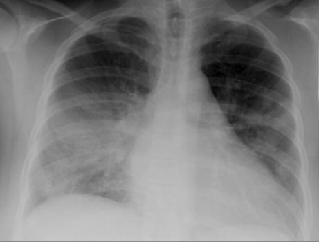
23 Kerley B Air Bronchogram

24
25
26 CHRONIC PASSIVE HYPEREMIA/CONGESTION, LUNG

27 Acute Passive Congestion, Liver

28 Acute Passive Congestion, Liver

29 CHRONIC PASSIVE HYPEREMIA/CONGESTION, LIVER
30
31
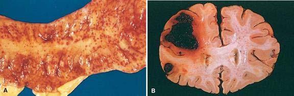
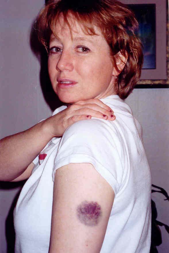
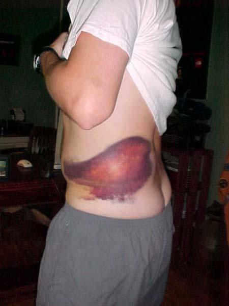
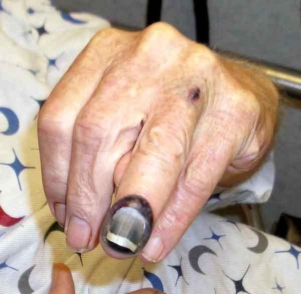
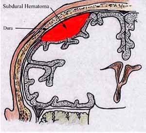
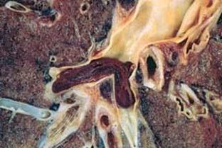
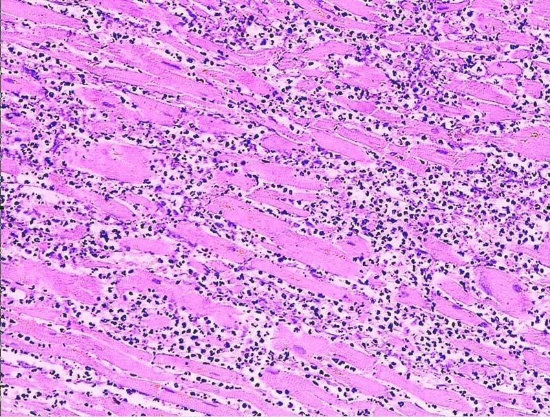
32 HEMORRHAGE EXTRAVASATION beyond vessel HEMORRHAGIC DIATHESIS HEMATOMA (implies MASS effect) DISSECTION PETECHIAE (1-2mm) (PLATELETS) PURPURA <1cm ECCHYMOSES >1cm (BRUISE) HEMO-: -thorax, -pericardium, -peritoneum, HEMARTHROSIS ACUTE, CHRONIC
33 EVOLUTION of HEMORRHAGE ACUTE CHRONIC PURPLE GREEN BROWN HGB BILIRUBIN HEMOSIDERIN
34
35
36
37
38
39 HEMATOMA vs. CLOT
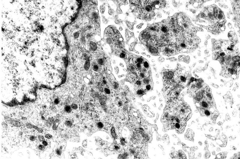
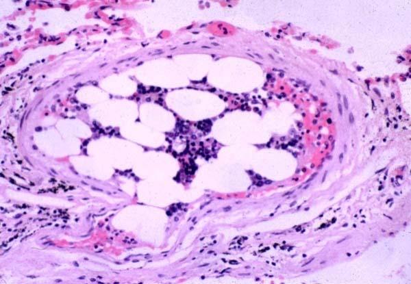
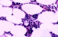
40 HEMOSTASIS OPPOSITE of THROMBOSIS PRESERVE LIQUIDITY OF BLOOD PLUG sites of vascular injury THREE COMPONENTS VASCULAR WALL, i.e., endoth/ecm PLATELETS COAGULATION CASCADE
41 SEQUENCE of EVENTS following VASCULAR INJURY ARTERIOLAR VASOCONSTRICTION Reflex Neurogenic Endothelin, from endothelial cells THROMBOGENIC ECM at injury site Adhere and activate platelets Platelet aggregation (1 HEMOSTASIS) TISSUE FACTOR released by endothelium Activates coagulation cascade thrombin fibrin (2 HEMOSTASIS) FIBRIN polymerizes, TPA limits plug
42 PLAYERS ENDOTHELIUM PLATELETS COAGULATION CASCADE
43 ENDOTHELIUM NORMALLY ANTIPLATELET PROPERTIES ANTICOAGULANT PROPERTIES FIBRINOLYTIC PROPERTIES IN INJURY PRO-COAGULANT PROPERTIES

44
45 ENDOTHELIUM ANTI-Platelet PROPERTIES Protection from the subendothelial ECM Degrades ADP (inhib. Aggregation) ANTI-Coagulant PROPERTIES Membrane HEPARIN-like molecules Makes THROMBOMODULIN Protein-C TISSUE FACTOR PATHWAY INHIBITOR FIBRINOLYTIC PROPERTIES (TPA)
46 ENDOTHELIUM PROTHROMBOTIC PROPERTIES Makes vwf, which binds Plats Coll Makes TISSUE FACTOR (with plats) Makes Plasminogen inhibitors
47 ENDOTHELIUM ACTIVATED by INFECTIOUS AGENTS ACTIVATED by HEMODYNAMICS ACTIVATED by PLASMA
48 PLATELETS ALPHA GRANULES Fibrinogen Fibronectin Factor-V, Factor-VIII Platelet factor 4, TGF-beta DELTA GRANULES (DENSE BODIES) ADP/ATP, Ca+, Histamine, Serotonin, Epineph. With endothelium, form TISSUE FACTOR
49


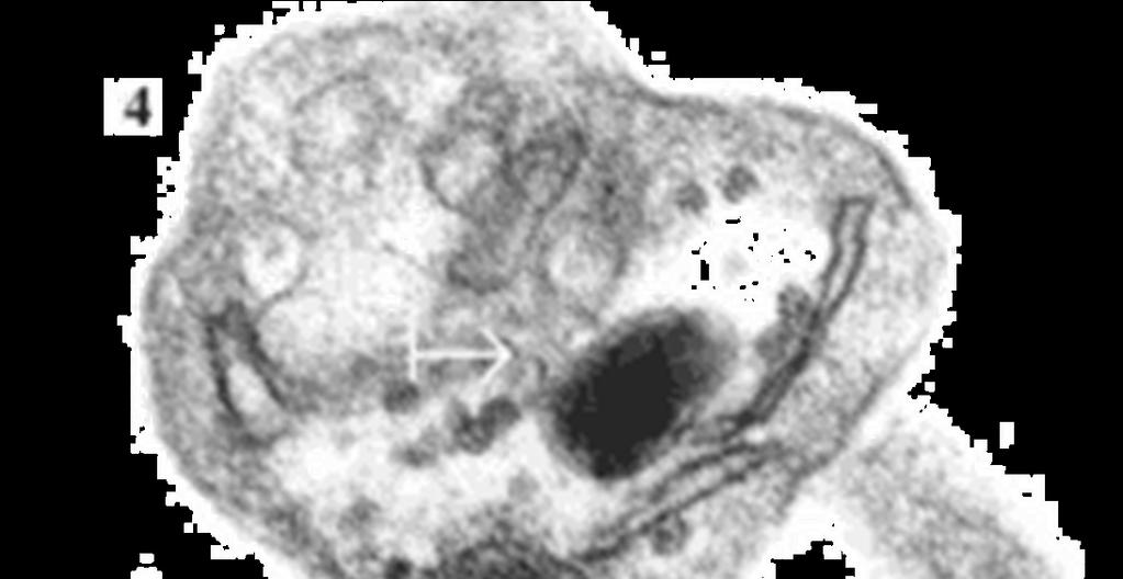
50 NORMAL platelet on LEFT, DEGRANULATING ALPHA GRANULE ON RIGHT AT OPEN WHITE ARROW
51
52 PLATELET PHASES ADHESION SECRETION (i.e., release or activation or degranulation ) AGGREGATION
53 PLATELET ADHESION Primarily to the subendothelial ECM Regulated by vwf, which bridges platelet surface receptors to ECM collagen
54 PLATELET SECRETION BOTH granules, α and δ Binding of agonists to platelet surface receptors AND intracellular protein PHOSPHORYLATION
55 PLATELET AGGREGATION ADP TxA2 (Thromboxane A2) THROMBIN from coagulation cascade also FIBRIN further strengthens and hardens and contracts the platelet plug
56 PLATELET EVENTS ADHERENCE to ECM SECRETION of ADP and TxA2 EXPOSE phospholipid complexes Express TISSUE FACTOR PRIMARY SECONDARY PLUG STRENGTHENED by FIBRIN
57 COAGULATION CASCADE INTRINSIC(contact)/EXTRINSIC(TissFac) Proenzymes Enzymes Prothrombin(II) Thrombin(IIa) Fibrinogen(I) Fibrin(Ia) Cofactors Ca++ Phospholipid (from platelet membranes) Vit-K dep. factors: II, VII, IX, X, Prot. S, C, Z
58
59 COAGULATION TESTS (a)ptt INTRINSIC (HEP Rx) PT (INR) EXTRINSIC (COUM Rx) BLEEDING TIME (PLATS) (2-9min) Platelet count (150, ,000/mm3) Fibrinogen Factor assays
60 THROMBOSIS Pathogenesis Endothelial Injury Alterations in Flow Hypercoagulability Morphology Fate Clinical Correlations Venous Arterial (Mural)
61 THROMBOSIS Virchow s TRIANGLE ENDOTHELIAL INJURY ABNORMAL FLOW (NON-LAMINAR) HYPER- COAGULATION
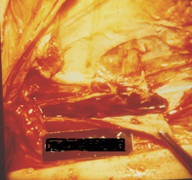
62 ENDOTHELIAL INJURY Jekyll/Hyde disruption any perturbation in the dynamic balance of the pro- and antithrombotic effects of endothelium, not only physical damage
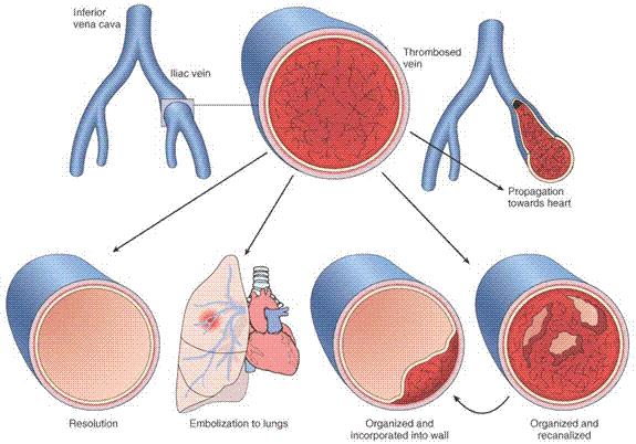
63 ENDOTHELIUM ANTI-Platelet PROPERTIES Protection from the subendothelial ECM Degrades ADP (inhib. Aggregation) ANTI-Coagulant PROPERTIES Membrane HEPARIN-like molecules Makes THROMBOMODULIN Protein-C TISSUE FACTOR PATHWAY INHIBITOR FIBRINOLYTIC PROPERTIES (TPA)
64 ENDOTHELIUM PROTHROMBOTIC PROPERTIES Makes vwf, which binds Plats Coll Makes TISSUE FACTOR (with plats) Makes Plasminogen inhibitors
65 ABNORMAL FLOW NON-LAMINAR FLOW TURBULENCE EDDIES STASIS DISRUPTED ENDOTHELIUM ALL of these factors may bring platelets into contact with endothelium and/or ECF
66 1 HYPERCOAGULABILITY (INHERITED) COMMONEST: Factor V and Prothrombin defects Common: Mutation in prothrombin gene, Mutation in methyltetrahydrofolate gene Rare: Antithrombin III deficiency, Protein C deficiency, Protein S deficiency Very rare: Fibrinolysis defects
67
68 2 HYPERCOAGULABILITY (ACQUIRED) Prolonged bed rest or immobilization Myocardial infarction Atrial fibrillation Tissue damage (surgery, fracture, burns) Cancer (TROUSSEAU syndrome, i.e., migratory thrombophlebitis) Prosthetic cardiac valves Disseminated intravascular coagulation Heparin-induced thrombocytopenia Antiphospholipid antibody syndrome (lupus anticoagulant syndrome) Lower risk for thrombosis: Cardiomyopathy Nephrotic syndrome Hyperestrogenic states (pregnancy) Oral contraceptive use Sickle cell anemia Smoking, Obesity
69 MORPHOLOGY ADHERENCE TO VESSEL WALL HEART (MURAL) ARTERY (OCCLUSIVE/INFARCT) VEIN OBSTRUCTIVE vs. NON-OBSTRUCTIVE RED, YELLOW, GREY/WHITE ACUTE, ORGANIZING, OLD

70 MURAL THROMBI, HEART
71 FATE of THROMBI PROPAGATION (Downstream) EMBOLIZATION DISSOLUTION ORGANIZATION RECANALIZATION
72

73 OCCLUSIVE ARTERIAL THROMBUS
74 D. (CALF, THIGH, PELVIC) V.T. CHF a huge factor INACTIVITY!!! Trauma Surgery Burns Injury to vessels, Procoagulant substances from tissues Reduced t-pa activity
75 ARTERIAL/CARDIAC THROMBI ACUTE MYOCARDIAL INFARCTION = OLD ATHEROSCLEROSIS + FRESH THROMBOSIS ARTERIAL THROMBI also may send fragments DOWNSTREAM, but these fragments may contain flecks of PLAQUE also LODGING is PROPORTIONAL to the % of cardiac output the organ receives, i.e., brain, kidneys, spleen, legs, or the diameter of the downstream vessel

76 ATHEROEMBOLI CHOLESTEROL clefts are components of atherosclerotic plaques, NOT thrombi!!!
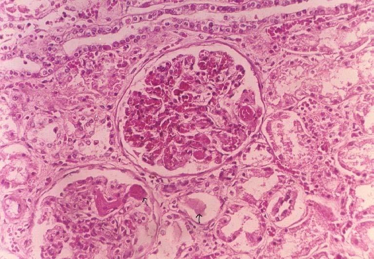
77 Disseminated Intravascular Coagulation D.I.C. OBSTETRIC COMPLICATIONS ADVANCED MALIGNANCY NOT a primary disease CONSUMPTIVE coagulopathy, e.g., reduced platelets, fibrinogen, F-VIII and other consumable clotting factors, brain, heart, lungs, kidneys, MICROSCOPIC ONLY
78
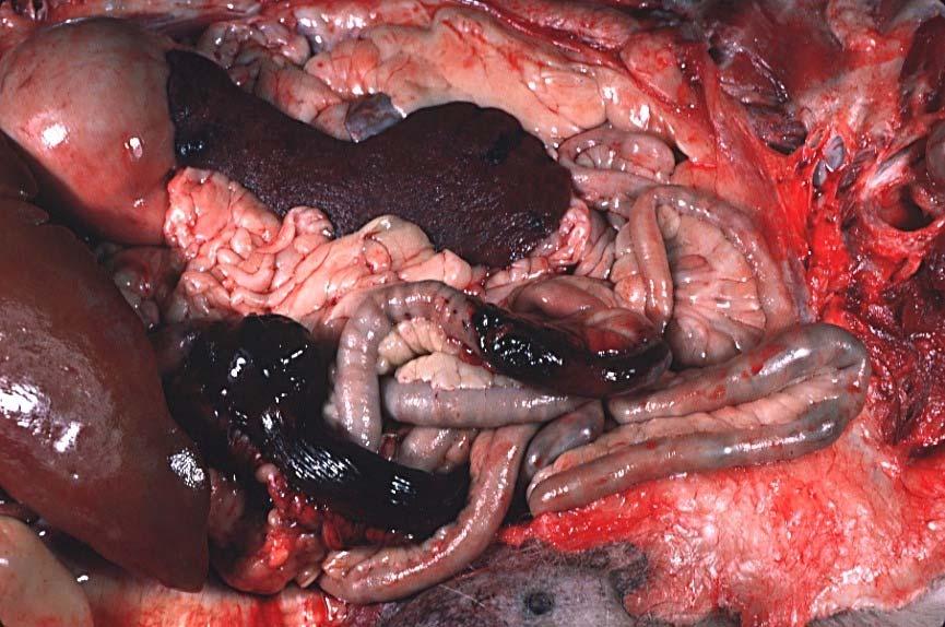
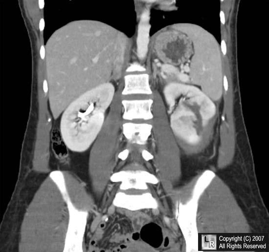
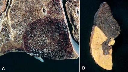
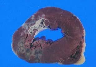
79 EMBOLISM Pulmonary Systemic (Mural Thrombi and Aneurysms) Fat Air Amniotic Fluid
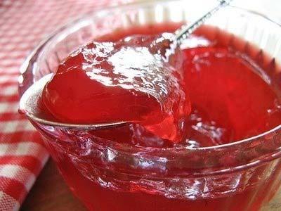
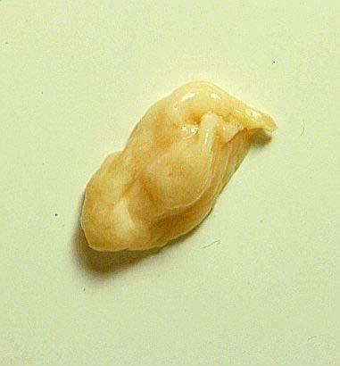
80 PULMONARY EMBOLISM USUALLY SILENT CHEST PAIN, LOW PO2, S.O.B. Sudden OCCLUSION of >60% of pulmonary vasculature, presents a HIGH risk for sudden death, i.e., acute cor pulmonale, ACUTE right heart failure SADDLE embolism often/usually fatal PRE vs. POST mortem blood clot: PRE: Friable, adherent, lines of ZAHN POST: Current jelly or chicken fat
81
82
83 SYSTEMIC EMBOLI PARADOXICAL EMBOLI 80% cardiac/20% aortic Embolization lodging site is proportional to the degree of flow (cardiac output) that area or organ gets, i.e., brain, kidneys, legs
84 OTHER EMBOLI FAT (long bone fx s ) AIR (SCUBA bends) AMNIOTIC FLUID, very prolonged or difficult delivery, high mortality
85
86
87 INFARCTION Defined as an area of necrosis* secondary to decreased blood flow HEMORRHAGIC vs. ANEMIC RED vs. WHITE END ARTERIES vs. NO END ARTERIES ACUTE ORGANIZATION FIBROSIS
88 INFARCTION FACTORS NATURE of VASCULAR SUPPLY RATE of DEVELOPMENT SLOW (BETTER) FAST (WORSE) VULNERABILITY to HYPOXIA MYOCYTE vs. FIBROBLAST CHF vs. NO CHF
89
90
91
92 SHOCK Pathogenesis Cardiac Septic Hypovolemic Morphology Clinical Course
93 SHOCK Definition: CARDIOVASCULAR COLLAPSE Common pathophysiologic features: INADEQUATE CARDIAC OUTPUT and/or INADEQUATE BLOOD VOLUME
94 GENERAL RESULTS INADEQUATE TISSUE PERFUSION CELLULAR HYPOXIA UN-corrected, a FATAL outcome
95 TYPES of SHOCK CARDIOGENIC: (Acute, Chronic Heart Failure) HYPOVOLEMIC: (Hemorrhage or Leakage) SEPTIC: ( ENDOTOXIC shock, #1 killer in ICU) NEUROGENIC: (loss of vascular tone) ANAPHYLACTIC: (IgE mediated systemic vasodilation and increased vascular permeability)
96 CARDIOGENIC shock MI VENTRICULAR RUPTURE ARRHYTHMIA CARDIAC TAMPONADE PULMONARY EMBOLISM (acute RIGHT heart failure or cor pulmonale )
97 HYPOVOLEMIC shock HEMORRHAGE, Vasc. compartment H2O VOMITING, Vasc. compartment H2O DIARRHEA, Vasc. compartment H2O BURNS, Vasc. compartment H2O
98 SEPTIC shock OVERWHELMING INFECTION ENDOTOXINS, i.e., LPS (Usually Gm-) Gm+ FUNGAL SUPERANTIGENS, (Superantigens are polyclonal T-lymphocyte activators that induce systemic inflammatory cytokine cascades similar to those occurring downstream in septic shock, toxic shock antigens by staph are the prime example.)
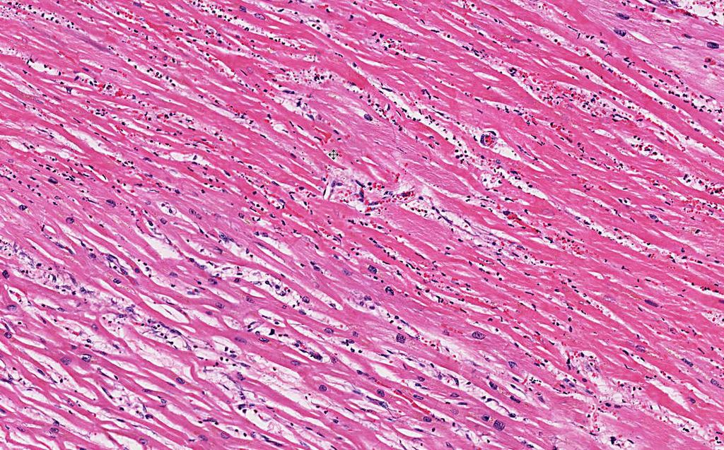
99 SEPTIC shock events* (overwhelming infection) Peripheral vasodilation Pooling Endothelial Activation DIC * Think of this as a TOTAL BODY inflammatory response
100 ENDOTOXINS Usually Gm- Degraded bacterial cell wall products Also called LPS, because they are Lipo- Poly-Saccharides Attach to a cell surface antigen known as CD-14
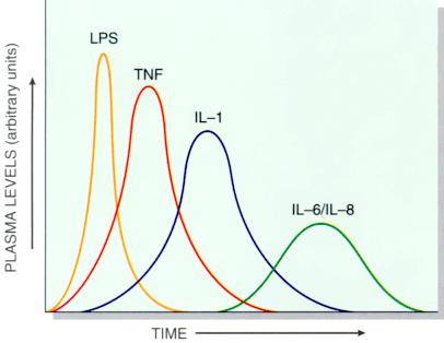
101 ENDOTOXINS
102 SEPTIC shock events (linear sequence) SYSTEMIC VASODILATION (hypotension) MYOCARDIAL CONTRACTILITY DIFFUSE ENDOTHELIAL ACTIVATION LEUKOCYTE ADHESION ALVEOLAR DAMAGE (ARDS) DIC VITAL ORGAN FAILURE CNS
103 CLINICAL STAGES of shock NON-PROGRESSIVE PROGRESSIVE IRREVERSIBLE
104 NON-PROGRESSIVE COMPENSATORY MECHANISMS CATECHOLAMINES VITAL ORGANS PERFUSED
105 PROGRESSIVE HYPOPERFUSION EARLY VITAL ORGAN FAILURE OLIGURIA ACIDOSIS
106 IRREVERSIBLE HEMODYNAMIC CORRECTIONS of no use
107 PATHOLOGY MULTIPLE ORGAN FAILURE SUBENDOCARDIAL HEMORRHAGE (why?) ACUTE TUBULAR NECROSIS (why?) DAD (Diffuse Alveolar Damage, lung) (why?) GI MUCOSAL HEMORRHAGES (why?) LIVER NECROSIS (why?) DIC (why?)
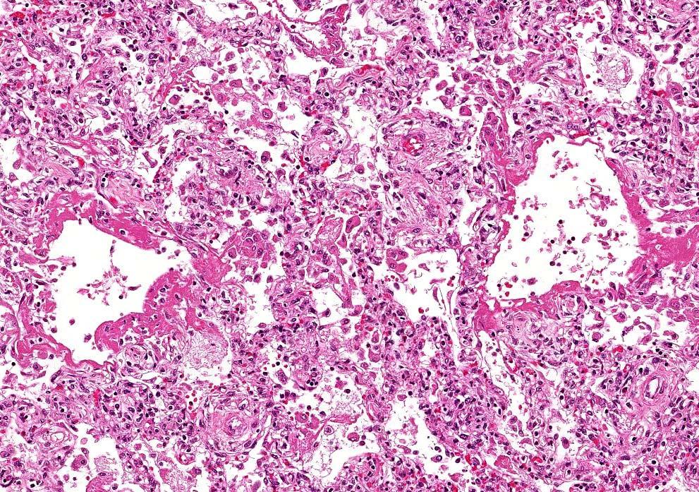
108 ARDS/DAD
109 MYOCARDIAL NECROSIS
110 ATN
111 DIC
112 CLINICAL PROGRESSION of SYMPTOMS Hypotension Tachycardia Tachypnea Warm skin Cool skin Cyanosis Renal insufficiency Obtundance Death
Hemodynamic Disorders, Thrombosis, and Shock. Richard A. McPherson, M.D.
 Hemodynamic Disorders, Thrombosis, and Shock Richard A. McPherson, M.D. Edema The accumulation of abnormal amounts of fluid in intercellular spaces of body cavities. Inflammation and release of mediators
Shock, Hemorrhage and Thrombosis
 Shock, Hemorrhage and Thrombosis 1 Shock Systemic hypoperfusion due to: Reduction in cardiac output Reduction in effective circulating blood volume Hypotension Impaired tissue perfusion Cellular hypoxia
HEMODYNAMIC DISORDERS
 HEMODYNAMIC DISORDERS Normal fluid homeostasis requires vessel wall integrity as well as maintenance of intravascular pressure and osmolarity within certain physiologic ranges. Increases in vascular volume
Disturbance of Circulation Hemodynamic Disorder
 Disturbance of Circulation Hemodynamic Disorder 2/17/2017 By Dr. Hemn Hassan Othman PhD, Pathology Fall 2016 1 Thrombosis Definition: Thrombosis is the formation of solid or semisolid blood clot within
Hemodynamic Disorders Thrombosis and Shock. 1. Interstitial, between the cells, but outside of the vascular system. - water making up the blood and
 Hemodynamic Disorders Thrombosis and Shock I. Body water, where is it and what keeps it there? A. Intracellular B. Extracellular (intercellular) 1. Interstitial, between the cells, but outside of the vascular
Thrombosis. Dr. László Terézia
 Thrombosis Dr. László Terézia HYPERCOAGULABILITY THROMBOSIS BLOODFLOW ENDOTHEL VIRCHOW ENDOTHEL INJURY L. ventricle: Arteries: surgery infection prosthetic valve hypertension irradiation chemical: cigarette
Bachelor of Chinese Medicine Shock
 BCM Year 2 Dr. Irene Ng Jan 28, 2003 9:30 am 1:00 pm Rm 004 UPB Bachelor of Chinese Medicine 2002 2003 Shock Learning objectives Be able to: know the definition of shock know the classification and causes
Hyperemia, Congestion, and Edema
 Hyperemia, Congestion, and Edema Hyperemia Acute, actively increased blood flow Tissues look red (erythema) Congestion Chronic, passively reduced outflow Tissues look pale or blue (cyanosis) Edema Water
Pathophysiology. Tutorial 3 Hemodynamic Disorders
 Pathophysiology Tutorial 3 Hemodynamic Disorders ILOs Recall different causes of thrombosis. Explain different types of embolism and their predisposing factors. Differentiate between hemorrhage types.
HYPEREMIA AND CONGESTION
 HYPEREMIA AND CONGESTION Learning Objectives Define congestion and hyperemia Differentiate between the two with regard to: Mechanisms / underlying causes Appearance (gross and histologic) Effects Differentiate
THROMBOSIS. Dr. Nisreen Abu Shahin Assistant Professor of Pathology Pathology Department University of Jordan
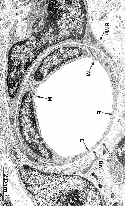
 THROMBOSIS Dr. Nisreen Abu Shahin Assistant Professor of Pathology Pathology Department University of Jordan NORMAL BLOOD VESSEL HISTOLOGY THROMBOSIS Pathogenesis (called Virchow's triad): 1. Endothelial*
Hemodynamic Disorders, Thromboembolic Disease, and Shock
 Hemodynamic Disorders, Thromboembolic Disease, and Shock Kumar et al: Robbins & Cotran Pathologic Basis of Disease 7E Figure 4-1 Factors affecting fluid balance across capillary walls. Capillary hydrostatic
Hemodynamic derangement. Komson Wannasai, M.D.,FRCPath. Department of Pathology Faculty of Medicine Chiang Mai University
 Hemodynamic derangement Komson Wannasai, M.D.,FRCPath. Department of Pathology Faculty of Medicine Chiang Mai University Objective The students should be able to Explain normal body fluid homeostasis Explain
Hemodynamic Disorders Thrombosis and Shock
 Hemodynamic Disorders Thrombosis and Shock SCPA 202 Basic Pathology Somphong Narkpinit, M.D. Department of Pathobiology, Faculty of Science, Mahidol University Email : somphong.nar@mahidol.ac.th Hemodynamic
Thrombosis and emboli. Peter Nagy
 Thrombosis and emboli Peter Nagy A thrombus is any solid object developing from the blood in vivo within the vascular system or heart. Thrombosis is hemostasis in the wrong place. Major components, forms:
Circulatory Disturbances 5: Thrombosis, Embolism, Infarction, Shock
 Circulatory Disturbances 5: Thrombosis, Embolism, Infarction, Shock Shannon Martinson, Feb 2016 http://people.upei.ca/smartinson/ VPM 152 General Pathology Thrombosis, Embolism, Infarction, Shock Learning
Pathology of pulmonary vascular disease. Dr.Ashraf Abdelfatah Deyab. Assistant Professor of Pathology Faculty of Medicine Almajma ah University
 Pathology of pulmonary vascular disease Dr.Ashraf Abdelfatah Deyab Assistant Professor of Pathology Faculty of Medicine Almajma ah University Pulmonary vascular disease Type of pulmonary circulation: Types
What are blood clots?
 What are blood clots? Dr Matthew Fay GP Principal The Willows Medical Practice- Queensbury GPwSI and Co-Founder Westcliffe Cardiology Service GP Partner Westcliffe Medical Group Created 5/31/18 Dr. Matthew
Hemostasis and Thrombosis
 Hemostasis Hemostasis and Thrombosis Normal hemostasis is a consequence of tightly regulated processes that maintain blood in a fluid state in normal vessels, yet also permit the rapid formation of a hemostatic
Wheater: Part 1: Thrombosis, embolism and infarction. Laboratory assignment: C601/C602 Histopathology manual, hemodynamic unit.
 Pathology C 601 Hemodynamic Derangements Assignment page. Reading: Robbins: Chapter 4 Clinical Lab Source: - Protime (PT) Know about INR - Activated partial thrmboplastin time (APTT) - Activated coagulation
Chapter 4: Haemodynamic disorders, shock
 Chapter 4: Haemodynamic disorders, shock 1. Regarding platelets (2006) (a) They are the main source of thrombin (b) they number 150-300 x10 3 per microlitre (c) They contain a nucleus (d) They are biconcave
Thrombosis. Jeffrey Jhang, M.D.
 Thrombosis Jeffrey Jhang, M.D. Introduction The human hemostatic system has evolved to maintain blood flow under normal physiologic conditions while remaining primed to rapidly respond to vascular injury
Topics of today lectures: Hemostasis
 Topics of today lectures: Hemostasis Meaning of hemostasis Mechanisms of hemostasis - Vascular contraction - Platelets plug - Blood coagulation (clotting) - Structure and functions of platelets - Blood
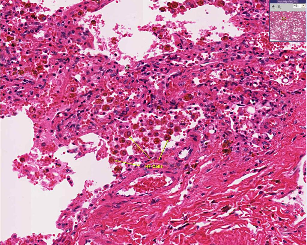
This slide belongs to iron lecture and it is to clarify the iron cycle in the body and the effect of hypoxia on erythropoitein secretion
 This slide belongs to iron lecture and it is to clarify the iron cycle in the body and the effect of hypoxia on erythropoitein secretion Topics of today lectures: Hemostasis Meaning of hemostasis Mechanisms
Aneurysms & a Brief Discussion on Embolism
 Aneurysms & a Brief Discussion on Embolism Aneurysms, overview = congenital or acquired dilations of blood vessels or the heart True aneurysms -involve all three layers of the artery (intima, media, and
According to the etiology, edema may be:
 What is edema? Edema : It refers to the accumulation of excess liquid in the interstitial (extracellular) spaces of a tissue or in pre-existing cavities. It may affect any organ, but most often it appears
Disseminated Intravascular Coagulation. M.Bahmanpour MD Assistant professor IUMS
 به نام خدا Disseminated Intravascular Coagulation M.Bahmanpour MD Assistant professor IUMS Algorithm for Diagnosis of DIC DIC Score factor score Presence of known underlying disorder No= 0 yes=2 Coagolation
SHOCK AETIOLOGY OF SHOCK (1) Inadequate circulating blood volume ) Loss of Autonomic control of the vasculature (3) Impaired cardiac function
 Inadequate circulating blood volume ) Loss of Autonomic control of the vasculature (3) Impaired cardiac function") SHOCK Shock is a condition in which the metabolic needs of the body are not met because of an inadequate cardiac output. If tissue perfusion can be restored in an expeditious fashion, cellular injury may
SHOCK Shock is a condition in which the metabolic needs of the body are not met because of an inadequate cardiac output. If tissue perfusion can be restored in an expeditious fashion, cellular injury may
UNIT VI. Chapter 37: Platelets Hemostasis and Blood Coagulation Presented by Dr. Diksha Yadav. Copyright 2011 by Saunders, an imprint of Elsevier Inc.
 UNIT VI Chapter 37: Platelets Hemostasis and Blood Coagulation Presented by Dr. Diksha Yadav Hemostasis: Prevention of Blood Loss Vascular constriction Formation of a platelet plug Formation of a blood
Physiological Response to Hypovolemic Shock Dr Khwaja Mohammed Amir MD Assistant Professor(Physiology) Objectives At the end of the session the
 Objectives At the end of the session the") Physiological Response to Hypovolemic Shock Dr Khwaja Mohammed Amir MD Assistant Professor(Physiology) Objectives At the end of the session the students should be able to: List causes of shock including
Physiological Response to Hypovolemic Shock Dr Khwaja Mohammed Amir MD Assistant Professor(Physiology) Objectives At the end of the session the students should be able to: List causes of shock including
Chapter 19. Hemostasis
 Chapter 19 Hemostasis Hemostasis Hemostasis is the cessation of bleeding stopping potentially fatal leaks important in small blood vessels not effective in hemorrhage excessive bleeding from large blood
HEME 10 Bleeding Disorders
 HEME 10 Bleeding Disorders When injury occurs, three mechanisms occur Blood vessels Primary hemostasis Secondary hemostasis Diseases of the blood vessels Platelet disorders Thrombocytopenia Functional
Ischemic heart disease
 Ischemic heart disease Introduction In > 90% of cases: the cause is: reduced coronary blood flow secondary to: obstructive atherosclerotic vascular disease so most of the time it is called: coronary artery
Chapter 3 Disorder of Local Blood Circulation
 Chapter 3 Disorder of Local Blood Circulation Disorder of Circulation Disorder of vascular flow may be divided into general and local categories. The local disorders contain: 1Derangement of local blood
Coagulation Disorders. Dr. Muhammad Shamim Assistant Professor, BMU
 Coagulation Disorders Dr. Muhammad Shamim Assistant Professor, BMU 1 Introduction Local Vs. General Hematoma & Joint bleed Coagulation Skin/Mucosal Petechiae & Purpura PLT wound / surgical bleeding Immediate
Disorder of Local Blood Circulation. Pathology Department, Zhejiang University School of Medicine,
 Disorder of Local Blood Circulation Pathology Department, Zhejiang University School of Medicine, maliqin198@zju.edu.cn Hyperemia Hemorrhage Thrombosis Embolism Infarction Edema Conception: Disorder of
Circulation. Blood Pressure and Antihypertensive Medications. Venous Return. Arterial flow. Regulation of Cardiac Output.
 Circulation Blood Pressure and Antihypertensive Medications Two systems Pulmonary (low pressure) Systemic (high pressure) Aorta 120 mmhg Large arteries 110 mmhg Arterioles 40 mmhg Arteriolar capillaries
DVT Pathophysiology and Prophylaxis in Medically Hospitalized Patients. David Liff MD Oklahoma Heart Institute Vascular Center
 DVT Pathophysiology and Prophylaxis in Medically Hospitalized Patients David Liff MD Oklahoma Heart Institute Vascular Center Overview Pathophysiology of DVT Epidemiology and risk factors for DVT in the
Shock Management. Seyed Tayeb Moradian MSc, Critical Care Nursing Ph.D Candidate. PDF created with pdffactory Pro trial version
 Shock Management Seyed Tayeb Moradian MSc, Critical Care Nursing Ph.D Candidate Definition of Shock The definition of shock does not involve low blood pressure, rapid pulse or cool clammy skin - these
Primary Exam Physiology lecture 5. Haemostasis
 Primary Exam Physiology lecture 5 Haemostasis Haemostasis Body s response for the prevention and cessation of bleeding. Broadly consists of: Primary Haemostasis - vascular spasm and platlet plug formation
Hemodynamic Disorders, Thromboembolic Disease and Shock (part 1)
") Hemodynamic Disorders, Thromboembolic Disease and Shock (part 1) Lilla Madaras Semmelweis University 2 nd Department of Pathology 17 th September 2018 1 Normal fluid homeostasis Vessel wall integrity Intravascular
Hemodynamic Disorders, Thromboembolic Disease and Shock (part 1) Lilla Madaras Semmelweis University 2 nd Department of Pathology 17 th September 2018 1 Normal fluid homeostasis Vessel wall integrity Intravascular
Circulatory disorders
 Circulatory disorders Edema = fluid in interstitium transudate s.w.1.012, exudate 1.020) generalized x local prominent cavities hydrothorax, hydropericardium, ascites subcutaneous tissue (pitting edema)
Lung diseases of Vascular Origin. By: Shefaa Qa qqa
 Lung diseases of Vascular Origin By: Shefaa Qa qqa Pulmonary Hypertension Pulmonary hypertension is defined as a mean pulmonary artery pressure greater than or equal to 25 mm Hg at rest. Based on underlying
Thromboembolismand Shock 血管栓塞和休克
 Thromboembolismand Shock 血管栓塞和休克 Major Hemodynamic Disorders Edema Hypermia and Congestion 充血 Haemorrhage Hemostasis 止血 and Blood Coagulation 血液凝固 Thrombosis 血栓形成 Embolism 栓塞 Infarction 梗死 Disseminated
Thrombosis, Embolism and Infarction
 Thrombosis, Embolism and Infarction THROMBOSIS Thrombus formation (called Virchow's triad): (1) endothelial injury, (2) stasis or turbulent blood flow (3) hypercoagulability of the blood Endothelial Injury
PHM142 Lecture 4: Platelets + Endothelial Cells
 PHM142 Lecture 4: Platelets + Endothelial Cells 1 Hematopoiesis 2 Platelets Critical in clotting - activated by subendothelial matrix proteins (e.g. collagen, fibronectin, von Willebrand factor) and thrombin
EDEMA. Learning Objectives
 EDEMA Learning Objectives Define edema Recognize and be able to describe the gross and microscopic appearance of edema Know the four pathophysiological mechanisms by which edema develops Understand the
Thursday, February 26, :00 am. Regulation of Coagulation/Disseminated Intravascular Coagulation HEMOSTASIS/THROMBOSIS III
 REGULATION OF COAGULATION Introduction HEMOSTASIS/THROMBOSIS III Regulation of Coagulation/Disseminated Coagulation necessary for maintenance of vascular integrity Enough fibrinogen to clot all vessels
Schematic Of Heparin Induced Thrombocytopenia Platelet Count
 Schematic Of Heparin Induced Thrombocytopenia Platelet Count Normal IgG and IgG2 differentially inhibit HIT antibody-dependent platelet activation that platelet counts were lower in FcγRIIA 131RR patients
Haemodynamic Disorders
 Haemodynamic Disorders ZHANG WEI 张伟 Ph.D., A.P. Institute of Pathology & Forensic Medicine Department of Pathology & Patho-physiology Zhejiang University School of Medicine Email:zwei72@zju.edu.cn Thrombosis
Myocardial Infarction
 Myocardial Infarction MI = heart attack Defined as necrosis of heart muscle resulting from ischemia. A very significant cause of death worldwide. of these deaths, 33% -50% die before they can reach the
Hemostasis. Learning objectives Dr. Mária Dux. Components: blood vessel wall thrombocytes (platelets) plasma proteins
 plasma proteins") Hemostasis Learning objectives 14-16 Dr. Mária Dux Components: blood vessel wall thrombocytes (platelets) plasma proteins Hemostatic balance! procoagulating activity anticoagulating activity 1 Thrombocytes
Hemostasis Learning objectives 14-16 Dr. Mária Dux Components: blood vessel wall thrombocytes (platelets) plasma proteins Hemostatic balance! procoagulating activity anticoagulating activity 1 Thrombocytes
DIC. Bert Vandewiele Fellow Critical Care 23 May 2011
 DIC Bert Vandewiele Fellow Critical Care 23 May 2011 Dissiminated Intravascular Coagulopathie 11/3/2011 Dr. Bert Vandewiele 2 Dissiminated Intravascular Coagulopathie = Consumption coagulopathie = Defibrination
Means failure of heart to pump enough blood to satisfy the need of the body.
 Means failure of heart to pump enough blood to satisfy the need of the body. Due to an impaired ability of the heart to adequately to fill or eject blood. HEART FAILURE Heart failure (HF) means decreased
Thrombosis. By Dr. Sara Mohamed Abuelgasim
 Thrombosis By Dr. Sara Mohamed Abuelgasim 1 Thrombosis Unchecked, blood coagulation would lead to dangerous occlusion of blood vessels if the protective mechanisms of coagulation factor inhibitors, blood
PE and DVT. Dr Anzo William Adiga WatsApp or Call Medical Officer/RHEMA MEDICAL GROUP
 PE and DVT Dr Anzo William Adiga WatsApp or Call +256777363201 Medical Officer/RHEMA MEDICAL GROUP OBJECTIVES DEFINE DVT AND P.E PATHOPHYSIOLOGY OF DVT CLINICAL PRESENTATION OF DVT/PE INVESTIGATE DVT MANAGEMENT
ATHEROSCLEROSIS. Secondary changes are found in other coats of the vessel wall.
 ATHEROSCLEROSIS Atherosclerosis Atherosclerosis is a disease process affecting the intima of the aorta and large and medium arteries, taking the form of focal thickening or plaques of fibrous tissue and
Moath Darweesh. Omar Sami. Saleem Khreisha. 1 P a g e
 7 Moath Darweesh Omar Sami Saleem Khreisha 1 P a g e -First of all, I want to give a quick revision to simplify the whole hemostasis mechanism, it will be much easier here with me. Enjoy (you can skip
BLEEDING DISORDERS Simple complement:
 BLEEDING DISORDERS Simple complement: 1. Select the statement that describe the thrombocytopenia definition: A. Marked decrease of the Von Willebrandt factor B. Absence of antihemophilic factor A C. Disorder
5 DISTURBANCES IN CIRCULATION. Congestion / Hyperemia Haemorrhage Thrombosis Embolism Ischemia Infarction Oedema Shock Sludged blood Model Questions
 5 DISTURBANCES IN CIRCULATION Congestion / Hyperemia Haemorrhage Thrombosis Embolism Ischemia Infarction Oedema Shock Sludged blood Model Questions CONGESTION/ HYPEREMIA Hyperemia is increased amount of
Outline Anti-coagulant and anti-thrombotic drugs Haemostasis and Thrombosis Year 3 Dentistry
 Outline Anti-coagulant and anti-thrombotic drugs Year 3 Dentistry Professor Yotis Senis Cellular Haemostasis y.senis@bham.ac.uk I. Haemostasis and II. Coagulation and anti-coagulants III. Platelets and
-Hashim ahmed is the one who wrote this sheet. I just edited it according to our record.
 * Subjects of this lecture : - Hemostasis - Platelets, general information, their ultrastructure and role in hemostasis. - Definitions: Thrombus, Embolus, Arteriosclerosis and Atherosclerosis. *NOTE: Prof
12/1/2009. Chapter 19: Hemorrhage. Hemorrhage and Shock Occurs when there is a disruption or leak in the vascular system Internal hemorrhage
 Chapter 19: Hemorrhage Hemorrhage and Shock Occurs when there is a disruption or leak in the vascular system External hemorrhage Internal hemorrhage Associated with higher morbidity and mortality than
Anticoagulants. Pathological formation of a haemostatic plug Arterial associated with atherosclerosis Venous blood stasis e.g. DVT
 Haemostasis Thrombosis Phases Endogenous anticoagulants Stopping blood loss Pathological formation of a haemostatic plug Arterial associated with atherosclerosis Venous blood stasis e.g. DVT Vascular Platelet
Part IV Antithrombotics, Anticoagulants and Fibrinolytics
 Part IV Antithrombotics, Anticoagulants and Fibrinolytics "The meaning of good and bad, of better and worse, is simply helping or hurting" Emerson Chapter 16: Blood Coagulation and Fibrinolytic System
SPECIAL PATHOPHYSIOLOGY CARDIO-VASCULAR SYSTEM
 1. Myocardia lischemia is mainly a result of: 1.Coronary hypoxemia. 2. Coronary artery disease (CAD). 3. Acute coronaritis. 4. Coronary anemia. 5. Heart remodelling. SPECIAL PATHOPHYSIOLOGY CARDIO-VASCULAR
Physiology of. The Blood hemostasis. By prof. Israa f. jaafar
 Physiology of The Blood hemostasis By prof. Israa f. jaafar Learning objectives Understand the Platelet structure and function Explane the Platelet production Understand the phases of hemostasis: vascular
Circulatory Disturbances 1: Introduction and Edema
 Circulatory Disturbances 1: Introduction and Edema Shannon Martinson, January 2016 http://people.upei.ca/smartinson/ VPM 152 General Pathology INTRODUCTION NORMAL CIRCULATORY SYSTEM Important concepts
Bleeding and Haemostasis. Saman W.Boskani HDD, FIBMS Maxillofacial Surgeon
 Bleeding and Haemostasis Saman W.Boskani HDD, FIBMS Maxillofacial Surgeon 1 Beeding Its escaping or extravasation of blood contents from blood vessels Types: - Arterial - Venous - Capillary Differences
CIRCULATORY DISTURBANCES
 CIRCULATORY DISTURBANCES Shannon Martinson, January 2017 Office: 418N Email: smartinson@upei.ca All lecture notes and slide shows are available online: http://people.upei.ca/smartinson REFERENCE TEXTS:
1) Severe, crushing substernal chest pain 2) radiate to the neck, jaw, epigastrium, or left arm. 3- rapid and weak pulse 4- nausea (posterior MI).
 Severe, crushing substernal chest pain 2) radiate to the neck, jaw, epigastrium, or left arm. 3- rapid and weak pulse 4- nausea (posterior MI).") 1) Severe, crushing substernal chest pain 2) radiate to the neck, jaw, epigastrium, or left arm. 3- rapid and weak pulse 4- nausea (posterior MI). 5- cardiogenic shock (massive MIs >40% of the left ventricle)
1) Severe, crushing substernal chest pain 2) radiate to the neck, jaw, epigastrium, or left arm. 3- rapid and weak pulse 4- nausea (posterior MI). 5- cardiogenic shock (massive MIs >40% of the left ventricle)
1 Functions of endothelial cells include all the following EXCEPT. 2 Response to vascular injury is characterised by
 airns ase Hospital mergency epartment Part 1 FM MQs 1 Functions of endothelial cells include all the following XPT Formation of von-willebrand factor Formation of collagen and proteoglycans Formation of
-Cardiogenic: shock state resulting from impairment or failure of myocardium
 Shock chapter Shock -Condition in which tissue perfusion is inadequate to deliver oxygen, nutrients to support vital organs, cellular function -Affects all body systems -Classic signs of early shock: Tachycardia,tachypnea,restlessness,anxiety,
Veins. VENOUS RETURN = PRELOAD = End Diastolic Volume= Blood returning to heart per cardiac cycle (EDV) or per minute (Venous Return)
 or per minute (Venous Return)") Veins Venous system transports blood back to heart (VENOUS RETURN) Capillaries drain into venules Venules converge to form small veins that exit organs Smaller veins merge to form larger vessels Veins
Veins Venous system transports blood back to heart (VENOUS RETURN) Capillaries drain into venules Venules converge to form small veins that exit organs Smaller veins merge to form larger vessels Veins
CIRCULATORY DISTURBANCES
 CIRCULATORY DISTURBANCES Shannon Martinson, January 2016 Email: smartinson@upei.ca All lecture notes and slide shows are available online: http://people.upei.ca/smartinson Office: 418N REFERENCE TEXTS:
Shock. Shao Mian Emergency Department,Zhongshan Hospital
 Shock Shao Mian Emergency Department,Zhongshan Hospital What is shock THE BEGINNINGS OF UNDERSTANDING: THE LATE 19TH CENTURY THE AGE OF REASON: 1890 1925 THE MODERN ERA: BLALOCK S EPIPHANY POSTMODERNISM:
Diagnosis of hypercoagulability is by. Molecular markers
 Agenda limitations of clinical laboratories to evaluate hypercoagulability and the underlying cause for thrombosis what is the INR the lupus anticoagulant and the antiphospholipid antibody syndrome hassouna
1- Thromboembolism. 2- fat embolism. 3- air embolism. 4- amniotic fluid embolism.
 Embolism Definition:- An embolus is a detached intravascular solid, liquid or gaseous mass that is carried by blood to sites distant from its point of origin. After traveling via the blood, the embolus
Hemostasis Haemostasis means prevention of blood loss from blood vessels.
 ١ Hemostasis Haemostasis means prevention of blood loss from blood vessels. Bleeding is stopped by several mechanisms, which are: 1. Local vasoconstriction 2. Formation of platelet plug 3. Blood coagulation
Disseminated Intravascular Coagulation (DIC) Seminar. Ron Kopilov 4 th year Medical Student, Tel Aviv University Internal Medicine A 8.3.
 Seminar. Ron Kopilov 4 th year Medical Student, Tel Aviv University Internal Medicine A 8.3.") Disseminated Intravascular Coagulation (DIC) Seminar Ron Kopilov 4 th year Medical Student, Tel Aviv University Internal Medicine A 8.3.2012 1 Our plan: Understand the pathophysiology Identify risk factors
Disseminated Intravascular Coagulation (DIC) Seminar Ron Kopilov 4 th year Medical Student, Tel Aviv University Internal Medicine A 8.3.2012 1 Our plan: Understand the pathophysiology Identify risk factors
Cardiac Pathology & Rehabilitation
 Cardiac Pathology & Rehabilitation Which of the following best describes the physical activity performed in my leisure time? A. I perform vigorous physical activity 3X/week for 20 minutes each time B.
HYPERTENSIVE VASCULAR DISEASE
 HYPERTENSIVE VASCULAR DISEASE Cutoffs in diagnosing hypertension in clinical practice sustained diastolic pressures >90 mm Hg, or sustained systolic pressures >140 mm Hg Malignant hypertension A small
What would be the response of the sympathetic system to this patient s decrease in arterial pressure?
 CASE 51 A 62-year-old man undergoes surgery to correct a herniated disc in his spine. The patient is thought to have an uncomplicated surgery until he complains of extreme abdominal distention and pain
LAMA SHATAT TTP, ITP, DIC
 TTP, ITP, DIC Reduction in platelet number (thrombocytopenia) constitutes an important cause of generalized bleeding. A count less than 100,000 platelets/μl is generally considered to constitute thrombocytopenia.
Pathophysiology of Cardiovascular System. Dr. Hemn Hassan Othman, PhD
 Pathophysiology of Cardiovascular System Dr. Hemn Hassan Othman, PhD hemn.othman@univsul.edu.iq What is the circulatory system? The circulatory system carries blood and dissolved substances to and from
Written 01/09/17 Rewritten 3/29/17 for Interior Regional EMS Symposium
 Written 01/09/17 Rewritten 3/29/17 for Interior Regional EMS Symposium MARIA E. MANDICH MD Fairbanks Memorial Hospital Emergency Department Attending Physician Interior Region EMS Council Medical Director
Thrombosis (lec#1) * Pathogenesis (called Virchow's triad ): Endothelial Injury Stasis Blood Hypercoagulability. stimulated Stasis:
 * Pathogenesis (called Virchow's triad ): Endothelial Injury Stasis Blood Hypercoagulability. stimulated Stasis:") Thrombosis (lec#1) * Pathogenesis (called Virchow's triad): Endothelial Injury ( Heart, Arteries), Stasis (abnormal blood flow),blood Hypercoagulability. * Endothelial cells can be stimulated by direct
Thrombosis (lec#1) * Pathogenesis (called Virchow's triad): Endothelial Injury ( Heart, Arteries), Stasis (abnormal blood flow),blood Hypercoagulability. * Endothelial cells can be stimulated by direct
Approach to disseminated intravascular coagulation
 Approach to disseminated intravascular coagulation Khaire Ananta Shankarrao 1, Anil Burley 2, Deshmukh 3 1.MD Scholar, [kayachikitsa] 2.Professor,MD kayachikitsa. 3.Professor and HOD,Kayachikitsa. CSMSS
Disseminated intravascular coagulation (DIC) Dr. Klara Vezendi Szeged University Transfusiology Department
 Dr. Klara Vezendi Szeged University Transfusiology Department") Disseminated intravascular coagulation (DIC) Dr. Klara Vezendi Szeged University Transfusiology Department Disseminated intravascular coagulation (DIC, consumptive coagulopathy) is a clinicopathologic
Disseminated intravascular coagulation (DIC) Dr. Klara Vezendi Szeged University Transfusiology Department Disseminated intravascular coagulation (DIC, consumptive coagulopathy) is a clinicopathologic
HEMODYNAMIC DISORDERS
 CHAPTER 3 HEMODYNAMIC DISORDERS Ivan Damjanov, MD, PhD EDEMA 1. How is body water distributed? Body water is divided into two main compartments: & Intracellular, comprising two thirds of total body fluid.
Jessica Bryan, Natalia Evans, Karlyn Henderson, & Whitney Parks
 Jessica Bryan, Natalia Evans, Karlyn Henderson, & Whitney Parks 1. What is the most common cause of death in hospitalized patients? 1. Hospital-acquired infection 2. Pulmonary embolism 3. Myocardial infarction
Dr. MUBARAK ABDELRAHMAN MD PEDIATRICS AND CHILD HEALTH Assistant Professor FACULTY OF MEDICINE -JAZAN
 Dr. MUBARAK ABDELRAHMAN MD PEDIATRICS AND CHILD HEALTH Assistant Professor FACULTY OF MEDICINE -JAZAN The student should be able:» To identify the mechanism of homeostasis and the role of vessels, platelets
HEART HEALTH WEEK 2 SUPPLEMENT. A Beginner s Guide to Cardiovascular Disease ATHEROSCLEROSIS. Fatty deposits can narrow and harden the artery
 WEEK 2 SUPPLEMENT HEART HEALTH A Beginner s Guide to Cardiovascular Disease ATHEROSCLEROSIS FIGURE 1 Atherosclerosis is an inflammatory process where cholesterol is deposited in the wall of arteries and
Hematologic Disorders. Assistant professor of anesthesia
 Preoperative Evaluation Hematologic Disorders Dr M.Razavi Assistant professor of anesthesia Anemia Evaluation needs to consider the extent and type of surgery, the anticipated blood loss, and the patient's
Pathology note 8 BLEEDING DISORDER
 Pathology note 8 BLEEDING DISORDER Slide75 ( Types of clotting factors deficiency): Today we will talk about public public factor deficiency it could be acquired or inherited, acquired diseases are more
Cardiovascular Disorders. Heart Disorders. Diagnostic Tests for CV Function. Bio 375. Pathophysiology
 Cardiovascular Disorders Bio 375 Pathophysiology Heart Disorders Heart disease is ranked as a major cause of death in the U.S. Common heart diseases include: Congenital heart defects Hypertensive heart
Circulatory shock. Types, Etiology, Pathophysiology. Physiology of Circulation: The Vessels. 600,000 miles of vessels containing 5-6 liters of blood
 Circulatory shock Types, Etiology, Pathophysiology Blagoi Marinov, MD, PhD Pathophysiology Dept. Physiology of Circulation: The Vessels 600,000 miles of vessels containing 5-6 liters of blood Vessel tone
Pulmonary-Vascular Disease. Howard J. Sachs, MD.
 Pulmonary-Vascular Disease Howard J. Sachs, MD www.12daysinmarch.com The Disorders COPD/ILD Chronic Hypoxia Vasoconstrictive Obliterative PPH Obstructive Hyperkinetic LEFT right Shunt Passive 2 nd to LV