Cardiovascular hemodynamics in the stress echo lab with open-source software
|
|
|
- Christina Isabella Flowers
- 5 years ago
- Views:
Transcription


1 Cardiovascular hemodynamics in the stress echo lab with open-source software T. Bombardini, D. Cini, E. Picano Institute of Clinical Physiology of CNR, Pisa, Italy no conflict of interest
2 Background Stress echocardiographic evaluation of volumes is ideally suited for the quantitative calculation of a set of parameters allowing a complete characterization of cardiovascular hemodynamics of established pathophysiological and potential clinical relevance, including cardiac output, systemic vascular resistance, left ventricular elastance, arterial elastance, and ventricular arterial coupling However, this is a tedious and time-consuming
3 Aim to build a web-based computing software program for self-instruction and calculation of hemodynamic parameters in the stress echo lab to test the software with skilled echocardiographers to distribute the software to the cardiological community
4 Methods A website with the informatics infrastructure was built (at Ten skilled echocardiographers (American Society of Echocardiography class III) were asked to calculate hemodynamic parameters by using personal knowledge (Mode 1) and by using the dedicated software called CCtrainer (Mode 2) After testing, the software was offered free on the web
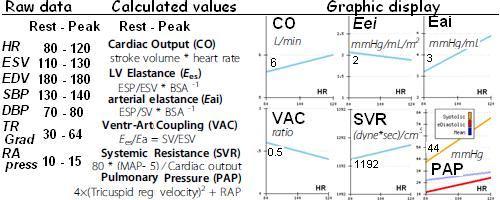
5 Results The software was inserted in a dedicated web domain. After linking in, the cardiologist is asked to fill in the rest and stress data set. In a few seconds the program completes baseline and peak stress data, providing both numerical and graphical display of results The computation time of manual Mode 1 (55 ± 63 min) was substantially reduced with softwareassisted Mode 2 (2 ± 0.5 min, p < 0.05 vs Mode 1)
6 Conclusions Cardiovascular hemodynamics are important but their calculation remains time-consuming and demanding However, they can be made simple, rapid and easy in the echo lab with a user-friendly, open-source program fed by simple raw echo data
7
8 Accesses to the web site

9 Blood pressure analysis. One investigator records all blood pressures at rest and during exercise during the study. Echocardiography is performed using 3-D or conventional 2-D echocardiography and left ventricular end-systolic volume is measured. The contractility is determined at each stress step as the ratio of the systolic pressure (cuff sphygmomanometer)/end-systolic volume index (end-systolic volume/body surface area). Bombardini T. Myocardial contractility in the echo lab: molecular, cellular and pathophysiological basis. Cardiovascular ultrasound 2005, 3:27
 when the peak exercise SP/ESV index is higher than baseline and")

10 Contractility changes with heart rate changes. The force-frequency relationship is defined up-sloping (upper panel) when the peak exercise SP/ESV index is higher than baseline and intermediate stress values; flat or negative (lower panel), when the peak exercise systolic pressure/end-systolic volume index is equal to or lower than baseline stress values. The critical heart rate (or optimum stimulation frequency) is defined as the heart rate at which systolic pressure/endsystolic volume index reaches the maximum value during progressive increase in heart rate.

11 Preload, afterload, heart rate: the dark side of the force. When a stress echo is scheduled, interest is focused on wall motion segmental contraction abnormality to diagnose ischemic response to stress and on LVEF to assess contractile reserve. However, ejection fraction is a very gross index of left ventricular performance. It is affected by pre-load and after-load changes and heart rate. Bombardini et al. Cardiovascular Ultrasound :48
.")
12 Arterial elastance In the cartoon (left panel) a compliant and no resistive young hungry snake eats a big sheep easily. In the cardiovascular system the hydraulic load, namely effective arterial elastance, is described by the formula: End Systolic Pressure/Stroke Volume/Body Surface Area (right panel). Intuitively a well compliant and no resistive vascular system easily accomplish a big stroke volume without great increase of systemic pressure. Bombardini et al. Cardiovascular Ultrasound :48
 the skinny snake is a thin pipe with high strength, the snake stretched by food is a low resistance conduit.")
13 The steady (Systemic Vascular Resistance) and the pulsatile (Systemic Arterial Compliance) components of Arterial Elastance. In the cartoon (left panel) the skinny snake is a thin pipe with high strength, the snake stretched by food is a low resistance conduit. In the cartoon (middle panel) the compliant snake easily dilates itself and pushes the food at pulsatile waves. In the cartoon (right panel) the blind zoo guardian unsuccessfully try to use the snake tail (extremely resistive and no compliant) as a water conduct. Increased systemic vascular resistance and decreased total arterial compliance both contribute to the high arterial load in hypertensive patients. Bombardini et al. Cardiovascular Ultrasound :48
, able to easily accommodate large stroke volume energetically ejected; small fish mouth means higher arterial elastance")
14 Ventricular-arterial coupling. In the cartoon, arterial elastance is the "mouth" of the whale. Big mouth means low arterial elastance (no resistive and well compliant), able to easily accommodate large stroke volume energetically ejected; small fish mouth means higher arterial elastance (highly resistive and no compliant), able to accommodate only small stroke volume (low contractile-energy launched). Interaction of the LV with the arterial system, termed ventricular-arterial coupling, is a central determinant of cardiovascular performance and cardiac energetics. Ventricular arterial coupling is indexed by the ratio of left ventricular systolic elastance index (systolic pressure/end-systolic volume) to arterial elastance (Ea, ratio of end-systolic pressure by stroke volume). Bombardini et al. Cardiovascular Ultrasound :48


15 The mechanical events in the cardiac cycle. Cardiological systole is demarcated by the interval between the first and the second heart sounds, lasting from the first heart sound to the closure of the aortic valve. The remainder of the cardiac cycle automatically becomes cardiological diastole

16 Operator-independent cardiologic systole and diastole quantification. An analog peak-to-peak detector scans the first 150 ms following the R wave to record first heart sound force vibrations and the 100 ms following the T wave to record second heart sound force vibrations. A stable, reproducible, and consistent first heart sound and second heart sound signal is obtained and utilized as time markers to continuously assess cardiologic systole and diastole during exercise, or pharmacological stress echo.


17 Implementation of the computational training software. Opening a new example by the web linking to and clicking on "insert case" button a electronic sheet appears. Rest and peak data sets should be filled by the trainer in the fourth and fifth columns


18 Once linked to after filling the "insert case sheet" and clicking on the "rest and stress data calculator" button, the calculation results sheet opens. Calculation algorithms are shown in column 2 and software calculated results appear in the third "rest" and in the fourth "peak "column".

19 Training example with a "normal subject" data set. Clicking on the "view graphics" button opens the graph results sheet.
20 Training example with a "DCM subject" data set. Clicking on the "view graphics" button opens the graph results sheet
21 Inotropic reserve in the stress echo lab EXERCISE DOB DIP PACEMAKER Contractility Inotropic reserve Bowditch treppe + + =/+ ++ User-friendly Requisites Capability to exercise Intravenous line Intravenou s line Permanent pacemaker Bombardini et al JASE 2003 Int J Cardiol 2007 JNC 2008 Bombardini et al EHJ 2005 Bombardini et al J Heart Lung Transpl 2009 Bombardini et al Eur J Heart Failure 2004
Stress Echocardiography in VHD: is myocardial contrac8on a reliable marker for ischemia?
 Stress Echocardiography in VHD: is myocardial contrac8on a reliable marker for ischemia? Rosa Sicari CNR, Ins8tute of Clinical Physiology, Pisa, Italy Milano 8 maggio 2012 Stress Echo and Cardiac death
Cardiac Output Monitoring - 6
 Cardiac Output Monitoring - 6 How to use Wrexham s Cardiac Output Monitors. Wrexham Maelor Critical Care Version 02.05.16 Introduction Types of Devices: NICOM - Cheetah Oesophageal Doppler +/- Pulse Contour
Electrical Conduction
 Sinoatrial (SA) node Electrical Conduction Sets the pace of the heartbeat at 70 bpm AV node (50 bpm) and Purkinje fibers (25 40 bpm) can act as pacemakers under some conditions Internodal pathway from
Advanced Multi-Layer Speckle Strain Permits Transmural Myocardial Function Analysis in Health and Disease:
 Advanced Multi-Layer Speckle Strain Permits Transmural Myocardial Function Analysis in Health and Disease: Clinical Case Examples Jeffrey C. Hill, BS, RDCS Echocardiography Laboratory, University of Massachusetts
Adult Echocardiography Examination Content Outline
 Adult Echocardiography Examination Content Outline (Outline Summary) # Domain Subdomain Percentage 1 2 3 4 5 Anatomy and Physiology Pathology Clinical Care and Safety Measurement Techniques, Maneuvers,
Rotation: Echocardiography: Transthoracic Echocardiography (TTE)
") Rotation: Echocardiography: Transthoracic Echocardiography (TTE) Rotation Format and Responsibilities: Fellows rotate in the echocardiography laboratory in each clinical year. Rotations during the first
Rotation: Echocardiography: Transthoracic Echocardiography (TTE) Rotation Format and Responsibilities: Fellows rotate in the echocardiography laboratory in each clinical year. Rotations during the first
Title: Second-Opinion Stress Tele-Echocardiography for Aged Donor Heart Selection
 Author's response to reviews Title: Second-Opinion Stress Tele-Echocardiography for Aged Donor Heart Selection Authors: Daniele Franchi (franchid@ifc.cnr.it) Davide Cini (davide.cini@cnr.it) Giorgio Arpesella
LV FUNCTION ASSESSMENT: WHAT IS BEYOND EJECTION FRACTION
 LV FUNCTION ASSESSMENT: WHAT IS BEYOND EJECTION FRACTION Jamilah S AlRahimi Assistant Professor, KSU-HS Consultant Noninvasive Cardiology KFCC, MNGHA-WR Introduction LV function assessment in Heart Failure:
Mechanisms of heart failure with normal EF Arterial stiffness and ventricular-arterial coupling. What is the pathophysiology at presentation?
 Mechanisms of heart failure with normal EF Arterial stiffness and ventricular-arterial coupling What is the pathophysiology at presentation? Ventricular-arterial coupling elastance Central arterial pressure
Left atrial function. Aliakbar Arvandi MD
 In the clinic Left atrial function Abstract The left atrium (LA) is a left posterior cardiac chamber which is located adjacent to the esophagus. It is separated from the right atrium by the inter-atrial
Cardiovascular Imaging Endpoints in Oncology Clinical Trials
 Cardiovascular Imaging Endpoints in Oncology Clinical Trials Bonnie Ky, MD, MSCE Assistant Professor of Medicine and Epidemiology Director, Penn Cardio-Oncology Center of Excellence Director, Penn Center
RV dysfunction and failure PATHOPHYSIOLOGY. Adam Torbicki MD, Dept Chest Medicine Institute of Tuberculosis and Lung Diseases Warszawa, Poland
 RV dysfunction and failure PATHOPHYSIOLOGY Adam Torbicki MD, Dept Chest Medicine Institute of Tuberculosis and Lung Diseases Warszawa, Poland Normal Right Ventricle (RV) Thinner wall Weaker myocytes Differences
Cardiac Cycle MCQ. Professor of Cardiovascular Physiology. Cairo University 2007
 Cardiac Cycle MCQ Abdel Moniem Ibrahim Ahmed, MD Professor of Cardiovascular Physiology Cairo University 2007 1- Regarding the length of systole and diastole: a- At heart rate 75 b/min, the duration of
Assessment of LV systolic function
 Tutorial 5 - Assessment of LV systolic function Assessment of LV systolic function A knowledge of the LV systolic function is crucial in the undertanding of and management of unstable hemodynamics or a
powerful versatile mobile
 M9 Premium Compact Ultrasound System powerful versatile mobile M9 Premium Compact Ultrasound System powerful versatile mobile Powerful Platform With the next generation of technologies, the Mindray M9
From PV loop to Starling curve. S Magder Division of Critical Care, McGill University Health Centre
 From PV loop to Starling curve S Magder Division of Critical Care, McGill University Health Centre Otto Frank 1890 s Frank-Starling Relationship ( The Law of the Heart ) The greater the initial stretch
LA Function analysis Marcia Barbosa Vice Presidente - Brazilian Soc of Cardiology President-elect - Interamerican Soc of Cardiology
 LA Function analysis Marcia Barbosa Vice Presidente - Brazilian Soc of Cardiology President-elect - Interamerican Soc of Cardiology Belo Horizonte Brazil DECLARATION OF CONFLICT OF INTEREST Nothing to
SymBioSys Exercise 2 Cardiac Function Revised and reformatted by C. S. Tritt, Ph.D. Last updated March 20, 2006
 SymBioSys Exercise 2 Cardiac Function Revised and reformatted by C. S. Tritt, Ph.D. Last updated March 20, 2006 The goal of this exercise to explore the behavior of the heart as a mechanical pump. For
Advanced imaging of the left atrium - strain, CT, 3D, MRI -
 Advanced imaging of the left atrium - strain, CT, 3D, MRI - Monica Rosca, MD Carol Davila University of Medicine and Pharmacy, Bucharest, Romania Declaration of interest: I have nothing to declare Case
How does the heart pump? From sarcomere to ejection volume
 How does the heart pump? From sarcomere to ejection volume Piet Claus Cardiovascular Imaging and Dynamics Department of Cardiovascular Diseases University Leuven, Leuven, Belgium Course on deformation
Echo assessment of the failing heart
 Echo assessment of the failing heart Mark K. Friedberg, MD The Labatt Family Heart Center The Hospital for Sick Children Toronto, Ontario, Canada Cardiac function- definitions Cardiovascular function:
Index of subjects. effect on ventricular tachycardia 30 treatment with 101, 116 boosterpump 80 Brockenbrough phenomenon 55, 125
 145 Index of subjects A accessory pathways 3 amiodarone 4, 5, 6, 23, 30, 97, 102 angina pectoris 4, 24, 1l0, 137, 139, 140 angulation, of cavity 73, 74 aorta aortic flow velocity 2 aortic insufficiency
Vivid T8 Ultrasound. Cardiac and shared services. Together like never before from GE.
 Vivid T8 Ultrasound Cardiac and shared services. Together like never before from GE. Vivid cardiac. Shared services. Paired for excellent performance. In the Vivid T8, we ve taken the established cardiac
Transesophageal Echocardiography
 N. Kolev, G. Huemer, M. Zimpfer Transesophageal Echocardiography A New Monitoring Technique Springer-Verlag Wien New York Nikolai Kolev, MD, FACC Research Cardiologist Associate in Anesthesia and Consultant
Diastology State of The Art Assessment
 Diastology State of The Art Assessment Dr. Mohammad AlGhamdi Assistant professor, KSAU-HS Consultant Cardiologist King AbdulAziz Cardiac Center Ministry of National Guard Health Affairs Diagnostic Clinical
Blood Pressure Laboratory
 Introduction The blood that circulates throughout the body maintains a flow and pressure. The nervous system can change the flow and pressure based on the particular needs at a given time. For example,
Coronary Artery Bypass Graft: Monitoring Patients and Detecting Complications
 Coronary Artery Bypass Graft: Monitoring Patients and Detecting Complications Madhav Swaminathan, MD, FASE Professor of Anesthesiology Division of Cardiothoracic Anesthesia & Critical Care Duke University
Abstract ESC Pisa
 Abstract ESC 82441 Maximal left ventricular mass-to-power output: A novel index to assess left ventricular performance and to predict outcome in patients with advanced heart failure FL. Dini 1, D. Mele
Vivid T8 Ultrasound. Cardiac and shared services. Together like never before from GE. Take rugged reliability to challenging conditions.
 Vivid T8 Ultrasound Cardiac and shared services. Together like never before from GE. Vivid cardiac. Shared services. Paired for excellent performance. In the Vivid T8, we ve taken the established cardiac
Echocardiographic assessment of the right ventricle in paediatric pulmonary hypertension.
 Echocardiographic assessment of the right ventricle in paediatric pulmonary hypertension. Mark K. Friedberg, MD No disclosures Outline RV response to increased afterload Echo assessment of RV function
TEACH Lesson Plan Manual for Herlihy s The Human Body in Health and Illness 5 th edition
 TEACH Lesson Plan Manual for Herlihy s The Human Body in Health and Illness 5 th edition Chapter 17 Function of the Heart Lesson 17.1 Function of the Heart 1. Define cardiac cycle with respect to systole
Cardiovascular Physiology. Heart Physiology. Introduction. The heart. Electrophysiology of the heart
 Cardiovascular Physiology Heart Physiology Introduction The cardiovascular system consists of the heart and two vascular systems, the systemic and pulmonary circulations. The heart pumps blood through
Impedance Cardiography (ICG) Method, Technology and Validity
 Method, Technology and Validity") Method, Technology and Validity Hemodynamic Basics Cardiovascular System Cardiac Output (CO) Mean arterial pressure (MAP) Variable resistance (SVR) Aortic valve Left ventricle Elastic arteries / Aorta
Method, Technology and Validity Hemodynamic Basics Cardiovascular System Cardiac Output (CO) Mean arterial pressure (MAP) Variable resistance (SVR) Aortic valve Left ventricle Elastic arteries / Aorta
Ventriculo-arterial coupling and diastolic elastance. MasterclassIC Schiermonnikoog 2015
 Ventriculo-arterial coupling and diastolic elastance MasterclassIC Schiermonnikoog 2015 Ventriculo-arterial coupling Dynamic interaction between heart and systemic circulation (modulation of compliance
10/7/2013. Systolic Function How to Measure, How Accurate is Echo, Role of Contrast. Thanks to our Course Director: Neil J.
 Systolic Function How to Measure, How Accurate is Echo, Role of Contrast Neil J. Weissman, MD MedStar Health Research Institute & Professor of Medicine Georgetown University Washington, D.C. No Disclosures
Evaluation of Left Ventricular Function and Hypertrophy Gerard P. Aurigemma MD
 Evaluation of Left Ventricular Function and Hypertrophy Gerard P. Aurigemma MD Board Review Course 2017 43 year old health assistant Severe resistant HTN LT BSA 2 Height 64 1 Here is the M mode echocardiogram
Μαρία Μπόνου Διευθύντρια ΕΣΥ, ΓΝΑ Λαϊκό
 Μαρία Μπόνου Διευθύντρια ΕΣΥ, ΓΝΑ Λαϊκό Diastolic HF DD: Diastolic Dysfunction DHF: Diastolic HF HFpEF: HF with preserved EF DD Pathophysiologic condition: impaired relaxation, LV compliance, LV filling
The circulatory system
 Introduction to Physiology (Course # 72336) 1 הלב עקרונות בסיסיים (הכנה למעבדת לב) Adi Mizrahi mizrahia@cc.huji.ac.il Textbook Chapter 12 2 The circulatory system To the heart Away from the heart 3 L 2.5
Appendix II: ECHOCARDIOGRAPHY ANALYSIS
 Appendix II: ECHOCARDIOGRAPHY ANALYSIS Two-Dimensional (2D) imaging was performed using the Vivid 7 Advantage cardiovascular ultrasound system (GE Medical Systems, Milwaukee) with a frame rate of 400 frames
Introduction to Physiology (Course # 72336) 1. Adi Mizrahi Textbook Chapter 12
 1. Adi Mizrahi Textbook Chapter 12") Introduction to Physiology (Course # 72336) 1 עקרונות בסיסיים (הכנה למעבדת לב) הלב Adi Mizrahi mizrahia@cc.huji.ac.il Textbook Chapter 12 2 The circulatory system To the heart Away from the heart 3 L 2.5
Introduction to Physiology (Course # 72336) 1 עקרונות בסיסיים (הכנה למעבדת לב) הלב Adi Mizrahi mizrahia@cc.huji.ac.il Textbook Chapter 12 2 The circulatory system To the heart Away from the heart 3 L 2.5
Impedance Cardiography (ICG) Application of ICG for Hypertension Management
 Application of ICG for Hypertension Management") Application of ICG for Hypertension Management 1mA @ 100 khz Impedance Cardiography (ICG) Non-invasive Beat-to-beat Hemodynamic Monitoring Diastole Systole Aortic valve is closed No blood flow in the aorta
Application of ICG for Hypertension Management 1mA @ 100 khz Impedance Cardiography (ICG) Non-invasive Beat-to-beat Hemodynamic Monitoring Diastole Systole Aortic valve is closed No blood flow in the aorta
Vevo 2100 System Cardio Measurements. Dieter Fuchs, PhD FUJIFILM VisualSonics, Inc.
 Vevo 2100 System Cardio Measurements Dieter Fuchs, PhD FUJIFILM VisualSonics, Inc. dfuchs@visualsonics.com Instructions This document is a guideline on how to assess cardiac function in rodents imaged
Hemodynamic Assessment. Assessment of Systolic Function Doppler Hemodynamics
 Hemodynamic Assessment Matt M. Umland, RDCS, FASE Aurora Medical Group Milwaukee, WI Assessment of Systolic Function Doppler Hemodynamics Stroke Volume Cardiac Output Cardiac Index Tei Index/Index of myocardial
SUPPLEMENTAL MATERIAL
 SUPPLEMENTAL MATERIAL Supplemental methods Pericardium In several studies, it has been shown that the pericardium significantly modulates ventricular interaction. 1-4 Since ventricular interaction has
Quantitation of right ventricular dimensions and function
 SCCS Basics of cardiac assessment Quantitation of right ventricular dimensions and function Tomasz Kukulski, MD PhD Dept of Cardiology, Congenital Heart Disease and Electrotherapy Silesian Medical University
Basic Assessment of Left Ventricular Systolic Function
 WINFOCUS BASIC ECHO (WBE) Basic Assessment of Left Ventricular Systolic Function Ritesh Dhar, MD Director, Echocardiography Lab and Staff Cardiologist Intermountain Medical Center Murray, Utah Outline
A STUDY OF LEFT VENTRICULAR DIASTOLIC DYSFUNCTION IN HYPERTENSION Ravi Keerthy M 1
 A STUDY OF LEFT VENTRICULAR DIASTOLIC DYSFUNCTION IN HYPERTENSION Ravi Keerthy M 1 HOW TO CITE THIS ARTICLE: Ravi Keerthy M. A Study of Left Ventricular Diastolic Dysfunction in Hypertension. Journal of
Doppler Basic & Hemodynamic Calculations
 Doppler Basic & Hemodynamic Calculations August 19, 2017 Smonporn Boonyaratavej MD Division of Cardiology, Department of Medicine Chulalongkorn University Cardiac Center, King Chulalongkorn Memorial Hospital
Acute impairment of basal left ventricular rotation but not twist and untwist are involved in the pathogenesis of acute hypertensive pulmonary oedema
 Acute impairment of basal left ventricular rotation but not twist and untwist are involved in the pathogenesis of acute hypertensive pulmonary oedema A.D. Margulescu 1,2, R.C. Sisu 1,2, M. Florescu 2,
QCVC Committees Scientific Activities Central Hall General Information FAC. SPECT tomography has the advantage of quantifying biventricular volumes.
 QCVC Committees Scientific Activities Central Hall General Information FAC Thematic Units Arrhythmias and Electrophysiology Basic Research Bioengineering and Medical Informatics Cardiac Surgical Intensive
Multiple Gated Acquisition (MUGA) Scanning
 Scanning") Multiple Gated Acquisition (MUGA) Scanning Dmitry Beyder MPA, CNMT Nuclear Medicine, Radiology Barnes-Jewish Hospital / Washington University St. Louis, MO Disclaimers/Relationships Standard of care research
Multiple Gated Acquisition (MUGA) Scanning Dmitry Beyder MPA, CNMT Nuclear Medicine, Radiology Barnes-Jewish Hospital / Washington University St. Louis, MO Disclaimers/Relationships Standard of care research
Index. K Knobology, TTE artifact, image resolution, ultrasound, 14
 A Acute aortic regurgitation (AR), 124 128 Acute aortic syndrome (AAS) classic aortic dissection diagnosis, 251 263 evolutive patterns, 253 255 pathology, 250 251 classifications, 247 248 incomplete aortic
Cardiac MRI in ACHD What We. ACHD Patients
 Cardiac MRI in ACHD What We Have Learned to Apply to ACHD Patients Faris Al Mousily, MBChB, FAAC, FACC Consultant, Pediatric Cardiology, KFSH&RC/Jeddah Adjunct Faculty, Division of Pediatric Cardiology
ENVIRONMENT Operating Room, Simulation Suite, Echo Lab. Operating Room, Simulation Suite. Simulation Suite, Echo Lab.
 Goals and Objectives, Perioperative Transesophageal Echocardiography, CA-3 year UCSD DEPARTMENT OF ANESTHESIOLOGY PERIOPERATIVE TRANSESOPHAGEAL ECHOCARDIOGRAPHY GOALS AND OBJECTIVES, CA-3 YEAR PATIENT
Aortic Stenosis: UPDATE Anjan Sinha, MD Krannert Institute of Cardiology
 Aortic Stenosis: UPDATE 2010 Anjan Sinha, MD Krannert Institute of Cardiology None Disclosures 67-Year-Old Male Dyspnea and angina Class III heart failure No PND or orthopnea 3/6 late peak SEM Diminished
Fetal cardiac function: what to use and does it make a difference?
 17 th International Conference on Prenatal Diagnosis and Therapy Lisbon, June 2013 Fetal cardiac function: what to use and does it make a difference? Fàtima Crispi Department of Maternal-Fetal Medicine,
LV geometric and functional changes in VHD: How to assess? Mi-Seung Shin M.D., Ph.D. Gachon University Gil Hospital
 LV geometric and functional changes in VHD: How to assess? Mi-Seung Shin M.D., Ph.D. Gachon University Gil Hospital LV inflow across MV LV LV outflow across AV LV LV geometric changes Pressure overload
Value of echocardiography in chronic dyspnea
 Value of echocardiography in chronic dyspnea Jahrestagung Schweizerische Gesellschaft für /Schweizerische Gesellschaft für Pneumologie B. Kaufmann 16.06.2016 Chronic dyspnea Shortness of breath lasting
PRELIMINARY STUDIES OF LEFT VENTRICULAR WALL THICKNESS AND MASS OF NORMOTENSIVE AND HYPERTENSIVE SUBJECTS USING M-MODE ECHOCARDIOGRAPHY
 Malaysian Journal of Medical Sciences, Vol. 9, No. 1, January 22 (28-33) ORIGINAL ARTICLE PRELIMINARY STUDIES OF LEFT VENTRICULAR WALL THICKNESS AND MASS OF NORMOTENSIVE AND HYPERTENSIVE SUBJECTS USING
The importance of left atrium in LV diastolic function
 II Baltic Heart Failure Meeting and Congress of Latvian Society of Cardiology The importance of left atrium in LV diastolic function Dr. Artem Kalinin Eastern Clinical University Hospital Riga 30.09.2010.
Evaluation of Left Ventricular Diastolic Dysfunction by Doppler and 2D Speckle-tracking Imaging in Patients with Primary Pulmonary Hypertension
 ESC Congress 2011.No 85975 Evaluation of Left Ventricular Diastolic Dysfunction by Doppler and 2D Speckle-tracking Imaging in Patients with Primary Pulmonary Hypertension Second Department of Internal
Aortic valve Stenosis: Insights in the evaluation of LV function. Erwan DONAL Cardiologie CHU Rennes
 Aortic valve Stenosis: Insights in the evaluation of LV function Erwan DONAL Cardiologie CHU Rennes erwan.donal@chu-rennes.fr Preload Afterload Myocardial Fiber Shortening Circumferential Longitudinal
Evalua&on)of)Le-)Ventricular)Diastolic) Dysfunc&on)by)Echocardiography:) Role)of)Ejec&on)Frac&on)
of)Le-)Ventricular)Diastolic) Dysfunc&on)by)Echocardiography:) Role)of)Ejec&on)Frac&on)") Evalua&on)of)Le-)Ventricular)Diastolic) Dysfunc&on)by)Echocardiography:) Role)of)Ejec&on)Frac&on) N.Koutsogiannis) Department)of)Cardiology) University)Hospital)of)Patras)! I have no conflicts of interest
Evalua&on)of)Le-)Ventricular)Diastolic) Dysfunc&on)by)Echocardiography:) Role)of)Ejec&on)Frac&on) N.Koutsogiannis) Department)of)Cardiology) University)Hospital)of)Patras)! I have no conflicts of interest
Scisense ADV500. Pressure-Volume Measurement System for Cardiac Function Research in All Sizes of Hearts. Pressure-Volume
 Pressure-Volume Scisense ADV500 Pressure-Volume Measurement System for Cardiac Function Research in All Sizes of Hearts True volume in real-time with Admittance technology Variable Segment Length (VSL)
3/27/2014. Introduction.
 Introduction. Myocardial perfusion & contractility becomes abnormal immediately after the onset of ischaemia, even before the development of the symptoms & ST segment changes. 1 Myocardial Wall Motion
HUMAN ANATOMY AND PHYSIOLOGY
 HUMAN ANATOMY AND PHYSIOLOGY NAME Detection of heart sounds. Clean the ear pieces of the stethoscope before using. The ear pieces should be pointing slightly forward when inserted into the ears because
Ventricular Systolic Function (Echocardiography Ilustrated) (Volume 5) By Bernard E Bulwer MD READ ONLINE
 (Volume 5) By Bernard E Bulwer MD READ ONLINE") Ventricular Systolic Function (Echocardiography Ilustrated) (Volume 5) By Bernard E Bulwer MD READ ONLINE If you are searching for a ebook by Bernard E Bulwer MD Ventricular Systolic Function (Echocardiography
Ventricular Systolic Function (Echocardiography Ilustrated) (Volume 5) By Bernard E Bulwer MD READ ONLINE If you are searching for a ebook by Bernard E Bulwer MD Ventricular Systolic Function (Echocardiography
E/Ea is NOT an essential estimator of LV filling pressures
 Euroecho Kopenhagen Echo in Resynchronization in 2010 E/Ea is NOT an essential estimator of LV filling pressures Wilfried Mullens, MD, PhD December 10, 2010 Ziekenhuis Oost Limburg Genk University Hasselt
A New Method to Rapidly Evaluate LVEF from a Contractility Polar Map. Lebeau et al.
 A New Method to Rapidly Evaluate LVEF from a Contractility Polar Map Lebeau et al. Good afternoon It is my pleasure to present to you a new method to rapidly evaluate LVEF from a contractility polar map
Das recht Ventrikel ist auch noch da! RV function The RV operates as. Physiology Not very sensitive to preload Good compliance of the free wall
 Das recht Ventrikel ist auch noch da! I.Michaux Intensive Care Medicine University Hospital CHU UCL Namur Mont-Godinne Belgium RV function The RV operates as a low pressure, volume pump, moving the blood
Basic Approach to the Echocardiographic Evaluation of Ventricular Diastolic Function
 Basic Approach to the Echocardiographic Evaluation of Ventricular Diastolic Function J A F E R A L I, M D U N I V E R S I T Y H O S P I T A L S C A S E M E D I C A L C E N T E R S T A F F C A R D I O T
Aortic Valve Practice Guidelines: What Has Changed and What You Need to Know
 Aortic Valve Practice Guidelines: What Has Changed and What You Need to Know James F. Burke, MD Program Director Cardiovascular Disease Fellowship Lankenau Medical Center Disclosure Dr. Burke has no conflicts
Ejection across stenotic aortic valve requires a systolic pressure gradient between the LV and aorta. This places a pressure load on the LV.
 Valvular Heart Disease Etiology General Principles Cellular and molecular mechanism of valve damage Structural pathology Functional pathology - stenosis/regurgitation Loading conditions - pressure/volume
Automated Image Biometrics Speeds Ultrasound Workflow
 Whitepaper Automated Image Biometrics Speeds Ultrasound Workflow ACUSON SC2000 Volume Imaging Ultrasound System S. Kevin Zhou, Ph.D. Siemens Corporate Research Princeton, New Jersey USA Answers for life.
The Pathophysiology of Cardiogenic Shock Knowledge Gaps & Opportunities
 The Pathophysiology of Cardiogenic Shock Knowledge Gaps & Opportunities Navin K. Kapur, MD, FACC, FSCAI, FAHA Associate Professor, Department of Medicine Interventional Cardiology & Advanced Heart Failure
Atrial dyssynchrony syndrome: An overlooked cause of heart failure with normal ejection fraction
 Atrial dyssynchrony syndrome: An overlooked cause of heart failure with normal ejection fraction JC Eicher, G Laurent, O Barthez, A Mathé, G Bertaux, JE Wolf Heart Failure Treatment Unit, Rhythmology and
2.6 Cardiovascular Computer Simulation
 2.6 Cardiovascular Computer Simulation ROOM 23G22 Contents 1. INTRODUCTION... 4 1.1. GENERAL REMARKS... 4 1.2. LEARNING GOALS... 4 1.3. PHYSIOLOGICAL PARAMETERS... 5 1.4. GLOSSARY... 5 2. USING THE COMPUTER
The cardiovascular system is composed of a pump the heart and blood
 5 E X E R C I S E Cardiovascular Dynamics O B J E C T I V E S 1. To understand the relationships among blood flow, pressure gradient, and resistance 2. To define resistance and describe the main factors
Age-related changes in cardiovascular system. Dr. Rehab Gwada
 Age-related changes in cardiovascular system Dr. Rehab Gwada Objectives explain the main structural and functional changes in cardiovascular system associated with normal aging Introduction aging results
The Arterial and Venous Systems Roland Pittman, Ph.D.
 The Arterial and Venous Systems Roland Pittman, Ph.D. OBJECTIVES: 1. State the primary characteristics of the arterial and venous systems. 2. Describe the elastic properties of arteries in terms of pressure,
Georgios C. Bompotis Cardiologist, Director of Cardiological Department, Papageorgiou Hospital,
 Georgios C. Bompotis Cardiologist, Director of Cardiological Department, Papageorgiou Hospital, Disclosure Statement of Financial Interest I, Georgios Bompotis DO NOT have a financial interest/arrangement
Strain/Untwisting/Diastolic Suction
 What Is Diastole and How to Assess It? Strain/Untwisting/Diastolic Suction James D. Thomas, M.D., F.A.C.C. Cardiovascular Imaging Center Department of Cardiology Cleveland Clinic Foundation Cleveland,
3D-stress echocardiography Bernard Cosyns, MD, PhD
 3D-stress echocardiography Bernard Cosyns, MD, PhD No Disclosure The Pro-Technology bias Sicari et al. Cardiovascular Ultrasound 2006, 4:11 Overview 2D stress echocardiography: main limitations 3D echocardiography:
Ejection across stenotic aortic valve requires a systolic pressure gradient between the LV and aorta. This places a pressure load on the LV.
 Valvular Heart Disease General Principles Etiology Cellular and molecular mechanism of valve damage Structural pathology Functional pathology - stenosis/regurgitation Loading conditions - pressure/volume
Objectives. Diastology: What the Radiologist Needs to Know. LV Diastolic Function: Introduction. LV Diastolic Function: Introduction
 Objectives Diastology: What the Radiologist Needs to Know. Jacobo Kirsch, MD Cardiopulmonary Imaging, Section Head Division of Radiology Cleveland Clinic Florida Weston, FL To review the physiology and
Vital Signs. Vital Signs. Pulse. Temperature. Respiration. Blood Pressure
 Vital Signs Jarvis, Chapter 9 Vital Signs Classic Vital Signs TPR/BP Temperature Pulse Respirations Blood Pressure Additional Vital Signs Height Weight BMI (Kg/m2) or (702Xlbs/in2) Supine, orthostatic
OPO Best Practices. Optimal Recovery of Donor Hearts, Lungs, Livers & Kidneys
 OPO Best Practices Optimal Recovery of Donor Hearts, Lungs, Livers & Kidneys What is the Cost of Delayed/Missed Organs? Serial ECHO impacts hearts recovered per donor & increases recovery of multiple transplantable
Disclosures. Afterload on the PV loop. RV Afterload THE PULMONARY VASCULATURE AND ASSESSMENT OF THE RIGHT VENTRICLE
 THE PULMONARY VASCULATURE AND ASSESSMENT OF THE RIGHT VENTRICLE Ryan J. Tedford, MD Heart Failure, Mechanical Circulatory Support, and Cardiac Transplantation Division of Cardiology, Department of Medicine
PROBLEM SET 2. Assigned: February 10, 2004 Due: February 19, 2004
 Harvard-MIT Division of Health Sciences and Technology HST.542J: Quantitative Physiology: Organ Transport Systems Instructors: Roger Mark and Jose Venegas MASSACHUSETTS INSTITUTE OF TECHNOLOGY Departments
Cardiac Resynchronization Therapy: Improving Patient Selection and Outcomes
 The Journal of Innovations in Cardiac Rhythm Management, 3 (2012), 899 904 DEVICE THERAPY CLINICAL DECISION MAKING Cardiac Resynchronization Therapy: Improving Patient Selection and Outcomes GURINDER S.
Chapter 13 The Cardiovascular System: Cardiac Function
 Chapter 13 The Cardiovascular System: Cardiac Function Overview of the Cardiovascular System The Path of Blood Flow through the Heart and Vasculature Anatomy of the Heart Electrical Activity of the Heart
A case of post myocardial infarction ventricular septal rupture CHRISTOFOROS KOBOROZOS, MD
 A case of post myocardial infarction ventricular septal rupture CHRISTOFOROS KOBOROZOS, MD NAVAL HOSPITAL OF ATHENS case presentation Female, 81yo Hx: diabetes mellitus, hypertension, chronic anaemia presented
Effect of physiological heart rate changes on left ventricular dimensions and mitral blood flow velocities in the normal fetus
 ELSEVIER Early Human Development 40 (1995) 109-114 Effect of physiological heart rate changes on left ventricular dimensions and mitral blood flow velocities in the normal fetus P.B. Tsyvian a, K.V. Malkin
Heart Failure in Women: Dr Goh Ping Ping Cardiologist Asian Heart & Vascular Centre
 Heart Failure in Women: More than EF? Dr Goh Ping Ping Cardiologist Asian Heart & Vascular Centre Overview Review pathophysiology as it relates to diagnosis and management Rational approach to workup:
Cardiac Output. Graphics are used with permission of: adam.com ( Benjamin Cummings Publishing Co (
 Interactive Physiology Cardiac Output Graphics are used with permission of: adam.com (http://www.adam.com/) Benjamin Cummings Publishing Co (http://www.aw.com/bc) Page 1. Introduction Cardiac output is
SUPPLEMENTAL MATERIAL
 SUPPLEMENTAL MATERIAL Table S1: Number and percentage of patients by age category Distribution of age Age
Δυναμική υπερηχοκαρδιογραφία στις μυοκαρδιοπάθειες : έχει θέση και ποια ;
 Ελληνική Καρδιολογική Εταιρεία Σεμινάρια ομάδων εργασίας Θεσσαλονίκη, 8-10 Φεβρουαρίου 2018 Ομάδα εργασίας Ηχωκαρδιολογίας Δυναμική υπερηχοκαρδιογραφία στις μυοκαρδιοπάθειες : έχει θέση και ποια ; ΑΓΑΘΗ-ΡΟΖΑ
BIOL 219 Spring Chapters 14&15 Cardiovascular System
 1 BIOL 219 Spring 2013 Chapters 14&15 Cardiovascular System Outline: Components of the CV system Heart anatomy Layers of the heart wall Pericardium Heart chambers, valves, blood vessels, septum Atrioventricular
Aortic stenosis aetiology: morphology of calcific AS,
 How to improve patient selection in aortic stenosis? Fausto J. Pinto, FESC Aortic stenosis aetiology: morphology of calcific AS, bicuspid valve, and rheumatic AS (Adapted from C. Otto, Principles of