A Cardiologist s Approach to Thoracic Radiology. Outline. Technique. Technique. Principles of interpretation. Case Examples. Optimize image quality
|
|
|
- Doris Houston
- 5 years ago
- Views:
Transcription
1 A Cardiologist s Approach to Thoracic Radiology Kacie Schmitt Felber, DVM, DACVIM Cardiology Thursday, May 17 th, 2018 Mid Atlantic States Veterinary Clinic Conference Outline Technique Principles of interpretation Case Examples Technique Optimize image quality Machine settings Positioning Artifacts Effect of recumbency Effect of aeration Patient movement Patient positioning 1
2 Technique: Optimize Image Quality Thorax has high inherent subject contrast Primarily air and soft tissue Looking for subtle changes in opacity Utilize long gray scale High kvp and low mas settings High kvp affords latitude and reduces total mas required for optimal film blackness Low mas minimizes respiratory motion What s wrong? How to correct? Technique: Positioning Keys to success Sternum directly over vertebrae (use padded trough) Legs pulled forward and parallel Ribs superimposed (may need foam pad under sternum) Inspiratory phase of respiration Collimatefor thorax only Contrast Safety for team and patient Minimize artifact 2

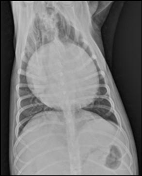
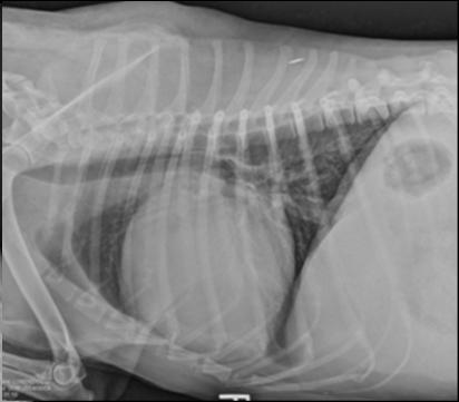
3 What s wrong with these images? Technique: Patient Positioning 3


4 Technique: Patient Positioning Cat in a Box Radiograph Trick Technique: Positioning Recumbency Problem Profound effect on cardiopulmonary physiology Results in differences in the appearance of R vs L and DV vs VD Results in reduced aeration in down lung Silhouette sign Exacerbated by sedation, anesthesia, and patient size Solution Take on full inspiration Minimize sedation PPV when anesthetized Take orthogonal views Minimal 3 views R and L laterals VD +/ DV 4
5 Technique: Aeration Technique: Aeration Technique: Patient Movement 5
6 Importance of multiple views Importance of multiple views Principles of Interpretation Knowledge of normal anatomy Age, breed/conformation, species, cardiac cycle Understanding of radiographic signs of pathology Ex. Heart failure vs. Pulmonary hypertension Develop a systematic approach Is the study of diagnostic quality? 6


7 What is normal? <3 ICS Avg Dog <3.5 Small Breeds < 2.5 Deep Chested Cats <70% height of chest; < 2 3 ICS Greatest horizontal dimension should be <2/3 chest dimension at that location Cats <50% width of chest Vertebral Heart Scale Normal Anatomy: Cat 7
 Long/oval heart Vertical position in thorax")
 Shorter/rounder heart Increased sternal contact(mimics RHE) Rounded LV/RV borders Apex to")
8 Normal Anatomy: Dog Normal Anatomy: Dog Breed Variations Thorax Type Lateral View D/V View Wide/shallow Dachshund, Shi Tzu, Boston Terrier, Bulldog Deep/narrow Greyhound, Doberman, Whippet Intermediate German Shepherd, Lab, Golden Shorter/rounder heart Large inclination to the spine Long contact with sternum (mimics RHE) Long/oval heart Vertical position in thorax almost perpendicular to spine Ovoid or lop sided eggshaped Rounded RV/LV borders Apex usually well to the left of the spine Circular silhouette due to upright position Apex close to median plan Similar to lateral Apex is usually slightly to left of spine Wide/Shallow (Barrel) Shorter/rounder heart Increased sternal contact(mimics RHE) Rounded LV/RV borders Apex to left of sternum 8
9 Wide/Shallow Yorkie Chest Malformation Yorkie Doberman Upright cardiac silhouette Deep/Narrow Greyhound Rounded silhouette Apex on midline Borzoi Elongated cardiac silhouette 9


10 Intermediate Dogs Increased sternal contact mimicking rightsided cardiomegaly Cardiac size appears normal Apex displaced into left hemithorax Cardiac Cycle Systole Ventricles contracted/smaller Atria dilated Diastole Entire heart more rounded Atrial/auricular bulges less prominent Age 10
11 Obesity Principles of Interpretation Consider false positive findings Right heart enlargement Dorso caudal rotation of apex in left lateral view Breed variation Alveolar disease Atelectasis Pneumothorax Skin folds Esophageal dilation Sedation Pleural effusion Pleural thickening Cardiomegaly Pericardial effusion PPDH Principles of Interpretation Consider false negative findings Assessing dynamic problem with static images Tracheal collapse / airway disease Hiatal hernia Disclose a hidden dynamic lesion Vary posture or position Inspiration vs expiration Other imaging modality 11
12 Clinical Cases: Sometimes there is little need for interpretation but more often than not it s not that obvious Clinical Cases Degenerative valve disease Hypertrophic cardiomyopathy Dilated cardiomyopathy Upper and lower airway disease Pulmonary hypertension Pleural effusion Pericardial effusion Pneumothorax Pulmonary masses/nodules 12
13 13
14 14
15 15
16 Heart failure or not? Is heart big or does technique/conformation give artifactual appearance of enlargement? Are pulmonary veins dilated? Is infiltrate consistent with CHF? Is there a murmur/gallop and where? If small dog and no loud murmur on left, unlikely CHF If murmur louder on R side, think PHT Is the heart rate fast? Unless hypothermic or Cocker/Schnauzer Is NT probnp elevated? Is there clinical/radiographic response to furosemide? 16
17 Tracheal Plasmacytoma 17
18 18

19 2 wk Recheck 19
20 20
21 21
22 22
23 23


24 Comments / Questions Contact Information: kacie.schmitt@cvcavets.com info@cvcavets.com 24
Concepts in Small Animal Thoracic Radiology Thoracic Radiology
 Concepts in Small Animal Thoracic Radiology + Radiology of the Pleural Space VMB 960 2/21/2011 Optimizing Image Quality Inherent subject contrast Thorax has high inherent subject contrast c/f abdomen Primarily
Corso Base di Cardiologia Unisvet 2012
 Principi di Radiologia del torace Dr Luca Ferasin DVM PhD CertVC PGCert(HE) DipECVIM-CA (Cardiology) MRCVS European and RCVS Recognised Specialist in Veterinary Cardiology Introduction Thoracic radiography
Small Animal Teaching Hospital, Leahurst Campus, University of Liverpool, Chester High Road, Neston, Wirral, CH64 7TE
 Thoracic Imaging: taking and reading a great X-ray J. Fraser McConnell BVM&S CertSAM DVR DipECVDI MRCVS Small Animal Teaching Hospital, Leahurst Campus, University of Liverpool, Chester High Road, Neston,
Calvin 9 year old NM DLH. Dr. Norman Ackerman Memorial Radiography Case Challenge
 September 2014 Dr. Norman Ackerman served the University of Florida, College of Veterinary Medicine with distinction as Professor of Radiology from 1979 to 1994. A concerned teacher of veterinary students
Manage TB Dr. A. Chitrakumar Madras Medical College and RGGGH Institute of Thoracic Medicine, Chennai
 Manage TB Dr. A. Chitrakumar Madras Medical College and RGGGH Institute of Thoracic Medicine, Chennai Lecture 16 Radiology in diagnosis of Tuberculosis Session 01 So, welcome to the session Radiology in
Cardiology Diagnostics Radiographs, Echo, and Biomarkers Introduction Thoracic Radiographs Cardiac Sze Cardiac Shape
 Cardiology Diagnostics Radiographs, Echo, and Biomarkers John E Rush, DVM, DACVECC, DACVIM (Cardiology) Cummings School of Veterinary Medicine at Tufts University, North Grafton, MA, USA Introduction Respiratory
Objectives. What is a Chest X Ray? CXR Workshop. Definition (diagnostic tool/internal PE) Types. Cost
 Types. Cost") Objectives CAPA 2011 Christy Wilson, PA C Georgia Lung Associates Identify the radiographic landmarks on a chest radiograph Recognize identifiers of poor quality on the chest radiograph Outline an approach
Objectives CAPA 2011 Christy Wilson, PA C Georgia Lung Associates Identify the radiographic landmarks on a chest radiograph Recognize identifiers of poor quality on the chest radiograph Outline an approach
What s Your Diagnosis? Signalment: Species: Ferret, Mustela putorius furo Sex: Female Spayed Date of Birth: 03/01/02 History of Adrenal Disease
 What s Your Diagnosis? Signalment: Species: Ferret, Mustela putorius furo Sex: Female Spayed Date of Birth: 03/01/02 History of Adrenal Disease Presenting Complaint: Diarrhea; Acute Dyspnea. For a couple
Pulmonary Patterns & Correlated Pathology
 Pulmonary Patterns & Correlated Pathology Russell Tucker, DVM, DACVR Washington State University College of Veterinary Medicine Objective: correlate radiographic findings of common lung diseases to actual
NIGERIAN VETERINARY JOURNAL
 NIGERIAN VETERINARY JOURNAL ISSN 0331-3026 Nig. Vet. J., December 2015 Vol. 36 (4): 1283-1287. ORIGINAL ARTICLE Preliminary Assessment of Vertebral Heart Score of the Nigerian Mongrel Dog Olatunji-Akioye,
Diagnosis of heart failure in dogs with mitral valve disease
 Vet Times The website for the veterinary profession https://www.vettimes.co.uk Diagnosis of heart failure in dogs with mitral valve disease Author : PHILLIP SPEER Categories : Vets Date : March 31, 2014
Incidentally-detected heart murmurs in dogs and cats: executive summary 2015
 Incidentally-detected heart murmurs in dogs and cats: executive summary 2015 E Côté, NJ Edwards, SJ Ettinger, VL Fuentes, KA MacDonald, BA Scansen, DD Sisson, JA Abbott.* An incidentally-detected heart
Chest X-ray (CXR) Interpretation Brent Burbridge, MD, FRCPC
 Interpretation Brent Burbridge, MD, FRCPC") Chest X-ray (CXR) Interpretation Brent Burbridge, MD, FRCPC An approach to reviewing a chest x-ray will create a foundation that will facilitate the detection of abnormalities. You should create your own
Chest X-ray (CXR) Interpretation Brent Burbridge, MD, FRCPC An approach to reviewing a chest x-ray will create a foundation that will facilitate the detection of abnormalities. You should create your own
Spinal radiographs are indicated for: THORACIC SPINE RADIOGRAPHY SMALL ANIMAL SPINAL RADIOGRAPHY SERIES. ImagIng EssEntIals
 PEER REVIEWED ImagIng EssEntIals SMLL NIML SPINL RDIOGRPHY SERIES THORCIC SPINE RDIOGRPHY Danielle Mauragis, CVT, and Clifford R. erry, DVM, Diplomate CVR Imaging Essentials provides comprehensive information
Thorax Review and Revitalize
 Outline Thorax Review and Revitalize Anatomy Sarah Tibbs, BVetMed, DACVR April 2016 I will try to share little Tibbits along the way (Tibbs Tidbits) Patterns Review like students Lungs Pleura Heart Other
Practical radiography in small animal practice III: cases with a heart murmur
 Practical radiography in small animal practice III: cases with a heart murmur Francisco Llabrés-Díaz, Davies Veterinary Specialists, Manor Farm Business Park, Higham Gobion, Hertfordshire SG5 3HR, England
10/17/2016. Nuts and Bolts of Thoracic Radiology. Objectives. Techniques
 Nuts and Bolts of Thoracic Radiology October 20, 2016 Carleen Risaliti Objectives Understand the basics of chest radiograph Develop a system for interpreting chest radiographs Correctly identify thoracic
FUNDAMENTALS OF CXR INTERPRETATION THE BASICS
 FUNDAMENTALS OF CXR INTERPRETATION THE BASICS PART I QUALITY ASSESSMENT 1 PATIENT-DEPENDENT FACTORS 3 REVIEW OF IMPORTANT ANATOMY 7 LUNGS AND PLEURA 11 DIAPHRAGMS 13 BONES AND SOFT TISSUES 14 A BRIEF LOOK
Radiological Anatomy of Thorax. Dr. Jamila Elmedany & Prof. Saeed Abuel Makarem
 Radiological Anatomy of Thorax Dr. Jamila Elmedany & Prof. Saeed Abuel Makarem Indications for Chest x - A chest x-ray may be used to diagnose and plan treatment for various conditions, including: Diseases/Fractures
NOW RACE Certified Earn CE Credits for Lunch and Learns
 In this Issue COVER Recommendations on the Management of Incidentally Detected Heart Murmurs Page 2 Page 5 New Cardiologist 4 Days a Week For Veterinarian Section New Handouts For Tech Section Locations
Disclosure. Clinical Chest Radiography Interpretation Part I
 Clinical Chest Radiography Interpretation Part I Anthony M. Angelow, PhD(c), MSN, ACNPC, AGACNP-BC, CEN Associate Lecturer, Fitzgerald Health Education Associates Clinical practice Division of Trauma Surgery
DIAGNOSING HEART FAILURE IN DOGS
 Vet Times The website for the veterinary profession https://www.vettimes.co.uk DIAGNOSING HEART FAILURE IN DOGS Author : Mike Martin Categories : Vets Date : November 7, 2011 Mike Martin offers advice
4/16/2017. Learning Objectives. Interpretation of the Chest Radiograph. Components. Production of the Radiograph. Density & Appearance
 Interpretation of the Arthur Jones, EdD, RRT Learning Objectives Identify technical defects in chest radiographs Identify common radiographic abnormalities This Presentation is Approved for 1 CRCE Credit
The Thorax Excluding the Heart and Pulmonary Patterns
 The Thorax Excluding the Heart and Pulmonary Patterns Lisa G. Britt, DVM, MS, Diplomate American College of Veterinary Radiology, Clinical Assistant Professor @ University of Missouri s College of Veterinary
8/14/2017. Objective: correlate radiographic findings of common lung diseases to actual lung pathologic features
 What is that lung disease? Pulmonary Patterns & Correlated Pathology Dr. Russell Tucker, DACVR Objective: correlate radiographic findings of common lung diseases to actual lung pathologic features Improved
Cardiomyopathy and Less Common Canine Heart Disease
 Cardiomyopathy and Less Common Canine Heart Disease John E. Rush, DVM, MS, DACVIM (Cardiology), ACVECC Cummings School of Veterinary Medicine at Tufts University Dilated cardiomyopathy (DCM) is the second
Murmur diagnosis in cats. Your pet has a murmur! Meg Sleeper VMD, DACVIM (cardiology) Gainesville, FL. Reasons to work up the murmur in a cat
 Gainesville, FL. Reasons to work up the murmur in a cat") Murmur diagnosis in cats Your pet has a murmur! Meg Sleeper VMD, DACVIM (cardiology) Gainesville, FL Heart disease diagnosis in cats and kittens in general is challenging because: Approximately ½ of systolic
Murmur diagnosis in cats Your pet has a murmur! Meg Sleeper VMD, DACVIM (cardiology) Gainesville, FL Heart disease diagnosis in cats and kittens in general is challenging because: Approximately ½ of systolic
1/13/2014. Proper Radiographs. Proper Radiographs. A Review of Pulmonary Patterns
 Live Webinar A Review of Pulmonary Patterns Sofija R. Liles, DVM, DACVR Proper Radiographs Which views? One lateral plus ventrodorsal (at least) Left lateral is best for thorax Three views for full metastatic
Proceedings of the World Small Animal Veterinary Association Sydney, Australia 2007
 Proceedings of the World Small Animal Veterinary Association Sydney, Australia 2007 Hosted by: Australian Small Animal Veterinary Association (ASAVA) Australian Small Animal Veterinary Association (ASAVA)
Instructions. Print out all three tabs of the guide to be used as a reference.
 Instructions For the Learner 1) The purpose of the observation checklist is to help evaluate the skills obtained from the course. It is also a reference and reminder for what was learned in this course.
Disclosures. Cardiac Ultrasound. Introductory Case. 80 y/o male Syncope at home Emesis x 3 in ambulance Looks sick. No pain.
 Disclosures Cardiac Ultrasound Justin A Davis, MD MPH RDMS Subchief for Emergency Ultrasound Kaiser Permanente East Bay Medical Center I have nothing to disclose. Introductory Case HR 118 BP 65/43 RR 27
Bony Thorax. Anatomy and Procedures of the Bony Thorax Edited by M. Rhodes
 Bony Thorax Anatomy and Procedures of the Bony Thorax 10-526-191 Edited by M. Rhodes Anatomy Review Bony Thorax Formed by Sternum 12 pairs of ribs 12 thoracic vertebrae Conical in shape Narrow at top Posterior
Cardiology made easy. Dr. Markus Killich DipACVIM (Cardiology) DipECVIM-CA (Cardiology)
 DipECVIM-CA (Cardiology)") Cardiology made easy Dr. Markus Killich DipACVIM (Cardiology) DipECVIM-CA (Cardiology) www.kardiokonsult.de questions Does a patient have heart disease? What kind of heart disease does it have? What is
Cardiology made easy Dr. Markus Killich DipACVIM (Cardiology) DipECVIM-CA (Cardiology) www.kardiokonsult.de questions Does a patient have heart disease? What kind of heart disease does it have? What is
Atrioventricular Valve Dysplasia
 Atrioventricular Valve Dysplasia How does the heart work? The heart is the organ responsible for pumping blood to and from all tissues of the body. The heart is divided into right and left sides. The job
Techniques of examination of the thorax and lungs. Dr. Szathmári Miklós Semmelweis University First Department of Medicine 24. Sept
 Techniques of examination of the thorax and lungs Dr. Szathmári Miklós Semmelweis University First Department of Medicine 24. Sept. 2013. Inspection of the thorax Observe: the shape of chest Deformities
Brachycephalics: It is More Than Just a Short Nose
 OCTOBER 2018 Brachycephalics: It is More Than Just a Short Nose ELIZABETH ROZANSKI, DVM, DACVIM (SA-IM), DACVECC TUFTS UNIVERSITY, NORTH GRAFTON, MA Respiratory diseases as well as respiratory distress
Surface anatomy of Cardiovascular system
 Surface anatomy of Cardiovascular system Prof. Abdulameer Al-Nuaimi E-mail: a.al-nuaimi@sheffield.ac.uk E. mail: abdulameerh@yahoo.com The lines cover the front, side, and back of the thorax Midsternal
Chest X-ray Interpretation
 Chest X-ray Interpretation Introduction Routinely obtained Pulmonary specialist consultation Inherent physical exam limitations Chest x-ray limitations Physical exam and chest x-ray provide compliment
Practical Echocardiography: ECHOES in the REAL WORLD Know When to Hold Em and When to Fold Em
 Practical Echocardiography: ECHOES in the REAL WORLD Know When to Hold Em and When to Fold Em Introduction The use of ultrasound in private veterinary practice is continuing to grow. The popularity of
Undergraduate Teaching
 Prof. James F Meaney Undergraduate Teaching Chest X-Ray Understanding the normal anatomical by reference to cross sectional imaging Radiology? It s FUN! Cryptic puzzle Sudoku (Minecraft?) It s completely
MITRAL VALVE DISEASE IN CAVALIER KING CHARLES SPANIELS. Carroll Loyer, DVM, DACVIM
 MITRAL VALVE DISEASE IN CAVALIER KING CHARLES SPANIELS Carroll Loyer, DVM, DACVIM CAVIEPALOOZA!! MITRAL INSUFFICIENCY mitral regurgitation (MR) chronic degenerative mitral valve disease (CVD) myxomatous
Chest XRay interpretation INTERPRETATIONS Identifications: Name & Date Technical evaluation Basic Interpretations
 Chest XRay interpretation INTERPRETATIONS Identifications: Name & Date Technical evaluation Basic Interpretations TECHNICAL EVALUATION 1. Projection: AP/PA view To differentiate between AP & PA films,
!"#$%&'%()'*+,-%&&.'+('*/%)+%,#+0' 12/.,'3%)+"4#%52.
'*+,-%&&.'+('*/%)+%,#+0' 12/.,'3%)+4#%52.") !"#$%&'%()'*+,-%&&.'+('*/%)+%,#+0' 12/.,'3%)+"4#%52.!"#$%&'()$*+&,--#&$.//,0'1232'!-#0'45 *6 '7849!!"#$%&'"(&)*+),$-.*/*01) 2$34/&1)*+)5"-.3.(") 6%.(3")*+)7*(08/$)9(.:"%;.&1)) )?
!"#$%&'%()'*+,-%&&.'+('*/%)+%,#+0' 12/.,'3%)+"4#%52.!"#$%&'()$*+&,--#&$.//,0'1232'!-#0'45 *6 '7849!!"#$%&'"(&)*+),$-.*/*01) 2$34/&1)*+)5"-.3.(") 6%.(3")*+)7*(08/$)9(.:"%;.&1)) )?
Spinal radiographs are indicated for: CerviCal Spine radiography. Small animal Spinal RadiogRaphy SeRieS. ImagIng EssEnTIals
 ImagIng EssEnTIals Peer reviewed Small animal Spinal RadiogRaphy SeRieS CerviCal Spine radiography Danielle Mauragis, CVT, and Clifford R. erry, DVM, Diplomate CVR Imaging Essentials provides comprehensive
Interactive Lecture. Lecture 7 - Interactive. Radiology of cardiorespiratory disease. Editing File. Done By. Color Coding Important Notes Extra
 Lecture 7 - Interactive 436 Teams Interactive Lecture Radiology of cardiorespiratory disease Done By Team Leaders: Khalid Alshehri Hanin Bashaikh Team Members: Ghaida Alsaeed Maha Alissa Nawwaf AlHarbi
Tracheal normal sound heard over trachea loud tubular quality high-pitched expiration equal to or slightly longer than inspiration
 = listening for sounds produced in the body over chest to ID normal & abnormal lung sounds all BS made by turbulent flow in the airways useful in making initial D & evaluating effects of R 4 characteristics
The Veteducation International Online Veterinary Conference 2011
 The Veteducation International Online Veterinary Conference 2011 Part of the Veteducation Live Online Web-Seminar Series The Artefacts of Life! With Dr Angela Hartman DVM Dipl. ACVR Massey University New
Chest X rays and Case Studies. No disclosures. Outline 5/31/2018. Carlo Manalo, M.D. Department of Radiology Loma Linda University Children s Hospital
 Chest X rays and Case Studies Carlo Manalo, M.D. Department of Radiology Loma Linda University Children s Hospital No disclosures. Outline Importance of history Densities delineated on radiography An approach
Don t Panic! Dr. Karau s Guide to Respiratory Emergencies November 4, 2018
 Don t Panic! Dr. Karau s Guide to Respiratory Emergencies November 4, 2018 Objectives Oxygen delivery methods Emergent diagnostic tests Differentiating between upper and lower respiratory disease Respiratory
Atrioventricular Valve Endocardiosis Basics
 Atrioventricular Valve Endocardiosis Basics OVERVIEW Atrioventricular valve refers to the heart valves between the top chamber (known as the atrium ) and the bottom chamber (known as the ventricle ) of
Interpreting thoracic x-ray of the supine immobile patient: Syllabus
 Interpreting thoracic x-ray of the supine immobile patient: Syllabus Johannes Godt Dep. of Radiology and Nuclear Medicine Oslo University Hospital Ullevål NORDTER 2017, Helsinki Content - Why bedside chest
Eun-Young Kang, M.D., Jae Wook Lee, M.D., Ji Yung Choo, M.D., Hwan Seok Yong, M.D., Ki Yeol Lee, M.D., Yu-Whan Oh, M.D.
 Eun-Young Kang, M.D., Jae Wook Lee, M.D., Ji Yung Choo, M.D., Hwan Seok Yong, M.D., Ki Yeol Lee, M.D., Yu-Whan Oh, M.D. Department of Radiology, Korea University Guro Hospital, College of Medicine, Korea
Chapter 2 Cardiac Interpretation of Pediatric Chest X-Ray
 Chapter 2 Cardiac Interpretation of Pediatric Chest X-Ray Ra-id Abdulla and Douglas M. Luxenberg Key Facts The cardiac silhouette occupies 50 55% of the chest width on an anterior posterior chest X-ray
Proudly Presents: DIAGNOSTIC IMAGING
 Chicago Veterinary Medical Association Shaping the Future of Veterinary Medicine - Promoting the Human-Animal Bond Proudly Presents: DIAGNOSTIC IMAGING With: ROBERT O BRIEN DVM, MS, DACVR April 9, 2014
X-Rays. Kunal D Patel Research Fellow IMM
 X-Rays Kunal D Patel Research Fellow IMM The 12-Steps } 1: Name 2: Date 3: Old films 4: What type of view(s) 5: Penetration } Pre-read 6: Inspiration 7: Rotation Quality Control 8: Angulation 9: Soft tissues
Approach to a new murmur in a cat Terri DeFrancesco, DVM, DACVIM (Cardiology), DACVECC NCSU College of Veterinary Medicine, Raleigh, NC
, DACVECC NCSU College of Veterinary Medicine, Raleigh, NC") Approach to a new murmur in a cat Terri DeFrancesco, DVM, DACVIM (Cardiology), DACVECC NCSU College of Veterinary Medicine, Raleigh, NC One s approach to the diagnostic work up of an asymptomatic cat with
Approach to a new murmur in a cat Terri DeFrancesco, DVM, DACVIM (Cardiology), DACVECC NCSU College of Veterinary Medicine, Raleigh, NC One s approach to the diagnostic work up of an asymptomatic cat with
Proceedings of the 34th World Small Animal Veterinary Congress WSAVA 2009
 www.ivis.org Proceedings of the 34th World Small Animal Veterinary Congress WSAVA 2009 São Paulo, Brazil - 2009 Next WSAVA Congress : Reprinted in IVIS with the permission of the Congress Organizers MANAGEMENT
Shedding Light on Neonatal X-rays. Objectives. Indications for X-Rays 5/14/2018
 Shedding Light on Neonatal X-rays Barbara C. Mordue, MSN, NNP-BC Neonatal Nurse Practitioner LLUH Children s Hospital, NICU Objectives Utilize a systematic approach to neonatal x-ray interpretation Identify
Simplifying mitral valve disease diagnostics
 DIAGNOSIS Simplifying mitral valve disease diagnostics Nuala Summerfield Myxomatous mitral valve disease (MMVD) in dogs is a slowly progressive disease. Until recently, focus was aimed at the symptomatic
Chest Ultrasound: Pneumothorax
 WINFOCUS BASIC ECHO (WBE) Chest Ultrasound: Pneumothorax Mark Hamlin, MD, MS Associate Professor of Anesthesiology and Surgery University of Vermont College of Medicine Co-Director of Surgical Critical
Cardiac Examination. Pediatrics Clinical Examination
 Pediatrics Clinical Examination Symptoms of Cardiovascular Affection: Cardiac Examination 1. Perinatal history: Maternal DM, cyanosis, respiratory distress 2. Symptoms of lung congestion: Poor interrupted
Chest Radiology Interpretation: Findings of Tuberculosis
 Chest Radiology Interpretation: Findings of Tuberculosis Get out your laptops, smart phones or other devices pollev.com/chestradiology Case #1 1 Plombage Pneumonia Cancer 2 Reading the TB CXR Be systematic!
About the Cardiac Education Group (CEG) The CEG Mission. The CEG promotes and facilitates:
 The CEG Mission. The CEG promotes and facilitates:") About the Cardiac Education Group (CEG) The Cardiac Education Group is a group of board certified veterinary cardiologists from both academia and private practice that offers independent recommendations
About the Cardiac Education Group (CEG) The Cardiac Education Group is a group of board certified veterinary cardiologists from both academia and private practice that offers independent recommendations
B-I-2 CARDIAC AND VASCULAR RADIOLOGY
 (YEARS 1 3) CURRICULUM FOR RADIOLOGY 13 B-I-2 CARDIAC AND VASCULAR RADIOLOGY KNOWLEDGE To describe the normal anatomy of the heart and vessels including the lymphatic system as demonstrated by radiographs,
BIOE221. Session 5. Examination of Thorax- Respiratory system. Bioscience Department. Endeavour College of Natural Health endeavour.edu.
 BIOE221 Session 5 Examination of Thorax- Respiratory system Bioscience Department Session Objectives Understand the structure of the thorax and the organs contained in this cavity Understand the importance
Instructions. Print out all three tabs of the guide to be used as a reference.
 Instructions For the Learner 1) The purpose of the observation checklist is to help evaluate the skills obtained from the course. It is also a reference and reminder for what was learned in this course.
A Practical Approach to Ultrasound Assessment of Respiratory Distress
 A Practical Approach to Ultrasound Assessment of Respiratory Distress Yanick Beaulieu, MD, FRCPC Director, Bedside Ultrasound Curriculum Division of Cardiology and Critical Care Hôpital du Sacré-Coeur
CHEST & ABDOMINAL X-RAYS MALIKA IBRAHIM CORE MEDICAL TRAINEE BLACKPOOL VICTORIA HOSPITAL DATA INTERPRETATION COURSE FEB 20, 2017
 CHEST & ABDOMINAL X-RAYS MALIKA IBRAHIM CORE MEDICAL TRAINEE BLACKPOOL VICTORIA HOSPITAL DATA INTERPRETATION COURSE FEB 20, 2017 1. Sample x-rays 2. Basic chest x-ray interpretation skills 3. Chest x-ray
Eight-year-old toy poodle. Cough for 6 months. No murmur. Top differentials?
 What IS CHF? Congestive Heart Failure What s New? Bill Saxon ACVECC, ACVIM Idexx Our definition pulmonary venous congestion and/or edema This is a radiographic diagnosis Echocardiography for determination
Septal Defects. How does the heart work?
 Septal Defects How does the heart work? The heart is the organ responsible for pumping blood to and from all tissues of the body. The heart is divided into right and left sides. The job of the right side
ISPUB.COM. Cardiac Dimensions Derived From Helical Ct: Correlation With Plain Film Radiography
 ISPUB.COM The Internet Journal of Radiology Volume 1 Number 1 Cardiac Dimensions Derived From Helical Ct: Correlation With Plain Film Radiography J Miller, A Singer, C Hinrichs, S Contractor, S Doddakashi
Diagnosis is complicated
 Peer reviewed Cardiac Blood Tests in Cats Another Tool for Detection of Heart Disease Mark A. Oyama, DVM, Diplomate ACVIM (Cardiology) Detection of asymptomatic (occult) heart disease in cats is challenging.
CASE DISCUSSION. Dr JAYASREE VEERABOINA 2nd yr PG MS OBG
 CASE DISCUSSION Dr JAYASREE VEERABOINA 2nd yr PG MS OBG Normal Cardiovascular changes in Pregnancy CARDIAC OUTPUT 5 th wk -- starts 12 wks -- 30-35% 30-32 wks -- 40% During labour -- 50% After delivery
New Location! New Columbia, MD Location! Is it a seizure or syncope? Tips for differentiating. In this Issue. Opening in September 2016
 In this Issue COVER Is it seizure or syncope? Tips for differentiating. New Location! New Columbia, MD Location! Opening in September 2016 10000 Old Columbia Road Columbia, MD 21046 Page 2 Fairfax, VA
PLEURAE and PLEURAL RECESSES
 PLEURAE and PLEURAL RECESSES By Dr Farooq Aman Ullah Khan PMC 26 th April 2018 Introduction When sectioned transversely, it is apparent that the thoracic cavity is kidney shaped: a transversely ovoid space
Surgical Diseases of the Upper Airways. Michael Huber DVM, MS Diplomate American College of Veterinary Surgeons
 Surgical Diseases of the Upper Airways Michael Huber DVM, MS Diplomate American College of Veterinary Surgeons Surgical Diseases of the Upper Airways General Considerations Brachycephalic Syndrome Laryngeal
Pulmonary Embolism. Thoracic radiologist Helena Lauri
 Pulmonary Embolism Thoracic radiologist Helena Lauri 8.5.2017 Statistics 1-2 out of 1000 adults annually are diagnosed with deep vein thrombosis (DVT) and/or pulmonary embolism (PE) About half of patients
Close window to return to IVIS. in collaborazione con RICHIESTO ACCREDITAMENTO. organizzato da certificata ISO 9001:2000
 in collaborazione con Close window to return to IVIS RICHIESTO ACCREDITAMENTO SOCIETÀ CULTURALE ITALIANA VETERINARI PER ANIMALI DA COMPAGNIA SOCIETÀ FEDERATA ANMVI organizzato da certificata ISO 9001:2000
LUNG PATTERNS IN THE DOG NORMAL AND PATHOLOGICAL
 TRADITION AND MODERNITY IN VETERINARY MEDICINE, 2018, vol. 3, No 1(4): 7 14 LUNG PATTERNS IN THE DOG NORMAL AND PATHOLOGICAL Kalin Spasov 1, Michaela Kunovska 2, Dimo Dimov 3 1 University of Forestry,
d) Always ensure patient comfort. Be considerate and warm the diaphragm of your stethoscope with your hand before auscultation.
 Always ensure patient comfort. Be considerate and warm the diaphragm of your stethoscope with your hand before auscultation.") Auscultation Auscultation is perhaps the most important and effective clinical technique you will ever learn for evaluating a patient s respiratory function. Before you begin, there are certain things
Auscultation Auscultation is perhaps the most important and effective clinical technique you will ever learn for evaluating a patient s respiratory function. Before you begin, there are certain things
College of Veterinary Medicine Policies and Procedures
 College of Veterinary Medicine Policies and Procedures Subject: Scheduling. Requests and After-Hours Imaging Section: Diagnostic Imaging Number: CVM 6.10.02 Pages: 8 Date: 2012 Replaces Policy Dated: 1995
Constrictive Pericarditis Pitfalls in MR Diagnosis Cylen Javidan-Nejad Associate Professor Mallinckrodt Institute of Radiology Washington University
 Constrictive Pericarditis Pitfalls in MR Diagnosis Cylen Javidan-Nejad Associate Professor Mallinckrodt Institute of Radiology Washington University in St. Louis Goal o To review the imaging criteria of
2. The heart sounds are produced by a summed series of mechanical events, as follows:
 Heart Sounds. Phonocardiography 1 Objectives 1. Phonocardiography - Definition 2. What produces the heart sounds 3. Where to listen for the heart sounds 4. How to record a phonocardiogram 5. Normal heart
European Veterinary Dental College
 European Veterinary Dental College EVDC Training Support Document Preparation of Radiograph Sets (Cat and Dog) Document version : evdc-tsd-radiograph_positioning_(dog_and_cat)-20120121.docx page 1 of 13
Radiological conference. Left upper lobe collapse. Citation Hong Kong Practitioner, 1998, v. 20 n. 9, p
 Title Radiological conference. Left upper lobe collapse Author(s) Wong, LLS; Peh, WCG Citation Hong Kong Practitioner, 1998, v. 20 n. 9, p. 513-517 Issued Date 1998 URL http://hdl.handle.net/10722/44672
Radiology of the respiratory/cardiac diseases (part 2)
") Cardiology Cycle - Lecture 6 436 Teams Radiology of the respiratory/cardiac diseases (part 2) Objectives Done By Team Leaders: Khalid Alshehri Hanin Bashaikh Team Members: Leena Alwakeel Aroob Alhuthail
Cardiology Cycle - Lecture 6 436 Teams Radiology of the respiratory/cardiac diseases (part 2) Objectives Done By Team Leaders: Khalid Alshehri Hanin Bashaikh Team Members: Leena Alwakeel Aroob Alhuthail
Proceedings of the Southern European Veterinary Conference and Congreso Nacional de AVEPA
 www.ivis.org Proceedings of the Southern European Veterinary Conference and Congreso Nacional de AVEPA Oct. 18-21, 2012 - Barcelona, Spain Next Conference: Oct. 17-19, 2013 - Barcelona, Spain Reprinted
UERMMMC Department of Radiology. Basic Chest Radiology
 UERMMMC Department of Radiology Basic Chest Radiology PHYSICS DENSITIES BONE SOFT TISSUES WATER FAT AIR TELEROENTGENOGRAM Criteria for an Ideal Chest Radiograph 1. Upright 2. Posteroanterior View 3. Full
Auscultation of the lung
 Auscultation of the lung Auscultation of the lung by the stethoscope. *Compositions of the stethoscope: 1-chest piece 2-Ear piece 3-Rubber tubs *Auscultation area of the lung(triangle of auscultation).
Chest X-Ray: the essentials
 Chest X-Ray: the essentials Poster No.: C-1264 Congress: ECR 2017 Type: Educational Exhibit Authors: J. J. Delgado Moraleda, A. ALEGRE DELGADO, R. M. Piqueras Olmeda, E. Chacón Avilés, J. F. Melo Villamarín,
Radiology of the respiratory disease
 Radiology of the respiratory disease [ Color index: Important Notes Extra ] [ Editing file Feedback Share your notes Shared notes ] Resources: - 435 Slides - 434 Team - 435 Notes Done by: - Mai Alageel
FUNDAMENTAL ISSUES. Clinical Approach to Respiratory Disease in the Dog and the Cat
 Clinical Approach to Respiratory Disease in the Dog and the Cat Philip Padrid DVM Medical Director VCA Vet Care Specialty Referral Center Regional Medical Director VCA SW Region Associate Professor of
Echocardiography as a diagnostic and management tool in medical emergencies
 Echocardiography as a diagnostic and management tool in medical emergencies Frank van der Heusen MD Department of Anesthesia and perioperative Care UCSF Medical Center Objective of this presentation Indications
Imaging of Thoracic Trauma: Tips and Traps. Arun C. Nachiappan, MD Associate Professor of Clinical Radiology University of Pennsylvania
 Imaging of Thoracic Trauma: Tips and Traps Arun C. Nachiappan, MD Associate Professor of Clinical Radiology University of Pennsylvania None Disclosures Objectives Describe blunt and penetrating traumatic
X-Rays. Prepared by Prof.Dr. Magda Hassab Allah Assist.lecturer Marwa Al Hady
 X-Rays Prepared by Prof.Dr. Magda Hassab Allah Assist.lecturer Marwa Al Hady CHEST X-RAYS Normal Chest X-ray Comments on chest X ray includes examination of 1- Bony cage (ribs,clavicles &vertebral column
Initially for cardiac echo Subsequent studies non-cardiac applications
 No disclosures But Heavy accent Initially for cardiac echo Subsequent studies non-cardiac applications 1973: Goldberg et al in JCUS 30 mediastinal masses in pts. age 1-84 yrs. 1977: Kangarloo et al in
Persistent right aortic arch (PRAA) is the most common
 is the most common") J Vet Intern Med 2004;18:510 514 Tracheal Signs and Associated Vascular Anomalies in Dogs with Persistent Right Aortic Arch James W. Buchanan Medical records of 55 dogs with 1 or more vascular rings around
J Vet Intern Med 2004;18:510 514 Tracheal Signs and Associated Vascular Anomalies in Dogs with Persistent Right Aortic Arch James W. Buchanan Medical records of 55 dogs with 1 or more vascular rings around
Anesthesia Monitoring
 Anesthesia Monitoring Horatiu V. Vinerean, DVM, DACLAM Anesthesia Monitoring Anesthesia can be divided into four progressive phases. The signs relating to a certain phase are based upon the presence or
Country Health SA Medical Imaging
 Country Health SA Medical Imaging REMOTE OPERATORS POSITIONING GUIDE Contents Image Evaluation Page 4 Positioning Guides Section 1 - THORAX 1.1 Chest Page 5 1.2 Bedside Chest Page 7 1.3 Ribs Page 8 Section