Constrictive Pericarditis Pitfalls in MR Diagnosis Cylen Javidan-Nejad Associate Professor Mallinckrodt Institute of Radiology Washington University
|
|
|
- Tracy Shepherd
- 5 years ago
- Views:
Transcription

1 Constrictive Pericarditis Pitfalls in MR Diagnosis Cylen Javidan-Nejad Associate Professor Mallinckrodt Institute of Radiology Washington University in St. Louis
2 Goal o To review the imaging criteria of constrictive pericarditis o To discuss the diagnostic pitfalls & technical challenges o Tricks to improve diagnosis o Future directions o None Disclosures
3 Introduction Slide 1 o Definition: diastolic fxn impaired by thickening or fibrosis usually identified by echo most common indication for evaluation by MR o Clinical presentation confused w/ restrictive CMP, heart failure, & hepatic dis symptoms & signs of right heart failure (not d/t left heart dysfxn or valve dis) o Causes TB mc cause worldwide A series from Mayo Clinic examined 143 surgical patients idiopathic (49%) iatrogenic, including prior pericardiotomy (30%) radiation (11%) other (10%): viral pericarditis, conn tiss dis, uremia, neoplasm Glockner JF. Imaging of pericardial disease. Magn Reson Imaging Clin N Am 2003; (11):
4 MR Technique Slide 2 o Morphology T2 -Black blood imaging double inversion recovery Black blood imaging-double inversion recovery single shot Haste, T1 weighted : not routinely performed precontrast VIBE o Function Cine Balanced steady state free precession (bssfp), cine Turbo Flash - True FISP Tagged myocardium - not routinely performed o Enhancement 3D (VIBE, Timing needs to be optimized o Respiratory maneuvers Real time SSFP MR Signal Intensity of Pericardium: o o o double inversion recovery sequences Intermediate T2-weighted Lower than H2O SSFP Low signal


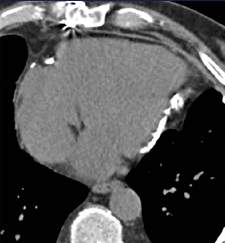
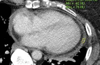
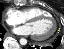
5 Slide 3 Diagnostic Hallmark o Pericardial thickening with or without calcifications in setting of suggestive clinical signs & symptoms Thickening difficult to detect by echocardiography Thickness abnormal when > 3mm ; only 1/3 Ca ++ CT > MR for identifying Ca ++ MR > CT :distinguishing tiny effusion vs smooth thickening
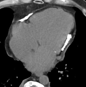
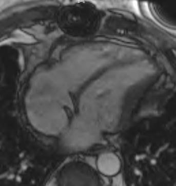
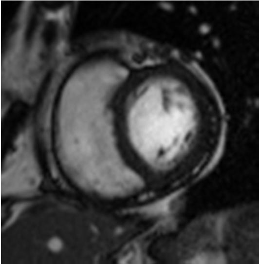
6 Slide 4


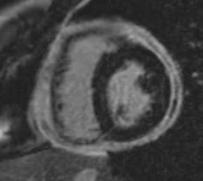
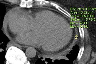
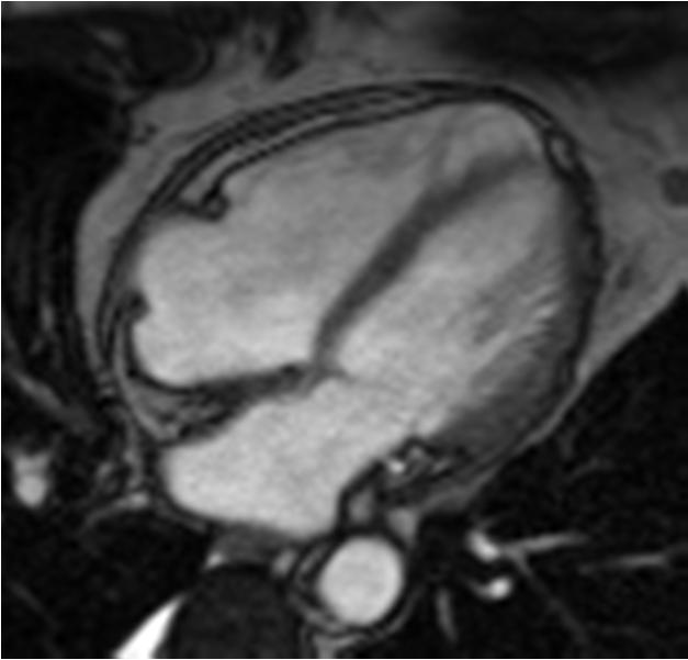
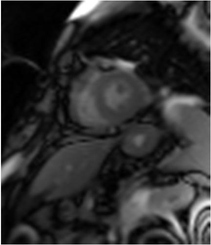
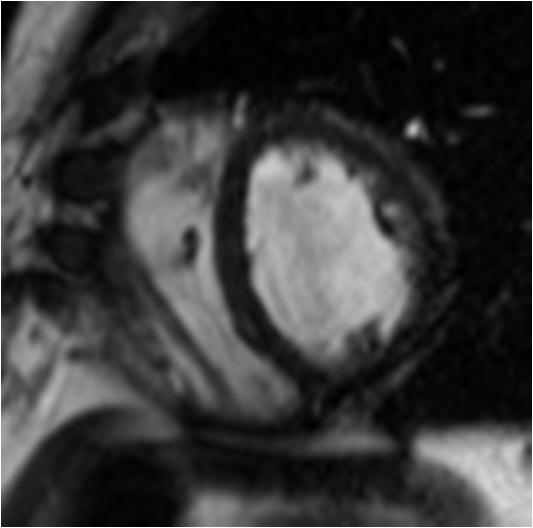
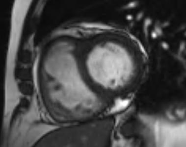
7 MR Pericardial Thickness Slide 5 o Diagnostic Pitfalls Detecting calcification Not every thickening causes compressive physiology SSFP black outline difficult to exclude smooth thickening Susceptibilty artifact simulates calcium or thickening


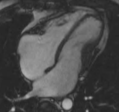
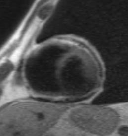
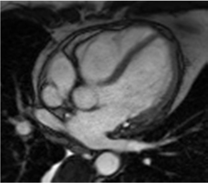
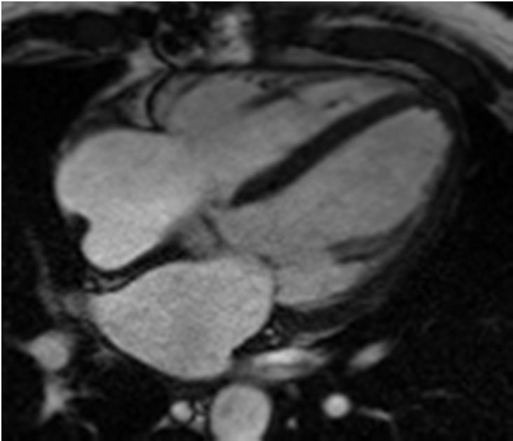
8 Slide 7 Morphologic Findings of Constriction o Elongated tubular or Bullet-shaped ventricles not always present Pitfall: restrictive CMP o Straightening of septum Pitfall: pulm hypertension o Dilated atria & IVC o * Pitfall: many causes of Right-sided heart failure


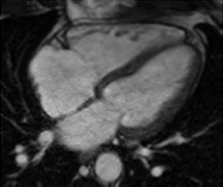
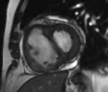
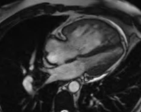
9 Slide 8 Morphologic Findings of Constriction o Contour deformity RV free wall or atrioventricular groove Rarely RVOT subpulmonic stenosis absent in malignant pericardial thickening w/ constrictive physiology o Pericardial adhesions Pitfall: can be present w/o constrictive physiology hard to detect


10 Slide 9 Morphologic Findings of Constriction o Contour deformity Usually RV free wall, atrioventricular groove, or rarely RVOT absent in malignant pericardial thickening w/ constrictive physiology o Poor relaxation Rarely RVOT subpulmonic stenosis


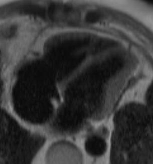
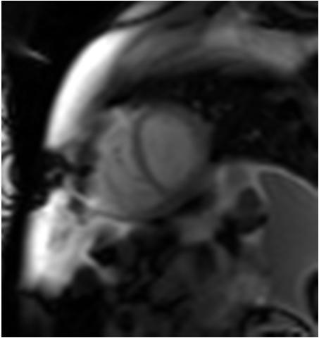
11 Slide 10 o Cardiac tamponade Caused by: Large pleural effusion Blood Rarely air Important Mimiker Early diastolic notching Atrium or ventricle hard to detect Treatment pericardial window / drain


12 Slide 11 Morphologic Findings of Constriction o Dilated IVC & hepatic veins o Nutmeg liver Passive congestion o Ascites & anasarca o Pleural effusions o Cardiac cirrhosis o All above also Restrictive CMP Tricuspid regurgitation Pulm HTN RV systolic failure


13 Effusive Constrictive Pericarditis Slide 12 o Effusive constrictive pericarditis. Acute to subacute pericarditis fat stranding Small or large pericardial effusion Visceral pericardium compresses the heart No significant relief with pericardiotomy needs ectomy Can be reversible Caused by infection, lupus, radiation, collagen vascular dis


14 Slide 13 Constrictive Pericardial Metastases o Can occur : sarcoma, melanoma, & breast ca Nodular thickening protrude into epicardial fat No fat stranding Enhance RV & /or RA compression
15 Enhancement-Technique Slide 14 o Contrast-enhanced MR clearly separates fluid from pericardial thickening


16 Enhancement-Technique Slide 15 o Enhancement can be subtle when chronic & independent of the severity of constriction


 after 10-15 sec")
17 Slide 16 Enhancement-Technique o Enhancement increases with time scan with 10 min delay (more important for CT) after sec After 90 sec After 5 min


18 Slide 17 Functional Analysis o Poor relaxation on diastole Diagnostic dilemma: restrictive CMP Also dilemma with echocardiography Myocardial tagging Systole: slippage between the myocardium & pericardium Absent when pericardial adhesions exist


19 Mimicker Cardiac Tamponade Slide 18 o Diastolic notching & RV or RA flattening


20 Septal Bouncing Slide 19 o Septal bouncing or paradoxical septal motion Bowing of septum towards LV Both 2 chamber short axis & 4 chamber views


21 Ventricular Interdependence Slide 21 o Absent in restrictive CMP o RV free wall cannot expand o In early diastole RV pressure exceeds LV press o septum bounces towards the LV


22 Slide 22 Pitfall: Septal Bouncing w/o Constriction o Pressure overloads Pulmonary Hypertension End systolic bounce o Volume overload state Diastolic bounce Pulmonic regurgitation Aortic regurgitation Pulmonary Hypertensions Cardiac Amyloidosis
23
24
25

26 Respiratory Manuever Slide 23 o Ventricular interdependence becomes more pronounced with inspiration o Inflow to the RA & RV increases with inspiration Septum flattens with inspiration

27 Respiratory Manuever Slide 23 o Ventricular interdependence becomes more pronounced with inspiration o Inflow to the RA & RV increases with inspiration Septum flattens with inspiration


28 Respiratory Manuever Slide 24 Courtesy of Travis L. Henry, MD


29 Respiratory Manuever Slide 24 Courtesy of Travis L. Henry, MD
30 Take home points Slide 23 o Not every morphologic change is consistent with constriction diagnosis made in conjunction with other imaging modalities and clinical findings o Pericardial enhancement increases with time Scan 10 minutes post injection o Septal bounce occurs with BBB, pulm HTN, RV volume overload, e.g. pulmonary regurgitation o Inspiration makes septal bouncing more pronounced (due to ventricular interdependance) Real-time imaging
31 Thank you o javidanc@mir.wustl.edu
Adel Hasanin Ahmed 1
 Adel Hasanin Ahmed 1 PERICARDIAL DISEASE The pericardial effusion ends anteriorly to the descending aorta and is best visualised in the PLAX. PSAX is actually very useful sometimes for looking at posterior
Cardiac MRI: Clinical Application to Disease
 Cardiac MRI: Clinical Application to Disease Jessi Smith, MD Cardiothoracic imaging, Indiana University Slides courtesy of Stacy Rissing, MD Outline Imaging planes Disease findings Pulse sequences used
Cardiac MRI: Clinical Application to Disease
 Cardiac MRI: Clinical Application to Disease Stacy Rissing, MD! Cardiothoracic imaging, Indiana University! Outline Imaging planes Disease findings Pulse sequences used for each indication Pathophysiology
Pericardial Diseases. Smonporn Boonyaratavej, MD. Division of Cardiology, Department of Medicine Chulalongkorn University
 Pericardial Diseases Smonporn Boonyaratavej, MD Division of Cardiology, Department of Medicine Chulalongkorn University Cardiac Center, King Chulalongkorn Memorial Hospital 21 AUGUST 2016 Pericardial
Cardiovascular manifestations of HIV
 Cardiovascular manifestations of HIV Prabhakar Rajiah, MBBS, MD, FRCR Associate Professor of Radiology Associate Director, Cardiac CT and MRI University of Texas Southwestern Medical Center, Dallas, USA
Pericardial disease. Se-Jung Yoon Cardiology division NHIS Ilsan hospital
 Pericardial disease Se-Jung Yoon Cardiology division NHIS Ilsan hospital Normal pericardial effusion Normal pericardium Normal pericardium Pericardial Layers: Visceral layer Parietal layer Fibrous pericardium
Constriction vs Restriction Case-based Discussion
 Mayo Clinic Department of Cardiovascular Diseases Mayo Clinic Echocardiography Review Course for Boards and Recertification Constriction vs Restriction Case-based Discussion Jae K. Oh, MD Samsung Professor
Can SCMR CMR protocol recommendations
 Can SCMR CMR protocol recommendations V1.3 - April 2009 CanSCMR CMR Protocol and SOP Recommendation 2009 (15 minutes) 2 Planning of LV fct. real time multiple axes Realtime 3 cine long axis 6 long axes
Constrictive pericarditis: Morphological, functional and haemodynamic evaluation
 Constrictive pericarditis: Morphological, functional and haemodynamic evaluation Poster No.: C-0743 Congress: ECR 2010 Type: Educational Exhibit Topic: Cardiac Authors: B. Graca, P. Donato, M. Ferreira,
Echocardiography Conference
 Echocardiography Conference David Stultz, MD Cardiology Fellow, PGY-6 September 20, 2005 Atrial Septal Aneurysm Bulging of Fossa Ovalis Associated commonly with Atrial septal defect or small perforations
Certificate in Clinician Performed Ultrasound (CCPU) Syllabus. Rapid Cardiac Echo (RCE)
 Syllabus. Rapid Cardiac Echo (RCE)") Certificate in Clinician Performed Ultrasound (CCPU) Syllabus Rapid Cardiac Echo (RCE) Purpose: Rapid Cardiac Echocardiography (RCE) This unit is designed to cover the theoretical and practical curriculum
Certificate in Clinician Performed Ultrasound (CCPU) Syllabus Rapid Cardiac Echo (RCE) Purpose: Rapid Cardiac Echocardiography (RCE) This unit is designed to cover the theoretical and practical curriculum
Echocardiography as a diagnostic and management tool in medical emergencies
 Echocardiography as a diagnostic and management tool in medical emergencies Frank van der Heusen MD Department of Anesthesia and perioperative Care UCSF Medical Center Objective of this presentation Indications
Pericardial Disease: Case Examples. Echo Fiesta 2017
 Pericardial Disease: Case Examples Echo Fiesta 2017 2014 2014 MFMER MFMER 3346252-1 slide-1 Objectives Have a systematic approach to evaluation of constriction 2014 MFMER 3346252-2 CASE 1 2013 MFMER 3248567-3
Outline. Echocardiographic Assessment of Pericardial Effusion/Tamponade: The Essentials
 Echocardiographic Assessment of Pericardial Effusion/Tamponade: The Essentials John R Schairer DO FACC Henry Ford Heart and Vascular Institute No Disclosures Outline Normal Anatomy and Physiology Pathophysiology
Imaging in Heart Failure: A Multimodality Approach. Thomas Ryan, MD
 Imaging in Heart Failure: A Multimodality Approach Thomas Ryan, MD Heart Failure HFrEF HFpEF EF50% Lifetime risk 20% Prevalence 6M Americans Societal costs - $30B 50% 5-year survival 1 Systolic
CMS Limitations Guide - Radiology Services
 CMS Limitations Guide - Radiology Services Starting October 1, 2015, CMS will update their existing medical necessity limitations on tests and procedures to correspond to ICD-10 codes. This limitations
THE PERICARDIUM: LOOKING OUTSIDE THE HEART
 THE PERICARDIUM: LOOKING OUTSIDE THE HEART DISCLOSURE Relevant relationships with commercial entities none Potential for conflicts of interest within this presentation none Steps taken to review and mitigate
Adult Echocardiography Examination Content Outline
 Adult Echocardiography Examination Content Outline (Outline Summary) # Domain Subdomain Percentage 1 2 3 4 5 Anatomy and Physiology Pathology Clinical Care and Safety Measurement Techniques, Maneuvers,
PERICARDIAL DIAESE. Kaijun Cui Associated professor Sichuan University
 PERICARDIAL DIAESE Kaijun Cui Associated professor Sichuan University CLASSIFICATION acute pericarditis pericardial effusion cardiac tamponade constrictive pericarditis congenitally absent pericardium
Cardiac ultrasound protocols
 Cardiac ultrasound protocols IDEXX Telemedicine Consultants Two-dimensional and M-mode imaging planes Right parasternal long axis four chamber Obtained from the right side Displays the relative proportions
Restrictive Cardiomyopathy
 ESC Congress 2011, Paris Imaging Unusual Causes of Cardiomyopathy Restrictive Cardiomyopathy Kazuaki Tanabe, MD, PhD Professor of Medicine Chair, Division of Cardiology Izumo, Japan I Have No Disclosures
cardiac imaging planes planning basic cardiac & aortic views for MR
 cardiac imaging planes planning basic cardiac & aortic views for MR Dianna M. E. Bardo, M. D. Assistant Professor of Radiology & Cardiovascular Medicine Director of Cardiac Imaging cardiac imaging planes
The Cardiovascular System Part I: Heart Outline of class lecture After studying part I of this chapter you should be able to:
 The Cardiovascular System Part I: Heart Outline of class lecture After studying part I of this chapter you should be able to: 1. Describe the functions of the heart 2. Describe the location of the heart,
An Uncommon Cardiac Etiology of Liver Cirrhosis, Recurrent Ascites, Atrial Fibrillation and Congestive Heart Failure
 Cronicon OPEN ACCESS EC CARDIOLOGY Case Report An Uncommon Cardiac Etiology of Liver Cirrhosis, Recurrent Ascites, Atrial Fibrillation and Congestive Heart Failure Montaser Y Ismail 1 *, Mohammed I Nassar
Constrictive/Restrictive Cardiomyopathies: Diagnosis and Management Update; Radiation Induced Heart Disease. Alexander (Sandy) Dick, MD
 Dick, MD") Constrictive/Restrictive Cardiomyopathies: Diagnosis and Management Update; Radiation Induced Heart Disease Alexander (Sandy) Dick, MD Outline Pericardial Constriction Diagnosis: Imaging, Hemodynamics
Constrictive/Restrictive Cardiomyopathies: Diagnosis and Management Update; Radiation Induced Heart Disease Alexander (Sandy) Dick, MD Outline Pericardial Constriction Diagnosis: Imaging, Hemodynamics
Review of Cardiac Imaging Modalities in the Renal Patient. George Youssef
 Review of Cardiac Imaging Modalities in the Renal Patient George Youssef ECHO Left ventricular hypertrophy (LVH) assessment Diastolic dysfunction Stress ECHO Cardiac CT angiography Echocardiography - positives
2/4/2011. Nathan Kerner, M.D.
 Nathan Kerner, M.D. Definition Elevated pressures - cut off usually >40 mmhg pulmonary artery systolic pressure (PASP) Usually associated with elevated pulmonary vascular resistance (PVR) measured in dynessec/cm
Images in Cardiovascular Medicine
 Images in Cardiovascular Medicine Constrictive Pericarditis Presenting as a Large Mediastinal Mass Causing Functional Tricuspid and Pulmonary Stenosis Niklas F. Ehl, MD; Simon Sündermann, MD; Reinhard
10/1/2016. Constrictive Pericarditis Unique Hemodynamics. What s New in Pericardial Disease? Case-based Discussion
 Mayo Clinic Department of Cardiovascular Diseases Mayo Clinic Echocardiography Review Course for Boards and Recertification What s New in Pericardial Disease? Case-based Discussion Jae K. Oh, MD Samsung
Department of Cardiac, Thoracic and Vascular Sciences University of Padua Cardiac Tamponade. Echocardiography in Diagnosis and Management
 Department of Cardiac, Thoracic and Vascular Sciences University of Padua Cardiac Tamponade. Echocardiography in Diagnosis and Management Luigi P. Badano, MD, FESC, FACC Declaration of interest **Dr. Badano
Cardiac MRI in ACHD What We. ACHD Patients
 Cardiac MRI in ACHD What We Have Learned to Apply to ACHD Patients Faris Al Mousily, MBChB, FAAC, FACC Consultant, Pediatric Cardiology, KFSH&RC/Jeddah Adjunct Faculty, Division of Pediatric Cardiology
TAMPONADE CARDIAQUE. Dr Cédrick Zaouter TUSAR 15 décembre 2015
 TAMPONADE CARDIAQUE Dr Cédrick Zaouter TUSAR 15 décembre 2015 OUTLINE History Incidence Definition Pathophysiology Aetiologies Investigations - Echocardiography Treatment of cardiac tamponade Pericardial
MRI Sequences: What to use for what
 MRI Sequences: What to use for what MRI basics T 1 and T 2 relaxation Common Imaging Protocols Mechanical function (cine) Tissue characterization LGE Edema imaging (T 2 weighted) T1 Special protocols MRA
The pericardial sac is composed of the outer fibrous pericardium
 Pericardiectomy for Constrictive or Recurrent Inflammatory Pericarditis Mauricio A. Villavicencio, MD, Joseph A. Dearani, MD, and Thoralf M. Sundt, III, MD Anatomy and Preoperative Considerations The pericardial
Presenter: Steven Brust, HCS-D, HCS-H Product Manager, Home Health Coding Center
 Presenter: Steven Brust, HCS-D, HCS-H Product Manager, Home Health Coding Center Pinpoint & properly assign the appropriate heart failure codes Left- vs. Right-sided Left ventricular failure (LVF) may
ADVANCED CARDIOVASCULAR IMAGING. Medical Knowledge. Goals and Objectives PF EF MF LF Aspirational
 Medical Knowledge Goals and Objectives PF EF MF LF Aspirational Know the basic principles of magnetic resonance imaging (MRI) including the role of the magnetic fields and gradient coil systems, generation
ΚΑΡΔΙΟΛΟΓΟΣ EUROPEAN ACCREDITATION IN TRANSTHORACIC AND TRANSESOPHAGEAL ECHOCARDIOGRAPHY
 1 ΚΑΡΔΙΟΛΟΓΟΣ EUROPEAN ACCREDITATION IN TRANSTHORACIC AND TRANSESOPHAGEAL ECHOCARDIOGRAPHY 2 Constrictive pericarditis (CP) is characterized by impaired ventricular filling due to a stiffened or noncompliant
COMPREHENSIVE EVALUATION OF FETAL HEART R. GOWDAMARAJAN MD
 COMPREHENSIVE EVALUATION OF FETAL HEART R. GOWDAMARAJAN MD Disclosure No Relevant Financial Relationships with Commercial Interests Fetal Echo: How to do it? Timing of Study -optimally between 22-24 weeks
M-Mode Echocardiography Is it still Alive? Itzhak Kronzon, MD,FASE. Sampling Rate M-Mode: 1800 / sec 2D: 30 / sec
 M-Mode Echocardiography Is it still Alive? Itzhak Kronzon, MD,FASE Honoraria: Philips Classical M-mode Echocardiography M-Mode offers better time and image resolution. Sampling Rate M-Mode: 1800 / sec
DISCLOSURE. Echocardiography in Systemic Diseases: Questions. Relevant Financial Relationship(s) None. Off Label Usage None 5/7/2018
 None. Off Label Usage None 5/7/2018") Echocardiography in Systemic Diseases: Questions Sunil Mankad, MD, FACC, FCCP, FASE Associate Professor of Medicine Mayo Clinic College of Medicine Director, Transesophageal Echocardiography Associate
Echocardiography in Systemic Diseases: Questions Sunil Mankad, MD, FACC, FCCP, FASE Associate Professor of Medicine Mayo Clinic College of Medicine Director, Transesophageal Echocardiography Associate
Copyright 2017 American College of Emergency Physicians. All rights reserved.
 POLICY Approved April 2017 Guidelines for the Use of Transesophageal Echocardiography (TEE) in the ED for Cardiac Arrest Approved by the ACEP Board of Directors April 2017 1. Introduction The American
CARDIOVASCULAR SYSTEM
 CARDIOVASCULAR SYSTEM Overview Heart and Vessels 2 Major Divisions Pulmonary Circuit Systemic Circuit Closed and Continuous Loop Location Aorta Superior vena cava Right lung Pulmonary trunk Base of heart
Constrictive Pericarditis
 Constrictive Pericarditis Never Confused with Anything Else Jae K. Oh, MD 2018 MFMER 3712003-1 ARS #1 CP Which of following patients has constrictive pericarditis? 1 2 3 Medial e 13 cm/s Medial e 3 cm/s
P = 4V 2. IVC Dimensions 10/20/2014. Comprehensive Hemodynamic Evaluation by Doppler Echocardiography. The Simplified Bernoulli Equation
 Comprehensive Hemodynamic Evaluation by Doppler Echocardiography Itzhak Kronzon, MD North Shore LIJ/ Lenox Hill Hospital New York, NY Disclosure: Philips Healthcare St. Jude Medical The Simplified Bernoulli
Tuberculous Pericarditis: A multimodality imaging approach
 Tuberculous Pericarditis: A multimodality imaging approach Poster No.: C-1612 Congress: ECR 2011 Type: Authors: Educational Exhibit A. S. Udare 1, P. K. Mondel 1, A. A. Raut 2 ; 1 Mumbai, Maharastra/IN,
Myocardial Strain Imaging in Cardiac Diseases and Cardiomyopathies.
 Myocardial Strain Imaging in Cardiac Diseases and Cardiomyopathies. Session: Cardiomyopathy Tarun Pandey MD, FRCR. Associate Professor University of Arkansas for Medical Sciences Disclosures No relevant
Right-Sided Congestive Heart Failure Basics
 Right-Sided Congestive Heart Failure Basics OVERVIEW Failure of the right side of the heart to pump blood at a sufficient rate to meet the needs of the body or to prevent blood from pooling within the
Introduction. Cardiac Imaging Modalities MRI. Overview. MRI (Continued) MRI (Continued) Arnaud Bistoquet 12/19/03
 MRI (Continued) Arnaud Bistoquet 12/19/03") Introduction Cardiac Imaging Modalities Arnaud Bistoquet 12/19/03 Coronary heart disease: the vessels that supply oxygen-carrying blood to the heart, become narrowed and unable to carry a normal amount
Introduction Cardiac Imaging Modalities Arnaud Bistoquet 12/19/03 Coronary heart disease: the vessels that supply oxygen-carrying blood to the heart, become narrowed and unable to carry a normal amount
Objectives 8/17/2011. Challenges in Cardiac Imaging. Challenges in Cardiac Imaging. Basic Cardiac MRI Sequences
 8/17/2011 Traditional Protocol Model for Tomographic Imaging Cardiac MRI Sequences and Protocols Frandics Chan, M.D., Ph.D. Stanford University Medical Center Interpretation Lucile Packard Children s Hospital
Ch.15 Cardiovascular System Pgs {15-12} {15-13}
 Ch.15 Cardiovascular System Pgs {15-12} {15-13} E. Skeleton of the Heart 1. The skeleton of the heart is composed of rings of dense connective tissue and other masses of connective tissue in the interventricular
SUPPLEMENTAL MATERIAL
 SUPPLEMENTAL MATERIAL Supplemental methods Pericardium In several studies, it has been shown that the pericardium significantly modulates ventricular interaction. 1-4 Since ventricular interaction has
MRI evidence of cardiovascular involvement in Erdheim-Chester disease
 MRI evidence of cardiovascular involvement in Erdheim-Chester disease Davide Gianfreda, Alessandro A. Palumbo, Enrica Rossi, Lorenzo Buttarelli, Gaia Manari, Chiara Martini, Massimo de Filippo and Augusto
Managing Hypertrophic Cardiomyopathy with Imaging. Gisela C. Mueller University of Michigan Department of Radiology
 Managing Hypertrophic Cardiomyopathy with Imaging Gisela C. Mueller University of Michigan Department of Radiology Disclosures Gadolinium contrast material for cardiac MRI Acronyms Afib CAD Atrial fibrillation
Atlas of Practical Cardiac Applications of MRI
 Atlas of Practical Cardiac Applications of MRI Atlas of Practical Cardiac Applications of MRI Guillcm Pons-LIado, MD. Director, Cardiac Imaging Unit, Cardiology Department, Hospital de la Santa Creu i
Choose the grading of diastolic function in 82 yo woman
 Question #1 Choose the grading of diastolic function in 82 yo woman E= 80 cm/s A= 70 cm/s LAVI < 34 ml/m 2 1= Grade 1 2= Grade 2 3= Grade 3 4= Normal 5= Indeterminate 2018 MFMER 3712003-1 Choose the grading
A Case of Impending Cardiac Tamponade Caused by Effusive Constrictive Pericarditis
 Archives of Clinical and Medical Case Reports doi: 10.26502/acmcr.96550038 Volume 2, Issue 5 Case Report A Case of Impending Cardiac Tamponade Caused by Effusive Constrictive Pericarditis Catalina Sanchez-Alvarez
Cardiac Radiology In-Training Test Questions for Diagnostic Radiology Residents
 Cardiac Radiology In-Training Test Questions for Diagnostic Radiology Residents March, 2013 Sponsored by: Commission on Education Committee on Residency Training in Diagnostic Radiology 2013 by American
Disclosures. Cardiac Ultrasound. Introductory Case. 80 y/o male Syncope at home Emesis x 3 in ambulance Looks sick. No pain.
 Disclosures Cardiac Ultrasound Justin A Davis, MD MPH RDMS Subchief for Emergency Ultrasound Kaiser Permanente East Bay Medical Center I have nothing to disclose. Introductory Case HR 118 BP 65/43 RR 27
Echo in Pulmonary HTN
 Echo in Pulmonary HTN Steven A. Goldstein MD FACC FASE Professor of Medicine Georgetown University Medical Center MedStar Heart Institute Washington Hospital Center Monday, October 10, 2017 Pulmonary Artery
Intro Case. Outline What We ll Cover. What we won t cover. Cardiac Ultrasound and The RUSH Exam: Bedside Ultrasound in Resuscitation and Shock
 Cardiac Ultrasound and The RUSH Exam: Bedside Ultrasound in Resuscitation and Shock Justin Davis, MD, MPH, RDMS Associate Physician Subchief for Emergency Ultrasound Services Kaiser Oakland Medical Center
Pulmonary Hypertension. Echocardiography: Pearls & Pitfalls
 Pulmonary Hypertension Echocardiography: Pearls & Pitfalls Αθανάσιος Γ. Κουτσάκης Ειδικευόμενος Καρδιολογίας Α Καρδιολογική Κλινική ΑΠΘ Σεμινάρια Ομάδων Εργασίας Ελληνικής Καρδιολογικής Εταιρείας Ιωάννινα,
HYPERTROPHY: Behind the curtain. V. Yotova St. Radboud Medical University Center, Nijmegen
 HYPERTROPHY: Behind the curtain V. Yotova St. Radboud Medical University Center, Nijmegen Disclosure of interest: none Relative wall thickness (cm) M 0.22 0.42 0.43 0.47 0.48 0.52 0.53 F 0.24 0.42 0.43
Alfonso López. Cardiac Hypertrophy and Dilation
 Cardiac Hypertrophy and Dilation Alfonso López Professor of Anatomic Pathology Dept. Pathology and Microbiology Atlantic Veterinary College University of Prince Edward Island Canada Jan 23, 2012 Compensatory
PROSTHETIC VALVE BOARD REVIEW
 PROSTHETIC VALVE BOARD REVIEW The correct answer D This two chamber view shows a porcine mitral prosthesis with the typical appearance of the struts although the leaflets are not well seen. The valve
MRI (AND CT) FOR REPAIRED TETRALOGY OF FALLOT
 FOR REPAIRED TETRALOGY OF FALLOT") MRI (AND CT) FOR REPAIRED TETRALOGY OF FALLOT Linda B Haramati MD, MS Departments of Radiology and Medicine Bronx, New York OUTLINE Pathogenesis Variants Initial surgical treatments Basic MR protocols
MRI (AND CT) FOR REPAIRED TETRALOGY OF FALLOT Linda B Haramati MD, MS Departments of Radiology and Medicine Bronx, New York OUTLINE Pathogenesis Variants Initial surgical treatments Basic MR protocols
Making the Black Box of the Heart More Transparent!
 Making the Black Box of the Heart More Transparent! Elena Peña MD Assistant Professor of Radiology University of Ottawa Cardiothoracic Radiologist The Ottawa Hospital 79th CAR Annual Scientific Meeting,
Evalua&on)of)Le-)Ventricular)Diastolic) Dysfunc&on)by)Echocardiography:) Role)of)Ejec&on)Frac&on)
of)Le-)Ventricular)Diastolic) Dysfunc&on)by)Echocardiography:) Role)of)Ejec&on)Frac&on)") Evalua&on)of)Le-)Ventricular)Diastolic) Dysfunc&on)by)Echocardiography:) Role)of)Ejec&on)Frac&on) N.Koutsogiannis) Department)of)Cardiology) University)Hospital)of)Patras)! I have no conflicts of interest
Evalua&on)of)Le-)Ventricular)Diastolic) Dysfunc&on)by)Echocardiography:) Role)of)Ejec&on)Frac&on) N.Koutsogiannis) Department)of)Cardiology) University)Hospital)of)Patras)! I have no conflicts of interest
The Doppler Examination. Katie Twomley, MD Wake Forest Baptist Health - Lexington
 The Doppler Examination Katie Twomley, MD Wake Forest Baptist Health - Lexington OUTLINE Principles/Physics Use in valvular assessment Aortic stenosis (continuity equation) Aortic regurgitation (pressure
Case # 1. Page: 8. DUKE: Adams
 Case # 1 Page: 8 1. The cardiac output in this patient is reduced because of: O a) tamponade physiology O b) restrictive physiology O c) coronary artery disease O d) left bundle branch block Page: 8 1.
MRI protocol for post-repaired TOF
 2012 NASCI MRI protocol for post-repaired TOF Taylor Chung, M.D. Associate Director, Body and Cardiovascular Imaging Department of Diagnostic Imaging Children s Hospital & Research Center Oakland Oakland,
The Cardiovascular System
 The Cardiovascular System The Manila Times College of Subic Prepared by: Stevens B. Badar, RN, MANc THE HEART Anatomy of the Heart Location and Size approx. the size of a person s fist, hollow and cone-shaped,
Echo in Systemic Disease
 Echo in Systemic Disease Vera H. Rigolin, MD, FASE, FACC, FAHA Professor of Medicine Northwestern University Feinberg School of Medicine Medical Director, Echocardiography Laboratory Northwestern Memorial
RIGHT VENTRICULAR SIZE AND FUNCTION
 RIGHT VENTRICULAR SIZE AND FUNCTION Edwin S. Tucay, MD, FPCC, FPCC, FPSE Philippine Society of Echocardiography Quezon City, Philippines Echo Mission, BRTTH, Legaspi City, July 1-2, 2016 NO DISCLOSURE
Echo Doppler Assessment of Right and Left Ventricular Hemodynamics.
 Echo Doppler Assessment of Right and Left Ventricular Hemodynamics. Itzhak Kronzon, MD, FASE, FACC, FESC, FAHA, FACP, FCCP Northwell, Lenox Hill Hospital, New York Professor of Cardiology Hofstra University
Cardiac Cycle MCQ. Professor of Cardiovascular Physiology. Cairo University 2007
 Cardiac Cycle MCQ Abdel Moniem Ibrahim Ahmed, MD Professor of Cardiovascular Physiology Cairo University 2007 1- Regarding the length of systole and diastole: a- At heart rate 75 b/min, the duration of
Atrial Septal Defects
 Supplementary ACHD Echo Acquisition Protocol for Atrial Septal Defects The following protocol for echo in adult patients with atrial septal defects (ASDs) is a guide for performing a comprehensive assessment
Πνευμονική υπέρταση και περικαρδιακή συλλογή. Τρόποι αντιμετώπισης
 Πνευμονική υπέρταση και περικαρδιακή συλλογή. Τρόποι αντιμετώπισης Γεώργιος Λάζαρος Καρδιολόγος, Διευθυντής ΕΣΥ Α Πανεπιστημιακή Καρδιολογική Κλινική Ιπποκράτειο Γ.Ν. Αθηνών Pericardial syndromes o Acute
Looking Outside the Box: Incidental Extracardiac Finding in Echo
 Looking Outside the Box: Incidental Extracardiac Finding in Echo Dr. Aijaz Shah Head of Division, Adult Echocardiography Laboratory Prince Sultan Cardiac Centre Riyadh Case 1 17 year old boy presented
We are now going to review the diagnosis and management of pericardial collections and tamponade
 We are now going to review the diagnosis and management of pericardial collections and tamponade FEEL COURSE PAGE 1 Paying particular attention to the difference between a collection and cardiac tamponade
UPDATE ON CONSTRICTIVE PERICARDITIS ECHOCARDIOGRAPHY AND CARDIAC CATHETERISATION
 Arsen D. Ristić, MD, PhD, FESC (no conflicts of interest to disclose regarding this presentation) UPDATE ON CONSTRICTIVE PERICARDITIS ECHOCARDIOGRAPHY AND CARDIAC CATHETERISATION Department of Cardiology,
THE HEART OBJECTIVES: LOCATION OF THE HEART IN THE THORACIC CAVITY CARDIOVASCULAR SYSTEM
 BIOLOGY II CARDIOVASCULAR SYSTEM ACTIVITY #3 NAME DATE HOUR THE HEART OBJECTIVES: Describe the anatomy of the heart and identify and give the functions of all parts. (pp. 356 363) Trace the flow of blood
Cath Lab Essentials: Basic Hemodynamics for the Cath Lab and ICU
 Cath Lab Essentials: Basic Hemodynamics for the Cath Lab and ICU Ailin Barseghian El-Farra, MD, FACC Assistant Professor, Interventional Cardiology University of California, Irvine Department of Cardiology
efferent fibers from t.. Heart Surface anatomy and heart sounds -Dry lecture -Gray s 169,
 A patient is diagnosed with ischemia (i.e., lack of blood flow) in a left lobar pulmonary vein. The attending physician determines that the ischemia is due to a vasospastic episode. Constriction of this
Pulmonary Embolism. Thoracic radiologist Helena Lauri
 Pulmonary Embolism Thoracic radiologist Helena Lauri 8.5.2017 Statistics 1-2 out of 1000 adults annually are diagnosed with deep vein thrombosis (DVT) and/or pulmonary embolism (PE) About half of patients
Case based learning: CMR in Heart Failure
 Case based learning: CMR in Heart Failure Milind Y Desai, MD FACC FAHA FESC Associate Professor of Medicine Heart and Vascular Institute, Cleveland Clinic Cleveland, OH Disclosures: none Use of Gadolinium
Noncoronary Cardiac MDCT
 Noncoronary Cardiac MDCT David A. Bluemke, M.D., Ph.D. Professor, of Radiology and Medicine Johns Hopkins University School of Medicine Baltimore, Maryland Toshiba Disclosures Grant support Noncoronary
Case 47 Clinical Presentation
 93 Case 47 C Clinical Presentation 45-year-old man presents with chest pain and new onset of a murmur. Echocardiography shows severe aortic insufficiency. 94 RadCases Cardiac Imaging Imaging Findings C
CARDIAC MRI. Cardiovascular Disease. Cardiovascular Disease. Cardiovascular Disease. Overview
 CARDIAC MRI Dr Yang Faridah A. Aziz Department of Biomedical Imaging University of Malaya Medical Centre Cardiovascular Disease Diseases of the circulatory system, also called cardiovascular disease (CVD),
Malignant Cardiac Tumors Rad-Path Correlation
 Malignant Cardiac Tumors Rad-Path Correlation Vincent B. Ho, M.D., M.B.A. 1 Jean Jeudy, M.D. 2 Aletta Ann Frazier, M.D. 2 1 Uniformed Services University of the Health Sciences 2 University of Maryland
Cardiovascular System. Heart Anatomy
 Cardiovascular System Heart Anatomy 1 The Heart Location & general description: Atria vs. ventricles Pulmonary vs. systemic circulation Coverings Walls The heart is found in the mediastinum, the medial
Data Collected: June 17, Reported: June 30, Survey Dates 05/24/ /07/2010
 Job Task Analysis for ARDMS Pediatric Echocardiography Data Collected: June 17, 2010 Reported: Analysis Summary For: Pediatric Echocardiography Exam Survey Dates 05/24/2010-06/07/2010 Invited Respondents
TAVR: Echo Measurements Pre, Post And Intra Procedure
 2017 ASE Florida, Orlando, FL October 10, 2017 8:00 8:25 AM 25 min TAVR: Echo Measurements Pre, Post And Intra Procedure Muhamed Sarić MD, PhD, MPA Director of Noninvasive Cardiology Echo Lab Associate
Miscellaneous Cardiology Topics pregnancy - congenital - myocarditis - pericardial disease. Pregnancy and Cardiovascular Disease MCQ
 Miscellaneous Cardiology Topics pregnancy - congenital - myocarditis - pericardial disease Maan Jokhadar, MD, FACC Emory Center for Advanced Heart Failure Therapy Emory Adult Congenital Heart Center Pregnancy
Management Options and Risks
 Session: Update on Constrictive Pericarditis Management Options and Risks Prof. Dr. Bernhard Maisch Director of Internal Medicine-Cardiology the UKGM GmbH and Philipps University Marburg Marburg, Germany
Little is known about the degree and time course of
 Differential Changes in Regional Right Ventricular Function Before and After a Bilateral Lung Transplantation: An Ultrasonic Strain and Strain Rate Study Virginija Dambrauskaite, MD, Lieven Herbots, MD,
Cardiac Tumors Sharon S. Brouha, MD
 Cardiac Tumors Sharon S. Brouha, MD CARDIAC TUMORS Imaging techniques Sharon Sudarshan Brouha, MD, MPH Assistant Clinical Professor Cardiothoracic Imaging Section University of California San Diego Cardiac
Normal Pericardial Physiology
 Normal Pericardial Physiology Normal pericardium contains 20-30 ml of lymphoid fluid lubricating function that facilitates normal myocardial rotation and translation during each cardiac cycle in that the
The Heart. Happy Friday! #takeoutyournotes #testnotgradedyet
 The Heart Happy Friday! #takeoutyournotes #testnotgradedyet Introduction Cardiovascular system distributes blood Pump (heart) Distribution areas (capillaries) Heart has 4 compartments 2 receive blood (atria)
Tricuspid and Pulmonary Valve Disease
 Tricuspid and Pulmonary Valve Disease Lawrence Rudski MD FRCPC FACC FASE Professor of Medicine Director, Division of Cardiology Jewish General Hospital McGill University Right Sided Failure Edema Gut congestion
Fig.1 Normal appearance of RV in SAX:
 Tutorial 7 - Assessment of the right heart Assessment of the Right heart The right heart assessment clinically and echocardiographically is not a very important part of mainstream cardiology. In the ICU,