A Practical Approach to Ultrasound Assessment of Respiratory Distress
|
|
|
- Marylou West
- 6 years ago
- Views:
Transcription
1 A Practical Approach to Ultrasound Assessment of Respiratory Distress Yanick Beaulieu, MD, FRCPC Director, Bedside Ultrasound Curriculum Division of Cardiology and Critical Care Hôpital du Sacré-Coeur de Montréal Assistant Professor, Department of Medicine University of Montreal, Qc, Canada
 Assessment of the cardiac and vascular systems 4-) Summary 5-)")
2 A Practical Approach to Respiratory Distress Plan 1-) Introduction 2-) Assessment of the thoracic space 3-) Assessment of the cardiac and vascular systems 4-) Summary 5-) Conclusion
3 A Practical Approach to Respiratory Distress Plan 1-) Introduction 2-) Assessment of the thoracic space 3-) Assessment of the cardiac and vascular systems 4-) Summary 5-) Conclusion
4 A Practical Approach to Respiratory Distress The challenge is to achieve an as accurate presumptive diagnosis as possible and to differentiate between the most common causes for acute respiratory failure Most of the diseases commonly seen in patients with acute respiratory symptoms can be diagnosed with sonography


5 A Practical Approach to Respiratory Distress The cause of acute respiratory failure most often originate from the heart, lungs, and deep veins of the leg. All three can be directly visualized using ultrasound: Focused Echocardiography Lung Sonography Limited Vascular Compression Ultrasonography

6 Goal-Focused Transthoracic Echo Protocols BLEEP CLUE FAST RUSH UHP FATE RACE FEEL HEART NBU FOCUS Royse et al. Core Review: Physician-performed ultrasound: the time has come for routine use in acute care medicine. Anaesth Analgesia 2012.
7
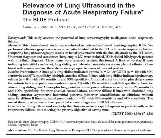
8 An examination combining: Anterior approach analyzing artifacts, lung sliding, alveolar consolidation Lateral subposterior search for posterolateral alveolar and/or pleural syndrome (PLAPS) Venous analysis
9 A profile designates anterior predominat bilateral A-lines associated with lung sliding B profile designates anterior predominat bilateral B-lines associated with lung sliding
10

11 Triple scan rapid 3-view sonographic evaluation of Heart Lungs Inferior vena cava
12
13

14 Laursen CB, Sloth E, Lassen AT, et al. Focused sonographic examination of the heart, lungs and deep veins in an unselected population of acute admitted patients with respiratory symptoms: a protocol for a prospective, blinded, randomised controlled trial. BMJ Open 2012;2:e doi: / bmjopen
15 Focused Ultrasound Study Goal-directed Systematic
16 Focused Ultrasound Study Specific clinical setting Differential diagnosis «Pre-test probability»
17 Main Causes of Acute Respiratory Failure Differential Diagnosis Cardiac etiologies Left heart failure Systolic / Diastolic Valvular problem Fluid overload Right heart failure Systolic / Diastolic Valvular problem Fluid overload Pulmonary emboli Tamponade Thoracic etiologies Lungs Pleura Pneumothorax Pleural effusion Interstitial syndromes - Hydrothorax - Pulmonary edema - Hemothorax - ARDS - Empyema - Interstitial diseases Pneumonia Asthma / Decompensation of chronic disease Other etiologies Metabolic acidosis Anxiety / Agitation Salicylate intoxication Sepsis Etc
18 Focused Ultrasound Study Specific ultrasound signs are sought to answer specific clinical questions
19 Focused Ultrasound Study Findings from the focused ultrasound examination have to be correctly integrated in clinical decision making
20 Focused Ultrasound Study for Respiratory Distress Main systems to be assessed: Cardiac Thoracic (lung + pleura) Vascular In which order should the exam proceed??
21 In which order should the exam proceed?? Undifferentiated shock start by the heart Undifferentiated respiratory distress start by the thorax
22 A Practical Approach to Respiratory Distress Plan 1-) Introduction 2-) Assessment of the thoracic space 3-) Assessment of the cardiac and vascular systems 4-) Summary 5-) Conclusion


23 Rapid bedside sonographic diagnosis of various conditions Pneumothorax Interstitial syndromes Atelectasis, consolidation Pleural effusion
24 Sonographic examination of the lung and pleural space Ultrasound findings Lung sliding / pneumothorax Normal aeration Alveolar interstitial pattern Alveolar consolidation Pleural effusions
25 Sonographic examination of the lung and pleural space Ultrasound findings Lung sliding / pneumothorax Normal aeration Alveolar interstitial pattern Alveolar consolidation Pleural effusions


26 Sonographic examination of the lung Scanning technique Sequential scan lines allow examiner to form a 3-D image based on multiple 2-D images
")
27 Sonographic examination of the lung Scanning technique Imaging controls Adjust gain to a lowmoderate level if too high, pleural line will not be well visualized Depth setting should be low ( cm)

28 Sonographic examination of the lung Basic terminology Sliding lung Represents the movement of visceral against parietal pleura Lichtenstein D, Menu Y. A Bedside Ultrasound Sign Ruling Out Pneumothorax in the Critically Ill: Lung Sliding, CHEST 1995;108:
29 Sonographic examination of the lung Pneumothorax Look for the absence of lung sliding


30 Sonographic examination of the lung Assess for lung sliding Normal lung Pneumothorax Lung sliding present Lung sliding absent
31 Sonographic examination of the lung Pneumothorax Look for the absence of lung sliding However, absence of lung sliding only indicates the possibility of pneumothorax Other causes of absent lung sliding: Apnea Mainstem intubation Mainstem occlusion Very severe parenchymal lung disease Pleural adhesions
32 Sonographic examination of the lung Pneumothorax Lung sliding means no pneumothorax with 100% certainty but only at the site of the transducer Absence of lung sliding means possible pneumothorax
33 Sonographic examination of the lung Ultrasound findings Lung sliding / pneumothorax Normal aeration Alveolar interstitial pattern Alveolar consolidation Pleural effusion
34 Sonographic examination of the lung Normal aeration A-lines Horizontal line that arise from the pleural line Localized at the exact distance separating the probe from the pleural line. Can be repeated multiple times Represents a reverberation of the pleural line

35 Sonographic examination of the lung Normal aeration A-lines Clinical utility of A-lines The presence of A-line pattern with sliding lung is a normal aeration pattern The presence of A-line pattern without sliding lung indicates the possibility of pneumothorax
36 Sonographic examination of the lung and pleural space Ultrasound findings Lung sliding / pneumothorax Normal aeration Alveolar interstitial pattern Alveolar consolidation Pleural effusions

37 Sonographic examination of the lung Alveolar interstitial pattern B-lines («lung rockets», «comet tail artifact») Long, vertical, hyperechoic, ray-like lines that originate at the pleural interface. Extend to the bottom of the screen Move with sliding lung Efface A-lines A few B lines may be normally found in the lower lateral thorax (in up to 27% of patients)

38 Sonographic examination of the lung Alveolar interstitial pattern B-lines («lung rockets», «comet tail artifact»)
39 Sonographic examination of the lung Alveolar interstitial pattern B-lines («lung rockets», «comet tail artifact») Clinical utility of B-lines The presence of B-lines is strongly associated with findings of alveolar interstitial pattern on chest CT Generalized B-lines indicate a «wet» lung: CHF, ARDS, ILD Useful for immediate evaluation of respiratory failure and to clarify ambiguous CXR B-lines excludes pneumothorax Pulmonary edema ARDS
40 Sonographic examination of the lung Alveolar interstitial pattern To be diagnostic of pulmonary edema (or other interstitial lung pathologies), the B-lines need to be present bilaterally in various areas of the chest


41 Sonographic examination of the lung Alveolar interstitial pattern To be diagnostic The of pulmonary more severe edema the interstitial (or other interstitial pathology, lung pathologies), the B-lines need to the be more present B-lines bilaterally will be in present various areas of the chest
42 Sonographic examination of the lung and pleural space Ultrasound findings Lung sliding / pneumothorax Normal aeration Alveolar interstitial pattern Alveolar consolidation Pleural effusions
 The findings of alveolar consolidation is purely descriptive; it does not imply a specific diagnosis such as")
43 Sonographic examination of the lung Alveolar consolidation Airless lung Airless lung appears with tissue density Liver Consolidated lung looks like liver («sonographic hepatization») The findings of alveolar consolidation is purely descriptive; it does not imply a specific diagnosis such as pneumonia.
44 Sonographic examination of the lung Alveolar consolidation Occurs with standard pneumonia or other alveolar filling process. Also occurs with atelectasis from any cause: - Compressive (pleural effusion) or - Resorptive (bronchial block) LLL pneumonia (radiology view) Right pleural effusion (radiology view)

45 Sonographic examination of the lung Alveolar consolidation Sonographic air bronchograms: - indicate whether the bronchus is open - appear as punctate echogenic foci
46 Sonographic examination of the lung and pleural space Ultrasound findings Lung sliding / pneumothorax Normal aeration Alveolar interstitial pattern Alveolar consolidation Pleural effusions
47 Sonographic examination of the lung Pleural effusion Right pleural effusion (radiology view)

48 Assessment of the pleural space for effusion Ultrasonographic diagnosis The sonographic appearance of a pleural effusion will depend on its cause, nature and chronicity. Simple vs Complex

49 Assessment of the pleural space for effusion Ultrasonographic diagnosis Simple effusion Typically are anechoic (echo-free) Effusion Lung Liver Variable etiologies - Fluid overload - Heart failure - Parapneumonic - Hepatic hydrothorax - Hypoalbuminemia
50 Assessment of the pleural space for effusion Ultrasonographic diagnosis Complex effusion These effusions may be «echogenic» because of proteinacious or highly cellular material in the exhudate ex: blood, pus, fibrin,

51 Assessment of the pleural space for effusion Ultrasonographic diagnosis Complex effusion with septations
52 CHEST 2009; 135:
53 ACCP / SRLF competency statement on critical care ultrasonography GCCU Pleural ultrasonography Lung ultrasonography Abdominal ultrasonography Vascular ultrasonography: guidance of vascular access Vascular ultrasonography: diagnosis of venous thrombosis CHEST 2009; 135:

54 Sonographic examination of the lung Intensive Care Med Apr;38(4):
55 A Practical Approach to Respiratory Distress Plan 1-) Introduction 2-) Assessment of the thoracic space 3-) Assessment of the cardiac and vascular systems 4-) Summary 5-) Conclusion

56 FOcused Cardiac Ultrasound Study Systematic assessment of - LV size and function - RV size and function - Pericardial space fluid tamponade - Inferior vena cava


57 Venous Focused Compression Technique Assessment of vessel collapsibility with probe pressure Presence of heterogenous, irregular echo-dense material (thrombus) in the lumen of the vein Not fully collapsible Normal vein Fully collapsible
58 A Practical Approach to Respiratory Distress Plan 1-) Introduction 2-) Assessment of the thoracic space 3-) Assessment of the cardiac and vascular systems 4-) Summary 5-) Conclusion
59 Summary Assessment of the thoracic space 1-) Is there a pneumothorax? Lung sliding Present Absent No pneumothorax Suspect pneumothorax ** Keep in mind the false +
60 Summary Assessment of the thoracic space 2-) Is there an interstitial syndrome? B lines Absence Presence Focal Diffuse - Normal - Local disease Assess LV function Cardiogenic Non-cardiogenic
61 Summary Assessment of the thoracic space 3-) Is there a pulmonary consolidation? Yes No Differential diagnosis
62 Summary Assessment of the thoracic space 4-) Is there a pleural effusion? Yes No Simple Complex Differential diagnosis
63 Summary 5-) Focused cardiac examination 6-) Focused vascular compression study 7-) If the complete ultrasound examination is normal, assess for non cardio-pulmonary causes of respiratory distress - Metabolic acidosis - Salicylate intoxication - Early sepsis - Neurologic cause
64 A Practical Approach to Respiratory Distress Plan 1-) Introduction 2-) Assessment of the thoracic space 3-) Assessment of the cardiac and vascular systems 4-) Summary 5-) Conclusion
65 A Practical Approach to Respiratory Distress Conclusion A systematic, algorithmic approach simplifying the use of ultrasound could lead to better and more efficient management of undifferentiated respiratory distress. Such an approach integrates concepts from the BLUE protocol, the focused cardiac exam, the E-FAST exam, and the focused compression technique for vessels.
Ultrasound. FAST Focused Assessment with Sonography in Trauma
 Ultrasound FAST Focused Assessment with Sonography in Trauma Rohit Patel, MD University of Florida Health Director, Critical Care Ultrasound Surgical ICU Center for Intensive Care Gainesville, Florida
Lung ultrasound in the critically ill patient Pleural Effusions
 Lung ultrasound in the critically ill patient Pleural Effusions Rohit Patel, MD University of Florida Health Director, Critical Care Ultrasound Surgical ICU Center for Intensive Care Gainesville, Florida
ASSESSMENT OF LUNG PARENCHYMAL ABNORMALITIES
 2016 by the author Thank you for viewing this presentation. We would like to remind you that this material is the property of the author. It is provided to you by the ERS for your personal use only, as
Lung ultrasound in the critically ill patient BASICS
 Lung ultrasound in the critically ill patient BASICS Rohit Patel, MD University of Florida Health Director, Critical Care Ultrasound Surgical ICU Center for Intensive Care Gainesville, Florida Introduction
Chest Ultrasound: Pneumothorax
 WINFOCUS BASIC ECHO (WBE) Chest Ultrasound: Pneumothorax Mark Hamlin, MD, MS Associate Professor of Anesthesiology and Surgery University of Vermont College of Medicine Co-Director of Surgical Critical
Contents& & & 1.! Ultrasound&basics& 1! 2.! Image&generation& 15!
 A l i n e press é % % % Contents& & & 1. Ultrasound&basics& 1 1.1. What,is,ultrasound?, 1 1.2. Ultrasound,probes,send,and,receive,ultrasound, 3 1.3. How,does,ultrasound,behave,travelling,through,tissue?,
Pediatric Lung Ultrasound (PLUS) In Diagnosis of Community Acquired Pneumonia (CAP)
 In Diagnosis of Community Acquired Pneumonia (CAP)") Pediatric Lung Ultrasound (PLUS) In Diagnosis of Community Acquired Pneumonia (CAP) Dr Neetu Talwar Senior Consultant, Pediatric Pulmonology Fortis Memorial Research Institute, Gurugram Study To compare
Pediatric Lung Ultrasound (PLUS) In Diagnosis of Community Acquired Pneumonia (CAP) Dr Neetu Talwar Senior Consultant, Pediatric Pulmonology Fortis Memorial Research Institute, Gurugram Study To compare
POCUS for the Internist: Lungs & Pericardial Effusions
 POCUS for the Internist: Lungs & Pericardial Effusions Jeremy S. Boyd, MD, FACEP Asst. Professor of Emergency Medicine Vanderbilt University Medical Illustrations courtesy of Robinson Ferre, MD, FACEP
Definitions and diagnostic implications of terms used in the chest radiograph and lung ultrasound diagnoses of pneumonia.
 Supplementary 1 Definitions and diagnostic implications of terms used in the chest radiograph and lung ultrasound diagnoses of pneumonia. Imaging finding Definition Implication CR Consolidation Interstitial
Lung sonography in the diagnosis of pneumothorax.
 Lung sonography in the diagnosis of pneumothorax. Poster No.: C-0526 Congress: ECR 2011 Type: Educational Exhibit Authors: K. Stefanidis, K. Vintzilaios, D. D. Cokkinos, E. Antypa, S. Dimopoulos, S. Nanas,
Bedside ultrasound - Lung ultrasound in the Intensive Care Unit
 Bedside ultrasound - Lung ultrasound in the Intensive Care Unit Kishore K. Pichamuthu, Professor, Department of Critical Care, Christian Medical College, Vellore. Summary In an ICU setting, ultrasonographic
Initially for cardiac echo Subsequent studies non-cardiac applications
 No disclosures But Heavy accent Initially for cardiac echo Subsequent studies non-cardiac applications 1973: Goldberg et al in JCUS 30 mediastinal masses in pts. age 1-84 yrs. 1977: Kangarloo et al in
OVERVIEW. Need for USG. Weaning assessment. Mechanics of USG. Pneumonia / VAP. Principles of lung USG. Prone position ventilation assessment
 OVERVIEW Need for USG Mechanics of USG Principles of lung USG BLUE protocol Alveolar syndrome Interstitial syndrome Weaning assessment Pneumonia / VAP Prone position ventilation assessment ETT positioning
This appendix was part of the submitted manuscript and has been peer reviewed. It is posted as supplied by the authors.
 This appendix was part of the submitted manuscript and has been peer reviewed. It is posted as supplied by the authors. - Figure S1: The four quadrant approach lung ultrasound at the bedside. * The anterolateral
The Shocked Patient. Adapted from Lichtenstein's FALLS protocol, with permission
 The Shocked Patient Adapted from Lichtenstein's FALLS protocol, with permission 1 Summary 1. (Ongoing resus) Clinical assessment: formulate the question 2. Rapid shock screen 3. Form a working diagnosis
Point-of-care lung ultrasound
 Ultrasound Point-of-care lung ultrasound Philips tutorial Michael B. Stone, MD, RDMS Director, Division of Emergency Ultrasound Department of Emergency Medicine Brigham and Women s Hospital, Boston, MA
NON INVASIVE LIFE SAVERS. Ultrasound in PICU
 VOL 1 NO.1 Jan March 2014 54 Table 1. Selected Applications of Point-of-Care Ultrasonography, According to Medical Specialty. Specialty Ultrasound Applications Anesthesia Cardiology Guidance for vascular
Diagnostic Bedside Ultrasound for the Hospitalist
 Diagnostic Bedside Ultrasound for the Hospitalist Trevor Jensen MD MS Assistant Professor, UCSF Nima Afshar MD Associate Professor, UCSF Diagnostic Bedside Ultrasound AKA Point-of-Care Ultrasound (POCUS)
EUROPEAN ASSOCIATION OF VETERINARY DIAGNOSTIC IMAGING EUROPEAN COLLEGE OF VETERINARY DIAGNOSTIC IMAGING
 EISAGOGIKO EUROPEAN ASSOCIATION OF VETERINARY DIAGNOSTIC IMAGING EUROPEAN COLLEGE OF VETERINARY DIAGNOSTIC IMAGING ARISTOTLE UNIVERSITY OF THESSALONIKI SCHOOL OF VETERINARY MEDICINE SECTION OF RADIOLOGY
Case 1. A 35-year-old male presented with fever, cough, and purulent sputum for one week. This was his CXR (Fig. 1.1). What is the diagnosis?
. What is the diagnosis?") 1 Interpreting Chest X-Rays CASE 1 Fig. 1.1 Case 1. A 35-year-old male presented with fever, cough, and purulent sputum for one week. This was his CXR (Fig. 1.1). What is the diagnosis? CASE 1 Interpreting
1 Interpreting Chest X-Rays CASE 1 Fig. 1.1 Case 1. A 35-year-old male presented with fever, cough, and purulent sputum for one week. This was his CXR (Fig. 1.1). What is the diagnosis? CASE 1 Interpreting
Ultrasound in the ICU
 Ultrasound in the ICU Kristine E. W. Breyer, MD Assistant Professor Anesthesia & Critical Care Medicine UCSF DISCLOSURES: NONE Definition The Ultrasound Exam Types & Uses Training Clinical Examples Objectives
Echocardiography as a diagnostic and management tool in medical emergencies
 Echocardiography as a diagnostic and management tool in medical emergencies Frank van der Heusen MD Department of Anesthesia and perioperative Care UCSF Medical Center Objective of this presentation Indications
Pulmonary Ultrasound in Emergency Medicine and Critical Care
 Pulmonary Ultrasound in Emergency Medicine and Critical Care www.rmgultrasound.com Author: Virginia M Stewart, MD RDMS RDCS RDMSK Dr Stewart is a practicing Emergency Physician in Eastern Virginia, USA.
Certificate in Clinician Performed Ultrasound (CCPU) Syllabus. Lung
 Syllabus. Lung") Certificate in Clinician Performed Ultrasound (CCPU) Syllabus Lung Page 1 of 8 12/15 Lung Syllabus Purpose: This unit is designed to cover the theoretical and practical curriculum for lung ultrasound in
Certificate in Clinician Performed Ultrasound (CCPU) Syllabus Lung Page 1 of 8 12/15 Lung Syllabus Purpose: This unit is designed to cover the theoretical and practical curriculum for lung ultrasound in
Certificate in Clinician Performed Ultrasound (CCPU) Syllabus. Lung
 Syllabus. Lung") Certificate in Clinician Performed Ultrasound (CCPU) Syllabus Lung Page 1 of 8 01/17 Lung Syllabus Purpose: This unit is designed to cover the theoretical and practical curriculum for lung ultrasound in
Certificate in Clinician Performed Ultrasound (CCPU) Syllabus Lung Page 1 of 8 01/17 Lung Syllabus Purpose: This unit is designed to cover the theoretical and practical curriculum for lung ultrasound in
MINERVA MEDICA COPYRIGHT REVIEW ARTICLE D. LICHTENSTEIN. Resuscitation Service, Ambroise-Paré Hospital, Boulogne, France ABSTRACT
 MINERVA ANESTESIOL 2009;75:313-7 REVIEW ARTICLE Lung ultrasound in acute respiratory failure an introduction to the BLUE-protocol D. Resuscitation Service, Ambroise-Paré Hospital, Boulogne, France ABSTRACT
Dr. Rami M. Adil Al-Hayali Assistant Professor in Medicine
 Dr. Rami M. Adil Al-Hayali Assistant Professor in Medicine Venous thromboembolism: pulmonary embolism (PE) deep vein thrombosis (DVT) 1% of all patients admitted to hospital 5% of in-hospital mortality
Lung Ultrasound in Diagnosis of Acute Respiratory Failure: BLUE Protocol Based Evaluation
 Lung Ultrasound in Diagnosis of Acute Respiratory Failure: BLUE Protocol Based Evaluation Niyas. K. Naseer 1, Muhammad Shafeek 2*, Rajani. M 3, Manoj. D. K 3 1Junior Resident, 2* Assistant Professor, 3
Certificate in Clinician Performed Ultrasound (CCPU) Syllabus. Lung
 Syllabus. Lung") Certificate in Clinician Performed Ultrasound (CCPU) Syllabus Lung ASUM Quality CCPU Syllabi Released: 21 March 2013 Approved by: CEO Lung Purpose: This unit is designed to cover the theoretical and practical
Certificate in Clinician Performed Ultrasound (CCPU) Syllabus Lung ASUM Quality CCPU Syllabi Released: 21 March 2013 Approved by: CEO Lung Purpose: This unit is designed to cover the theoretical and practical
Intro Case. Outline What We ll Cover. What we won t cover. Cardiac Ultrasound and The RUSH Exam: Bedside Ultrasound in Resuscitation and Shock
 Cardiac Ultrasound and The RUSH Exam: Bedside Ultrasound in Resuscitation and Shock Justin Davis, MD, MPH, RDMS Associate Physician Subchief for Emergency Ultrasound Services Kaiser Oakland Medical Center
Objectives. The Extended FAST Exam. Focused Assessment e With Sonography In. Trauma (FAST)
") Northern California Emergency Ultrasound Course Objectives The Extended FAST Exam Rimon Bengiamin, MD, RDMS UC SF Discuss the components of the EFAST exam Evaluate the utility of the EFAST Review how to
Northern California Emergency Ultrasound Course Objectives The Extended FAST Exam Rimon Bengiamin, MD, RDMS UC SF Discuss the components of the EFAST exam Evaluate the utility of the EFAST Review how to
ORIGINAL ARTICLE. Role of Ultrasound in Evaluation of Undifferentiated Shock in ICU Settings
 Journal of The Association of Physicians of India Vol. 66 August 2018 13 Role of Ultrasound in Evaluation of Undifferentiated Shock in ICU Settings Tanvi Vaidya 1*, Pradeep D costa 2, Satish Pande 3 ORIGINAL
Patient Management Code Blue in the CT Suite
 Patient Management Code Blue in the CT Suite David Stultz, MD November 28, 2001 Case Presentation A 53-year-old woman experienced acute respiratory distress during an IV contrast enhanced CT scan of the
B-I-2 CARDIAC AND VASCULAR RADIOLOGY
 (YEARS 1 3) CURRICULUM FOR RADIOLOGY 13 B-I-2 CARDIAC AND VASCULAR RADIOLOGY KNOWLEDGE To describe the normal anatomy of the heart and vessels including the lymphatic system as demonstrated by radiographs,
Shedding Light on Neonatal X-rays. Objectives. Indications for X-Rays 5/14/2018
 Shedding Light on Neonatal X-rays Barbara C. Mordue, MSN, NNP-BC Neonatal Nurse Practitioner LLUH Children s Hospital, NICU Objectives Utilize a systematic approach to neonatal x-ray interpretation Identify
CHEST Recent Advances in Chest Medicine
 CHEST Recent Advances in Chest Medicine Thoracic Ultrasonography for the Pulmonary Specialist Seth J. Koenig, MD; Mangala Narasimhan, DO, FCCP; and Paul H. Mayo, MD, FCCP Thoracic ultrasonography is a
Extended FAST Exam. Goal of Trauma Care. Golden Hour of Trauma
 Extended FAST Exam Goal of Trauma Care Golden Hour of Trauma Best INITIAL screening modality in trauma efast 2014 LLSA Article (ACEP Policy Statement) Level B Recommendation: In hemodynamically unstable
Current Opinion in Anesthesiology Basic concepts in the use of thoracic and lung ultrasound
 Manuscript Number: Current Opinion in Anesthesiology Basic concepts in the use of thoracic and lung ultrasound --Manuscript Draft-- Full Title: Article Type: Corresponding Author: Basic concepts in the
Looking Outside the Box: Incidental Extracardiac Finding in Echo
 Looking Outside the Box: Incidental Extracardiac Finding in Echo Dr. Aijaz Shah Head of Division, Adult Echocardiography Laboratory Prince Sultan Cardiac Centre Riyadh Case 1 17 year old boy presented
Introduction to Chest Radiography
 Introduction to Chest Radiography RSTH 366: DIAGNOSTIC TECHNIQUES Alan Alipoon BS, RCP, RRT Instructor Department of Cardiopulmonary Sciences 1 Introduction Discovered in 1895 by Wilhelm Roentgen Terminology
Point of Care Ultrasound (PoCUS)
") Point of Care Ultrasound (PoCUS) Competency Assessment Forms AORTA Competency A Focussed Assessment of the Aorta (AAA) Guidance Please follow this guidance as closely as possible to ensure consistency
Point of Care Ultrasound (PoCUS) Competency Assessment Forms AORTA Competency A Focussed Assessment of the Aorta (AAA) Guidance Please follow this guidance as closely as possible to ensure consistency
10/17/2016. Nuts and Bolts of Thoracic Radiology. Objectives. Techniques
 Nuts and Bolts of Thoracic Radiology October 20, 2016 Carleen Risaliti Objectives Understand the basics of chest radiograph Develop a system for interpreting chest radiographs Correctly identify thoracic
ARDS - a must know. Page 1 of 14
 ARDS - a must know Poster No.: C-1683 Congress: ECR 2016 Type: Authors: Keywords: DOI: Educational Exhibit M. Cristian; Turda/RO Education and training, Edema, Acute, Localisation, Education, Digital radiography,
USE OF LUNG ULTRASOUND IN SMALL ANIMALS - THE VET BLUE THE BASICS OF VET BLUE
 Ultrasound in the Respiratory Distress Patient: Using Vet Blue International Veterinary Emergency and Critical Care Symposium 2018 Gregory R. Lisciandro, DVM, DABVP, DACVECC Hill Country Veterinary Specialists
Non Invasive Hemodynamic Monitoring in the ED
 Is haemodinamic monitoring useful in the ED? Non Invasive Hemodynamic Monitoring in the ED Roberta PETRINO Director Emergency Medicine Unit S. Andrea Hospital, Vercelli - Italy EuSEM Vice-president Helps
The efficacy of bedside chest ultrasound: from accuracy to outcomes
 EUROPEAN RESPIRATORY UPDATE EFFICACY OF BEDSIDE CHEST ULTRASOUND The efficacy of bedside chest ultrasound: from accuracy to outcomes Mark Hew 1,2 and Tunn Ren Tay 1,3 Affiliations: 1 Allergy, Immunology
Transthoracic Echocardiography:
 Transthoracic Echocardiography: An essential tool for the obstetric anaesthetist? Brendan Carvalho MBBCh, FRCA Department of Anesthesiology Stanford University, California Focused TTE Stethoscope of the
POCUS is the future of the physical exam
 Diagnostic Point of Care Ultrasound For Hospitalists Nima Afshar MD Associate Professor Trevor Jensen MD MS Assistant Professor Department of Medicine, UCSF Oct 2018 POCUS is the future of the physical
Imaging of Pleural Effusion: Comparing Ultrasound, X-Ray and CT findings
 Imaging of Pleural Effusion: Comparing Ultrasound, X-Ray and CT findings Poster No.: C-2067 Congress: ECR 2017 Type: Educational Exhibit Authors: J. M. Almeida, N. Antunes, C. Leal, L. Figueiredo ; Lisboa/PT,
Identification of lung sliding: a basic ultrasound technique with a steep learning curve
 SIGNA VITAE 2013; 8(1): 31-35 ORIGINAL Identification of lung sliding: a basic ultrasound technique with a steep learning curve MATEJ STRNAD SABINA ZADEL ZALIKA KLEMENC-KETIS MATEJ STRNAD ( ) SABINA ZADEL
Introduction to Radiology for TB Nurses
 Introduction to Radiology for TB Nurses Juzar Ali, MD; FRCP(C); FCCP May 4, 2018 Essential Skills for the TB Nurse Case Manager Little Rock, AR May 3 4, 2017 Juzar Ali, MD; FRCP(C); FCCP has the following
The GATOR SIGN Basic Lung Ultrasound Orientation
 Case-based Vet BLUE and Its 6 Lung Ultrasound Signs Gregory R. Lisciandro, DVM, Dipl. ABVP, Dipl. ACVECC Hill Country Veterinary Specialists & FASTVet.com, Spicewood, Texas USA Email LearnGlobalFAST@gmail.com
We are IntechOpen, the world s leading publisher of Open Access books Built by scientists, for scientists. International authors and editors
 We are IntechOpen, the world s leading publisher of Open Access books Built by scientists, for scientists 3,800 116,000 120M Open access books available International authors and editors Downloads Our
Chest X rays and Case Studies. No disclosures. Outline 5/31/2018. Carlo Manalo, M.D. Department of Radiology Loma Linda University Children s Hospital
 Chest X rays and Case Studies Carlo Manalo, M.D. Department of Radiology Loma Linda University Children s Hospital No disclosures. Outline Importance of history Densities delineated on radiography An approach
For more information about how to cite these materials visit
 Project: Ghana Emergency Medicine Collaborative Document Title: Approach to the Dyspenic Adult Patient Author(s): Randall Ellis, MD MPH (Vanderbilt University) License: Unless otherwise noted, this material
Background & Indications Probe Selection
 Teresa S. Wu, MD, FACEP Director, EM Ultrasound Program & Fellowship Co-Director, Simulation Based Training Program & Fellowship Associate Program Director, EM Residency Program Maricopa Medical Center
Radiology of the respiratory/cardiac diseases (part 2)
") Cardiology Cycle - Lecture 6 436 Teams Radiology of the respiratory/cardiac diseases (part 2) Objectives Done By Team Leaders: Khalid Alshehri Hanin Bashaikh Team Members: Leena Alwakeel Aroob Alhuthail
Cardiology Cycle - Lecture 6 436 Teams Radiology of the respiratory/cardiac diseases (part 2) Objectives Done By Team Leaders: Khalid Alshehri Hanin Bashaikh Team Members: Leena Alwakeel Aroob Alhuthail
Management of Pleural Effusion
 Management of Pleural Effusion Development of Pleural Effusion pulmonary capillary pressure (CHF) capillary permeability (Pneumonia) intrapleural pressure (atelectasis) plasma oncotic pressure (hypoalbuminemia)
Role of Transthoracic Ultrasound in Detection of Pneumonia in ICU Patients
 Med. J. Cairo Univ., Vol. 83, No. 1, June: 307-314, 2015 www.medicaljournalofcairouniversity.net Role of Transthoracic Ultrasound in Detection of Pneumonia in ICU Patients FARES AUF, M.D.; AHMED ABO-NAGLH,
Point-of-Care Ultrasound Closer look at the Inferior Vena Cavae &
 Point-of-Care Ultrasound Closer look at the Inferior Vena Cavae & Brief Introduction to Gross Systolic Function Omar S. Darwish, MS, DO Certified in Point-of-Care Ultrasound Hospitalist University of California,
Unilateral pulmonary oedema, a forgotten presentation.
 Unilateral pulmonary oedema, a forgotten presentation. Poster No.: C-2146 Congress: ECR 2018 Type: Educational Exhibit Authors: C. A. Arboleda Vallejo, M. I. carvajal, M. Perez ; Medellin, 1 2 1 1 2 Antioquia/CO,
Lung ultrasound in follow-up of low birth weight with respiratory distress syndrome: clinical application and reduction of x-rays examinations
 Lung ultrasound in follow-up of low birth weight with respiratory distress syndrome: clinical application and reduction of x-rays examinations Poster No.: C-1724 Congress: ECR 2011 Type: Scientific Paper
Ultrasound Assessment of Pulmonary Embolism in Patients Receiving CT Pulmonary Angiography
 CHEST Ultrasound Assessment of Pulmonary Embolism in Patients Receiving CT Pulmonary Angiography Seth Koenig, MD, FCCP ; Subani Chandra, MD ; Artur Alaverdian, MD ; Christopher Dibello, MD ; Paul H. Mayo,
UERMMMC Department of Radiology. Basic Chest Radiology
 UERMMMC Department of Radiology Basic Chest Radiology PHYSICS DENSITIES BONE SOFT TISSUES WATER FAT AIR TELEROENTGENOGRAM Criteria for an Ideal Chest Radiograph 1. Upright 2. Posteroanterior View 3. Full
Bedside Sonographic Diagnosis of Pneumothorax in Pediatric Patients: A Preliminary Report Chia-Wang Tang 1, Kai-Sheng Hsieh 1 1
 ORIGINAL ARTICLE Bedside Sonographic Diagnosis of in Pediatric Patients: A Preliminary Report Chia-Wang Tang 1, Kai-Sheng Hsieh 1 1 Division of Pediatric Pulmonology, Department of Pediatrics, Kaohsiung
Impact of lung ultrasound on clinical decision making in critically ill patients
 Intensive Care Med DOI 10.1007/s00134-013-3133-3 ORIGINAL Nektaria Xirouchaki Eumorfia Kondili George Prinianakis Polychronis Malliotakis Dimitrios Georgopoulos Impact of lung ultrasound on clinical decision
Bedside Ultrasound. US Guided Fluid Resuscitation. Michiel J. van Veelen, Emergency Physician, DTM&H
 Bedside Ultrasound US Guided Fluid Resuscitation Michiel J. van Veelen, Emergency Physician, DTM&H Outline Shock and Fluid Resuscitation in ICU Ultrasound in Shock Ultrasound Guided Fluid Resuscitation
Perioperative Ultrasonography Ehab Farag, MD, FRCA Hesham Elsharkawy David G. Anthony, M.D.
 Perioperative Ultrasonography Ehab Farag, MD, FRCA Hesham Elsharkawy David G. Anthony, M.D. Cleveland Clinic, Cleveland OH 1 Complications during central venous catheterization (CVC) occur 2% -15% of the
AAENP US WORKSHOP 2/25/17
 Know the components of the Rapid Ultrasound for Shock & Hypotension & Extended Focused Assessment Sonography in Trauma & how they can help quickly determine diagnosis. Be comfortable obtaining and interpreting
Session 2: Ultrasonography for Primary Care Clinicians Learning Objectives
 Session 2: Ultrasonography for Primary Care Clinicians Learning Objectives 1. Assess the main components and functions of a portable ultrasound unit. 2. Identify three clinical applications of portable
Lung ultrasound: routine practice for the next generation of internists
 REVIEW Lung ultrasound: routine practice for the next generation of internists H.R.W. Touw 1,2, P.R. Tuinman* 1,3,4, H.P.M.M. Gelissen 1, E. Lust 1, P.W.G. Elbers 1,3,4 Departments of 1 Intensive Care
Chapter 17 Lung Ultrasound in Anaesthesia and Critical Care Medicine
 Chapter 17 Lung Ultrasound in Anaesthesia and Critical Care Medicine David Canty, Kavi Haji, André Denault, and Alistair Royse Abstract Lung ultrasound (respiratory or thoracic ultrasound) has traditionally
Point of Care Ultrasound in the ICU
 Point of Care Ultrasound in the ICU JENNIFER P. KANAAN, M.D. ASSISTANT PROFESSOR OF MEDICINE UNIVERSITY OF CONNECTICUT I have no disclosures 1 Ultrasound Ultrasound imaging is among the fastest, safest
Practical Echocardiography and Ultrasound in Critical Care
 WINFOCUS BASIC ECHO (WBE) Practical Echocardiography and Ultrasound in Critical Care Colin K. Grissom, MD Critical Care Medicine, Shock Trauma ICU Intermountain Medical Center, Murray, Utah Professor of
Case Study #2. Case Study #1 cont 9/28/2011. CAPA 2011 Christy Wilson PA C. LH is 78 yowf with PMHx of metz breast CA presents
 Case Study #1 CAPA 2011 Christy Wilson PA C 46 yo female presents with community acquired PNA (CAP). Her condition worsened and she was transferred to the ICU and placed on mechanical ventilation. Describe
Interactive Lecture. Lecture 7 - Interactive. Radiology of cardiorespiratory disease. Editing File. Done By. Color Coding Important Notes Extra
 Lecture 7 - Interactive 436 Teams Interactive Lecture Radiology of cardiorespiratory disease Done By Team Leaders: Khalid Alshehri Hanin Bashaikh Team Members: Ghaida Alsaeed Maha Alissa Nawwaf AlHarbi
Lung Ultrasound in Small Animals: The Vet BLUE SM
 Lung Ultrasound in Small Animals: The Vet BLUE SM Gregory R. Lisciandro, DVM, Dipl. ABVP, Dipl. ACVECC Hill Country Veterinary Specialists & FASTVet.com, San Antonio, Texas USA FASTVet TM and FAST Saves
Introduction to transthoracic ultrasound for the pulmonologist
 Review Introduction to transthoracic ultrasound for the pulmonologist Clementine Bostantzoglou 1, Charalampos Moschos 2 1 7 th Pulmonary Department, Sotiria Hospital for Chest Diseases, Athens, Greece
The faculty will include physicians with international reputations as outstanding ultrasound educators.
 Ultrasound Courses Course Description Whether you re a beginner or a seasoned sonographer, this year s AAEM pre-conference ultrasound course will be worth your time. We will be offering a half day course
Small animal point of care ultrasound techniques
 Small animal point of care ultrasound techniques The role of veterinary point of care ultrasound in determining the presence or absence of specific pathologies is examined by Jantina McMurray DVM; Søren
Lung Ultrasound in COPD and Bronchial Asthma-Measurements. of Lung Deformation with our Novel. Imaging Approach.
 Editorial imedpub Journals http://www.imedpub.com Insights in Allergy, Asthma & Bronchitis DOI: 10.21767/2471-304X.100006 Abstract Lung Ultrasound in COPD and Bronchial Asthma-Measurements of Lung Deformation
ECBSE Chest 29/01/2014 / 1:52 1. EFSUMB European Course Book Student Edition Editors: Jan Tuma, Radu Badea, Christoph F. Dietrich.
 ECBSE Chest 29/01/2014 / 1:52 1 EFSUMB European Course Book Student Edition Editors: Jan Tuma, Radu Badea, Christoph F. Dietrich Chest Gebhard Mathis Internistische Praxis, Rankweil, Austria Corresponding
My patient has no blood pressure: point-of-care ultrasound in the hypotensive patient FAST and RELIABLE
 PoCUS Series My patient has no blood pressure: point-of-care ultrasound in the hypotensive patient FAST and RELIABLE Andrew Liteplo 1, Vicki Noble 1 and Paul Atkinson 2 1 Department of Emergency Medicine,
Patients with sustained hypotension and shock are at
 Bedside Ultrasound Reduces Diagnostic Uncertainty and Guides Resuscitation in Patients With Undifferentiated Hypotension* Hamid Shokoohi, MD, MPH, RDMS, RDCS, FACEP 1 ; Keith S. Boniface, MD, RDMS, RDCS
Diagnostic Approach to Pleural Effusion
 Diagnostic Approach to Pleural Effusion Objectives Define the leading causes of pleural effusion Classify the type of effusion Identify procedures and tests associated with diagnosis 2 Agenda Basic anatomy
TB Radiology for Nurses Garold O. Minns, MD
 TB Nurse Case Management Salina, Kansas March 31-April 1, 2010 TB Radiology for Nurses Garold O. Minns, MD April 1, 2010 TB Radiology for Nurses Highway Patrol Training Center Salina, KS April 1, 2010
Lecture 3. Inflammatory Processes
 Lecture 3 Inflammatory Processes Process: Increased vascular permeability Water and cellular infiltrations Results: Abscess, ulceration, cavitation Penetration, perforation and fistula formation Scarring,
Interpreting thoracic x-ray of the supine immobile patient: Syllabus
 Interpreting thoracic x-ray of the supine immobile patient: Syllabus Johannes Godt Dep. of Radiology and Nuclear Medicine Oslo University Hospital Ullevål NORDTER 2017, Helsinki Content - Why bedside chest
Unusual new signs of pneumothorax at lung ultrasound
 Unusual new signs of pneumothorax at lung ultrasound Volpicelli et al. Volpicelli et al. Critical Ultrasound Journal 2013, 5:10 Volpicelli et al. Critical Ultrasound Journal 2013, 5:10 SHORT COMMUNICATION
EFAST. Extended Focussed Assessment with Sonography for Trauma. Ultrasound Logbook. Name
 EFAST Extended Focussed Assessment with Sonography for Trauma Ultrasound Logbook ame Contents EFAST Accreditation Requirements 25 Abdominal Aorta Report Forms 3 Formative Assessments 1 Summative Assessment
Imaging of Thoracic Trauma: Tips and Traps. Arun C. Nachiappan, MD Associate Professor of Clinical Radiology University of Pennsylvania
 Imaging of Thoracic Trauma: Tips and Traps Arun C. Nachiappan, MD Associate Professor of Clinical Radiology University of Pennsylvania None Disclosures Objectives Describe blunt and penetrating traumatic
Background Focused Assessment with Sonography in Trauma. Johann Baptist Dormagen, MD, PhD
 Focused Assessment with Sonography in Trauma Johann Baptist Dormagen, MD, PhD Unit of Abdominal and Oncologic Radiology Department of Radiology and Nuclear Medicine Oslo University Hospital, Norway 8 th
Shock. Undifferentiated Shock: Beyond Blood Pressure. Shock. Epidemiology. Matthew Strehlow, MD Stanford University
 Shock Undifferentiated Shock: Beyond Blood Pressure Matthew Strehlow, MD Stanford University Shock Shock - The rude unhinging of the machinery of life -SD Gross 1872 Epidemiology Shock - inadequate tissue
In ESH we usually see blunt chest trauma but penetrating injuries also treated here (usually as single injuries, like stab wound)
") Chest Trauma Dr Csaba Dioszeghy MD PhD FRCEM FFICM FERC East Surrey Hospital Emergency Department Scope Thoracic injuries are common and can be life threatening In ESH we usually see blunt chest trauma
Chest Trauma Dr Csaba Dioszeghy MD PhD FRCEM FFICM FERC East Surrey Hospital Emergency Department Scope Thoracic injuries are common and can be life threatening In ESH we usually see blunt chest trauma
Ultrasound basics Part 1
 Ultrasound basics Part 1 'Ultrasound enhanced critical care medicine' Rohit Patel, MD University of Florida Health Director, Critical Care Ultrasound Surgical ICU Center for Intensive Care Gainesville,
ACUTE RESPIRATORY DISTRESS SYNDROME
 ACUTE RESPIRATORY DISTRESS SYNDROME Angel Coz MD, FCCP, DCE Assistant Professor of Medicine UCSF Fresno November 4, 2017 No disclosures OBJECTIVES Identify current trends and risk factors of ARDS Describe
Ultrasound-guided Aspiration of the Iatrogenic Pneumothorax Caused by Paravertebral Block
 Case Report Korean J Pain 2012 January; Vol. 25, No. 1: 33-37 pissn 2005-9159 eissn 2093-0569 http://dx.doi.org/10.3344/kjp.2012.25.1.33 Ultrasound-guided Aspiration of the Iatrogenic Pneumothorax Caused
Background & Indications Probe Selection
 Teresa S. Wu, MD, FACEP Director, EM Ultrasound Program & Fellowship Co-Director, Simulation Based Training Program & Fellowship Associate Program Director, EM Residency Program Maricopa Medical Center
ERDHEIM-CHESTER DISEASE LUNG & HEART ISSUES
 ERDHEIM-CHESTER DISEASE LUNG & HEART ISSUES GIULIO CAVALLI, M.D. INTERNAL MEDICINE AND CLINICAL IMMUNOLOGY IRCCS SAN RAFFAELE HOSPITAL VITA-SALUTE SAN RAFFAELE UNIVERSITY MILAN, ITALY cavalli.giulio@hsr.it
Cardiac tamponade and Pericardiocentesis Made Easy
 Cardiac tamponade and Pericardiocentesis Made Easy www.cardiconcept.com Etiology of pericardial diseases. Non Infectious cause Infectious cause European Heart Journal (2015) 36, 2921 2964 Recommendations