Initially for cardiac echo Subsequent studies non-cardiac applications
|
|
|
- Eugene Henry
- 5 years ago
- Views:
Transcription


1
2 No disclosures But


3 Heavy accent
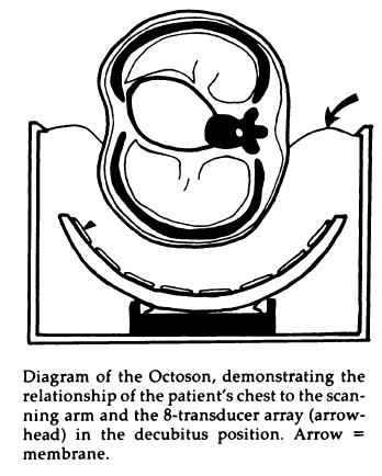
4 Initially for cardiac echo Subsequent studies non-cardiac applications 1973: Goldberg et al in JCUS 30 mediastinal masses in pts. age 1-84 yrs. 1977: Kangarloo et al in Radiology Juxtadiaphragmatic lesions in children, value of liver window 1980: Haller et al in AJR 28 children - 93% success rate; evaluation of opaque hemithorax, characterizing pleural fluid, guiding drainage, integrity of diaphragm 1984: Claus and Coppens in Ann Radiol Value of thymus as a window for US for mediastinal masses 1984: Miller et al in Radiology Water path ultrasound of chest disease in childhood 82 children placed in Octoson to expand windows
5
6 1986: Rosenberg HK in RG Complimentary role of US and CXR in differentiating pediatric chest abnormalities 1989: Glasier et al in J Pediatr Modality of choice for opaque chest, mediastinal masses, and pleural disease 1993: Ben -Ami TE et al in RCNA Review 2005: Coley BD in RCNA Review
7
8 Generic Non invasive, inexpensive, no contrast, realtime, Doppler, bedside/portable, some tissue characterization Pediatric No radiation Paucity of fat Smaller, more superficial structures Pediatric chest Portable Cartilagenous sternum and ribs Thymic window Pediatric chest Aerated lung Requires parenchymal lesion to be superficial Older children Sternum ossified, thymus small Problem= we believe that AIR is the enemy of US But when pathology is present, air is replaced Both, normal and abnormal lung cause artifacts
9 Problem= we believe that AIR is the enemy of US But when pathology is present, air is replaced Both, normal and abnormal lung cause artifacts Intensivists and ER docs learned to read these artifacts similarly to how they read an EKG Pediatric chest Aerated lung Requires parenchymal lesion to be superficial Older children Sternum ossified, thymus small

10


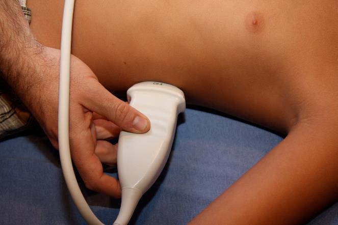
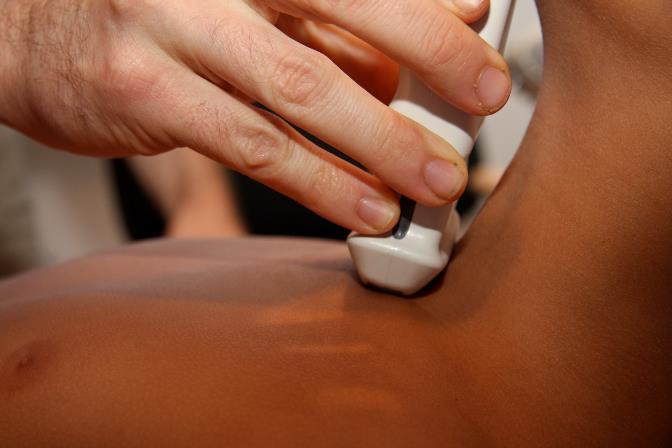
11 Parasternal Subxyphoid Haller et al AJR 1980 Suprasternal Transdiapragmatic Mong, Epelman, Darge Pediatr Radiol 2012

12 Parasternal Haller et al AJR anterior superior 2- anterior basal 3- lateral superior 4- lateral basal Coley BD 2011 Radiol Clinics NA
13 1. Pleural line 2. Lung sliding 3. A lines 4. B lines 5. Pneumothorax 6. M-mode normal lung: seashore sign 7. Pneumothorax real time/m-mode: barcode sign 8. Pneumonia: dynamic air bronchogram sign 9. Estimation of volume of pleural fluid

, and the")
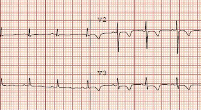
14 Pleural line: Echogenic line acoustic interface between the soft tissues of the chest wall and the air-filled lung R R R R Lung sliding: Dynamic finding Visceral and parietal surfaces are apposed and the visceral layer moves freely with respirations Subtle shimmering at the pleural line Absence: PTX, massive atelectasis, pleurodesis, apnea, pneumonia A lines: Echogenic, horizontal, usually evenly spaced lines parallel to the pleural interface Reverberation artifacts created by repetitive reflection of the US waves between the pleural line (a strong reflector due to the significant change Normal in acoustic lung in impedance a 2-week-old at the pleura-lung girl interface), and the transducer


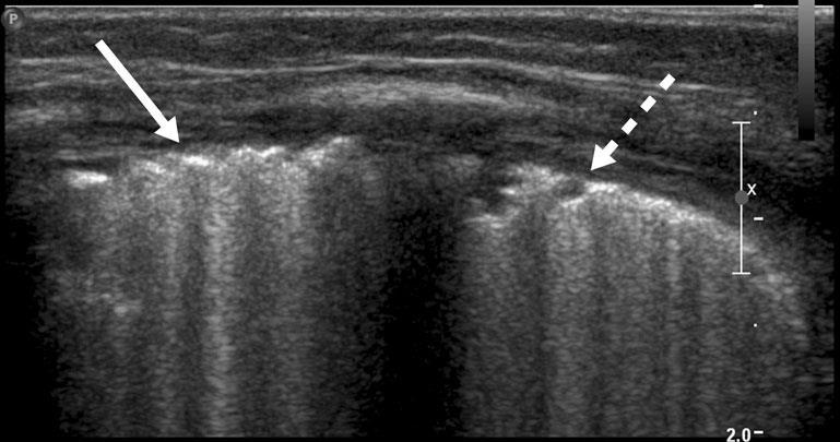
15 R R R Vertically oriented comet-tail artifacts Originate from irregularities at the lung-pleura interface Move with lung sliding Obscure A lines Up to 3 per interspace Normal


")
16 Vertically oriented comet-tail artifacts Originate from irregularities at the lungpleura interface Move with lung sliding Obscure A lines Up to 3 per interspace Normal More than 3 nonspecific markers of several lung disorders (pulm edema, interstitial lung dz, RDS, TTN, etc) Absent with pneumothorax


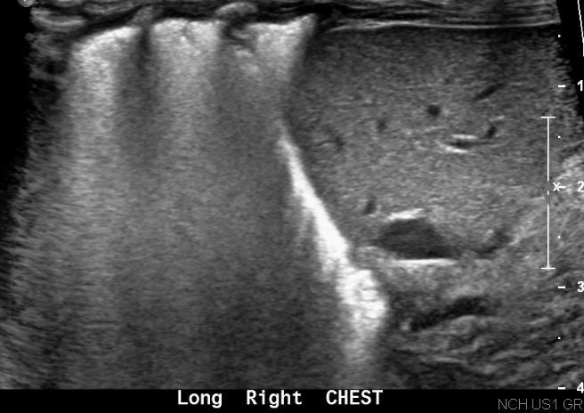
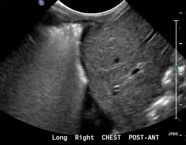
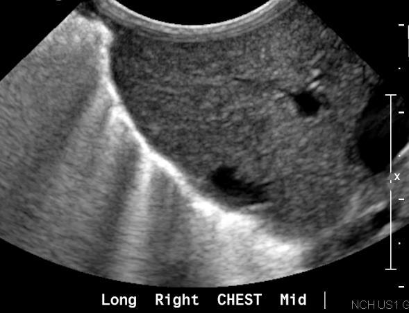

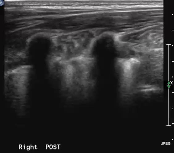
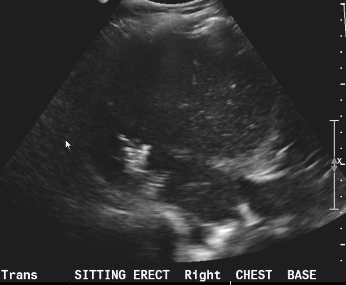
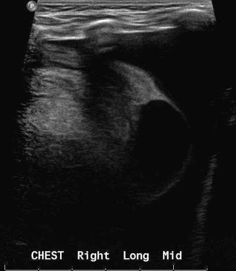
17 Ex 24 wk, now 25 days old, R/O pleural effusion


18 Grading system for lung ultrasound from Raimondi et al.1 3 Left: type 1 or white-out lung, significant liquid retention associated with respiratory distress syndrome (type 1) is seen between the acoustic shadow cast by the ribs (R). Douglas A Blank et al. Arch Dis Child Fetal Neonatal Ed 2018;103:F157-F162 Copyright BMJ Publishing Group Ltd & Royal College of Paediatrics and Child Health. All rights reserved.


19 Absent lung sliding Accentuated A lines Absent B lines Normal Lung sliding - Movement with respiration B lines -Comet tail artifacts Vague A lines


20 Normal pleura Pneumothorax Lung sliding Movement with respiration B lines Comet tail artifacts Case courtesy Dr. Olga Brook Beth Israel- Boston- MA


21 Considered fairly specific for pneumothorax Found at interface between inflated lung and pleural air Hard to identify Images courtesy of Dr. M. Jagla
 Normal lung sliding")
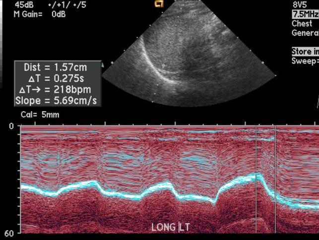
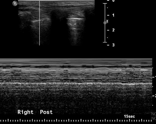
22 Pleural line echogenic Soft tissues echog. Lines between pleural line and transducer (no motion) Normal lung sliding grainy or sandy appearance Seashore sign
23 Strong interface between pleura/pneumothorax extensive reverberation artifacts barcode or stratosphere sign deeper reverberation artifacts lines Pneumothorax: barcode or stratosphere sign

24 Pneumothorax: barcode or stratosphere sign

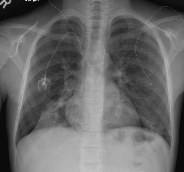
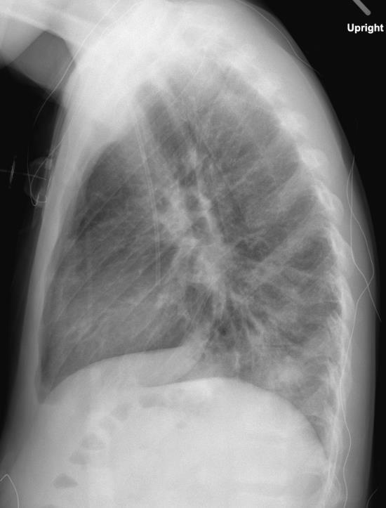
25 11 yo boy with CF exacerbation

26
27


28 In consolidation: Air- or fluid-filled bronchograms can be seen as branching echogenic lines, giving it a hepatized appearance Pleural line is less echogenic than usual Lung sliding may be decreased and even absent


29 3 y.o. F with a Hx of SCD, fever, cough, dyspnea

30 Differentiating consolidation from atelectasis is challenging on CXR US Lichtenstein et al dynamic air bronchogram If dynamic motion of gas bubbles is seen within the air bronchograms indicative of pneumonia PPV of 97% Lichtenstein D et al. Chest 2009; 135:
31

32 Principi et al. BMC Pulmonary Medicine (2017) 17:212


33 A B L A 19 month old with a background history of hypoxic ischemic injury and cerebral palsy L Beware!
34 Beware!
35
36 Beware!

37
38


39 Transverse section perpendicular to the body axis The maximal distance between parietal and visceral pleura was measured in endexpiration. The lung base is often consolidated and positioned posteriorly. Therefore, the maximum separation is frequently found lateral, rather than posterior V (ml): SEP (mm) x 20

40
41 Bedside study Mainly being done as POC studies by non-radiologists Easier than we think The beautiful images provided by current CT and MR imaging techniques are aesthetically seductive, but it should be remembered that US often provides the clinically needed information at lesser cost, without sedation or radiation exposure» From Coley B, RCNA 2005

42
Initially for cardiac echo Subsequent studies non-cardiac applications
 Initially for cardiac echo Subsequent studies non-cardiac applications 1973: Goldberg et al in JCUS 30 mediastinal masses in pts. age 1-84 yrs. 1977: Kangarloo et al in Radiology Juxtadiaphragmatic lesions
Initially for cardiac echo Subsequent studies non-cardiac applications
 Initially for cardiac echo Subsequent studies non-cardiac applications 1973: Goldberg et al in JCUS 30 mediastinal masses in pts. age 1-84 yrs. 1977: Kangarloo et al in Radiology Juxtadiaphragmatic lesions
This appendix was part of the submitted manuscript and has been peer reviewed. It is posted as supplied by the authors.
 This appendix was part of the submitted manuscript and has been peer reviewed. It is posted as supplied by the authors. - Figure S1: The four quadrant approach lung ultrasound at the bedside. * The anterolateral
Pediatric Lung Ultrasound (PLUS) In Diagnosis of Community Acquired Pneumonia (CAP)
 In Diagnosis of Community Acquired Pneumonia (CAP)") Pediatric Lung Ultrasound (PLUS) In Diagnosis of Community Acquired Pneumonia (CAP) Dr Neetu Talwar Senior Consultant, Pediatric Pulmonology Fortis Memorial Research Institute, Gurugram Study To compare
Pediatric Lung Ultrasound (PLUS) In Diagnosis of Community Acquired Pneumonia (CAP) Dr Neetu Talwar Senior Consultant, Pediatric Pulmonology Fortis Memorial Research Institute, Gurugram Study To compare
Chest Ultrasound: Pneumothorax
 WINFOCUS BASIC ECHO (WBE) Chest Ultrasound: Pneumothorax Mark Hamlin, MD, MS Associate Professor of Anesthesiology and Surgery University of Vermont College of Medicine Co-Director of Surgical Critical
Lung ultrasound in the critically ill patient Pleural Effusions
 Lung ultrasound in the critically ill patient Pleural Effusions Rohit Patel, MD University of Florida Health Director, Critical Care Ultrasound Surgical ICU Center for Intensive Care Gainesville, Florida
Lung sonography in the diagnosis of pneumothorax.
 Lung sonography in the diagnosis of pneumothorax. Poster No.: C-0526 Congress: ECR 2011 Type: Educational Exhibit Authors: K. Stefanidis, K. Vintzilaios, D. D. Cokkinos, E. Antypa, S. Dimopoulos, S. Nanas,
A Practical Approach to Ultrasound Assessment of Respiratory Distress
 A Practical Approach to Ultrasound Assessment of Respiratory Distress Yanick Beaulieu, MD, FRCPC Director, Bedside Ultrasound Curriculum Division of Cardiology and Critical Care Hôpital du Sacré-Coeur
UERMMMC Department of Radiology. Basic Chest Radiology
 UERMMMC Department of Radiology Basic Chest Radiology PHYSICS DENSITIES BONE SOFT TISSUES WATER FAT AIR TELEROENTGENOGRAM Criteria for an Ideal Chest Radiograph 1. Upright 2. Posteroanterior View 3. Full
Lung ultrasound in the critically ill patient BASICS
 Lung ultrasound in the critically ill patient BASICS Rohit Patel, MD University of Florida Health Director, Critical Care Ultrasound Surgical ICU Center for Intensive Care Gainesville, Florida Introduction
Bedside ultrasound - Lung ultrasound in the Intensive Care Unit
 Bedside ultrasound - Lung ultrasound in the Intensive Care Unit Kishore K. Pichamuthu, Professor, Department of Critical Care, Christian Medical College, Vellore. Summary In an ICU setting, ultrasonographic
NON INVASIVE LIFE SAVERS. Ultrasound in PICU
 VOL 1 NO.1 Jan March 2014 54 Table 1. Selected Applications of Point-of-Care Ultrasonography, According to Medical Specialty. Specialty Ultrasound Applications Anesthesia Cardiology Guidance for vascular
Definitions and diagnostic implications of terms used in the chest radiograph and lung ultrasound diagnoses of pneumonia.
 Supplementary 1 Definitions and diagnostic implications of terms used in the chest radiograph and lung ultrasound diagnoses of pneumonia. Imaging finding Definition Implication CR Consolidation Interstitial
Contents& & & 1.! Ultrasound&basics& 1! 2.! Image&generation& 15!
 A l i n e press é % % % Contents& & & 1. Ultrasound&basics& 1 1.1. What,is,ultrasound?, 1 1.2. Ultrasound,probes,send,and,receive,ultrasound, 3 1.3. How,does,ultrasound,behave,travelling,through,tissue?,
OVERVIEW. Need for USG. Weaning assessment. Mechanics of USG. Pneumonia / VAP. Principles of lung USG. Prone position ventilation assessment
 OVERVIEW Need for USG Mechanics of USG Principles of lung USG BLUE protocol Alveolar syndrome Interstitial syndrome Weaning assessment Pneumonia / VAP Prone position ventilation assessment ETT positioning
Point-of-care lung ultrasound
 Ultrasound Point-of-care lung ultrasound Philips tutorial Michael B. Stone, MD, RDMS Director, Division of Emergency Ultrasound Department of Emergency Medicine Brigham and Women s Hospital, Boston, MA
POCUS for the Internist: Lungs & Pericardial Effusions
 POCUS for the Internist: Lungs & Pericardial Effusions Jeremy S. Boyd, MD, FACEP Asst. Professor of Emergency Medicine Vanderbilt University Medical Illustrations courtesy of Robinson Ferre, MD, FACEP
Chest X rays and Case Studies. No disclosures. Outline 5/31/2018. Carlo Manalo, M.D. Department of Radiology Loma Linda University Children s Hospital
 Chest X rays and Case Studies Carlo Manalo, M.D. Department of Radiology Loma Linda University Children s Hospital No disclosures. Outline Importance of history Densities delineated on radiography An approach
Bedside Sonographic Diagnosis of Pneumothorax in Pediatric Patients: A Preliminary Report Chia-Wang Tang 1, Kai-Sheng Hsieh 1 1
 ORIGINAL ARTICLE Bedside Sonographic Diagnosis of in Pediatric Patients: A Preliminary Report Chia-Wang Tang 1, Kai-Sheng Hsieh 1 1 Division of Pediatric Pulmonology, Department of Pediatrics, Kaohsiung
Thoracic Ultrasound: Pictorial review of pneumothorax, the fastest and easiest method to diagnose.
 Thoracic Ultrasound: Pictorial review of pneumothorax, the fastest and easiest method to diagnose. Poster No.: C-1588 Congress: ECR 2014 Type: Educational Exhibit Authors: J. A. Guirola, V. Mayoral Campos,
Objectives. The Extended FAST Exam. Focused Assessment e With Sonography In. Trauma (FAST)
") Northern California Emergency Ultrasound Course Objectives The Extended FAST Exam Rimon Bengiamin, MD, RDMS UC SF Discuss the components of the EFAST exam Evaluate the utility of the EFAST Review how to
Northern California Emergency Ultrasound Course Objectives The Extended FAST Exam Rimon Bengiamin, MD, RDMS UC SF Discuss the components of the EFAST exam Evaluate the utility of the EFAST Review how to
We are IntechOpen, the world s leading publisher of Open Access books Built by scientists, for scientists. International authors and editors
 We are IntechOpen, the world s leading publisher of Open Access books Built by scientists, for scientists 3,800 116,000 120M Open access books available International authors and editors Downloads Our
EUROPEAN ASSOCIATION OF VETERINARY DIAGNOSTIC IMAGING EUROPEAN COLLEGE OF VETERINARY DIAGNOSTIC IMAGING
 EISAGOGIKO EUROPEAN ASSOCIATION OF VETERINARY DIAGNOSTIC IMAGING EUROPEAN COLLEGE OF VETERINARY DIAGNOSTIC IMAGING ARISTOTLE UNIVERSITY OF THESSALONIKI SCHOOL OF VETERINARY MEDICINE SECTION OF RADIOLOGY
Ultrasound. FAST Focused Assessment with Sonography in Trauma
 Ultrasound FAST Focused Assessment with Sonography in Trauma Rohit Patel, MD University of Florida Health Director, Critical Care Ultrasound Surgical ICU Center for Intensive Care Gainesville, Florida
Introduction & Physics of ED Ultrasound. Objectives. What? - Limited Studies. Who? - ED Docs
 Introduction & Physics of ED Ultrasound Martine Sargent, MD Ultrasound Director, Assistant Professor UCSF Department of Emergency Medicine San Francisco General Hospital & Trauma Center Objectives Who?
Certificate in Clinician Performed Ultrasound (CCPU) Syllabus. Lung
 Syllabus. Lung") Certificate in Clinician Performed Ultrasound (CCPU) Syllabus Lung Page 1 of 8 01/17 Lung Syllabus Purpose: This unit is designed to cover the theoretical and practical curriculum for lung ultrasound in
Certificate in Clinician Performed Ultrasound (CCPU) Syllabus Lung Page 1 of 8 01/17 Lung Syllabus Purpose: This unit is designed to cover the theoretical and practical curriculum for lung ultrasound in
ASSESSMENT OF LUNG PARENCHYMAL ABNORMALITIES
 2016 by the author Thank you for viewing this presentation. We would like to remind you that this material is the property of the author. It is provided to you by the ERS for your personal use only, as
!"#$%&'%()'*+,-%&&.'+('*/%)+%,#+0' 12/.,'3%)+"4#%52.
'*+,-%&&.'+('*/%)+%,#+0' 12/.,'3%)+4#%52.") !"#$%&'%()'*+,-%&&.'+('*/%)+%,#+0' 12/.,'3%)+"4#%52.!"#$%&'()$*+&,--#&$.//,0'1232'!-#0'45 *6 '7849!!"#$%&'"(&)*+),$-.*/*01) 2$34/&1)*+)5"-.3.(") 6%.(3")*+)7*(08/$)9(.:"%;.&1)) )?
!"#$%&'%()'*+,-%&&.'+('*/%)+%,#+0' 12/.,'3%)+"4#%52.!"#$%&'()$*+&,--#&$.//,0'1232'!-#0'45 *6 '7849!!"#$%&'"(&)*+),$-.*/*01) 2$34/&1)*+)5"-.3.(") 6%.(3")*+)7*(08/$)9(.:"%;.&1)) )?
Certificate in Clinician Performed Ultrasound (CCPU) Syllabus. Lung
 Syllabus. Lung") Certificate in Clinician Performed Ultrasound (CCPU) Syllabus Lung ASUM Quality CCPU Syllabi Released: 21 March 2013 Approved by: CEO Lung Purpose: This unit is designed to cover the theoretical and practical
Certificate in Clinician Performed Ultrasound (CCPU) Syllabus Lung ASUM Quality CCPU Syllabi Released: 21 March 2013 Approved by: CEO Lung Purpose: This unit is designed to cover the theoretical and practical
Extended FAST Exam. Goal of Trauma Care. Golden Hour of Trauma
 Extended FAST Exam Goal of Trauma Care Golden Hour of Trauma Best INITIAL screening modality in trauma efast 2014 LLSA Article (ACEP Policy Statement) Level B Recommendation: In hemodynamically unstable
Case 1. A 35-year-old male presented with fever, cough, and purulent sputum for one week. This was his CXR (Fig. 1.1). What is the diagnosis?
. What is the diagnosis?") 1 Interpreting Chest X-Rays CASE 1 Fig. 1.1 Case 1. A 35-year-old male presented with fever, cough, and purulent sputum for one week. This was his CXR (Fig. 1.1). What is the diagnosis? CASE 1 Interpreting
1 Interpreting Chest X-Rays CASE 1 Fig. 1.1 Case 1. A 35-year-old male presented with fever, cough, and purulent sputum for one week. This was his CXR (Fig. 1.1). What is the diagnosis? CASE 1 Interpreting
Ultrasound in the ICU
 Ultrasound in the ICU Kristine E. W. Breyer, MD Assistant Professor Anesthesia & Critical Care Medicine UCSF DISCLOSURES: NONE Definition The Ultrasound Exam Types & Uses Training Clinical Examples Objectives
Perioperative Ultrasonography Ehab Farag, MD, FRCA Hesham Elsharkawy David G. Anthony, M.D.
 Perioperative Ultrasonography Ehab Farag, MD, FRCA Hesham Elsharkawy David G. Anthony, M.D. Cleveland Clinic, Cleveland OH 1 Complications during central venous catheterization (CVC) occur 2% -15% of the
FAST Focused Assessment with Sonography in Trauma
 FAST Focused Assessment with Sonography in Trauma Wilma Rodriguez Mojica,MD,FACR Professor of Radiology UPR School of Medicine Ultrasound Section - Radiological Sciences Department OBJECTIVES Understand
"Pediatric chest: From x-ray to ultrasound. A pictorial review of 173 patients"
 "Pediatric chest: From x-ray to ultrasound. A pictorial review of 173 patients" Poster No.: C-0451 Congress: ECR 2014 Type: Educational Exhibit Authors: S. FYGETAKI, I. Tritou, S. Stefanaki, S. Antonopoulos,
Pulmonary Ultrasound in Emergency Medicine and Critical Care
 Pulmonary Ultrasound in Emergency Medicine and Critical Care www.rmgultrasound.com Author: Virginia M Stewart, MD RDMS RDCS RDMSK Dr Stewart is a practicing Emergency Physician in Eastern Virginia, USA.
10/17/2016. Nuts and Bolts of Thoracic Radiology. Objectives. Techniques
 Nuts and Bolts of Thoracic Radiology October 20, 2016 Carleen Risaliti Objectives Understand the basics of chest radiograph Develop a system for interpreting chest radiographs Correctly identify thoracic
Lung ultrasound in follow-up of low birth weight with respiratory distress syndrome: clinical application and reduction of x-rays examinations
 Lung ultrasound in follow-up of low birth weight with respiratory distress syndrome: clinical application and reduction of x-rays examinations Poster No.: C-1724 Congress: ECR 2011 Type: Scientific Paper
Ultrasound-guided Aspiration of the Iatrogenic Pneumothorax Caused by Paravertebral Block
 Case Report Korean J Pain 2012 January; Vol. 25, No. 1: 33-37 pissn 2005-9159 eissn 2093-0569 http://dx.doi.org/10.3344/kjp.2012.25.1.33 Ultrasound-guided Aspiration of the Iatrogenic Pneumothorax Caused
Certificate in Clinician Performed Ultrasound (CCPU) Syllabus. Lung
 Syllabus. Lung") Certificate in Clinician Performed Ultrasound (CCPU) Syllabus Lung Page 1 of 8 12/15 Lung Syllabus Purpose: This unit is designed to cover the theoretical and practical curriculum for lung ultrasound in
Certificate in Clinician Performed Ultrasound (CCPU) Syllabus Lung Page 1 of 8 12/15 Lung Syllabus Purpose: This unit is designed to cover the theoretical and practical curriculum for lung ultrasound in
Introduction to Chest Radiography
 Introduction to Chest Radiography RSTH 366: DIAGNOSTIC TECHNIQUES Alan Alipoon BS, RCP, RRT Instructor Department of Cardiopulmonary Sciences 1 Introduction Discovered in 1895 by Wilhelm Roentgen Terminology
Role of Transthoracic Ultrasound in Detection of Pneumonia in ICU Patients
 Med. J. Cairo Univ., Vol. 83, No. 1, June: 307-314, 2015 www.medicaljournalofcairouniversity.net Role of Transthoracic Ultrasound in Detection of Pneumonia in ICU Patients FARES AUF, M.D.; AHMED ABO-NAGLH,
Introduction to transthoracic ultrasound for the pulmonologist
 Review Introduction to transthoracic ultrasound for the pulmonologist Clementine Bostantzoglou 1, Charalampos Moschos 2 1 7 th Pulmonary Department, Sotiria Hospital for Chest Diseases, Athens, Greece
How, why and when - Chest US guided biopsy?
 How, why and when - Chest US guided biopsy? Poster No.: P-0092 Congress: ESTI 2014 Type: Educational Poster Authors: D. Penha, E. Pinto, A. M. D. Costa ; Lisbon/PT, Amadora/PT Keywords: Thorax, Oncology,
Basic of Ultrasound Physics E FAST & Renal Examination. Dr Muhammad Umer Ihsan MBBS,MD, DCH CCPU,DDU1,FACEM
 Basic of Ultrasound Physics E FAST & Renal Examination Dr Muhammad Umer Ihsan MBBS,MD, DCH CCPU,DDU1,FACEM What is Sound? Sound is Mechanical pressure waves What is Ultrasound? Ultrasounds are sound waves
Shedding Light on Neonatal X-rays. Objectives. Indications for X-Rays 5/14/2018
 Shedding Light on Neonatal X-rays Barbara C. Mordue, MSN, NNP-BC Neonatal Nurse Practitioner LLUH Children s Hospital, NICU Objectives Utilize a systematic approach to neonatal x-ray interpretation Identify
Focused Assessment Sonography of Trauma (FAST) Scanning Protocol
 Scanning Protocol") Focused Assessment Sonography of Trauma (FAST) Scanning Protocol Romolo Gaspari CHAPTER 3 GOAL OF THE FAST EXAM Demonstrate free fluid in abdomen, pleural space, or pericardial space. EMERGENCY ULTRASOUND
Focused Assessment Sonography of Trauma (FAST) Scanning Protocol Romolo Gaspari CHAPTER 3 GOAL OF THE FAST EXAM Demonstrate free fluid in abdomen, pleural space, or pericardial space. EMERGENCY ULTRASOUND
Ultrasound Principles cycle Frequency Wavelength Period Velocity
 ! Teresa S. Wu, MD, FACEP Director, EM Ultrasound Program & Fellowship Co-Director, Simulation Based Training Program & Fellowship Associate Program Director, EM Residency Program Maricopa Medical Center
Lesson 03: Sound Wave Propagation and Reflection. This lesson contains 15 slides plus 14 multiple-choice questions.
 Lesson 03: Sound Wave Propagation and Reflection This lesson contains 15 slides plus 14 multiple-choice questions. Accompanying text for the slides in this lesson can be found on pages 8 through 14 in
Imaging of Respiratory Disorders: M2 Pathology correlated with Radiology
 Imaging of Respiratory Disorders: M2 Pathology correlated with Radiology by (c) Dr Goh Poh Sun MBBS(Melb), FRCR(UK), FAMS(Singapore), MHPE(Maastricht) Senior Consultant Radiologist and Associate Professor
The Intensive Care Unit
 Imaging of the ADRS patient: Risk of transportation and alternative to repetitive radiation exposure Jean-Jacques Rouby Pitié-Salpêtrière Hospital The Intensive Care Unit The Intensive Care Unit Multidisciplinary
CHEST Recent Advances in Chest Medicine
 CHEST Recent Advances in Chest Medicine Thoracic Ultrasonography for the Pulmonary Specialist Seth J. Koenig, MD; Mangala Narasimhan, DO, FCCP; and Paul H. Mayo, MD, FCCP Thoracic ultrasonography is a
A Cardiologist s Approach to Thoracic Radiology. Outline. Technique. Technique. Principles of interpretation. Case Examples. Optimize image quality
 A Cardiologist s Approach to Thoracic Radiology Kacie Schmitt Felber, DVM, DACVIM Cardiology Thursday, May 17 th, 2018 Mid Atlantic States Veterinary Clinic Conference Outline Technique Principles of interpretation
The efficacy of bedside chest ultrasound: from accuracy to outcomes
 EUROPEAN RESPIRATORY UPDATE EFFICACY OF BEDSIDE CHEST ULTRASOUND The efficacy of bedside chest ultrasound: from accuracy to outcomes Mark Hew 1,2 and Tunn Ren Tay 1,3 Affiliations: 1 Allergy, Immunology
Pediatric TB Intensive Houston, Texas October 14, 2013
 Pediatric TB Intensive Houston, Texas October 14, 2013 Radiologic Presentation of Childhood TB Susan D. John, MD, FACR October 14, 2013 Disclosures I have no disclosures or conflicts of interest to report
Interpreting thoracic x-ray of the supine immobile patient: Syllabus
 Interpreting thoracic x-ray of the supine immobile patient: Syllabus Johannes Godt Dep. of Radiology and Nuclear Medicine Oslo University Hospital Ullevål NORDTER 2017, Helsinki Content - Why bedside chest
Ultrasonography of the Neck as an Adjunct to FNA. Nicole Massoll M.D.
 Ultrasonography of the Neck as an Adjunct to FNA Nicole Massoll M.D. Basic Features of Head and Neck Ultrasound and Anatomy Nicole Massoll M.D. University of Arkansas for Medical Sciences, Little Rock
WELCOME! Introduction to Bedside Ultrasound
 WELCOME! Introduction to Bedside Ultrasound TEACHERS University of California-Irvine School of Medicine Nathan Molina nathan.d.molina@gmail.com Trevor Plescia taplescia90@gmail.com Jack Silva jpsilva42@gmail.com
Chest X-ray Interpretation
 Chest X-ray Interpretation Introduction Routinely obtained Pulmonary specialist consultation Inherent physical exam limitations Chest x-ray limitations Physical exam and chest x-ray provide compliment
Chest Ultrasonography - A Quick and Accurate Diagnostic Tool in Pediatric Emergency Department and Intensive Care Unit
 Review Article Chest Ultrasonography - A Quick and Accurate Diagnostic Tool in Pediatric Emergency Department and Intensive Care Unit Dinakara Prithviraj 1, Suresh A 2 1 Associate Professor, Chief Neonatologist,
Pediatric TB Intensive Houston, Texas
 Pediatric TB Intensive Houston, Texas November 13, 2009 Radiographic Manifestations of Pediatric TB Susan D. John, MD, FACR November 13, 2009 Radiologic Presentation of Childhood TB Susan D. John, MD,
Pediatric High-Resolution Chest CT
 Pediatric High-Resolution Chest CT Alan S. Brody, MD Professor of Radiology and Pediatrics Chief, Thoracic Imaging Cincinnati Children s s Hospital Cincinnati, Ohio, USA Pediatric High-Resolution CT Short
Background & Indications
 Teresa S. Wu, MD, FACEP Director, EM Ultrasound Program & Fellowship Co-Director, Simulation Based Training Program & Fellowship Maricopa Medical Center Simulation Curriculum Director Associate Professor,
Diagnostic Approach to Pleural Effusion
 Diagnostic Approach to Pleural Effusion Objectives Define the leading causes of pleural effusion Classify the type of effusion Identify procedures and tests associated with diagnosis 2 Agenda Basic anatomy
Chest X-ray (CXR) Interpretation Brent Burbridge, MD, FRCPC
 Interpretation Brent Burbridge, MD, FRCPC") Chest X-ray (CXR) Interpretation Brent Burbridge, MD, FRCPC An approach to reviewing a chest x-ray will create a foundation that will facilitate the detection of abnormalities. You should create your own
Chest X-ray (CXR) Interpretation Brent Burbridge, MD, FRCPC An approach to reviewing a chest x-ray will create a foundation that will facilitate the detection of abnormalities. You should create your own
Accuracy of Lung Ultrasonography in Diagnosis of Community Acquired Pneumonia as Compared to Chest X-Ray in Pediatric Age Group
 The Egyptian Journal of Hospital Medicine (July 2018) Vol. 72 (8), Page 4977-4983 Accuracy of Lung Ultrasonography in Diagnosis of Community Acquired Pneumonia as Compared to Chest X-Ray in Pediatric Age
A Case of Pediatric Plasma Cell Granuloma
 August 2001 A Case of Pediatric Plasma Cell Granuloma Nii Tetteh, Harvard Medical School Year IV Our Patient 8 year old male with history of recurrent left lower lobe and lingular pneumonias since 1994.
THE BASICS OF POINT-OF-CARE ULTRASOUND FOR THE GENERAL PEDIATRICIAN
 THE BASICS OF POINT-OF-CARE ULTRASOUND FOR THE GENERAL PEDIATRICIAN Table of Contents Section 1. Introduction and Knobology 2 Section 2. Pulmonary POC Ultrasound 8 Section 3. efast 14 Section 4. Skin/Soft
Ultrasound basics Part 1
 Ultrasound basics Part 1 'Ultrasound enhanced critical care medicine' Rohit Patel, MD University of Florida Health Director, Critical Care Ultrasound Surgical ICU Center for Intensive Care Gainesville,
Identification of lung sliding: a basic ultrasound technique with a steep learning curve
 SIGNA VITAE 2013; 8(1): 31-35 ORIGINAL Identification of lung sliding: a basic ultrasound technique with a steep learning curve MATEJ STRNAD SABINA ZADEL ZALIKA KLEMENC-KETIS MATEJ STRNAD ( ) SABINA ZADEL
Sonographic Approach to Diagnosing Pulmonary Consolidation
 Sonographic Approach to Diagnosing Pulmonary Consolidation Remi Targhetta, MD, Roseline Chavagneux, MD, Jean-Marie Bourgeois, MD, Michel Dauzat, MD, Pierre Balmes, MD, Leandre Pourcelot, MD Thirty-nine
Objectives. What is a Chest X Ray? CXR Workshop. Definition (diagnostic tool/internal PE) Types. Cost
 Types. Cost") Objectives CAPA 2011 Christy Wilson, PA C Georgia Lung Associates Identify the radiographic landmarks on a chest radiograph Recognize identifiers of poor quality on the chest radiograph Outline an approach
Objectives CAPA 2011 Christy Wilson, PA C Georgia Lung Associates Identify the radiographic landmarks on a chest radiograph Recognize identifiers of poor quality on the chest radiograph Outline an approach
Chapter 17 Lung Ultrasound in Anaesthesia and Critical Care Medicine
 Chapter 17 Lung Ultrasound in Anaesthesia and Critical Care Medicine David Canty, Kavi Haji, André Denault, and Alistair Royse Abstract Lung ultrasound (respiratory or thoracic ultrasound) has traditionally
Imaging of Pleural Effusion: Comparing Ultrasound, X-Ray and CT findings
 Imaging of Pleural Effusion: Comparing Ultrasound, X-Ray and CT findings Poster No.: C-2067 Congress: ECR 2017 Type: Educational Exhibit Authors: J. M. Almeida, N. Antunes, C. Leal, L. Figueiredo ; Lisboa/PT,
Principles of Ultrasound. Cara C. Prideaux, M.D. University of Utah PM&R Sports Medicine Fellow March 14, 2012
 Principles of Ultrasound Cara C. Prideaux, M.D. University of Utah PM&R Sports Medicine Fellow March 14, 2012 None Disclosures Outline Introduction Benefits and Limitations of US Ultrasound (US) Physics
The Role of the FAST exam in the EDRU
 The Role of the FAST exam in the EDRU A. Robb McLean, MD, MHCM Vice Chair of Clinical Operations, Department of Emergency Medicine Joint Trauma Conference June 20, 2017 Disclosures Goals Describe the performance,
Ultrasonography for Novices
 module 03 Ultrasonography for Novices Stephanie J. Doniger, MD, RDMS, FAAP, FACEP Lei Chen, MD, FAAP Objectives 1Understand the basic principles of ultrasound physics. 2Be familiar with basic controls
Preamble (disclaimer)
") Preamble (disclaimer) PHYSICS AND PRINCIPLES OF HEAD/NECK ULTRASOUND Joseph C. Sniezek, MD FACS LTC, MC, USA Otolaryngology/H&N Surgery Tripler Army Medical Center 1. I am not a physicist 2. ACS has recommended
Preamble (disclaimer) PHYSICS AND PRINCIPLES OF HEAD/NECK ULTRASOUND Joseph C. Sniezek, MD FACS LTC, MC, USA Otolaryngology/H&N Surgery Tripler Army Medical Center 1. I am not a physicist 2. ACS has recommended
October Paediatric Respiratory Workbook APCP RESPIRATORY COMMITTEE
 October 2017 Paediatric Respiratory Workbook APCP RESPIRATORY COMMITTEE This workbook is designed to introduce to you the difference between paediatric and adult anatomy and physiology. It will also give
ANATOMY OF THE PLEURA. Dr Oluwadiya KS
 ANATOMY OF THE PLEURA Dr Oluwadiya KS www.oluwadiya.sitesled.com Introduction The thoracic cavity is divided mainly into: Right pleural cavity Mediastinum Left Pleural cavity Pleural cavity The pleural
Neonatal Chest X-Ray Interpretation
 CHAPTER 7 Neonatal Chest X-Ray Interpretation Prof. Praveen Kumar Neonatal unit, Department of Pediatrics, PGIMER, Chandigarh Learning Objectives At the end of this session, you should be able to: 1. Schematically
RSV infection and lung ultrasound
 RSV infection and lung ultrasound Neonatology Clinic, University Hospital, Krakow Joanna Hurkała Joanna Pietras Agnieszka Ochoda-Mazur Poznań, 28.09.2018 1 Disclosure In relations to this presentation,
Hepatobiliary Ultrasound Rimon Bengiamin, MD, RDMS Assistant Clinical Professor Director of Emergency Ultrasound UCSF Fresno. Objectives. Why?
 Hepatobiliary Ultrasound Rimon Bengiamin, MD, RDMS Assistant Clinical Professor Director of Emergency Ultrasound UCSF Fresno Objectives Discuss the goals of point-of-care biliary ultrasound Review the
TB Radiology for Nurses Garold O. Minns, MD
 TB Nurse Case Management Salina, Kansas March 31-April 1, 2010 TB Radiology for Nurses Garold O. Minns, MD April 1, 2010 TB Radiology for Nurses Highway Patrol Training Center Salina, KS April 1, 2010
MINERVA MEDICA COPYRIGHT REVIEW ARTICLE D. LICHTENSTEIN. Resuscitation Service, Ambroise-Paré Hospital, Boulogne, France ABSTRACT
 MINERVA ANESTESIOL 2009;75:313-7 REVIEW ARTICLE Lung ultrasound in acute respiratory failure an introduction to the BLUE-protocol D. Resuscitation Service, Ambroise-Paré Hospital, Boulogne, France ABSTRACT
Causes of pleural effusion and its imaging approach in pediatrics. M. Mearadji International Foundation for Pediatric Imaging Aid
 Causes of pleural effusion and its imaging approach in pediatrics M. Mearadji International Foundation for Pediatric Imaging Aid Pleural fluid A tiny amount of fluid in the pleural cavity is physiological.
The value of bedside Lung Ultrasonography in diagnosis of neonatal pneumonia
 The Egyptian Journal of Radiology and Nuclear Medicine (2013) 44, 339 347 Egyptian Society of Radiology and Nuclear Medicine The Egyptian Journal of Radiology and Nuclear Medicine www.elsevier.com/locate/ejrnm
4/16/2017. Learning Objectives. Interpretation of the Chest Radiograph. Components. Production of the Radiograph. Density & Appearance
 Interpretation of the Arthur Jones, EdD, RRT Learning Objectives Identify technical defects in chest radiographs Identify common radiographic abnormalities This Presentation is Approved for 1 CRCE Credit
Background & Indications Probe Selection
 Teresa S. Wu, MD, FACEP Director, EM Ultrasound Program & Fellowship Co-Director, Simulation Based Training Program & Fellowship Associate Program Director, EM Residency Program Maricopa Medical Center
White hemithorax in children
 DOI 10.1007/s00247-011-2065-8 PICTORIAL ESSAY White hemithorax in children Javier Lucaya & Enrique F. Garcés-Iñigo & Pilar García-Peña & Joaquim Piqueras & Goya Enriquez Received: 28 July 2010 /Revised:
The Physics of Ultrasound. The Physics of Ultrasound. Claus G. Roehrborn. Professor and Chairman. Ultrasound Physics
 The Physics of Ultrasound Pipe Organ 10-8000 Emission Dog 452-1080 Man 85-1100 Spectrum Bat 10,000-120,000 Porpoise 7000-120,000 Claus G. Roehrborn Professor and Chairman 10 20 Cycles per second Reception
Prenatal and Postnatal Evaluation of Lymphatic Disorders
 Prenatal and Postnatal Evaluation of Lymphatic Disorders David M Biko, MD Director, Section of Cardiovascular and Lymphatic Imaging Children s Hospital of Philadelphia Assistant Professor of Radiology
Dana Alrafaiah. - Moayyad Al-Shafei. -Mohammad H. Al-Mohtaseb. 1 P a g e
 - 6 - Dana Alrafaiah - Moayyad Al-Shafei -Mohammad H. Al-Mohtaseb 1 P a g e Quick recap: Both lungs have an apex, base, mediastinal and costal surfaces, anterior and posterior borders. The right lung,
Concepts in Small Animal Thoracic Radiology Thoracic Radiology
 Concepts in Small Animal Thoracic Radiology + Radiology of the Pleural Space VMB 960 2/21/2011 Optimizing Image Quality Inherent subject contrast Thorax has high inherent subject contrast c/f abdomen Primarily
Dr Emma Chung. Safety first - Physical principles for excellent imaging
 Safety first - Physical principles for excellent imaging Dr Emma Chung Lecturer in Medical Physics, University of Leicester Clinical Scientist, University Hospitals of Leicester NHS Trust Thanks to Caroline
Do you want to be an excellent Radiologist? - Focus on the thoracic aorta on lateral chest image!!!
 The lateral chest radiograph: Challenging area around the thoracic aorta!!! Do you want to be an excellent Radiologist? - Focus on the thoracic aorta on lateral chest image!!! Dong Yoon Han 1, So Youn
Neonatal Hypoxic-Ischemic Injury: Ultrasound and Dynamic Color Doppler Sonography perfusion of the Brain and Abdomen with pathologic correlation.
 Neonatal Hypoxic-Ischemic Injury: Ultrasound and Dynamic Color Doppler Sonography perfusion of the Brain and Abdomen with pathologic correlation. Ricardo Faingold,MD Montreal Children s Hospital Medical
Pitfalls of the Pediatric Chest and Abdomen SPR 2017
 Pitfalls of the Pediatric Chest and Abdomen SPR 2017 Richard I. Markowitz, MD, FACR Children s Hospital of Philadelphia Perelman School of Medicine University of Pennsylvania No Disclosures Cognitive Perceptual
Unusual new signs of pneumothorax at lung ultrasound
 Unusual new signs of pneumothorax at lung ultrasound Volpicelli et al. Volpicelli et al. Critical Ultrasound Journal 2013, 5:10 Volpicelli et al. Critical Ultrasound Journal 2013, 5:10 SHORT COMMUNICATION
Lung sequestration and Scimitar syndrome
 Lung sequestration and Scimitar syndrome Imaging approaches M. Mearadji International Foundation for Pediatric Imaging Aid Rotterdam, The Netherlands Pulmonary sequestration Pulmonary sequestration (PS)