Cytopathology Study Day 16 April RCPath - BAC. Digital cytology: EUS FNA pancreas and head and neck
|
|
|
- Brian Lambert
- 5 years ago
- Views:
Transcription


1 Cytopathology Study Day 16 April 2017 Guy s Hospital London RCPath - BAC Digital cytology: EUS FNA pancreas and head and neck R. Dina MD, FIAC, FRCPath Consultant Cyto/Histopathologist Hon Sen Lecturer Hammersmith Hospital Imperial College NHS Trust
2 Advantages of whole slide imaging in cytopathology practice. From :Patholog Res Int. 2011; 2011: Walid E. Khalbuss, 1, 2 * Liron Pantanowitz, 1, 2 and Anil V. Parwani 1 (1) Primary diagnosis (telecytology) (2) Remote second opinion consultation (3) Educational activity within the institution or remotely (4) Archiving interesting and legal cases (digital cytology slides replication) (5) Quality assurance (6) Educational conferences such as tumor boards (locally or remotely) (7) Online cytology proficiency testing (8) Online board exam or certification (9) Detailed image analysis and cytomorphometry (10) Annotation of various entities on the slides for teaching purpose (11) Easy acquisition of static images from whole-slide images (12) Provide cytopathology services to remote hospitals (13) Gains access to cytology subspecialty expertise (14) Remote on-site evaluation and triage (15) Synchronous consultation
3 Disadvantages of whole slide imaging in cytopathology practice (1) Costly: an expensive initial setup and storages (2) Limited focusing functions at present (3) Scanning time (4) Storage: large file size (5) Training requirements (6) Limited validation studies (7) Lack of standardization: multiple vendors, software, and lack of interoperability (8) Information technology infrastructure support (bandwidth limitation of networks) (9) Professional reluctance to adopt Patholog Res Int. 2011; 2011: Walid E. Khalbuss, 1, 2 * Liron Pantanowitz, 1, 2 and Anil V. Parwani 1 1 Division of Pathology Informatics, Department of Pathology, University of Pittsburgh Medical Center, Pittsburgh, PA 15232, USA 2 Division of Cytology, UPMC Shadyside Hospital, 5150 Centre Avenue, POB2, Suite 201, Pittsburgh, PA 15232, USA
4 Use of Digital Imaging At Imperial College we have been using in the past fixed digital images for cytology tests during our MSc in Cytopathology Cytology mock exams during our Advanced Courses which prepare for the MRCPath examination WSI for research purposes and testing. We currently informally review WSI of cases but we do not issue a formal report on them. This is because although technology on the different platforms available on the market has markedly improved we do not feel that there is an agreed standardised practice for it.
5 Conclusions WSI is here to stay and is fast improving and getting cheaper It is an important teaching and training tool It is used for EQA schemes and Quality Assurance It is used in MDT meetings (Tumour Boards) It helps retaining a screening component to all assessment tests BUT it is one of the many tools!
6 Digital Histology vs digital Cytology Digital Histology and digital Cytology need a different technical approach for many reasons
7 Digital Histology vs digital Dimension: Cytology


8 Digital Histology vs digital Dimension: Cytology
9 Digital Histology vs digital Cytology The nature of the material is different: Histology Cytology


10 Digital Histology vs digital Cytology The microscopy is different: A histological slide requires minimal focus adjustment Micro focusing is the essence of cytological screening
11 Digital Histology vs digital Cytology The scanning technique is different: If the scanner autofocus works well, a single layer virtual slide allows a high quality screen of a histological preparation. A multi level scanning is compulsory to get an acceptable cytological virtual slide.


12 Digital Histology vs digital Cytology
13 Digital Histology vs digital In essence: Cytology Digital Histology is two dimensional Digital Cytology is three-dimensional. This entails at least four problems.
14 The first: Digital Cytology: a 3D problem How many levels are needed to define "acceptable" a virtual slide? An immediate and seemingly logical answer is: The more the better
15 Digital Cytology: a 3D The second problem: problem Which is the optimal distance between each level?

16 Digital Cytology: a 3D problem Strictly related to the first two parameters comes the third problem: the size of the file.
17 Digital Cytology: a 3D problem The relationship between file dimension and number of levels is linear. Just for example: In four years in the Ljnkoeping University Hospital Pathology department (Sweden) about histological slides have been scanned. The space occupied is 400TB. The same number of cytological cases scanned with just 5 levels would need 400 x 5 TB = 2000TB Actually a huge amount of space!
18 Digital Cytology: a 3D problem Finally the fourth problem: the time needed for a multi level scanning. A 20x20 mm wide area can be scanned in about 50 seconds. The same area scanned with 5 z-stack levels takes more than 4 minutes
19 Digital Cytology: a 3D problem This technique consists 3 steps: 1. dividing in small areas (tiles) the image resulting from the scanning of each level 1. taking the best-focused tile from each layer 1. building a new virtual slide where all the objects result in focus

20 Leve l 1 New single level image Leve l 2 Leve
21 Digital Cytology: a 3D problem The final result is a single level virtual slides where all the tiles are perfectly in focus. Pros: - small dimension of the file - good visual results Cons: - long processing time - a lot of unnecessary data generated

22 Digital Cytology: a 3D problem

23 Digital Cytology: a 3D problem A second interesting method is proposed in

24 Digital Cytology: a 3D problem A specific software generates during the scanning a three dimensional focus map of the cells in the slide. Following this map the scanner takes only the images of the cells avoiding the generation of unnecessary and unwanted data.
25 How many web sites use digital cytology? 1) The research has used the Google search engine: 2) Searched nouns as keyword: nouns had to be the most concise as possible. The used keywords are: cytology web sites, cytology atlas, cytology and cytopathology journal, and cytology societies;
26 Criteria 1) Sponsor, scientific society, personal web page, academic institution or commercial site: whether a website is sponsored by a Society, a particular product or interest group, the owner of the web site. Personal web page web sites can list the author of the information and biographical information. 2) Society: the name of the involved Society. 3) Purpose: to provide educational information, professional advice, promoting the profession of cytologists, encouraging the science of cytology. Many web sites provide information on topics of interest to the owner, as well as tutorials or opinions. 4) Topic: FNA, gynaecologic or non-gynaecology cytology. 5) Target groups: whether the web site is recommended to cytologists, cytotechnologists, cytology trainees or students, laboratory personnel.
27 Criteria 6) Access: public, only registered members, any payment fees required. 7) Educational resources: each web site has been checked whether with or without educational purpose or to improve academic success. 8) Imaging: static or dynamic as virtual slides. 9) Passive or interactive: some web sites have just slides to look but no possibility to have an interactive approach. Other web sites allow the visitors to take quizzes or view solutions previously hidden, in order to test trainees or students.
28 Results The number of web sites is about 671,000 results for each keyword. Sites with only histopathology have been excluded. Based on the above mentioned criteria, the number of web sites considered adequate is 31.
29 There are numerous web sites available Aims are different Conclusions Few are available in multiple languages Cytology is notoriously more difficult to comprehensively scan Too few web sites are completely free to use Few offer interactive e-training However it is getting better all the time!
30 Incidence of Pancreatic Tumours Ductal adenocarcinoma - 80% include all the variants, then 90% Other tumours - 10% MCN - 2% PET - 2% IPMN - 1% Acinar carcinoma - 1% Serous cystadenoma - 1% SPPT - 1% Pancreatoblastoma
31 Ductal Adenocarcinoma of the Pancreas 85% of all pancreatic malignancies Increasing incidence pa in UK M1.6:1F years (average 60) 2% < 40 years

32 Incidence of Pancreatic Cancer
 FNA Biliary")
33 Ductal Adenocarcinoma of the Pancreas- Investigations CA19.9 >70IU/mL Biopsy - Core needle (histology) FNA Biliary brushings
34 Why cytology? Resectable - just take it out? Medical-legal issues related to a bad outcome with benign disease 10% of jaundiced patients with an obvious malignant mass prove to have a benign lesion Potential for lymphoma diagnosis, a non-surgical disease Cystic lesions Patient compliance
35 Why cytology? Unresectable, just leave it in? Not all large masses that appear unresectable are ductal adenocarcinoma advances in surgical and anaesthetic practices have improved surgical outcomes even in older, less fit patients a positive tissue diagnosis is mandatory before chemotherapy or radiation therapy can be instituted
36 Pancreatic Mass: Solid or Cystic? Solid Pancreatic masses - ductal adenocarcinoma typical variant - chronic pancreatitis - Acinar cell carcinoma - pancreatic endocrine tumour Cystic pancreatic masses - pseudocyst - serous cystadenoma - solid pseudopapillary tumour - mucinous cyst MCN IPMN - pancreatoblastoma

37 Endosonography High frequency miniature ultrasound transducer is incorporated into the tip of a conventional endoscope resulting in enhanced resolution of the GI wall and structures with close proximity to the GI wall
38 USS advantages High intrinsic spatial resolution No ionizing radiation Inexpensive and easily portable
39 USS Disadvantages Gas and bone impede the passage of USS waves As good as the operator
40 Types of Echoendoscopes Radial Linear Miniprobes
41 Advantages of EUS and EUS Guided FNAB Biopsy Not percutaneous FNAB no reported cases of needle tract seeding with EUS FNAB Small trajectory to target compared to percutaneous method More sensitive than CT for small masses (0.5 cm vs 2cm) cost effective relative to CT biopsy Staging/determining resectability distant metastases or SMA invasion=unresectable peripancreatic nodes and accessible liver lesions can be biopsied during the same procedure
42 Disadvantages to EUS and EUS Guided FNAB Expensive equipment Technically difficult and requires significant expertise low tissue yield with inexperience Currently no good core biopsy method GI contamination of cytology specimens particularly a problem with cystic lesions
43 EUS-guided FNA for diagnosis of solid pancreatic neoplasms False ve results up to % False positive very rare
44 Optimizing diagnostic yield from EUS-FNA. Cytopathology June 2013 ROSE increases diagnostic sensitivity and accuracy of FNA for solid pancreatic masses by up to % Meta-analysis of 34 studies with 3644 patients : ROSE : p=0.001 for accuracy

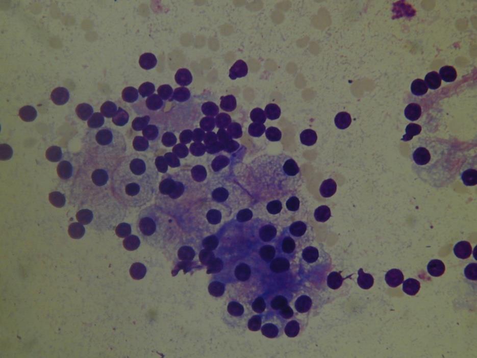
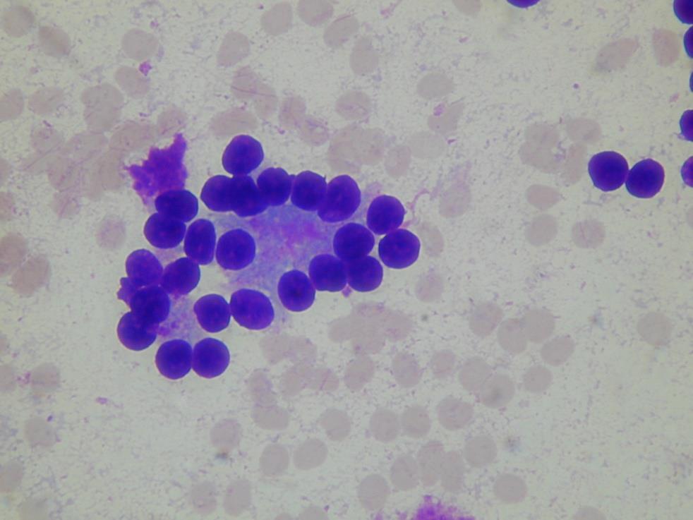
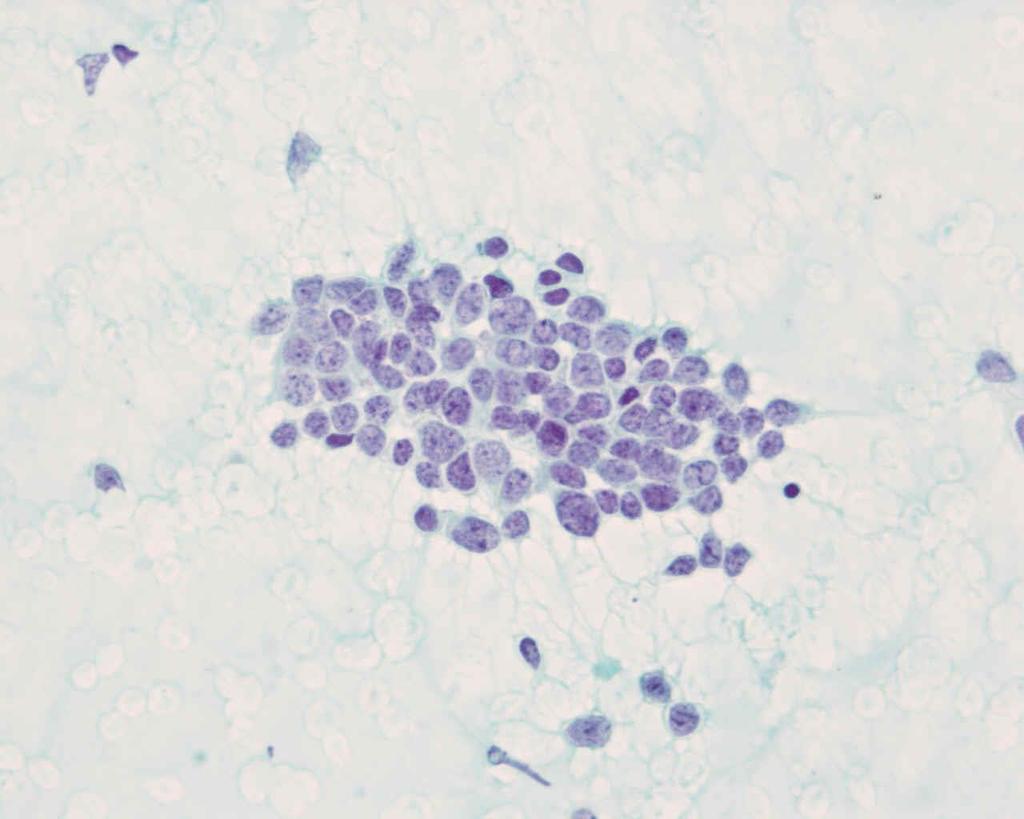
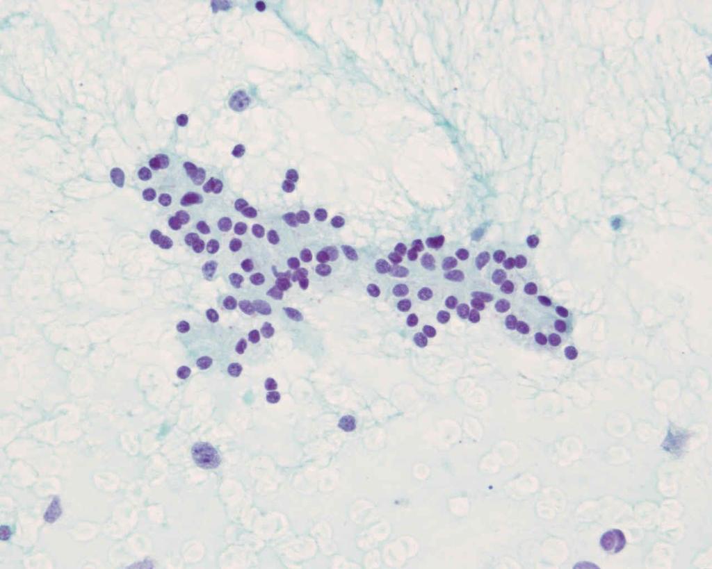
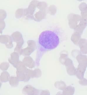
45 High Grade Adenocarcinoma Marked nuclear atypia hyperchromasia pleomorphism overlapping Prominent nucleoli Single atypical cells Mitoses Coagulative Necrosis

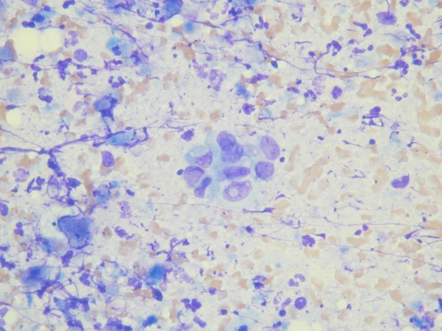
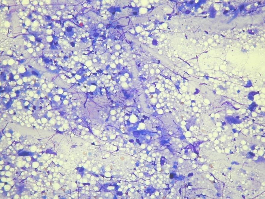
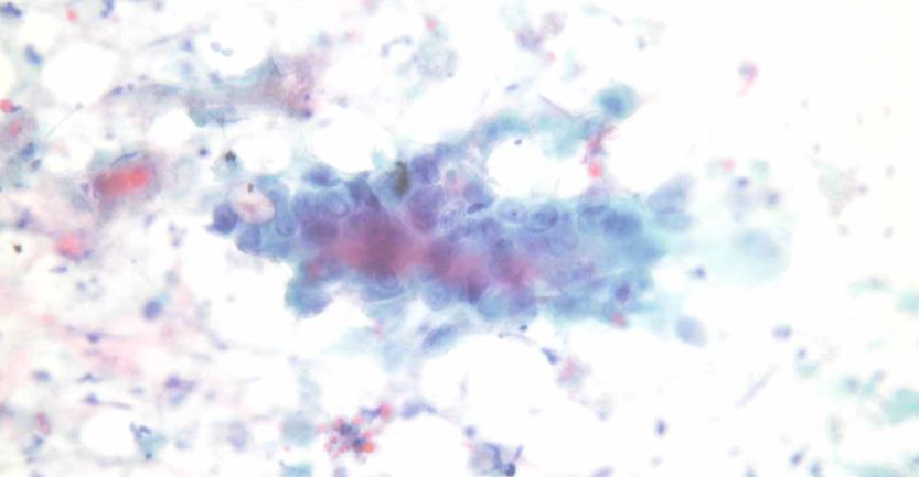
46 High Grade Adenocarcinoma
47 win.eurocytology.eu/virtualslides/git-eus/vs-064


48 Pitfalls Liver cells Intestinal cells Mesothelial cells Endothelial cells

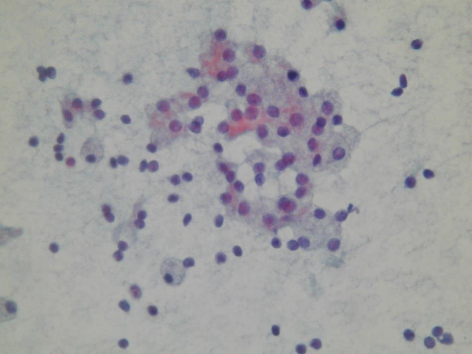
49 Early stages of Chronic active pancreatitis Both ductal and acinar cells Background inflammation Granulation tissue Fat necrosis


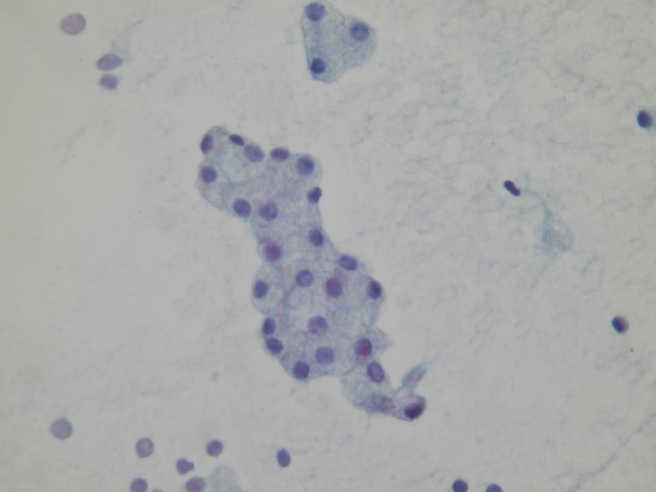
50 Late Chronic Pancreatitis mostly ductal cells few to no acinar cells some islet cells monolayered sheets cohesive, few single cells maintained polarity minimal nuclear overlap mild anisonucleosis smooth nuclear membranes rare/normal mitoses no coagulative necrosis
 Mostly adult men but can be seen in children Presentation variable but generally non-jaundiced (in contrast to ductal ca.")
51 Acinic cell Carcinoma Rare primary tumour Highly aggressive but better 5 year survival than ductal carcinoma (50% vs. <10%) Mostly adult men but can be seen in children Presentation variable but generally non-jaundiced (in contrast to ductal ca.) Small %- syndrome of disseminated fat necrosis/ polyarthralgia due to serum lipase secretion by tumour
52


53 Acinic cell carcinoma High cellularity No ducts No fatty stroma Poorly formed acini Variable cells Atypia variable


54 Pancreatic Endocrine Tumours PET can be cystic due to central necrosis PET, cystic or solid, located most commonly the body and tail Most cystic PET are non-functioning
55 Neuroendocrine Cytology
56 win.eurocytology.eu/virtualslides/git-eus/vs-052


57 Pancreatic Endocrine Tumours homogenous small cell population loosely cohesive clusters and single cells plasmacytoid morphology not uncommon round to oval nuclei coarse, speckled chromatin nucleoli also not uncommon chromogranin should be positive
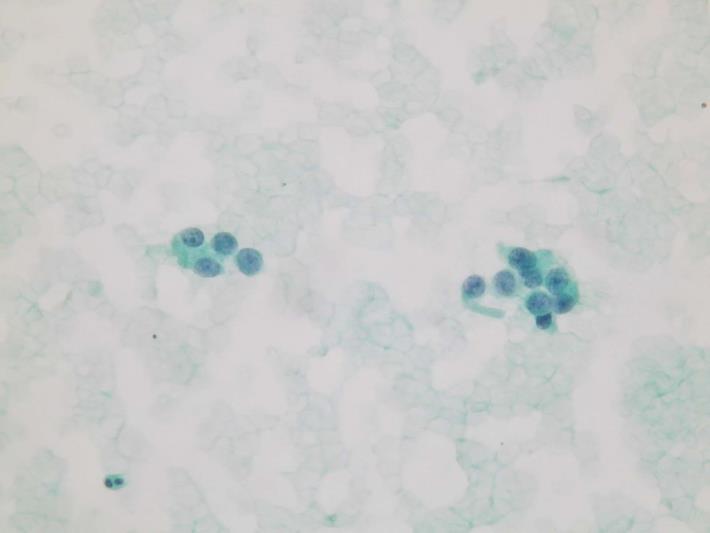
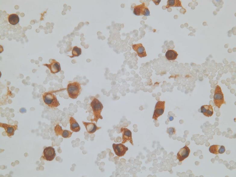
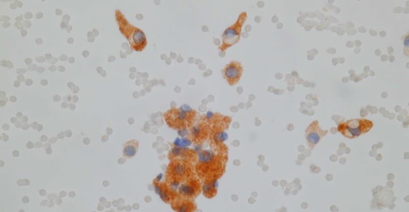
58 (Pancreatic?) Endocrine Tumours Chromogranin Calcitonin
59 Pancreatic cysts (most common and clinically relevant) Pseudocyst Serous cystadenoma Solid pseudopapillary tumour Mucinous cysts mucinous cystic neoplasm intraductal papillary mucinous neoplasm
60 win.eurocytology.eu/virtualslides/git-eus/vs-097
61 Pancreatic Pseudocyst Most common cystic lesion in the pancreas (75-90%) Associated with pancreatitis, trauma, surgery Thick walled, unilocular, +/- communication with duct Fluid aspirated is often dark and not viscous

")
62 Pancreatic Pseudocyst cytology Cyst debris with blood, proteinacous material and sometimes bile variable inflammation NO cyst lining epithelium (beware of contamination, mucin and epithelium)
63 Serous Cystadenoma benign neoplasm in the head and tail of elderly men and women star-burst calcifications within a central scar diagnostic on imaging when present, but this is rarely present most tumours are microcystic with multiple, <2cm cysts, but can be unilocular due to specific variant or due to haemorrhagic degeneration, causing problems with imaging diagnosis

64 Serous Cystadenoma Watery, non-mucinous fluid scant cellularity clean, proteinaceous or bloody background monolayered sheets or small, flat clusters bland, uniform, round nuclei scant but visible non-mucinous cytoplasm
65 Mucinous Cysts of the Pancreas WHO Classification Mucinous cystic neoplasm Mucinous cystadenoma Borderline mucinous cystic neoplasm Mucinous cystadenocarcinoma Intraductal papillary mucinous neoplasm Intraductal papillary mucinous adenoma Intraductal papillary mucinous neoplasm of borderline malignancy Intraductal papillary mucinous carcinoma Intraductal papillary mucinous neoplasm with invasive carcinoma: tubular type or colloid carcinoma
66 Mucinous Cystic Neoplasms (MCN) Lined by mucinous, generally non-papillary epithelium, but can be focally papillary Associated with a subepithelial ovarian-like stroma (females) Predominantly in middle aged females Mostly in the pancreatic tail Cysts do not communicate with the pancreatic ductal system Thin septae

")

67 Mucinous Cystic Neoplasms (MCN)
68 Intraductal papillary mucinous tumour (IPMT) Main duct or branch duct types Macroscopic papillae or mucin Focal or diffuse > 1cm PanIN < 5mm M>F (Main Duct Equal) 60 years average

69 Thank you!
40th European Congress of Cytology Liverpool, UK, 2-5 th October 2016
 40th European Congress of Cytology Liverpool, UK, 2-5 th October 2016 EUS FNA of abdominal organs: An approach to reporting and triage for ancillary testing Date and time: Sunday 2 nd October 2016 15.00-16.30
ROSE in EUS guided FNA of Pancreatic Lesions
 ROSE in EUS guided FNA of Pancreatic Lesions Guy s Hospital, London, 16 April 2018 Laxmi Batav Imperial College NHS Trust Imperial College NHS Trust Cytology Workload Cervical Cytology 57,500 (decreases
Outline 11/2/2017. Pancreatic EUS-FNA general aspects. Cytomorphologic features of solid neoplasms/lesions of the pancreas
 ENDOSCOPIC ULTRASOUND GUIDED-FINE NEEDLE ASPIRATION CYTOLOGY OF PANCREAS Khalid Amin M.D. Assistant Professor Department of Laboratory Medicine and Pathology University of Minnesota Outline Pancreatic
Pancreatitis: A Potential Pitfall in Endoscopic Ultrasound Guided Pancreatic FNA
 Pancreatitis: A Potential Pitfall in Endoscopic Ultrasound Guided Pancreatic FNA Jack Yang, MD Department of Pathology, Medical University of South Carolina Objectives Understand the indication of EUS
Video Microscopy Tutorial 19
 Video Microscopy Tutorial 19 EUS FNA of Pancreatic Cysts Martha Pitman, MD There are no disclosures necessary. EUS-FNA of Pancreatic Cysts Martha Bishop Pitman, M.D. Massachusetts General Hospital Harvard
Evaluation and Management of Cystic Lesions of the Pancreas: When to Resect, When to Follow and When to Forget
 Evaluation and Management of Cystic Lesions of the Pancreas: When to Resect, When to Follow and When to Forget Randall Brand, MD Professor of Medicine Division of Gastroenterology, Hepatology and Nutrition
Pancreatic Cytopathology: A pragmatic approach. By Dr Miguel Perez-Machado MD. PhD. MRCPath Royal Free Hospital
 Pancreatic Cytopathology: A pragmatic approach. By Dr Miguel Perez-Machado MD. PhD. MRCPath Royal Free Hospital Indications of Cytology Sampling To document malignancy in patients with malignant appearing
Neoplasias Quisticas del Páncreas
 SEAP -Aproximación Práctica a la Patología Gastrointestinal- Madrid, 26 de mayo, 2006 Neoplasias Quisticas del Páncreas Gregory Y. Lauwers, M.D. Director, Service Massachusetts General Hospital Harvard
Select problems in cystic pancreatic lesions
 Disclosure Select problems in cystic pancreatic lesions Five Prime Therapeutics shareholder Adicet Bio shareholder Bristol-Meyer Squibb advisory board grace.kim@ucsf.edu Pancreatic cystic lesions Intraductal
Cystic Pancreatic Lesions: Approach to Diagnosis
 Cystic Pancreatic Lesions: Approach to Diagnosis Poster No.: R-0130 Congress: RANZCR-AOCR 2012 Type: Educational Exhibit Authors: A. AGARWAL, R. M. Mendelson; Perth/AU Keywords: Cysts, Biopsy, Endoscopy,
Pancreatico-biliary cytology: a practical approach to diagnosis. Corina Cotoi
 Pancreatico-biliary cytology: a practical approach to diagnosis Corina Cotoi Pancreatico-biliary lesions Solid: Ductal adenocarcinoma Cholangiocarcinoma Acinar cell carcinoma Neuroendocrine tumour / carcinoma
Dr Claire Smith, Consultant Radiologist St James University Hospital Leeds
 Dr Claire Smith, Consultant Radiologist St James University Hospital Leeds Imaging in jaundice and 2ww pathway Image protocol Staging Limitations Pancreatic cancer 1.2.4 Refer people using a suspected
Standardized Terminology in Pancreatobiliary Cytology: The Papanicolaou Society Guidelines
 Standardized Terminology in Pancreatobiliary Cytology: The Papanicolaou Society Guidelines Barbara Ann Centeno. M.D. Vice-Chair, Clinical Services, Anatomic Pathology Assistant Chief, Pathology Service
Case 1. Case 1: EUS Report 5/1/2017. Interesting Cases of Pancreatic Masses
 Interesting Cases of Pancreatic Masses Martha Bishop Pitman, MD Professor of Pathology Harvard Medical School Director of Cytopathology Massachusetts General Hospital Boston, MA MASSACHUSETTS GENERAL PHYSICIANS
Cytyc Corporation - Case Presentation Archive - March 2002
 FirstCyte Ductal Lavage History: 68 Year Old Female Gail Index: Unknown Clinical History: Negative Mammogram in 1995 6 yrs. later presents with bloody nipple discharge Subsequent suspicious mammogram Suspicious
DIAGNOSTIC CHALLENGES Pancreas FNAB. Dr. M. Weir Oct 2017
 DIAGNOSTIC CHALLENGES Pancreas FNAB Dr. M. Weir Oct 2017 CONFLICT OF INTEREST DISCLOSURE I have not had in the past 3 years, a financial interest, arrangement or affiliation with one or more organizations
Thyroid follicular neoplasms in cytology. Ulrika Klopčič Institute of Oncology, Department of Cytopathology, Ljubljana, Slovenia
 Thyroid follicular neoplasms in cytology Ulrika Klopčič Institute of Oncology, Department of Cytopathology, Ljubljana, Slovenia Lecture overview importance of FNAB in assessing thyroid lesions follicular
Salivary Glands 3/7/2017
 Salivary Glands 3/7/2017 Goals and objectives Focus on the entities unique to H&N Common board type facts Information for your future practice Salivary Glands Salivary Glands Major gland. Paratid. Submandibular.
Pancreatic Cystic Lesions 원자력병원
 Pancreatic Cystic Lesions 원자력병원 박선 후 Lines of cellular differentiation Ductal Acinar Undetermined Ductal adenocarcinoma Serous/ mucinous tumor Intraductal papillary mucinous neoplasm Acinar cell carcinoma
Pancreatic Cysts. Darius C. Desai, MD FACS St. Luke s University Health Network
 Pancreatic Cysts Darius C. Desai, MD FACS St. Luke s University Health Network None Disclosures Incidence Widespread use of cross sectional imaging Seen in over 2% of patients having abdominal imaging
Respiratory Tract Cytology
 Respiratory Tract Cytology 40 th European Congress of Cytology Liverpool, UK Momin T. Siddiqui M.D. Professor of Pathology and Laboratory Medicine Director of Cytopathology Emory University Hospital, Atlanta,
Matthew McCollough, M.D. April 9, 2009 University of Louisville
 Matthew McCollough, M.D. April 9, 2009 University of Louisville List the differential diagnosis for pancreatic cysts Review the epidemiology Illustrate the types of cysts through case discussions Discuss
ACG Clinical Guideline: Diagnosis and Management of Pancreatic Cysts
 ACG Clinical Guideline: Diagnosis and Management of Pancreatic Cysts Grace H. Elta, MD, FACG 1, Brintha K. Enestvedt, MD, MBA 2, Bryan G. Sauer, MD, MSc, FACG (GRADE Methodologist) 3 and Anne Marie Lennon,
Patient History. A 58 year old man presents with a 16 mm cyst in the pancreatic tail. The cyst is unilocular with a thick wall and no mural nodule.
 Case 1 Martha Bishop Pitman, MD Director of Cytopathology Massachusetts General Hospital Associate Professor of Pathology Harvard Medical School Boston, MA Patient History A 58 year old man presents with
Appendix 4: WHO Classification of Tumours of the pancreas 17
 S3.01 The WHO histological tumour type must be recorded. CS3.01a The histological type of the tumour should be recorded based on the current WHO classification 17 (refer to Appendices 4-7). Appendix 4:
Pancreatic Cytopathology: The Solid Neoplasms
 Pancreatic Cytopathology: The Solid Neoplasms Syed Z. Ali, M.D. Professor of Pathology and Radiology Director of Cytopathology The Johns Hopkins Hospital Baltimore, Maryland Pancreatic Cytopathology: Past,
The role of endoscopy in the diagnosis and treatment of cystic pancreatic neoplasms
 The role of endoscopy in the diagnosis and treatment of cystic pancreatic neoplasms CYSTIC LESIONS AND FLUID COLLECTIONS OF THE PANCREAS Their pathology ranges from pseudocysts and pancreatic necrosis
Biliary cytolgy and pancreatic endoscopic ultrasound-guided FNA. Leena Krogerus Helsinki, FINLAND
 Biliary cytolgy and pancreatic endoscopic ultrasound-guided FNA Leena Krogerus Helsinki, FINLAND Reasons for biliary cytology PSC- is a pre-neoplastc condition in youg individulas, the cure of which is
Solid pseudopapillary tumour of the pancreas: Report of five cases
 ISPUB.COM The Internet Journal of Pathology Volume 8 Number 2 Solid pseudopapillary tumour of the pancreas: Report of five cases P Srilatha, V Manna, P Kanthilatha Citation P Srilatha, V Manna, P Kanthilatha..
Salivary Gland Cytology
 Salivary Gland Cytology Diagnostic challenges and potential pitfalls Tarik M. Elsheikh, MD Professor and Medical Director Anatomic Pathology Cleveland Clinic FNA Salivary Gland Lesions Indications Distinguish
An Approach to Pancreatic Cysts. Introduction
 An Approach to Pancreatic Cysts Nalini M. Guda, MD Aurora St. Luke s Medical Center, Milwaukee Clinical Adjunct Professor of Medicine, University of Wisconsin School of Medicine and Public Health Introduction
CYTOLOGY OF EUS- GUIDED FNA OF THE PANCREAS AND THE UPPER GI TRACT
 CYTOLOGY OF EUS- GUIDED FNA OF THE PANCREAS AND THE UPPER GI TRACT Barbara A. Centeno, M.D. Vice-Chair, Clinical Services Assistant Chief of Pathology Director of Cytopathology Department of Anatomic Pathology/Moffitt
FNA of Thyroid. Toward a Uniform Terminology With Management Guidelines. NCI NCI Thyroid FNA State of the Science Conference
 FNA of Thyroid NCI NCI Thyroid FNA State of the Science Conference Toward a Uniform Terminology With Management Guidelines Thyroid Thyroid FNA Cytomorphology NCI Thyroid FNA State of the Science Conference
Biliary Tract Neoplasia: A Cyto-histologic Review. Michelle Reid, MD, MSc Professor of Pathology Director of Cytopathology Emory University Hospital
 Biliary Tract Neoplasia: A Cyto-histologic Review Michelle Reid, MD, MSc Professor of Pathology Director of Cytopathology Emory University Hospital Bile Duct Brushings (BDB) BDBs are the initial diagnostic
Salivary gland tumor cytologic and histologic correlation: Algorithmic and risk stratification based approaches
 Salivary gland tumor cytologic and histologic correlation: Algorithmic and risk stratification based approaches Christopher C. Griffith, MD, PhD Raja R. Seethala, MD 1. Salivary gland tumor cytology: A
The Pancreas. Basic Anatomy. Endocrine pancreas. Exocrine pancreas. Pancreas vasculature. Islets of Langerhans. Acinar cells Ductal System
 SGNA: Back to Basics Rogelio G. Silva, MD Assistant Clinical Professor of Medicine University of Illinois at Chicago Department of Medicine Division of Gastroenterology Advocate Christ Medical Center GI
The role of the cytologist in breast cancer screening
 The role of the cytologist in breast cancer screening I.Seili-Bekafigo, MD, PhD Clinical cytologist KBC Rijeka Croatian Society for Clinical Cytology Fine needle aspiration (FNA, FNAB, FNAC) Fine needle
CT 101 :Pancreas and Spleen
 CT 101 :Pancreas and Spleen Shikha Khullar,, MD, MPH Division of Radiology University of South Alabama The Pancreas Normal Pancreas 3 Phase Pancreatic CT Non contrast Arterial phase : 30-35 35 second
Radiology Pathology Conference
 Radiology Pathology Conference Nadia F. Yusaf, M.D. PGY-3 1/29/2010 Presentation material is for education purposes only. All rights reserved. 2010 URMC Radiology Page 1 of 90 Case 1 60 year- old man presents
Pancreatobiliary Frozen Section Nightmares
 Pancreatobiliary Frozen Section Nightmares Aatur D. Singhi, MD PhD Assistant Professor University of Pittsburgh Medical Center Department of Pathology singhiad@upmc.edu Objectives Briefly give an overview
Biliary tract tumors
 Short Course 2010 Annual Fall Meeting of the Korean Society for Pathologists Biliary tract tumors Joon Hyuk Choi, M.D., Ph.D. Professor, Department of Pathology, Yeungnam Univ. College of Medicine, Daegu,
Chronic pancreatitis mimicking intraductal papillary mucinous neoplasm of the pancreas; Report of tow cases
 Jichi Medical University Journal Chronic pancreatitis mimicking intraductal papillary mucinous neoplasm of the pancreas; Report of tow cases Noritoshi Mizuta, Hiroshi Noda, Nao Kakizawa, Nobuyuki Toyama,
Intraductal Papillary Mucinous Neoplasms: We Still Have a Way to Go! Francesco M. Serafini, MD, FACS
 Intraductal Papillary Mucinous Neoplasms: We Still Have a Way to Go! Francesco M. Serafini, MD, FACS Brooklyn VAMC September 21 st GI Grand Rounds - What is it? - Clinical entity that has emerged from
Mody. AIS vs. Invasive Adenocarcinoma of the Cervix
 Common Problems in Gynecologic Pathology Michael T. Deavers, M.D. Houston Methodist Hospital, Houston, Texas Common Problems in Gynecologic Pathology Adenocarcinoma in-situ (AIS) of the Cervix vs. Invasive
Intro to Gallbladder & Pancreas Pathology
 Cholecystitis acute chronic Gallbladder tumors Adenomyoma (benign) Adenocarcinoma Pancreatitis acute chronic Pancreatic tumors Intro to Gallbladder & Pancreas Pathology Helen Remotti M.D. Gallstones (Cholelithiasis)
Biliary cytolgy and pancreatic endoscopic ultrasound-guided. Helsinki, FINLAND
 Biliary cytolgy and pancreatic endoscopic ultrasound-guided d FNA Leena Krogerus Helsinki, FINLAND Reasons for biliary cytology Obstructions PSC, a premalignant condition, the cure of which is transplantation
EUS-FNA OF PANCREATIC EXOCRINE TUMORS COMPARISON OF EXPERIENCES WITH PATHOLOGICAL DIAGNOSIS
 EUS-FNA OF PANCREATIC EXOCRINE TUMORS COMPARISON OF EXPERIENCES WITH PATHOLOGICAL DIAGNOSIS Vincenzo Canzonieri, M.D. CRO - Aviano National Cancer Institute Dept of Pathology EUS European Cyto-Pathologist
Unusual Pancreatic Neoplasms RTC 2/11/2011
 Unusual Pancreatic Neoplasms RTC 2/11/2011 Objectives Intraductal Papillary Mucinous Neoplasm (IPMN) Mucinous Cystic Neoplasm (MCN) Islet Cell Tumors Insulinoma Glucagonoma VIPoma Somatostatinoma Gastrinoma
ENDOSCOPIC ULTRA SOUND GUIDED FNA OF GI TRACT AND PANCREAS
 ENDOSCOPIC ULTRA SOUND GUIDED FNA OF GI TRACT AND PANCREAS Prof. Fernando Schmitt Medical Faculty of Porto University, Porto, Portugal IPATIMUP General Secretary of the International Academy of Cytology
EUS FNA NEUROENDOCRINE TUMORS. A. Ginès Endocopy Unit Hospital Cínic. Barcelona (Spain)
") EUS FNA NEUROENDOCRINE TUMORS A. Ginès Endocopy Unit Hospital Cínic. Barcelona (Spain) GI NEUROENDOCRINE TUMORS GENERAL CONCEPTS Rare neoplasms arising from the neuroendocrine cells of the GI tract Include:
EUS FNA NEUROENDOCRINE TUMORS A. Ginès Endocopy Unit Hospital Cínic. Barcelona (Spain) GI NEUROENDOCRINE TUMORS GENERAL CONCEPTS Rare neoplasms arising from the neuroendocrine cells of the GI tract Include:
Proliferative Epithelial lesions of the Breast. Sami Shousha, MD, FRCPath Charing Cross Hospital & Imperial College, London
 Proliferative Epithelial lesions of the Breast Sami Shousha, MD, FRCPath Charing Cross Hospital & Imperial College, London Amman, November2013 Proliferative Epithelial Lesions of the Breast Usual type
Morphologic features in cystic lesions of pancreas-a retrospective analysis
 International Journal of Advances in Medicine Cicy PJ et al. Int J Adv Med. 2018 Feb;5(1):192-196 http://www.ijmedicine.com pissn 2349-3925 eissn 2349-3933 Original Research Article DOI: http://dx.doi.org/10.18203/2349-3933.ijam20180083
FNA OF SALIVARY GLANDS: A PRACTICAL APPROACH
 FNA OF SALIVARY GLANDS: A PRACTICAL APPROACH FNA of Salivary Glands: Challenges Wide range of neoplastic and non-neoplastic lesions Cytological overlap between the different benign and malignant tumors
X-ray Corner. Imaging of The Pancreas. Pantongrag-Brown L
 X-ray Corner 125 Imaging of The Pancreas Modern imaging modalities commonly used in pancreas include ultrasound (US), CT, and MRI. Pancreas is a retroperitoneal organ which makes it difficult to visualize
Management A Guideline Based Approach to the Incidental Pancreatic Cysts. Common Cystic Pancreatic Neoplasms.
 Management 2016 A Guideline Based Approach to the Incidental Pancreatic Cysts ISMRM 2016 Masoom Haider, MD, FRCP(C) Professor of Radiology, University of Toronto Clinician Scientist, Ontario Institute
Anatomy of the biliary tract
 Harvard-MIT Division of Health Sciences and Technology HST.121: Gastroenterology, Fall 2005 Instructors: Dr. Jonathan Glickman Anatomy of the biliary tract Figure removed due to copyright reasons. Biliary
Outline. Intraductal Papillary Mucinous Neoplasm (IPMN) Guideline Review 4/6/2017. Case Example Background Classification Histology Guidelines
 Guideline Review 4/6/2017. Case Example Background Classification Histology Guidelines") Intraductal Papillary Mucinous Neoplasm (IPMN) Guideline Review The Nurse Practitioner Association New York State Capital Region Teaching Day Matthew Warndorf MD Case Example Background Classification
Intraductal Papillary Mucinous Neoplasm (IPMN) Guideline Review The Nurse Practitioner Association New York State Capital Region Teaching Day Matthew Warndorf MD Case Example Background Classification
Objectives. Salivary Gland FNA: The Milan System. Role of Salivary Gland FNA 04/26/2018
 Salivary Gland FNA: The Milan System Dr. Jennifer Brainard Section Head Cytopathology Cleveland Clinic Objectives Introduce the Milan System for reporting salivary gland cytopathology Define cytologic
Team approach is essential incorporating: radiology, gastroenterology, surgery and pathology Successful performance is operator dependent
 Committee V Post Brushing and FNA Testing Managment Options Daniel Kurtycz, M.D. Chair Committee V Committee V - Post brushing and FNA management options Team approach is essential incorporating: radiology,
Breast pathology. 2nd Department of Pathology Semmelweis University
 Breast pathology 2nd Department of Pathology Semmelweis University Breast pathology - Summary - Benign lesions - Acute mastitis - Plasma cell mastitis / duct ectasia - Fat necrosis - Fibrocystic change/
Pancreatic neoplasms associated with significant amounts of extracellular. Fine-Needle Aspiration Cytology of Mucinous Tumors of the Pancreas CANCER
 92 CANCER CYTOPATHOLOGY Fine-Needle Aspiration Cytology of Tumors of the Pancreas Monica Recine, M.D. 1 Madhukar Kaw, M.D. 2 Douglas B. Evans, M.D. 3 Savitri Krishnamurthy, M.D. 1 1 Division of Pathology
Background to the Thyroid Nodule
 William C. Faquin, MD, PhD Professor of Pathology Harvard Medical School Director of Head and Neck Pathology Massachusetts Eye and Ear Massachusetts General Hospital THYROID FNA: PART I Background to the
The Role of Molecular Analysis in the Diagnosis and Surveillance of Pancreatic Cystic Neoplasms
 JOP. J Pancreas (Online) 20 Mar 20; (2):-9. RESEARCH ARTICLE The Role of Molecular Analysis in the Diagnosis and Surveillance of Pancreatic Cystic Neoplasms Megan Winner, Amrita Sethi 2, John M Poneros
Evaluation of AGA and Fukuoka Guidelines for EUS and surgical resection of incidental pancreatic cysts
 Evaluation of AGA and Fukuoka Guidelines for EUS and surgical resection of incidental pancreatic cysts Authors Alexander Lee 1, Vivek Kadiyala 2,LindaS.Lee 3 Institutions 1 Texas Digestive Disease Consultants,
DIAGNOSTIC SLIDE SEMINAR: PART 1 RENAL TUMOUR BIOPSY CASES
 DIAGNOSTIC SLIDE SEMINAR: PART 1 RENAL TUMOUR BIOPSY CASES Dr. Andrew J. Evans MD, PhD, FACP, FRCPC Consultant in Genitourinary Pathology University Health Network, Toronto, ON Case 1 43 year-old female,
Intro to Gallbladder & Pancreas Pathology
 Cholecystitis acute chronic Gallbladder tumors Adenomyoma (benign) Adenocarcinoma Pancreatitis i acute chronic Pancreatic tumors Intro to Gallbladder & Pancreas Pathology Helen Remotti M.D. Dept. of Pathology
Cytyc Corporation - Case Presentation Archive - October 2001
 ThinPrep Pap Test History: 82 Year Old Female Specimen Type: Peritoneal Washings Case provided by Dr. Berle Stratton, Southwest Washington Medical Center, Vancouver, Washington. *The images, analysis and
Intraductal carcinoma of the prostate on needle biopsy: histologic features and clinical significance
 & 2006 USCAP, Inc All rights reserved 0893-3952/06 $30.00 www.modernpathology.org Intraductal carcinoma of the prostate on needle biopsy: histologic features and clinical significance Charles C Guo 1 and
Objectives. Pancreatic Cysts. Benefits and Limitations of the Cytologic Assessment of Cystic Pancreatic Lesions and Masses
 Benefits and Limitations of the Cytologic Assessment of Cystic Pancreatic Lesions and Masses Michelle D. Reid, MD, MSc Associate 1/24/15 Professor Director of Cytology - EUHM Michelle D Reid MD, MS Emory
Kenneth D. Chi, MD Medical Director GI Lab Advocate Lutheran General Hospital
 Kenneth D. Chi, MD Medical Director GI Lab Advocate Lutheran General Hospital Advances in Digestive Health for the Primary Care Physician Symposium May 2, 2015 None Case Presentation Types of Pancreatic
PRELIMINARY CYTOLOGIC DIAGNOSIS: Suspicious for Acinic Cell Carcinoma. Cell Block: Immunohistochemical Studies CYTOLOGIC DIAGNOSIS:
 1 PRELIMINARY CYTOLOGIC DIAGNOSIS: Suspicious for Acinic Cell Carcinoma. Cell Block: Immunohistochemical Studies GCDFP-15 S-100 CYTOLOGIC DIAGNOSIS: Consistent with mammary analogue secretory carcinoma.
Medullary Thyroid Carcinoma. This case was provided by Treant Hospital, Bethesda, Hoogeveen, The Netherlands
 Medullary Thyroid Carcinoma This case was provided by Treant Hospital, Bethesda, Hoogeveen, The Netherlands ADS-01504 Rev. 001 2016 Hologic, Inc. All rights reserved. Overview Medullary Thyroid Carcinoma
Objectives. Atypical Glandular Cells. Atypical Endocervical Cells. Reactive Endocervical Cells
 2013 California Society of Pathologists 66 th Annual Meeting San Francisco, CA Atypical Glandular Cells to Early Invasive Adenocarcinoma: Cervical Cytology and Histology Christina S. Kong, MD Associate
Standardization of Nomenclature
 Standardized Terminology and Nomenclature for Pancreaticobiliary Cytopathology from the Papanicolaou Society of Cytopathology Lester J. Layfield, M.D. Professor and Chair University of Missouri Pathology
Neoplasia 2018 Lecture 2. Dr Heyam Awad MD, FRCPath
 Neoplasia 2018 Lecture 2 Dr Heyam Awad MD, FRCPath ILOS 1. List the differences between benign and malignant tumors. 2. Recognize the histological features of malignancy. 3. Define dysplasia and understand
INTRADUCTAL LESIONS OF THE PROSTATE. Jonathan I. Epstein
 INTRADUCTAL LESIONS OF THE PROSTATE Jonathan I. Epstein Topics Prostatic intraepithelial neoplasia (PIN) Intraductal adenocarcinoma (IDC-P) Intraductal urothelial carcinoma Ductal adenocarcinoma High Prostatic
Case year old female presented with asymmetric enlargement of the left lobe of the thyroid
 Case 4 22 year old female presented with asymmetric enlargement of the left lobe of the thyroid gland. No information available relative to a prior fine needle aspiration biopsy. A left lobectomy was performed.
Case 4 Diagnosis 2/21/2011 TGB
 Case 4 22 year old female presented with asymmetric enlargement of the left lobe of the thyroid gland. No information available relative to a prior fine needle aspiration biopsy. A left lobectomy was performed.
3/28/2017. Disclosure of Relevant Financial Relationships. GU Evening Subspecialty Case Conference. Differential Diagnosis:
 GU Evening Subspecialty Case Conference Rajal B. Shah, M.D. VP, Medical Director, Urologic Pathology Miraca Life Sciences, Irving, Texas Clinical Associate Professor of Pathology Baylor College of Medicine,
EUS-guided FNAB. Differential Diagnosis 5/1/2017. EUS-FNA of Solid and Cystic Lesions:
 EUS-FNA of Solid and Cystic Lesions: Part 1: Solid Masses Martha Bishop Pitman, M.D. Director, Cytopathology Massachusetts General Hospital Professor of Pathology Harvard Medical School Boston, MA EUS-guided
performed to help sway the clinician in what the appropriate diagnosis is, which can substantially alter the treatment of management.
 Hello, I am Maura Polansky at the University of Texas MD Anderson Cancer Center. I am a Physician Assistant in the Department of Gastrointestinal Medical Oncology and the Program Director for Physician
Disclosures. Parathyroid Pathology. Objectives. The normal parathyroid 11/10/2012
 Disclosures Parathyroid Pathology I have nothing to disclose Annemieke van Zante MD/PhD Assistant Professor of Clinical Pathology Associate Chief of Cytopathology Objectives 1. Review the pathologic features
G3.02 The malignant potential of the neoplasm should be recorded. CG3.02a
 G3.02 The malignant potential of the neoplasm should be recorded. CG3.02a Conventional adrenocortical neoplasm. Each of the below parameters is scored 0 when absent and 1 when present. 3 or more of these
BREAST PATHOLOGY. Fibrocystic Changes
 BREAST PATHOLOGY Lesions of the breast are very common, and they present as palpable, sometimes painful, nodules or masses. Most of these lesions are benign. Breast cancer is the 2 nd most common cause
Imaging in breast cancer. Mammography and Ultrasound Donya Farrokh.MD Radiologist Mashhad University of Medical Since
 Imaging in breast cancer Mammography and Ultrasound Donya Farrokh.MD Radiologist Mashhad University of Medical Since A mammogram report is a key component of the breast cancer diagnostic process. A mammogram
Oncocytic-Appearing Salivary Gland Tumors. Oncocytic, Cystic, Mucinous, and High Grade Salivary Gland Tumors SALIVARY GLAND FNA: PART II
 William C. Faquin, MD, PhD Professor of Pathology Harvard Medical School Director of Head and Neck Pathology Massachusetts Eye and Ear Massachusetts General Hospital SALIVARY GLAND FNA: PART II Oncocytic,
CPC 4 Breast Cancer. Rochelle Harwood, a 35 year old sales assistant, presents to her GP because she has noticed a painless lump in her left breast.
 CPC 4 Breast Cancer Rochelle Harwood, a 35 year old sales assistant, presents to her GP because she has noticed a painless lump in her left breast. 1. What are the most likely diagnoses of this lump? Fibroadenoma
Hepatobiliary and Pancreatic Malignancies
 Hepatobiliary and Pancreatic Malignancies Gareth Eeson MD MSc FRCSC Surgical Oncologist and General Surgeon Kelowna General Hospital Interior Health Consultant, Surgical Oncology BC Cancer Agency Centre
Endoscopic Ultrasound-guided FNA Cytology of the Pancreas
 Endoscopic Ultrasound-guided FNA Cytology of the Pancreas 67 th Annual California Society of Pathologists 2014 Laura Tabatabai, MD Professor of Pathology University of California, San Francisco Overview
XIII CONGRESSO NAZIONALE Roma, 7-9 novembre NODULO TIROIDEO: Agoaspirato o Core Needle Biopsy?
 XIII CONGRESSO NAZIONALE NODULO TIROIDEO: Agoaspirato o Core Needle Biopsy? Anna Crescenzi Policlinico Universitario Campus Bio-Medico Roma Indeterminate lesions are heterogeneous The gray zone CONSERVATIVE
Diagnostic performance of endoscopic ultrasound-guided fine-needle aspiration in pancreatic lesions
 European Review for Medical and Pharmacological Sciences 2018; 22: 1397-1401 Diagnostic performance of endoscopic in pancreatic lesions Q.-M. WU 1, Y.-N. GUO 1, Y.-Q. XU 1 Digestive Department of Beijing
Cystic pancreatic lesions A proposal for a network approach. Chris Briggs Consultant HPB Surgeon Peninsula HPB Unit Derriford Hospital, Plymouth
 Cystic pancreatic lesions A proposal for a network approach Chris Briggs Consultant HPB Surgeon Peninsula HPB Unit Derriford Hospital, Plymouth Aims Brief overview of cystic pancreatic lesions International
BOSNIAN-TURKISH CYTOPATHOLOGY SCHOOL June 18-19, 2016 Sarajevo. Case Discussions. 60 year old woman Routine gynecologic control LBC
 BOSNIAN-TURKISH CYTOPATHOLOGY SCHOOL June 18-19, 2016 Sarajevo Case Discussions Prof Dr Sıtkı Tuzlalı Tuzlalı Pathology Laboratory 60 year old woman Routine gynecologic control LBC 1 2 Endometrial thickening
Fine-Needle Aspiration Cytology of Low-Grade Cribriform Cystadenocarcinoma with Many Psammoma Bodies of the Salivary Gland
 The Korean Journal of Pathology 2013; 47: 481-485 CASE STUDY Fine-Needle Aspiration Cytology of Low-Grade Cribriform Cystadenocarcinoma with Many Psammoma Bodies of the Salivary Gland Ji Yun Jeong Dongbin
PSA. HMCK, p63, Racemase. HMCK, p63, Racemase
 Case 1 67 year old male presented with gross hematuria H/o acute prostatitis & BPH Urethroscopy: small, polypoid growth with a broad base emanating from the left side of the verumontanum Serum PSA :7 ng/ml
Introduction of Endoscopic Ultrasonography (EUS)
") Introduction of Endoscopic Ultrasonography (EUS) Dr. Yuk Tong LEE MBChB, MD(CUHK), FRCP (Edin), FRCP(Lond), FHKCP, FHKAM Specialist in Gastroenterology and Hepatology Endoscopic Ultrasonography (EUS) ª
Introduction of Endoscopic Ultrasonography (EUS) Dr. Yuk Tong LEE MBChB, MD(CUHK), FRCP (Edin), FRCP(Lond), FHKCP, FHKAM Specialist in Gastroenterology and Hepatology Endoscopic Ultrasonography (EUS) ª
Workshop 9. Cytopathology of Solid and Cystic Neoplasms of the Pancreas: Everything You Need To Know For Daily Practice.
 Workshop 9 Cytopathology of Solid and Cystic Neoplasms of the Pancreas: Everything You Need To Know For Daily Practice Michelle Reid, MD Momin Siddiqui, MD,FIAC There are no disclosures necessary. Cytopathology
TBSRTC 1- Probabilistic approach and Relationship to Clinical Algorithms
 The Benefits of a Uniform Reporting System for Thyroid Cytopathology BETHESDA REPORTING SYSTEM Prof. Fernando Schmitt Department of Pathology and Oncology, Medical Faculty of Porto University Head of Molecular
Almost any suspected tumor can be aspirated easily and safely. Some masses are more risky to aspirate including:
 DOES THIS PATIENT HAVE CANCER? USING IN-HOUSE CYTOLOGY TO HELP YOU MAKE THIS DIAGNOSIS. Joyce Obradovich, DVM, Diplomate, ACVIM (Oncology) Animal Cancer & Imaging Center, Canton, Michigan Almost every
NODULAR CYSTIC HIDRADENOMA OVER THE GLUTEAL REGION: A RARE CYTOMORPHOLOGICAL DIAGNOSIS
 NODULAR CYSTIC HIDRADENOMA OVER THE GLUTEAL REGION: A RARE CYTOMORPHOLOGICAL DIAGNOSIS Abstract: The primary as well as metastatic tumours of the skin can be diagnosed by fine needle aspiration cytology