Case 1. Case 1: EUS Report 5/1/2017. Interesting Cases of Pancreatic Masses
|
|
|
- Irene Bates
- 5 years ago
- Views:
Transcription

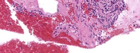
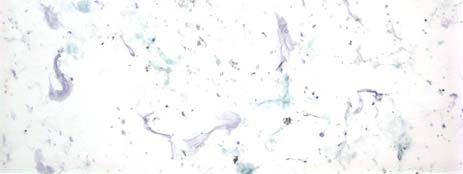
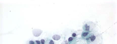
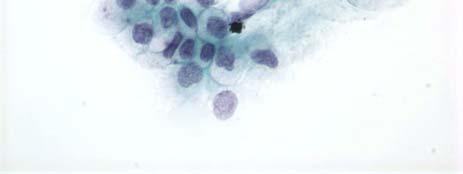
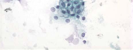
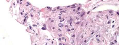
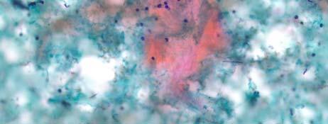
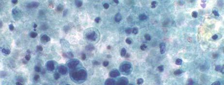
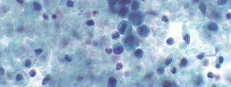
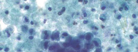
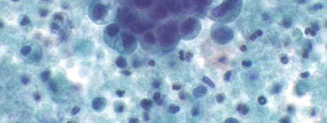
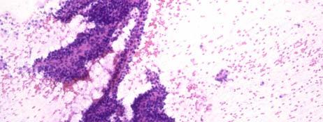
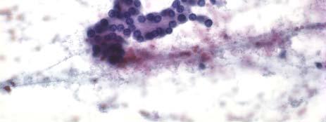
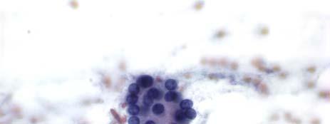
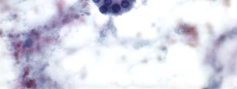
1 Interesting Cases of Pancreatic Masses Martha Bishop Pitman, MD Professor of Pathology Harvard Medical School Director of Cytopathology Massachusetts General Hospital Boston, MA MASSACHUSETTS GENERAL PHYSICIANS ORGANIZATION HARVARD MEDICAL SCHOOL Case 1 A 76 year old asymptomatic man was found to have a multiloculated cyst in the pancreatic tail. Approximately 1mL of slightly thin, slightly bloody fluid was removed. A cystospin was made of the cells and the supernatant was sent for CEA and amylase. Case 1: EUS Report A 3.5 cm well defined multiloculated, lobulated cystic mass was noted in the pancreatic tail. The cyst wall was not well visualized and the septations were thick. 1

2 Case 1 Case 1 Case 1 2
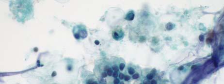
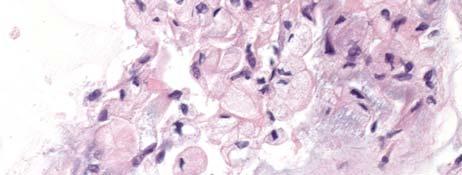
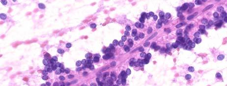
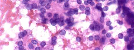
3 Case 1 Case 1: Cyst Fluid Analysis Amylase = 20 U/L CEA = 0.5 ng/ml. Case 1 Cytology Diagnosis: Satisfactory for Evaluation Neoplastic: Benign Nonmucinous cyst fluid with low amylase (20 U/L) and CEA (0.5 ng/ml.) consistent with serous cystadenoma. 3
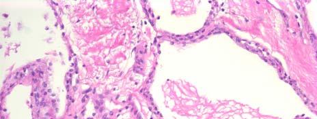
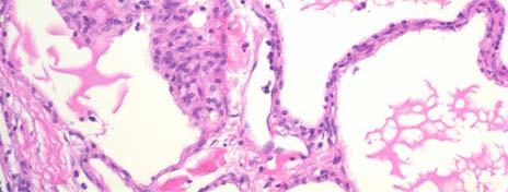
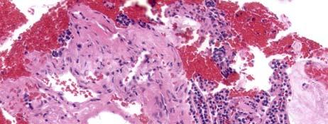
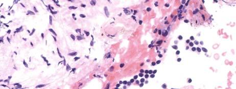
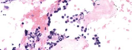
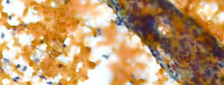
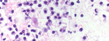
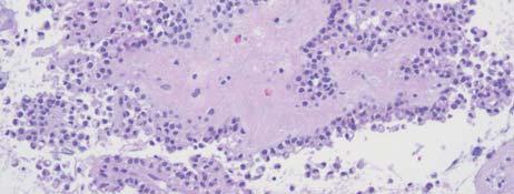
4 Serous Cystadenoma Shark Core Biopsy Moray Microforceps Biopsy 4


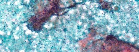
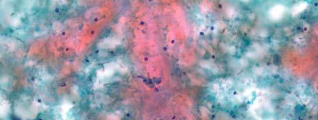
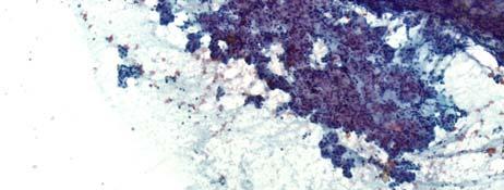
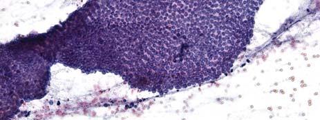
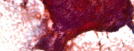
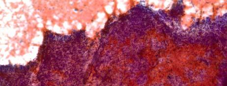
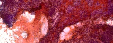
5 Serous Cystadenoma PAS dpas Case 1: Teaching Points SCA produce nonmucinous fluid Usually bloody or clear Hemosiderin-laden macrophages act as a surrogate marker to suggest the diagnosisi Epithelium is fragile and may not survive processing by smearing SCA have low amylase and CEA Case 2 A 64 year old woman being worked up for esophageal stricture is noted to have a cystic mass in the uncinate process. An EUS-FNA is scheduled. 5


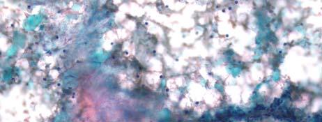
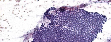
6 Case 2: EUS Report The body and tail of the pancreas is very atrophic. In the uncinate process, there is a fairly well circumscribed round cystic mass with thick cyst wall measuring about 30 X 20 mm. Aspirates are productive of yellow cyst fluid; cyst fluid CEA and amylase are assessed. Case 2 Case 2 6


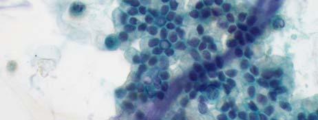
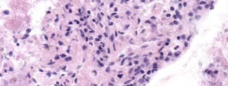
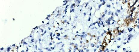
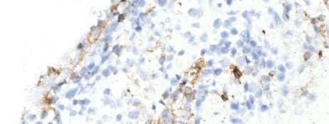
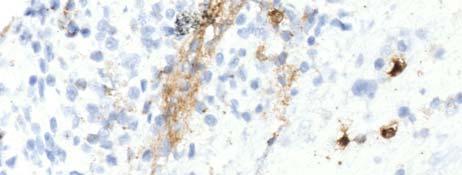
7 Case 2 Case 2: Cyst Fluid Analysis CEA = 6 mg/ml Amylase = 65 U/L Case 2: Molecular Analysis KRAS wild type GNAS wild type 7

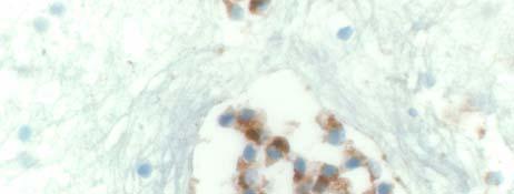
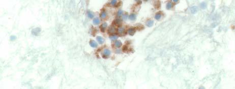
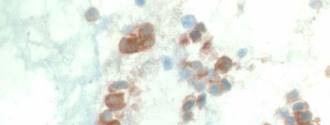
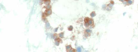
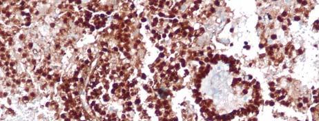
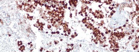
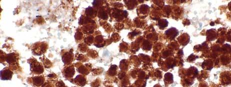
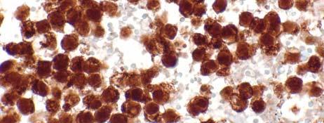
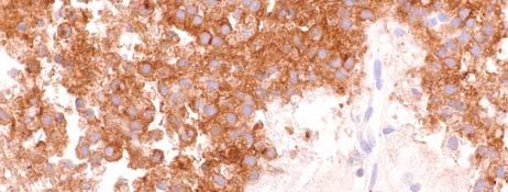
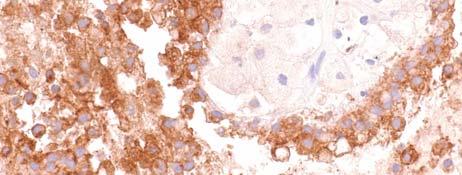
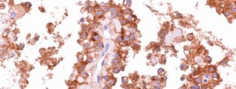
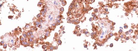
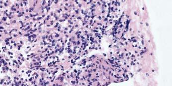
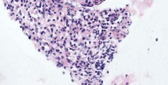
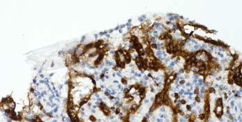
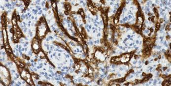
8 Case 2: CB Case 2: chromogranin Case 2: synaptophysin 8
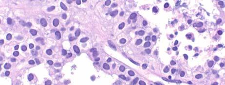
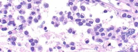
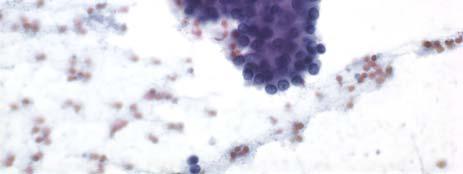
9 Case 2 Cytological Diagnosis Satisfactory for Evaluation Neoplastic: Other Cystic neuroendocrine tumor Case 2: Teaching Points cpannets mimic primary pancreatic cysts The cells of PanNET fall into the HGA category due to the small cell size and the abnormal chromatin Usually the endocrine features of the cells are apparent Cytology is THE test to diagnose cpannets: CEA and amylase are low and molecular analysis is negative Morales-Oyarvide V, Yoon WJ, Ingkakul T, Forcione DG, Casey BW, Brugge WR, Fernández-Del Castillo C, Pitman MB. Cystic pancreatic neuroendocrine tumors: The value of cytology in preoperative diagnosis. Cancer Cytopathol Case 2: Teaching Points If the diagnosis is indeterminate use the ATYPICAL category, not the SUSPICIOUS category Atypical is to Neoplastic as Suspicious is to Positive Similar for other neoplasms in the Other category 9


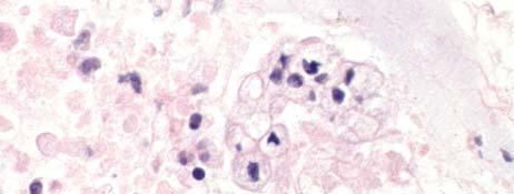
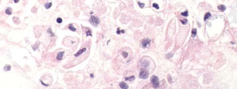
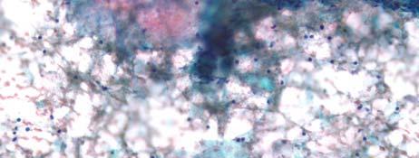
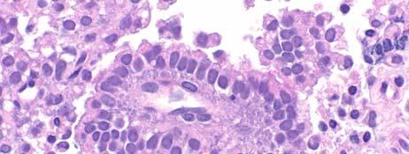
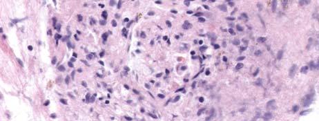
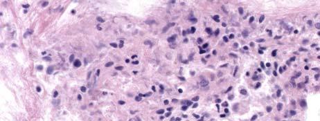
10 Case 3 A 54 year old woman was found to have a cyst in the pancreatic body following resolution of a bout of pancreatitis that brought her to the ER. Case 3: EUS Report A 25 mm cyst was identified in the pancreatic body. There was a single compartment without septae. The outer wall of the lesion was thick. There was a small mural nodule. Case 3 10

11 Case 3 Case 3: Cyst Fluid Analysis Amylase = 409 U/L CEA = 5165 ng/ml. Case 3: Molecular Analysis KRAS mutant GNAS mutant 11


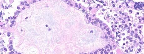
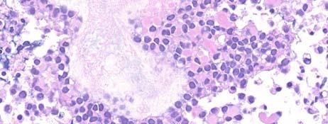
12 Case 3 Cytological Diagnosis Satisfactory for Evaluation Neoplastic: Other Mucinous cyst with high-grade grade atypia consistent with IPMN with at least high-grade dysplasia. See note. Case 3: IPMN-branch duct type with high-grade dysplasia Case 3: Teaching Points Cytology is the best test for determining cyst grade Distinguishing between low and high-grade is sufficient for patient management Intermediate-grade dysplasia is best placed with low-grade but features overlap with HGD. HGD cells are smaller than a 12 duodenal enterocyte, have abnormal chromatin and high N/C ratio; necrosis is often present 12

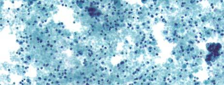
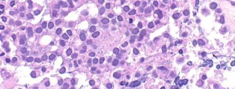
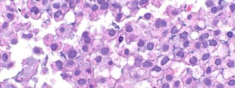
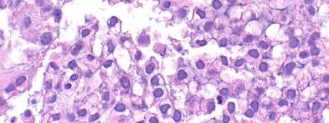
13 Case 4 An 84 year old female was found to have a 2.6 cm cyst in the body/tail of the pancreas during evaluation of her Barrett s esophagus. An FNA was performed. 1 ml of thin, clear fluid was submitted for cytology and biochemical testing. Case 4: EUS Report A 2.6 cm anechoic cyst was noted in the body-tail of the pancreas. There were no septations and the wall was thin. There was no associated mass lesion. A connection to the main pancreatic duct was visualized. Case 4: Cyst Fluid Analysis Amylase = 111,153 U/L y, CEA = 56 ng/ml. 13


14 Case 4 Case 4 Case 4 14
15 Case 4 Cytological Diagnosis Satisfactory for Evaluation Negative for Malignancy Mucoid cyst fluid; no high-grade g epithelial cells present. See note. Note: There appears to be extracellular mucin but its source is unclear and CEA is not elevated above our cut off level of 192 ng/ml to support a neoplastic mucinous cyst. Benign appearing mucinous epithelium is present, the distinction from gastric contamination is not possible. Case 4: Follow-up -In this elderly, asymptomatic woman with low-risk imaging that supports the diagnosis of an IPMN and low-risk cytology, conservative e observation was recommended -Follow-up at one year showed no growth Case 4: Teaching Points Aspiration of pancreatic cysts serves two main purposes: Determining if the cyst is mucinous Definitive criteria may not be present- choice between ND and negative, and correlation with clinical and imaging helps to determine which is best for the patient Determining if the cyst is high-risk by cytology, e.g. if there is HGA present HGA = HGD or adenocarcinoma No HGA + low-risk imaging = 99% NPV Wu RI, Yoon WJ, Brugge WR, Mino-Kenudson M, Pitman MB. Endoscopic ultrasound-guided fine needle aspiration (EUS-FNA) contributes to a triple-negative test in preoperative screening of pancreatic cysts. Cancer Cytopathol. 2014; 122:
16 Case 5 A 62 year old female is worked up for acute onset of abdominal pain. Ultrasound shows a markedly dilated pancreatic duct with sharp tapering in the pancreatic head where a 2 cm round cystic mass is identified. EUS with FNA was performed. Case 5: EUS Report A round hypoechoic, calcified and cystic mass is identified in the pancreatic head measuring 16 mm by 20 mm The endosonographic borders are welldefined. An intact interface is seen between the mass at the adjacent structures suggesting a lack of invasion. Diagnostic needle aspiration for fluid is performed into the cystic component of the mass and into the mass itself. The amount of brown-yellow fluid collected is 4 ml. The fluid is sent for biochemical and molecular analysis. Case 5: Cyst Fluid Analysis CEA = 35 mg/ml Amylase = 65,549 U/L KRAS mutant GNAS mutant 16


17 Case 5:cyst Case 5:cyst Case 5: mass TP 17


18 Case 5:mass TP Case 5:mass CB Case 5:mass CB 18


19 Case 5:mass CB-ki-67 Case 5:mass CB-SMAD4 Case 5 Diagnosis: Satisfactory for Evaluation Positive for Malignancy Adenocarcinoma consistent with IPMN with invasive carcinoma. See note. Note: The cyst is mucinous by CEA elevation and KRAS/GNAS mutations, the GNAS mutation supporting an IPMN. The cyst epithelium is low-grade but the associated mass is diagnostic of adenocarcinoma, supported by a brisk ki-67 positivity and loss of nuclear SMAD4 staining. 19


20 PDAC Case 5: Teaching Points Aspiration of both the cyst and associated mass is required for accurate diagnosis. A cyst may not show significant atypia whereas the mass will show adenocarcinoma Although these components of a single mass lesion may have discordant results, the overall result of the FNA is what counts This type of case should be considered a TP in studies A low CEA does not exclude a mucinous cyst GNAS mutations support the diagnosis of IPMN Cytology is the best test for grading the cyst/mass PDAC may be deceptively bland- the cytoplasm is the biggest clue to diagnosis!! Case 6 A 45 year old male was found to have a mass in the pancreatic tail during workup for non-specific abdominal pain. 20

21 Case 6: EUS Report EUS showed a 1.5 cm round, well-defined solid and cystic mass with a heterogenous echotexture in the pancreatic tail; 3cc of thick cyst fluid obtained Case 6: Cyst Fluid Analysis CEA = 235 mg/ml Amylase = 237 U/L Case 6: Molecular Analysis KRAS WT GNAS WT 21

22 Case 6 Case 6 Case 6 22


23 Case 6 Keratinous debris mimicking desiccated mucin Case 6 Cytological Diagnosis: Evaluation limited by scant cellularity Evaluation limited by scant cellularity Negative for Malignancy Lymphoepithelial Cyst 23
24 Case 6: Teaching Points Rare; 4:1 M:F; mean 56 years thick cheesy keratinous debris may cause the cyst to appear solid on imaging Recognition of keratinous debris is the key to diagnosis Elevated CEA a pitfall! Case 7 A 68 year old female presented to the emergency room for acute abdominal pain about 1 week following a dental procedure. A CT scan showed a ~3cm cyst in the pancreatic head. The patient was referred to a gastroenterologist. MASSACHUSETTS GENERAL PHYSICIANS ORGANIZATION HARVARD MEDICAL SCHOOL Case 7: Clinical History No history of alcohol abuse No history of pancreatitis Sudden onset of abdominal pain MASSACHUSETTS GENERAL PHYSICIANS ORGANIZATION HARVARD MEDICAL SCHOOL 24

25 Case 7:EUS Performed 36 x 26 mm cyst in the pancreatic head ~10 cc of thin purulent fluid was obtained. Cultures sent; no other CFA performed. Cytology = neutrophils only. A gram stain showed gram positive cocci in pairs and chains. and culture grew abundant Rothia Dentocariosa. 1 month of antibiotics was prescribed. Repeat EUS persistent cyst 30 x 25mm 12 cc of opaque, thin, yellow fluid obtained. Cytospin of cyst fluid- whole mount image 25

26 Case 7 Case 7 Case 7 26


27 Case 7 Case 7 Case 7: Diagnosis Neoplastic: Other Mucinous cyst with high-grade epithelial atypia consistent with branch-duct intraductal papillary mucinous neoplasm with at least high-grade dysplasia. 27

 Resect Case 8 A 56 year old man presents with abdominal pain.")
28 . IPMN, branch-duct type, with focal high-grade dysplasia. No invasion identified Case 7 Case 7: Teaching Points Clinical presentation Benign appearing BD-IPMN infected from dental procedure No high risk imaging features Small, no mural nodule, no associated dilated MPD CFA: Biochemical very elevated CEA supporting a mucinous etiology Cytology HGA (suspicious) Resect Case 8 A 56 year old man presents with abdominal pain. CT shows an 8 cm solid, partially cystic mass in the pancreatic head. An FNA is performed- smears and cellblock are made. 28


29 Case 8 Case 8 Case 8 29

30 Case 8 Case 8 Case 8 30
31 Case 8 Case 8 Beta-catenin 31
32 Beta-catenin CD10 CD56 32
33 Case 8:Diagnosis Satisfactory for Evaluation Neoplastic: Other Solid-pseudopapillary Neoplasm Case 8: Teaching Points SPN do occur in males (4-7%) with the same age mean and range as females- Mean age 30-35; Range 8-85 Frequent pseudopapillary pattern Formed by separation of vascular cores with adherent cells Typically thin walled vessels in cores >95% cured by resection Most recurrences or metastases associated with atypical features Rare non-atypical cases may metastasize May occur years later Most common metastatic sites Liver Peritoneum / omentum Local lymph nodes involved rarely Neoplastic: Other interpretation category rather than Malignant in the PSC PB terminology system Case 9 A 72 year old female is found incidentally to have a vague, questionable mass in the pancreatic body. The pancreas otherwise was normal. An EUS-FNA of the mass is performed. Direct smears fixed in alcohol are stained with a Papanicolaou stain. No EUS report was available as the case was sent to me in consultation. 33


34 Case 9 Case 9 Case 9 34

35 Case 9 Case 9 Case 9 35


36 Case 9 Cytological Diagnosis: Satisfactory for Evaluation Negative for Malignancy Benign pancreatic tissue. Acinar Tissue is often confused with NET Case 9: Teaching Points negative is an appropriate interpretation category when imaging does not indicate a definitive mass lesion nondiagnostic is the more appropriate category when imaging clearly defines a mass lesion Misdiagnosis of normal acinar tissue as NET is a significant pitfall. Mixture of ductal epithelium and?acinar/lesional? tissue is a clue to it being pancreatic acinar epithelium If uncertain, and NET is a possibility, use atypical category 36


37 Case 10 A 50 year old asymptomatic man is found to have a 1.5 cm round mass in the tail of the pancreas. Case 10: EUS 1.5 cm round well-circumscribed solid mass consistent with a neuroendocrine tumor Case 10 37

38 Case 10 Case 10 Case 10 38


39 Case 10 Case 10 Case 10: synaptophysin 39
40 Case 10: chromogranin Case 10: CD8 Diagnosis Negative Splenule (Accessory spleen) 40


41 Non-viscous and CEA c and -KRAS/GNAS Serous Cystadenoma Lymphoepithelial cyst GI contaminate in Pseudocyst Cystic neuroendocrine tumor Solid-pseudopapillary tumor Cystic acinar cell carcinoma Viscous or CEA c or +KRAS/GNAS Mucinous cystic neoplasm with lowintermediate grade dysplasia Intraductal papillary mucinous neoplasm with low-intermediate grade dysplasia Colloid carcinoma Mucinous cystic neoplasm with highgrade dysplasia or invasive carcinoma Intraductal papillary mucinous neoplasm with high-grade dysplasia or invasive carcinoma Colloid carcinoma Cystic ductal adenocarcinoma a small cell with high nuclear to cytoplasmic ratio and abnormal chromatin and/or nuclear membranes b large cells with low nuclear to cytoplasmic ratio and moderate to abundant cytoplasm, often mucinous c Cut off value of 192 ng/ml has an accuracy of ~80% for detecting a mucinous cyst 5/1/2017 Case 10: Teaching Points Splenule mimic NETs on images Cytologically, splenules mimic NETs or lymph node CD8 is diagnostic IHC stain along with negative endocrine markers Helpful Resources Chapter 3: Tissue handling and processing, including cellblock technique (collodian bag) Chapter 4: Ancillary Studies Chapter 7: Cystic Lesions Algorithm for interpretation Helpful Resources Appendix A: Template for signing out pancreatic cysts Appendix B: Mucinous Etiology Epithelium High grade a Low g grade b Absent Present 41
Patient History. A 58 year old man presents with a 16 mm cyst in the pancreatic tail. The cyst is unilocular with a thick wall and no mural nodule.
 Case 1 Martha Bishop Pitman, MD Director of Cytopathology Massachusetts General Hospital Associate Professor of Pathology Harvard Medical School Boston, MA Patient History A 58 year old man presents with
Video Microscopy Tutorial 19
 Video Microscopy Tutorial 19 EUS FNA of Pancreatic Cysts Martha Pitman, MD There are no disclosures necessary. EUS-FNA of Pancreatic Cysts Martha Bishop Pitman, M.D. Massachusetts General Hospital Harvard
Standardized Terminology in Pancreatobiliary Cytology: The Papanicolaou Society Guidelines
 Standardized Terminology in Pancreatobiliary Cytology: The Papanicolaou Society Guidelines Barbara Ann Centeno. M.D. Vice-Chair, Clinical Services, Anatomic Pathology Assistant Chief, Pathology Service
Pancreatico-biliary cytology: a practical approach to diagnosis. Corina Cotoi
 Pancreatico-biliary cytology: a practical approach to diagnosis Corina Cotoi Pancreatico-biliary lesions Solid: Ductal adenocarcinoma Cholangiocarcinoma Acinar cell carcinoma Neuroendocrine tumour / carcinoma
Evaluation and Management of Cystic Lesions of the Pancreas: When to Resect, When to Follow and When to Forget
 Evaluation and Management of Cystic Lesions of the Pancreas: When to Resect, When to Follow and When to Forget Randall Brand, MD Professor of Medicine Division of Gastroenterology, Hepatology and Nutrition
CYTOLOGY OF EUS- GUIDED FNA OF THE PANCREAS AND THE UPPER GI TRACT
 CYTOLOGY OF EUS- GUIDED FNA OF THE PANCREAS AND THE UPPER GI TRACT Barbara A. Centeno, M.D. Vice-Chair, Clinical Services Assistant Chief of Pathology Director of Cytopathology Department of Anatomic Pathology/Moffitt
The role of endoscopy in the diagnosis and treatment of cystic pancreatic neoplasms
 The role of endoscopy in the diagnosis and treatment of cystic pancreatic neoplasms CYSTIC LESIONS AND FLUID COLLECTIONS OF THE PANCREAS Their pathology ranges from pseudocysts and pancreatic necrosis
An Approach to Pancreatic Cysts. Introduction
 An Approach to Pancreatic Cysts Nalini M. Guda, MD Aurora St. Luke s Medical Center, Milwaukee Clinical Adjunct Professor of Medicine, University of Wisconsin School of Medicine and Public Health Introduction
40th European Congress of Cytology Liverpool, UK, 2-5 th October 2016
 40th European Congress of Cytology Liverpool, UK, 2-5 th October 2016 EUS FNA of abdominal organs: An approach to reporting and triage for ancillary testing Date and time: Sunday 2 nd October 2016 15.00-16.30
Standardization of Nomenclature
 Standardized Terminology and Nomenclature for Pancreaticobiliary Cytopathology from the Papanicolaou Society of Cytopathology Lester J. Layfield, M.D. Professor and Chair University of Missouri Pathology
Matthew McCollough, M.D. April 9, 2009 University of Louisville
 Matthew McCollough, M.D. April 9, 2009 University of Louisville List the differential diagnosis for pancreatic cysts Review the epidemiology Illustrate the types of cysts through case discussions Discuss
Advances in Pancreatic. Cytology. Martha B. Pitman, MD. Disclosure of Relevant Financial Relationships
 Advances in Pancreatic Cytology Martha B. Pitman, MD Disclosure of Relevant Financial Relationships USCAP requires that all planners (Education Committee) in a position to influence or control the content
ACG Clinical Guideline: Diagnosis and Management of Pancreatic Cysts
 ACG Clinical Guideline: Diagnosis and Management of Pancreatic Cysts Grace H. Elta, MD, FACG 1, Brintha K. Enestvedt, MD, MBA 2, Bryan G. Sauer, MD, MSc, FACG (GRADE Methodologist) 3 and Anne Marie Lennon,
Pancreatic Cytopathology: A pragmatic approach. By Dr Miguel Perez-Machado MD. PhD. MRCPath Royal Free Hospital
 Pancreatic Cytopathology: A pragmatic approach. By Dr Miguel Perez-Machado MD. PhD. MRCPath Royal Free Hospital Indications of Cytology Sampling To document malignancy in patients with malignant appearing
Pancreatitis: A Potential Pitfall in Endoscopic Ultrasound Guided Pancreatic FNA
 Pancreatitis: A Potential Pitfall in Endoscopic Ultrasound Guided Pancreatic FNA Jack Yang, MD Department of Pathology, Medical University of South Carolina Objectives Understand the indication of EUS
Pancreatic Cysts. Darius C. Desai, MD FACS St. Luke s University Health Network
 Pancreatic Cysts Darius C. Desai, MD FACS St. Luke s University Health Network None Disclosures Incidence Widespread use of cross sectional imaging Seen in over 2% of patients having abdominal imaging
Outline 11/2/2017. Pancreatic EUS-FNA general aspects. Cytomorphologic features of solid neoplasms/lesions of the pancreas
 ENDOSCOPIC ULTRASOUND GUIDED-FINE NEEDLE ASPIRATION CYTOLOGY OF PANCREAS Khalid Amin M.D. Assistant Professor Department of Laboratory Medicine and Pathology University of Minnesota Outline Pancreatic
Select problems in cystic pancreatic lesions
 Disclosure Select problems in cystic pancreatic lesions Five Prime Therapeutics shareholder Adicet Bio shareholder Bristol-Meyer Squibb advisory board grace.kim@ucsf.edu Pancreatic cystic lesions Intraductal
Neoplasias Quisticas del Páncreas
 SEAP -Aproximación Práctica a la Patología Gastrointestinal- Madrid, 26 de mayo, 2006 Neoplasias Quisticas del Páncreas Gregory Y. Lauwers, M.D. Director, Service Massachusetts General Hospital Harvard
Pancreatic Cysts. Pancreatic Cysts. Multidisciplinary and Multimodal Approach To the Pre-Operative Diagnosis of Pancreatic Cysts
 Multidisciplinary and Multimodal Approach To the Pre-Operative Diagnosis of Pancreatic Cysts Martha Bishop Pitman, M.D. Director of Cytopathology Massachusetts General Hospital Professor of Pathology Harvard
Evaluation of AGA and Fukuoka Guidelines for EUS and surgical resection of incidental pancreatic cysts
 Evaluation of AGA and Fukuoka Guidelines for EUS and surgical resection of incidental pancreatic cysts Authors Alexander Lee 1, Vivek Kadiyala 2,LindaS.Lee 3 Institutions 1 Texas Digestive Disease Consultants,
Conflicts of Interest
 Standardized Terminology and Nomenclature for Pancreaticobiliary Cytopathology from the Papanicolaou Society of Cytopathology Lester J. Layfield, M.D. Professor and Chair University of Missouri Pathology
Objectives. Pancreatic Cysts. Benefits and Limitations of the Cytologic Assessment of Cystic Pancreatic Lesions and Masses
 Benefits and Limitations of the Cytologic Assessment of Cystic Pancreatic Lesions and Masses Michelle D. Reid, MD, MSc Associate 1/24/15 Professor Director of Cytology - EUHM Michelle D Reid MD, MS Emory
Cystic Pancreatic Lesions: Approach to Diagnosis
 Cystic Pancreatic Lesions: Approach to Diagnosis Poster No.: R-0130 Congress: RANZCR-AOCR 2012 Type: Educational Exhibit Authors: A. AGARWAL, R. M. Mendelson; Perth/AU Keywords: Cysts, Biopsy, Endoscopy,
Pancreatic Cystic Lesions 원자력병원
 Pancreatic Cystic Lesions 원자력병원 박선 후 Lines of cellular differentiation Ductal Acinar Undetermined Ductal adenocarcinoma Serous/ mucinous tumor Intraductal papillary mucinous neoplasm Acinar cell carcinoma
EUS-FNAB of GI Tract: Specimen Handling and Triage
 EUS-FNAB of GI Tract: Specimen Handling and Triage Amy Ly, M.D. Director, Fine Needle Aspiration Service Massachusetts General Hospital Assistant Professor of Pathology Harvard Medical School Boston, MA
Management A Guideline Based Approach to the Incidental Pancreatic Cysts. Common Cystic Pancreatic Neoplasms.
 Management 2016 A Guideline Based Approach to the Incidental Pancreatic Cysts ISMRM 2016 Masoom Haider, MD, FRCP(C) Professor of Radiology, University of Toronto Clinician Scientist, Ontario Institute
Endoscopic Ultrasound-guided FNA Cytology of the Pancreas
 Endoscopic Ultrasound-guided FNA Cytology of the Pancreas 67 th Annual California Society of Pathologists 2014 Laura Tabatabai, MD Professor of Pathology University of California, San Francisco Overview
Pancreatic Cystic Neoplasms: Guidelines and beyond
 Pancreatic Cystic Neoplasms: Guidelines and beyond Kenneth J. Chang, MD, FACG, FASGE Executive Director, Comprehensive Digestive Disease Center Professor and Chief, Gastroenterology Vincent & Anna Kong
Outline. Intraductal Papillary Mucinous Neoplasm (IPMN) Guideline Review 4/6/2017. Case Example Background Classification Histology Guidelines
 Guideline Review 4/6/2017. Case Example Background Classification Histology Guidelines") Intraductal Papillary Mucinous Neoplasm (IPMN) Guideline Review The Nurse Practitioner Association New York State Capital Region Teaching Day Matthew Warndorf MD Case Example Background Classification
Intraductal Papillary Mucinous Neoplasm (IPMN) Guideline Review The Nurse Practitioner Association New York State Capital Region Teaching Day Matthew Warndorf MD Case Example Background Classification
Pancreatic Cytopathology: The Solid Neoplasms
 Pancreatic Cytopathology: The Solid Neoplasms Syed Z. Ali, M.D. Professor of Pathology and Radiology Director of Cytopathology The Johns Hopkins Hospital Baltimore, Maryland Pancreatic Cytopathology: Past,
DIAGNOSTIC CHALLENGES Pancreas FNAB. Dr. M. Weir Oct 2017
 DIAGNOSTIC CHALLENGES Pancreas FNAB Dr. M. Weir Oct 2017 CONFLICT OF INTEREST DISCLOSURE I have not had in the past 3 years, a financial interest, arrangement or affiliation with one or more organizations
ROSE in EUS guided FNA of Pancreatic Lesions
 ROSE in EUS guided FNA of Pancreatic Lesions Guy s Hospital, London, 16 April 2018 Laxmi Batav Imperial College NHS Trust Imperial College NHS Trust Cytology Workload Cervical Cytology 57,500 (decreases
Radiology Pathology Conference
 Radiology Pathology Conference Nadia F. Yusaf, M.D. PGY-3 1/29/2010 Presentation material is for education purposes only. All rights reserved. 2010 URMC Radiology Page 1 of 90 Case 1 60 year- old man presents
ENDOSCOPIC ULTRA SOUND GUIDED FNA OF GI TRACT AND PANCREAS
 ENDOSCOPIC ULTRA SOUND GUIDED FNA OF GI TRACT AND PANCREAS Prof. Fernando Schmitt Medical Faculty of Porto University, Porto, Portugal IPATIMUP General Secretary of the International Academy of Cytology
Suspicious Cytologic Diagnostic Category in Endoscopic Ultrasound-Guided FNA of the Pancreas: Follow-Up and Outcomes
 Suspicious Cytologic Diagnostic Category in Endoscopic Ultrasound-Guided FNA of the Pancreas: Follow-Up and Outcomes Evan A. Alston, MD 1 ; Sejong Bae, PhD 2 ; and Isam A. Eltoum, MD, MBA 1 BACKGROUND:
Chronic pancreatitis mimicking intraductal papillary mucinous neoplasm of the pancreas; Report of tow cases
 Jichi Medical University Journal Chronic pancreatitis mimicking intraductal papillary mucinous neoplasm of the pancreas; Report of tow cases Noritoshi Mizuta, Hiroshi Noda, Nao Kakizawa, Nobuyuki Toyama,
X-ray Corner. Imaging of The Pancreas. Pantongrag-Brown L
 X-ray Corner 125 Imaging of The Pancreas Modern imaging modalities commonly used in pancreas include ultrasound (US), CT, and MRI. Pancreas is a retroperitoneal organ which makes it difficult to visualize
Original article. Introduction!
 382 Original article Peritoneal seeding in intraductal papillary mucinous neoplasm of the pancreas patients who underwent endoscopic ultrasound-guided fine-needle aspiration: The PIPE Study Authors Won
State of the Art Symposium
 State of the Art Symposium Syed Z. Ali, MD There is no disclosure necessary Martha B. Pitman, MD There is no disclosure necessary Lester Layfield, MD There is no disclosure necessary Joseph Herman, MD
Biliary cytolgy and pancreatic endoscopic ultrasound-guided FNA. Leena Krogerus Helsinki, FINLAND
 Biliary cytolgy and pancreatic endoscopic ultrasound-guided FNA Leena Krogerus Helsinki, FINLAND Reasons for biliary cytology PSC- is a pre-neoplastc condition in youg individulas, the cure of which is
The Role of Molecular Analysis in the Diagnosis and Surveillance of Pancreatic Cystic Neoplasms
 JOP. J Pancreas (Online) 20 Mar 20; (2):-9. RESEARCH ARTICLE The Role of Molecular Analysis in the Diagnosis and Surveillance of Pancreatic Cystic Neoplasms Megan Winner, Amrita Sethi 2, John M Poneros
Oncocytic-Appearing Salivary Gland Tumors. Oncocytic, Cystic, Mucinous, and High Grade Salivary Gland Tumors SALIVARY GLAND FNA: PART II
 William C. Faquin, MD, PhD Professor of Pathology Harvard Medical School Director of Head and Neck Pathology Massachusetts Eye and Ear Massachusetts General Hospital SALIVARY GLAND FNA: PART II Oncocytic,
EUS-guided FNAB. Differential Diagnosis 5/1/2017. EUS-FNA of Solid and Cystic Lesions:
 EUS-FNA of Solid and Cystic Lesions: Part 1: Solid Masses Martha Bishop Pitman, M.D. Director, Cytopathology Massachusetts General Hospital Professor of Pathology Harvard Medical School Boston, MA EUS-guided
Diagnostic performance of endoscopic ultrasound-guided fine-needle aspiration in pancreatic lesions
 European Review for Medical and Pharmacological Sciences 2018; 22: 1397-1401 Diagnostic performance of endoscopic in pancreatic lesions Q.-M. WU 1, Y.-N. GUO 1, Y.-Q. XU 1 Digestive Department of Beijing
The Frozen Section: Diagnostic Challenges and Pitfalls
 The Frozen Section: Diagnostic Challenges and Pitfalls William C. Faquin, M.D., Ph.D. Director, Head and Neck Pathology Massachusetts General Hospital & Massachusetts Eye and Ear Infirmary Harvard Medical
Case Scenario 1. Discharge Summary
 Case Scenario 1 Discharge Summary A 69-year-old woman was on vacation and noted that she was becoming jaundiced. Two months prior to leaving on that trip, she had had a workup that included an abdominal
Guidelines for Pancreaticobiliary Cytology from the Papanicolaou Society of Cytopathology: A Review
 Guidelines for Pancreaticobiliary Cytology from the Papanicolaou Society of Cytopathology: A Review Martha B. Pitman, MD 1 and Lester J. Layfield, MD 2 The newest installment on state-of-the-art standards
04/10/2018. Intraductal Papillary Neoplasms Of Breast INTRADUCTAL PAPILLOMA
 Intraductal Papillary Neoplasms Of Breast Savitri Krishnamurthy MD Professor of Pathology Deputy Division Head The University of Texas MD Anderson Cancer Center 25 th Annual Seminar in Pathology Pittsburgh,
Predictors of Malignancy in Thyroid Fine-Needle Aspirates Cyst Fluid Only Cases
 Predictors of Malignancy in Thyroid Fine-Needle Aspirates Cyst Fluid Only Cases Can Potential Clues of Malignancy Be Identified? Mohammad Jaragh, MD 1 ; V. Bessie Carydis, MMedSci (Cytol) 1 ; Christina
Types of IPMN. Pancreas Cysts: An Incidental Finding or Harbinger of Malignancy. Cysts: Early Neoplasia. Mucinous Cystic Lesions. EUS-guided FNA EUS
 Pancreas Cysts: An Incidental Finding or Harbinger of Malignancy EUS-guided FNA William R. Brugge,, MD, FACG Professor of Medicine Harvard Medical School Director, GI Endoscopy Unit Massachusetts General
KRAS: ONE ACTOR, MANY POTENTIAL ROLES IN DIAGNOSIS
 UNIVERSITÀ DEGLI STUDI DI PALERMO Scuola di Specializzazione in Biochimica Clinica Direttore Prof. Marcello Ciaccio KRAS: ONE ACTOR, MANY POTENTIAL ROLES IN DIAGNOSIS Loredana Bruno KRAS gene Proto-oncogene
Research Article The Changing Spectrum of Surgically Treated Cystic Neoplasms of the Pancreas
 HPB Surgery Volume 2015, Article ID 791704, 7 pages http://dx.doi.org/10.1155/2015/791704 Research Article The Changing Spectrum of Surgically Treated Cystic Neoplasms of the Pancreas Jennifer K. Plichta,
The Pancreas. Basic Anatomy. Endocrine pancreas. Exocrine pancreas. Pancreas vasculature. Islets of Langerhans. Acinar cells Ductal System
 SGNA: Back to Basics Rogelio G. Silva, MD Assistant Clinical Professor of Medicine University of Illinois at Chicago Department of Medicine Division of Gastroenterology Advocate Christ Medical Center GI
Biliary Tract Neoplasia: A Cyto-histologic Review. Michelle Reid, MD, MSc Professor of Pathology Director of Cytopathology Emory University Hospital
 Biliary Tract Neoplasia: A Cyto-histologic Review Michelle Reid, MD, MSc Professor of Pathology Director of Cytopathology Emory University Hospital Bile Duct Brushings (BDB) BDBs are the initial diagnostic
Salivary Gland Cytology
 Salivary Gland Cytology Diagnostic challenges and potential pitfalls Tarik M. Elsheikh, MD Professor and Medical Director Anatomic Pathology Cleveland Clinic FNA Salivary Gland Lesions Indications Distinguish
Hepatobiliary and Pancreatic Malignancies
 Hepatobiliary and Pancreatic Malignancies Gareth Eeson MD MSc FRCSC Surgical Oncologist and General Surgeon Kelowna General Hospital Interior Health Consultant, Surgical Oncology BC Cancer Agency Centre
Kenneth D. Chi, MD Medical Director GI Lab Advocate Lutheran General Hospital
 Kenneth D. Chi, MD Medical Director GI Lab Advocate Lutheran General Hospital Advances in Digestive Health for the Primary Care Physician Symposium May 2, 2015 None Case Presentation Types of Pancreatic
Pancreatobiliary Frozen Section Nightmares
 Pancreatobiliary Frozen Section Nightmares Aatur D. Singhi, MD PhD Assistant Professor University of Pittsburgh Medical Center Department of Pathology singhiad@upmc.edu Objectives Briefly give an overview
Team approach is essential incorporating: radiology, gastroenterology, surgery and pathology Successful performance is operator dependent
 Committee V Post Brushing and FNA Testing Managment Options Daniel Kurtycz, M.D. Chair Committee V Committee V - Post brushing and FNA management options Team approach is essential incorporating: radiology,
Papillary Lesions of the Breast A Practical Approach to Diagnosis. (Arch Pathol Lab Med. 2016;140: ; doi: /arpa.
 Papillary Lesions of the Breast A Practical Approach to Diagnosis (Arch Pathol Lab Med. 2016;140:1052 1059; doi: 10.5858/arpa.2016-0219-RA) Papillary lesions of the breast Span the spectrum of benign,
Cytology Specimen Management, Triage and Standardized Reporting of Fine Needle Aspiration Biopsies of the Pancreas
 Cytology Specimen Management, Triage and Standardized Reporting of Fine Needle Aspiration Biopsies of the Pancreas The Harvard community has made this article openly available. Please share how this access
Cytyc Corporation - Case Presentation Archive - March 2002
 FirstCyte Ductal Lavage History: 68 Year Old Female Gail Index: Unknown Clinical History: Negative Mammogram in 1995 6 yrs. later presents with bloody nipple discharge Subsequent suspicious mammogram Suspicious
Update on Thyroid FNA The Bethesda System. Shikha Bose M.D. Associate Professor Cedars Sinai Medical Center
 Update on Thyroid FNA The Bethesda System Shikha Bose M.D. Associate Professor Cedars Sinai Medical Center Thyroid Nodules Frequent occurrence Palpable: 4-7% of adults Ultrasound: 10-31% Majority benign
Cystic lesions of the pancreas
 REVIEW ARTICLE Annals of Gastroenterology (2016) 29, 155-161 Cystic lesions of the pancreas Ioannis Karoumpalis a, Dimitrios K. Christodoulou b General Hospital of Athens G. Gennimatas, Athens; University
Cystic pancreatic lesions A proposal for a network approach. Chris Briggs Consultant HPB Surgeon Peninsula HPB Unit Derriford Hospital, Plymouth
 Cystic pancreatic lesions A proposal for a network approach Chris Briggs Consultant HPB Surgeon Peninsula HPB Unit Derriford Hospital, Plymouth Aims Brief overview of cystic pancreatic lesions International
Dr Claire Smith, Consultant Radiologist St James University Hospital Leeds
 Dr Claire Smith, Consultant Radiologist St James University Hospital Leeds Imaging in jaundice and 2ww pathway Image protocol Staging Limitations Pancreatic cancer 1.2.4 Refer people using a suspected
EUS FNA NEUROENDOCRINE TUMORS. A. Ginès Endocopy Unit Hospital Cínic. Barcelona (Spain)
") EUS FNA NEUROENDOCRINE TUMORS A. Ginès Endocopy Unit Hospital Cínic. Barcelona (Spain) GI NEUROENDOCRINE TUMORS GENERAL CONCEPTS Rare neoplasms arising from the neuroendocrine cells of the GI tract Include:
EUS FNA NEUROENDOCRINE TUMORS A. Ginès Endocopy Unit Hospital Cínic. Barcelona (Spain) GI NEUROENDOCRINE TUMORS GENERAL CONCEPTS Rare neoplasms arising from the neuroendocrine cells of the GI tract Include:
5/3/2017. Ahn et al N Engl J Med 2014; 371
 Alan Failor, M.D. Clinical Professor of Medicine Division of Metabolism, Endocrinology and Nutrition University of Washington April 20, 2017 No disclosures to report 1. Appropriately evaluate s in adult
Solid pseudopapillary tumour of the pancreas: Report of five cases
 ISPUB.COM The Internet Journal of Pathology Volume 8 Number 2 Solid pseudopapillary tumour of the pancreas: Report of five cases P Srilatha, V Manna, P Kanthilatha Citation P Srilatha, V Manna, P Kanthilatha..
CT 101 :Pancreas and Spleen
 CT 101 :Pancreas and Spleen Shikha Khullar,, MD, MPH Division of Radiology University of South Alabama The Pancreas Normal Pancreas 3 Phase Pancreatic CT Non contrast Arterial phase : 30-35 35 second
Biliary cytolgy and pancreatic endoscopic ultrasound-guided. Helsinki, FINLAND
 Biliary cytolgy and pancreatic endoscopic ultrasound-guided d FNA Leena Krogerus Helsinki, FINLAND Reasons for biliary cytology Obstructions PSC, a premalignant condition, the cure of which is transplantation
Unusual Pancreatic Neoplasms RTC 2/11/2011
 Unusual Pancreatic Neoplasms RTC 2/11/2011 Objectives Intraductal Papillary Mucinous Neoplasm (IPMN) Mucinous Cystic Neoplasm (MCN) Islet Cell Tumors Insulinoma Glucagonoma VIPoma Somatostatinoma Gastrinoma
EUS-guided FNAB. Differential Diagnosis 3/14/2018. EUS-FNA of Solid and Cystic Lesions: Part 1: Solid Masses
 EUS-FNA of Solid and Cystic Lesions: Part 1: Solid Masses Martha Bishop Pitman, M.D. Director, Cytopathology Massachusetts General Hospital Professor of Pathology Harvard Medical School Boston, MA EUS-guided
Endoscopic Ultrasound Guided Trucut Biopsy of the Cyst Wall for Diagnosing Cystic Pancreatic Tumors
 CLINICAL GASTROENTEROLOGY AND HEPATOLOGY 2005;3:974 979 Endoscopic Ultrasound Guided Trucut Biopsy of the Cyst Wall for Diagnosing Cystic Pancreatic Tumors MICHAEL J. LEVY,* THOMAS C. SMYRK, RAGHURAM P.
CN 925/15 History. Microscopic Findings
 CN 925/15 History 78 year old female. FNA indeterminate lesion right thyroid lobe. Previous THY1C (UK) Bethesda category 1 cyst fluid. Ultrasound showed part solid/cystic changes, indeterminate in nature
Intraductal Papillary Mucinous Neoplasms: We Still Have a Way to Go! Francesco M. Serafini, MD, FACS
 Intraductal Papillary Mucinous Neoplasms: We Still Have a Way to Go! Francesco M. Serafini, MD, FACS Brooklyn VAMC September 21 st GI Grand Rounds - What is it? - Clinical entity that has emerged from
Salivary Gland FNA ATYPICAL : Criteria and Controversies
 Salivary Gland FNA ATYPICAL : Criteria and Controversies W.C. Faquin, M.D., Ph.D. Director, Head and Neck Pathology Massachusetts General Hospital Massachusetts Eye and Ear Infirmary Harvard Medical School
Nonsurgical Management of Asymptomatic Incidental Pancreatic Cysts
 CLINICAL GASTROENTEROLOGY AND HEPATOLOGY 2007;5:813 817 Nonsurgical Management of Asymptomatic Incidental Pancreatic Cysts MAOR LAHAV, YAKOV MAOR, BENJAMIN AVIDAN, BEN NOVIS, and SIMON BAR MEIR Department
Cytopathology Study Day 16 April RCPath - BAC. Digital cytology: EUS FNA pancreas and head and neck
 Cytopathology Study Day 16 April 2017 Guy s Hospital London RCPath - BAC Digital cytology: EUS FNA pancreas and head and neck R. Dina MD, FIAC, FRCPath Consultant Cyto/Histopathologist Hon Sen Lecturer
Objectives. Salivary Gland FNA: The Milan System. Role of Salivary Gland FNA 04/26/2018
 Salivary Gland FNA: The Milan System Dr. Jennifer Brainard Section Head Cytopathology Cleveland Clinic Objectives Introduce the Milan System for reporting salivary gland cytopathology Define cytologic
Intraductal Papillary Mucinous Neoplasm of Pancreas
 Review Article Intraductal Papillary Mucinous Neoplasm of Pancreas Norman Oneil Machado, Hani al Qadhi, Khalifa al Wahibi Department of Surgery, Sultan Qaboos University Hospital, Muscat, Oman Abstract
INTRODUCTION TO PATHOLOGICAL TECHNIQUES. 1. Types of routine biopsy procedures 2. Special exams (IHC, FISH)
") INTRODUCTION TO PATHOLOGICAL TECHNIQUES 1. Types of routine biopsy procedures 2. Special exams (IHC, FISH) Biopsy-Indications Diffuse/multifocal lesions (neoplastic, inflammatory, etc) Etiology of the
INTRODUCTION TO PATHOLOGICAL TECHNIQUES 1. Types of routine biopsy procedures 2. Special exams (IHC, FISH) Biopsy-Indications Diffuse/multifocal lesions (neoplastic, inflammatory, etc) Etiology of the
Respiratory Tract Cytology
 Respiratory Tract Cytology 40 th European Congress of Cytology Liverpool, UK Momin T. Siddiqui M.D. Professor of Pathology and Laboratory Medicine Director of Cytopathology Emory University Hospital, Atlanta,
Patient with incidental pancreatic cyst
 Clinical problem Diego Aponte M, MD 1 1 Internal Medicine Gastroenterology Specialist. Gastroenterology Academic Coordinator for Graduate Level. Fundación Sanitas. Bogotá, Colombia.... Received: 07-09-10
Background to the Thyroid Nodule
 William C. Faquin, MD, PhD Professor of Pathology Harvard Medical School Director of Head and Neck Pathology Massachusetts Eye and Ear Massachusetts General Hospital THYROID FNA: PART I Background to the
EUS-FNA OF PANCREATIC EXOCRINE TUMORS COMPARISON OF EXPERIENCES WITH PATHOLOGICAL DIAGNOSIS
 EUS-FNA OF PANCREATIC EXOCRINE TUMORS COMPARISON OF EXPERIENCES WITH PATHOLOGICAL DIAGNOSIS Vincenzo Canzonieri, M.D. CRO - Aviano National Cancer Institute Dept of Pathology EUS European Cyto-Pathologist
Moving beyond Morphology: New Insights into the Characterization and Management of Cystic Pancreatic Lesions 1
 Note: This copy is for your personal non-commercial use only. To order presentation-ready copies for distribution to your colleagues or clients, contact us at www.rsna.org/rsnarights. Patrick C. Freeny,
Lymphoepithelial Cyst of Pancreas. Role of Endoscopic Ultrasound Guided Fine Needle Aspiration
 LETTER Lymphoepithelial Cyst of Pancreas. Role of Endoscopic Ultrasound Guided Fine Needle Aspiration Sushil K Ahlawat Division of Gastroenterology, University of Medicine and Dentistry of New Jersey.
Enterprise Interest None
 Enterprise Interest None Risk stratification of salivary gland lesions on cytology based on the proposed Milan System for reporting salivary gland cytopathology: A pilot study Kartik Viswanathan, M.D.,
A 60-year old Man with Left Jaw Mass. Simon Chiosea, MD University of Pittsburgh medical Center 3/15/2016
 ACCME/Disclosures The USCAP requires that anyone in a position to influence or control the content of CME disclose any relevant financial relationship WITH COMMERCIAL INTERESTS which they or their spouse/partner
Endoscopic Ultrasonography Clinical Impact. Giancarlo Caletti. Gastroenterologia Università di Bologna. Caletti
 Clinical Impact Giancarlo Gastroenterologia Università di Bologna AUSL di Imola,, Castel S. Pietro Terme (BO) 1982 Indications Diagnosis of Submucosal Tumors (SMT) Staging of Neoplasms Evaluation of Pancreato-Biliary
Citation American Journal of Surgery, 196(5)
") NAOSITE: Nagasaki University's Ac Title Author(s) Multifocal branch-duct pancreatic i neoplasms Tajima, Yoshitsugu; Kuroki, Tamotsu Amane; Adachi, Tomohiko; Mishima, T Kanematsu, Takashi Citation American
NAOSITE: Nagasaki University's Ac Title Author(s) Multifocal branch-duct pancreatic i neoplasms Tajima, Yoshitsugu; Kuroki, Tamotsu Amane; Adachi, Tomohiko; Mishima, T Kanematsu, Takashi Citation American
Diagnosis and Management of Cystic Pancreatic Lesions
 Gastrointestinal Imaging Best Practices/Review Sahani et al. Cystic Pancreatic Lesions Gastrointestinal Imaging Best Practices/Review Dushyant V. Sahani 1 Avinash Kambadakone 1 Michael Macari 2 Noaki Takahashi
3/28/2017. Disclosure of Relevant Financial Relationships. GU Evening Subspecialty Case Conference. Differential Diagnosis:
 GU Evening Subspecialty Case Conference Rajal B. Shah, M.D. VP, Medical Director, Urologic Pathology Miraca Life Sciences, Irving, Texas Clinical Associate Professor of Pathology Baylor College of Medicine,
DOWNLOAD ENTIRE DOCUMENT FROM
 PREVIEW ONLY 1 Atlas on Bethesda system for reporting Thyroid Cytology PREVIEW ONLY 2 OVERVIEW 1. Indications and goal of thyroid FNA 2. Contraindications 3. Procurement of cell sample 4. Staining methods
Salivary gland tumor cytologic and histologic correlation: Algorithmic and risk stratification based approaches
 Salivary gland tumor cytologic and histologic correlation: Algorithmic and risk stratification based approaches Christopher C. Griffith, MD, PhD Raja R. Seethala, MD 1. Salivary gland tumor cytology: A
Diagnosis and management of cystic lesions of the pancreas
 Review Article Diagnosis and management of cystic lesions of the pancreas William R. Brugge Massachusetts General Hospital, Boston, USA Correspondence to: William R. Brugge, MD. Massachusetts General Hospital,
PRELIMINARY CYTOLOGIC DIAGNOSIS: Suspicious for Acinic Cell Carcinoma. Cell Block: Immunohistochemical Studies CYTOLOGIC DIAGNOSIS:
 1 PRELIMINARY CYTOLOGIC DIAGNOSIS: Suspicious for Acinic Cell Carcinoma. Cell Block: Immunohistochemical Studies GCDFP-15 S-100 CYTOLOGIC DIAGNOSIS: Consistent with mammary analogue secretory carcinoma.
Pancreatic cystic neoplasms - A pictorial review
 Pancreatic cystic neoplasms - A pictorial review Poster No.: C-2386 Congress: ECR 2014 Type: Educational Exhibit Authors: J. Adu, A. McLean, A. Parsai, K. L. Shahabuddin; London/UK Keywords: Abdomen, Pancreas,