Dynamic MR Lymphangiography
|
|
|
- Barry Jones
- 6 years ago
- Views:
Transcription
1 Dynamic MR Lymphangiography Rajesh Krishnamurthy, MD EB Singleton Department of Radiology, Texas Children s Hospital, Baylor College of Medicine, Houston, TX
2 Acknowledgement: Dr. Sheena Pimpalwar, MD Interventional Radiologist, EB Singleton Department of Radiology, Texas Children s Hospital, Houston, TX
3 Disclosures Financial: None Will discuss off-label use of gadolinium contrast agents for MR lymphangiography
4 Dynamic MR Lymphangiography (d-mrl) Combining intra-nodal injection of contrast and MR imaging allows: Rapid 3D visualization of the central conducting lymphatics (CCL) Reliable visualization of the thoracic duct and cisterna chyli Dynamic visualization of contrast transit Lack of venous contamination Improved characterization of lymphatic abnormalities
5 Outline Planning and Patient Selection Technique and Image Acquisition Interpretation
6 Outline Planning and Patient Selection Technique and Image Acquisition Interpretation
7 Indications Most common indications Chylothorax Chylopericardium Chylous ascites Protein losing enteropathy Plastic bronchitis
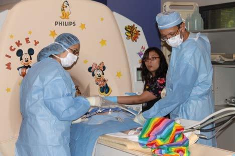
8 Patient selection Children as young as 3 months Less <1 year increased risk of failed study Right to left cardiac shunt not a contraindication General anesthesia Needle position and breath holds Inpatient and outpatient Combined Interventional and CV radiology procedure
9 Outline Planning and Patient Selection Technique and Image Acquisition Interpretation

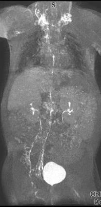
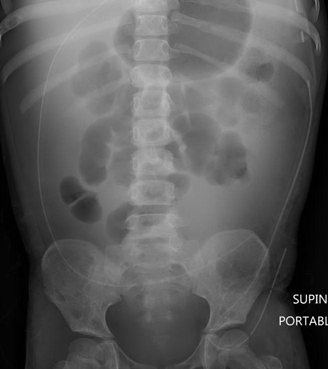
10 Case Example 1 20 year old female Gastroschisis SPEN tumor of pancreas Status post Whipple procedure with protein losing enteropathy Evaluate central lymphatics for chylolymphatic reflux

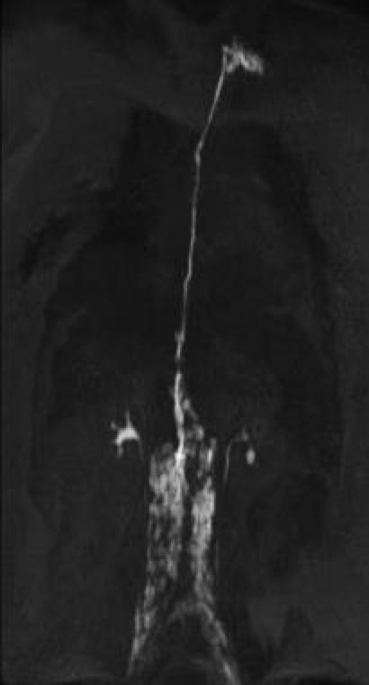
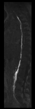
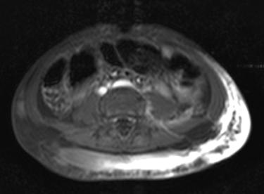
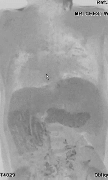
11 Normal d-mrl
12 Technique Formal consultation with Interventional Radiology physician Discuss procedure with patient and family Ultrasound of inguinal region Assess feasibility of lymph node injection Place on posterior coil on magnet table, and prep/drape


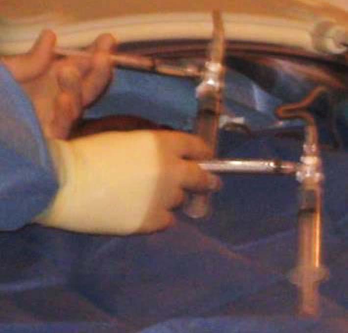
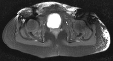
13 Technique Ultrasound guided placement of gauge needle into inguinal lymph node medulla Single capsule puncture Bilateral if possible Long subcutaneous tract Test injection to confirm positioning Lymph node slightly swells No fluid extravasation No securing to the skin Needle placement Post test injection
14 Technique Connected to long tubing which has been primed with gadolinium mixture 0.1 mmol/kg gadolinium dose (Magnevist, Dotarem) 1:1 (older) or 1:2 (younger) dilution with saline Saline chaser Feet Head

15 Technique Volume as important as concentration >10 y/o: 8 10 ml each side <10 y/o: 5-7 ml each side 1.5 x 2.0 gadolinium dosing and greater dilution, if needed Head Feet


16 Technique Plastic shield placed over abdomen followed by anterior coil Protects sterile field Lifts weight of coil off patient Torso phased array coil 55 cm field of view to cover from lower neck to groin
17 Technique
18 Technique: MRL protocol comprised of the following sequences: Breath held 3D THRIVE (VIBE/LAVA) sequence as a mask prior to contrast administration Repeat THRIVE every 2 minutes until contrast visualized in the retroperitoneal lymphatics Increase frequency to every minute to study transit from the retroperitoneal lymphatics to the cisterna chyli/thoracic duct STIR sequences optional
19 THRIVE Parameters Paramater Setting TR/TE: 4/1.9 msec Flip angle: 10 FOV: cm Fat suppression: Spectral adiabatic inversion Voxel size (acquired) x x mm3 Voxel size (reconstructed) x x mm3 Parallel Imaging Factor 2x1 or 2x3 Image acquisition duration: seconds per dynamic Krishnamurthy R et al. Radiology, 274(3), 2015


20 Normal appearance of the CCL on d-mrl 06:12 11:29 12:04 16:43 17:23 Time in minutes from the start of intranodal injection 25:53
21 Outline Planning and Patient Selection Technique and Image Acquisition Interpretation


22 Case 2: 4 year old female Hydrops fetalis and severe edema at birth Recurrent chylous ascites, bilateral pleural effusions, and protein losing enteropathy MRL to evaluate integrity of the central conducting lymphatics and to screen for pulmonary, retroperitoneal or intestinal lymphatic anomaly

23 Case 2: d-mrl
24 Nomenclature of d-mrl Recruitment Normal, from pressure injection into node Termination Non-visualization of a large channel due to occlusion, disruption, absent development, or lack of filling Collateral flow Diversion around obstruction / occlusion via normal channels in expected direction of flow Abnormal development Abnormal collection of dilated, serpiginous lymphatic channels Reflux Direction of flow away from expected direction of drainage into normal or abnormal channels Extravasation/leakage Pooling of contrast within anatomical space (pleural or pericardial cavity) or interstitium
25 Case 2: Interpretation Contrast passes through normal retroperitoneal lymphatics into the cisterna chyli Recruitment versus collateral flow into lymphatic channels along gonadal vein

26 Case 2: Interpretation Main central lymphatic channel extends to level of carina Termination of thoracic duct in lower chest Collateral channels in right paratracheal space, drain to cervical collaterals to reach left venous angle, and into right cervical nodes Transient reflux of contrast into right perihilar region that clears
27 CASE 2: Management Findings compatible with a generalized lymphatic anomaly Patient treated with sirolimus and furosemide No target for interventional therapy at this time Sirolimus therapy Improvement in edema Cessation of diuretic therapy


28 Case 3: 13 year old female History of recurrent chylopericardium and chylothorax Numerous other conditions including interstitial lung disease, pulmonary AVM, lytic bone lesions, and renal/splenic cysts

29 Case 3: 13 year old female History of recurrent chylopericardium and chylothorax Numerous other conditions including interstitial lung disease, pulmonary AVM, lytic bone lesions, and renal/splenic cysts


30 Case 3: Time resolved MRA for AVM

31 Case 3: Management Sirolimus Placement of a Denver shunt Coiling of left lower lobe AVM Significant Improvement in respiratory symptoms and quality of life
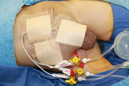
32 Case 3: Management Patient expired 2 years later secondary to acute pulmonary embolism Autopsy demonstrated proliferation of muscularized, dilated lymphatic channels through the mediastinum, pericardium, lung, and pleura

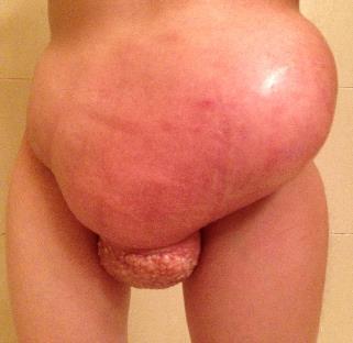
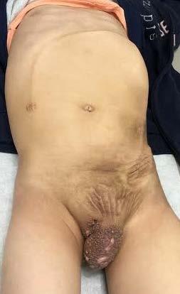
33 Case 4 6 year old girl with progressive swelling of the left flank and perineum Progressive Cutaneous vesicles Leaking lymphatic fluid One year

34 4 years 5 years

35 Case 4: Imaging 6 year old girl with progressive swelling of the left flank and perineum Diffuse osseous lesions Diffuse signal abnormality in abdominal and pelvic wall extending into perineum
36 Case 4: Sclerotherapy unsuccessful

37 Case 4: d-mrl


38 Case 4: Findings Needles are located within node with no evidence of extravasation New onset distention of subcutaneous lymphatic channels


39 Following Denver shunt placement from SQ space to peritoneal space
40
41 Case 4: Management Continued on sirolimus and interferon therapy Denver shunt placement Resolution of lymphatic leak from left flank/perineum Marked lifestyle improvement, able to attend school No sclerotherapy!

42 Case 5 29 year old woman Swelling of left extremity at 14 years Severe lymphedema by 22 Lymphorrhea Multiple surgical incisions, sclerotherapy, and radiotherapy at outside institution


43 Case 5: dmrl Right inguinal lymph node accessed Extensive retroperitoneal lymphatic abnormality
44 Case 5: Management Sclerotherapy discontinued No role for further interventional therapy Aggressive lymphatic massage Surgical debulking for fibrotic cutaneous masses Consideration of lymphovenous anastomosis

45 Case 6 11 year old male Heterotaxy syndrome with single right ventricle and TAPVC status post Fontan procedure Recurrent plastic bronchitis and pleural effusions
46 Case 6

47 Case 6 Management No interventional therapy offered Underwent heart transplant for failing Fontan Complete resolution of plastic bronchitis after transplant
48 Dynamic MR Lymphangiography (d-mrl) Combining intra-nodal injection of contrast and MR imaging allows: Rapid 3D visualization of central conducting lymphatics Reliable visualization of thoracic duct and cisterna chyli Dynamic visualization of contrast transit Lack of venous contamination
49 Dynamic MR Lymphangiography (d-mrl) Improved characterization of abnormalities of the CCL than previous modalities Development of nomenclature Differentiation of generalized lymphatic anomaly from lymphatic malformation Potential for mesenteric and extremity nodal access Indications for interventional versus medical therapy Identification of targets for interventions
Postoperative Chylothorax: the Use of Dynamic Magnetic Resonance Lymphangiography and Thoracic Duct Embolization
 pissn 2384-1095 eissn 2384-1109 imri 2018;22:182-186 Postoperative Chylothorax: the Use of Dynamic Magnetic Resonance Lymphangiography and Thoracic Duct Embolization Chae Woon Lee, Hyun Jung Koo, Ji Hoon
Interventional Management of Lymphatic Morbidity in Patients With CHD
 Interventional Management of Lymphatic Morbidity in Patients With CHD Maxim Itkin MD, FSIR Professor of Radiology and Pediatrics Hospital of University of Pennsylvania DISCLOSURE STATEMENT OF FINANCIAL
Director of HUP/CHOP Center for Lymphatic Imaging and Interventions
 Introduction Lymphatic Interventions: The Real Next Frontier Associate Professor of Radiology and Pediatrics Maxim Itkin MD, FSIR Director of HUP/CHOP Center for Lymphatic Imaging and Interventions Physiology
Prenatal and Postnatal Evaluation of Lymphatic Disorders
 Prenatal and Postnatal Evaluation of Lymphatic Disorders David M Biko, MD Director, Section of Cardiovascular and Lymphatic Imaging Children s Hospital of Philadelphia Assistant Professor of Radiology
REPEATED INTRANODAL LYMPHANGIOGRAPHY FOR THE TREATMENT OF LYMPHATIC LEAKAGE
 59 Lymphology 48 (2015) 59-63 REPEATED INTRANODAL LYMPHANGIOGRAPHY FOR THE TREATMENT OF LYMPHATIC LEAKAGE S. Kariya, M. Nakatani, R. Yoshida, Y. Ueno, A. Komemushi, N. Tanigawa Department of Radiology,
Thoracic Duct Embolization Technique & Results
 Thoracic Duct Embolization Technique & Results Edward W. Lee, MD, PhD, MSc Assistant Professor Director of Research Interventional Radiology Department of Radiology AGA AASLD JCCC CTSI CCTP TRIC AACR UCLA
Thoracic Duct Embolization for Nontraumatic Chylous Effusion. Gregory J. Nadolski, MD ; and Maxim Itkin, MD
 CHEST Thoracic Duct Embolization for Nontraumatic Chylous Effusion Experience in 34 Patients Gregory J. Nadolski, MD ; and Maxim Itkin, MD Original Research DISORDERS OF THE PLEURA Background: Thoracic
Lymphatic System and Immunity. Lymphatic System
 Lymphatic System and Immunity Lymphatic System Lymphatic System High hydrostatic pressure in the arterioles and capillaries at the arterial part of the circulation leads to move plasma fluid from the capillaries
Chylothorax Basics OVERVIEW GENETICS SIGNALMENT/DESCRIPTION OF PET
 Chylothorax Basics OVERVIEW Chylo- refers to chyle; thorax refers to the chest Chyle is a milky to slightly yellow fluid composed of lymph and fats (rich in triglycerides) taken up from the intestines
Trauma Activation 7/18/17
 Blunt Rupture of the Thoracic Duct after Severe Thoracic Trauma Samuel Brown, MD Trauma Activation 7/18/17 53 year old male, rear end MVC, exited vehicle and was struck by a semi truck. Denies LOC, complaints
Dr. Weyrich G07: Superior and Posterior Mediastina. Reading: 1. Gray s Anatomy for Students, chapter 3
 Dr. Weyrich G07: Superior and Posterior Mediastina Reading: 1. Gray s Anatomy for Students, chapter 3 Objectives: 1. Subdivisions of mediastinum 2. Structures in Superior mediastinum 3. Structures in Posterior
Large veins of the thorax Brachiocephalic veins
 Large veins of the thorax Brachiocephalic veins Right brachiocephalic vein: formed at the root of the neck by the union of the right subclavian & the right internal jugular veins. Left brachiocephalic
Case report description of a collaborative approach to thoracic duct embolization in patients with congenital heart disease
 Luangrath et al. Journal of Congenital Cardiology (2018)2:2 https://doi.org/10.1186/s40949-018-0016-z Journal of Congenital Cardiology CASE REPORT Open Access Case report description of a collaborative
Abdomen Sonography Examination Content Outline
 Abdomen Sonography Examination Content Outline (Outline Summary) # Domain Subdomain Percentage 1 2 3 Anatomy, Perfusion, and Function Pathology, Vascular Abnormalities, Trauma, and Postoperative Anatomy
Post-Operative Chylous Ascites. David Kashan, PGY-4 Richmond University Medical Center 7/30/15
 Post-Operative Chylous Ascites David Kashan, PGY-4 Richmond University Medical Center 7/30/15 HPI Patient is a 76 year old female p/w one day of worsening abdominal pain, +N/V, fevers and chills HPI PMHx:
(SKILLS/HANDS-ON) Chest Tubes. Rebecca Carman, MSN, ACNP-BC. Amanda Shumway, PA-C. Thomas W. White, MD, FACS, CNSC
 Chest Tubes. Rebecca Carman, MSN, ACNP-BC. Amanda Shumway, PA-C. Thomas W. White, MD, FACS, CNSC") (SKILLS/HANDS-ON) Chest Tubes Rebecca Carman, MSN, ACNP-BC Nurse Practitioner, Trauma Services, Intermountain Medical Center, Intermountain Healthcare Amanda Shumway, PA-C APC Trauma and Critical Care
(SKILLS/HANDS-ON) Chest Tubes Rebecca Carman, MSN, ACNP-BC Nurse Practitioner, Trauma Services, Intermountain Medical Center, Intermountain Healthcare Amanda Shumway, PA-C APC Trauma and Critical Care
Magnetic Resonance Lymphangiography for the Study of Lymphatic System in Lymphedema
 66 Invited Review Magnetic Resonance Lymphangiography for the Study of Lymphatic System in Lymphedema NingFei Liu, MD, PhD 1 Yixin Zhang, MD, PhD 1 1 Department of Plastic and Reconstructive Surgery, Shanghai
UNDERSTANDING CHYLE IN CATS
 Vet Times The website for the veterinary profession https://www.vettimes.co.uk UNDERSTANDING CHYLE IN CATS Author : DAN FORSTER Categories : Vets Date : February 11, 2008 DAN FORSTER discusses diagnosis
Returns fluids that leaked from blood vessels back to blood Consists of three parts
 Lymphatic System Returns fluids that leaked from blood vessels back to blood Consists of three parts 1. Network of lymphatic vessels (lymphatics) 2. Lymph fluid in vessels 3. Lymph cleanse lymph 1 Lymphoid
Mediastinum It is a thick movable partition between the two pleural sacs & lungs. It contains all the structures which lie
 Dr Jamila EL medany OBJECTIVES At the end of the lecture, students should be able to: Define the Mediastinum. Differentiate between the divisions of the mediastinum. List the boundaries and contents of
SETTING Fudan University Shanghai Cancer Center. RESPONSIBLE PARTY Haiquan Chen MD.
 OFFICIAL TITLE A Phase Ⅲ Study of Left Side Thoracotomy Approach (SweetProcedure) Versus Right Side Thoracotomy Plus Midline Laparotomy Approach (Ivor-Lewis Procedure) Esophagectomy in Middle or Lower
Disclosures: Image Guided Procedures Pearls, Pitfalls, and Disasters. Central Venous Access. Outline:
 Image Guided Procedures Pearls, Pitfalls, and Disasters Disclosures: I have nothing to disclose Miles B. Conrad MD, MPH Clinical Assoc. Prof of Radiology Section: IR Outline: Image Guided Procedures Pearls,
Job Task Analysis for ARDMS Abdomen Data Collected: June 30, 2011
 Job Task Analysis for ARDMS Abdomen Data Collected: June 30, 2011 Reported: Analysis Summary for: Abdomen Examination Survey Dates 06/13/2011-06/26/2011 Invited Respondents 6,000 Surveys with Demographics
Contents. Page 1. Homework 11 Chapter Blood Vessels Due: Week 6 Lec 11
 Page 1 Homework 11 Chapter 18-19 Blood Vessels Due: Week 6 Lec 11 Contents When printing, make sure that you specify the page range that you want to print out! Learning objectives for Lecture 11:...pg
Austin Radiological Association Nuclear Medicine Procedure LYMPHOSCINTIGRAPHY (Tc-99m-Sulfur Colloid [Filtered])
![Austin Radiological Association Nuclear Medicine Procedure LYMPHOSCINTIGRAPHY (Tc-99m-Sulfur Colloid [Filtered])](/thumbs/87/95258373.jpg "Austin Radiological Association Nuclear Medicine Procedure LYMPHOSCINTIGRAPHY (Tc-99m-Sulfur Colloid [Filtered])") Austin Radiological Association Nuclear Medicine Procedure LYMPHOSCINTIGRAPHY (Tc-99m-Sulfur Colloid [Filtered]) Overview Indications The Lymphoscintigraphy Study demonstrates the flow of lymph from the
Austin Radiological Association Nuclear Medicine Procedure LYMPHOSCINTIGRAPHY (Tc-99m-Sulfur Colloid [Filtered]) Overview Indications The Lymphoscintigraphy Study demonstrates the flow of lymph from the
Causes of pleural effusion and its imaging approach in pediatrics. M. Mearadji International Foundation for Pediatric Imaging Aid
 Causes of pleural effusion and its imaging approach in pediatrics M. Mearadji International Foundation for Pediatric Imaging Aid Pleural fluid A tiny amount of fluid in the pleural cavity is physiological.
Gastroschisis Sequelae and Management
 Gastroschisis Sequelae and Management Mary Finn Gillian Lieberman, MD Primary Care Radiology Beth Israel Deaconess Medical Center Harvard Medical School April 2014 Outline I. Definition and Epidemiology
What is Lymphedema? Primary lymphedema: a person is born with the condition where the lymphatic vessels are not formed completely or malformed
 Lymphedema What is Lymphedema? Lymphedema is a chronic health condition which causes localized swelling. There are 2 types: Primary lymphedema: a person is born with the condition where the lymphatic vessels
ANATOMY & PHYSIOLOGY ONLINE COURSE - SESSION 11 THE LYMPHATIC SYSTEM AND IMMUNITY
 ANATOMY & PHYSIOLOGY ONLINE COURSE - SESSION 11 THE LYMPHATIC SYSTEM AND IMMUNITY Functions of the Lymphatic System The lymphatic system has three primary functions. First of all, it returns excess interstitial
Copy Right- Hongqi ZHANG-Department of Anatomy-Fudan University. Systematic Anatomy
 Systematic Anatomy Department of Anatomy,Histology & Embryology Shanghai Medical College,Fudan University Dr.Hongqi Zhang ( 张红旗 ) Email: Zhanghq58@126.com Office: Building 9,Room308, 54237151-9308 Mobile:13761809799
Radiology- Pathology Conference 4/29/2012. Lymph Nodes. John McGrath
 Radiology- Pathology Conference 4/29/2012 Lymph Nodes John McGrath 1 Presentation material is for education purposes only. All rights reserved. 2012 URMC Radiology Page 1 of 24 Case 1: 51 year-old male
Esophageal Perforation
 Esophageal Perforation Dr. Carmine Simone Thoracic Surgeon, Division of General Surgery Head, Division of Critical Care May 15, 2006 Overview Case presentation Radiology Pre-operative management Operative
High-Resolution MR Lymphangiography in Patients with Primary and Secondary Lymphedema
 Lohrmann et al. MR Lymphangiog raphy of Patients with Lymphedem a Vascular Imaging Technical Innovation C M E D E N T U R I C L I M G I N G JR 2006; 187:556 561 0361 803X/06/1872 556 merican Roentgen Ray
Medical Review Guidelines Magnetic Resonance Angiography
 Medical Review Guidelines Magnetic Resonance Angiography Medical Guideline Number: MRG2001-05 Effective Date: 2/13/01 Revised Date: 2/14/2006 OHCA Reference OAC 317:30-5-24. Radiology. (f) Magnetic Resonance
Contrast-enhanced Breast MRI RSSA 2013
 Contrast-enhanced Breast MRI RSSA 2013 Prof. dr. Maurice van den Bosch University Medical Center Utrecht, the Netherlands Index 1) Breast cancer 2) Why MRI of the breast 3) Technique 4) Interpretation
A neonate is any patient less than 45 weeks post conception regardless of chronological age.
 Case Log Definitions: A Guide for Fellows and Program Directors Recommendations from the Pediatric Anesthesia Program Directors Association Case Log Task Force January 2013 These recommendations represent
High Field MR of the Spine
 Department of Radiology University of California San Diego 3T for MR Applications Advantages High Field MR of the Spine Increased signal-to-noise Better fat suppression Increased enhancement with gadolinium
Learning Radiology: Recognizing the Basics. Text with Student Consult Online Access Code
 Learning Radiology: Recognizing the Basics. Text with Student Consult Online Access Code Herring, W ISBN-13: 9780323074445 Table of Contents 1. Recognizing Anything The "colorful" world of radiology A
The Lymphoid System Pearson Education, Inc.
 23 The Lymphoid System Introduction The lymphoid system consists of: Lymph Lymphatic vessels Lymphoid organs An Overview of the Lymphoid System Lymph consists of: Interstitial fluid Lymphocytes Macrophages
10/14/2018 Dr. Shatarat
 2018 Objectives To discuss mediastina and its boundaries To discuss and explain the contents of the superior mediastinum To describe the great veins of the superior mediastinum To describe the Arch of
Lung sequestration and Scimitar syndrome
 Lung sequestration and Scimitar syndrome Imaging approaches M. Mearadji International Foundation for Pediatric Imaging Aid Rotterdam, The Netherlands Pulmonary sequestration Pulmonary sequestration (PS)
Raja Muthupillai, PhD. Department of Diagnostic and Interventional Radiology St. Luke s Episcopal Hospital. Research Support: Philips Healthcare
 3D Cardiac Imaging Raja Muthupillai, PhD Department of Diagnostic and Interventional Radiology St. Luke s Episcopal Hospital Houston, TX Disclosures Research Support: Philips Healthcare This presentation
ADDITIONS. The following codes have been added.
 ADDITIONS The following codes have been added. 99446 Interprofessional telephone/internet assessment and management service provided by treating/requesting physician or other qualified health care professional;
Noncontrast three-dimensional magnetic resonance imaging vs lymphoscintigraphy in the evaluation of lymph circulation disorders: A comparative study
 Noncontrast three-dimensional magnetic resonance imaging vs lymphoscintigraphy in the evaluation of lymph circulation disorders: A comparative study Ningfei Liu, MD, PhD, a Chenguang Wang, MD, PhD, b and
Appendix 5. EFSUMB Newsletter. Gastroenterological Ultrasound
 EFSUMB Newsletter 87 Examinations should encompass the full range of pathological conditions listed below A log book listing the types of examinations undertaken should be kept Training should usually
Anatomy The study of the body's structure.
 Anatomy The study of the body's structure. * 1. Systemic- Study of each of the body's systems. 2. Regional- Study of a specific area of the body 3. Surface- Study of external features. Physiology The study
Malignant Effusions. Anantham Devanand Respiratory and Critical Care Medicine Singapore General Hospital
 Malignant Effusions Anantham Devanand Respiratory and Critical Care Medicine Singapore General Hospital Malignant Effusions Definition: Presence of malignant cells in the pleural space 75% are caused by
Heart and Lungs. LUNG Coronal section demonstrates relationship of pulmonary parenchyma to heart and chest wall.
 Heart and Lungs Normal Sonographic Anatomy THORAX Axial and coronal sections demonstrate integrity of thorax, fetal breathing movements, and overall size and shape. LUNG Coronal section demonstrates relationship
QUESTIONS for the examination in surgery for 4 th -year students of the Faculty of foreign students
 QUESTIONS for the examination in surgery for 4 th -year students of the Faculty of foreign students 1. The main principles of surgical deontology and its founders. 2. Acute appendicitis. Anatomico-physiological
Pediatric TB Intensive Houston, Texas October 14, 2013
 Pediatric TB Intensive Houston, Texas October 14, 2013 Radiologic Presentation of Childhood TB Susan D. John, MD, FACR October 14, 2013 Disclosures I have no disclosures or conflicts of interest to report
Case Presentation Surgery Grand Round. Amid Keshavarzi, MD UCHSC 4/9/2006
 Case Presentation Surgery Grand Round Amid Keshavarzi, MD UCHSC 4/9/2006 Case Presentation 12 y/o female Presented to OSH after accidental swallowing of plastic fork in the bus, CXR/AXR form OSH did not
Proceedings of the World Small Animal Veterinary Association Sydney, Australia 2007
 Proceedings of the World Small Animal Sydney, Australia 2007 Hosted by: Next WSAVA Congress THE LAST GASP II: LUNGS AND THORAX David Holt, BVSc, Diplomate ACVS University of Pennsylvania School of Veterinary
MRI PEDIATRIC PROTOCOLS (Updated 6/19/2018)
") MRI PEDIATRIC PROTOCOLS (Updated 6/19/2018) *Please get or let us know where radiologist can review plain films. *For Texas Orthopedics and other Docs requesting only MSK section read for their pediatric
MRI PEDIATRIC PROTOCOLS (Updated 6/19/2018) *Please get or let us know where radiologist can review plain films. *For Texas Orthopedics and other Docs requesting only MSK section read for their pediatric
CHYLOPERITONEUM: DIAGNOSTIC AND THERAPEUTIC OPTIONS
 1 Lymphology 49 (2016) 1-7 CHYLOPERITONEUM: DIAGNOSTIC AND THERAPEUTIC OPTIONS S. Dessalvi, F. Boccardo, L. Molinari, S. Spinaci, C.C. Campisi, G.M. Ferrari, C. Campisi Department of Surgery, Unit of Lymphatic
Glenn Shunts Revisited
 Glenn Shunts Revisited What is a Super Glenn Patricia O Brien, MSN, CPNP-AC Nurse Practitioner, Pediatric Cardiology No Disclosures Single Ventricle Anatomy Glenn Shunt Cavopulmonary Anastomosis Anastomosis
Basics of Interventional Radiology Coding 2018
 Basics of Interventional Radiology Coding 2018 Prepared and Published By: MedLearn Publishing A Division of MedLearn Media, Inc. 445 Minnesota Street, Suite 514 St. Paul, MN 55101 1-800-252-1578 medlearnmedia.com
Portal System & Lymphatic System. When the vein of any organ of the body does not open in the caval vein or heart.
 1. Introduction of portal system 2. Renal portal system 3. Hepatic portal system 4. Hypophysial portal system 5. Introduction of lymphatic system 6. The lymph 7. Lymph vessels 8. Lymph nodes 9. Lymphoid
Management of Pleural Effusion
 Management of Pleural Effusion Development of Pleural Effusion pulmonary capillary pressure (CHF) capillary permeability (Pneumonia) intrapleural pressure (atelectasis) plasma oncotic pressure (hypoalbuminemia)
Cardiovascular system:
 Cardiovascular system: Mediastinum: The mediastinum: lies between the right and left pleura and lungs. It extends from the sternum in front to the vertebral column behind, and from the root of the neck
Pediatric TB Intensive Houston, Texas
 Pediatric TB Intensive Houston, Texas November 13, 2009 Radiographic Manifestations of Pediatric TB Susan D. John, MD, FACR November 13, 2009 Radiologic Presentation of Childhood TB Susan D. John, MD,
This appendix was part of the submitted manuscript and has been peer reviewed. It is posted as supplied by the authors.
 This appendix was part of the submitted manuscript and has been peer reviewed. It is posted as supplied by the authors. - Figure S1: The four quadrant approach lung ultrasound at the bedside. * The anterolateral
Imaging of the cisterna chyli on PET-CT in patients with known malignancy: Report of two cases
 Imaging of the cisterna chyli on PET-CT in patients with known malignancy: Report of two cases Natalie Burns, B.S., Jason Barksdale, M.D., Linh Ho, M.D., and Patrick M. Colletti, M.D. Citation: Burns N,
Can SCMR CMR protocol recommendations
 Can SCMR CMR protocol recommendations V1.3 - April 2009 CanSCMR CMR Protocol and SOP Recommendation 2009 (15 minutes) 2 Planning of LV fct. real time multiple axes Realtime 3 cine long axis 6 long axes
Chest X rays and Case Studies. No disclosures. Outline 5/31/2018. Carlo Manalo, M.D. Department of Radiology Loma Linda University Children s Hospital
 Chest X rays and Case Studies Carlo Manalo, M.D. Department of Radiology Loma Linda University Children s Hospital No disclosures. Outline Importance of history Densities delineated on radiography An approach
Procedure: Chest Tube Placement (Tube Thoracostomy)
") Procedure: Chest Tube Placement (Tube Thoracostomy) Basic Information: The insertion and placement of a chest tube into the pleural cavity for the purpose of removing air, blood, purulent drainage, or
Procedure: Chest Tube Placement (Tube Thoracostomy) Basic Information: The insertion and placement of a chest tube into the pleural cavity for the purpose of removing air, blood, purulent drainage, or
Children are not small adults Children are Not Small Adults Anatomic considerations Pliable bony & cartilagenous structures - Significant thoracic inj
 PEDIATRIC CHEST TRAUMA Children are not small adults Role of imaging Spectrum of injury Children are not small adults Children are Not Small Adults Anatomic considerations Pliable bony & cartilagenous
Basics of Interventional Radiology Coding 2017
 Basics of Interventional Radiology Coding 2017 Prepared and Published By: MedLearn Publishing A Division of Panacea Healthcare Solutions, Inc. 287 East Sixth Street, Suite 400 St. Paul, MN 55101 1-800-252-1578
Radiofrequency Ablation of Liver Tumors
 Radiofrequency Ablation of Liver Tumors Michael M. Awad, Michael A. Choti Indications and Contraindications Indications Unresectable malignant tumors of the liver (e.g., hepatocellular carcinoma, colorectal
When is Limb Edema Not Heart Failure
 When is Limb Edema Not Heart Failure An Approach to the Swollen Leg Greg Harding M.D. Vascular Surgeon Faculty/Presenter Disclosure Faculty: Greg Harding M.D. Relationships with commercial interests: None
Division of Diagnostic Imaging, The University of Texas M.D. Anderson Cancer Center, Houston, Texas, USA
 89 Lymphology 28 (1995) 89-94 Division of Diagnostic Imaging, The University of Texas M.D. Anderson Cancer Center, Houston, Texas, USA ABSTRACT The anatomy of the posterior intercostal lymphatics and lymph
This presentation is the intellectual property of the author. Contact them at for permission to reprint and/or distribute.
 MRI of the Knee Jennifer Swart, M.D. Musculoskeletal Radiology South Texas Radiology Group Financial Disclosure Dr. Jennifer Swart has no relevant financial relationships with commercial interests to disclose.
UNIVERSITY OF NAIROBI DEPARTMENT OF HUMAN ANATOMY LEVEL I MBChB/BDS REVISED TEACHING SCHEDULE 2016/2017 AFTER STRIKE
 20 th 24 th March Monday 2:00pm 6:00pm Tagged review of Head and Neck anatomy A Gross Anatomy lab All Tuesday 2:00pm 4:00pm Tagged review of Head and Neck anatomy A Gross Anatomy lab All 4:00pm 6:00pm
Mechanical Support in the Failing Fontan-Kreutzer
 Mechanical Support in the Failing Fontan-Kreutzer Stephanie Fuller MD, MS Thomas L. Spray Endowed Chair in Congenital Heart Surgery Associate Professor, The Perelman School of Medicine at the University
THE MANAGEMENT OF THE SWOLLEN ARM IN CARCINOMA OF THE BREAST
 THE MANAGEMENT OF THE SWOLLEN ARM IN CARCINOMA OF THE BREAST NORMAN TREVES, M.D. The terms "brawny arm" and "lymphedema" have been given to the swollen arm which may complicate the inoperable, recurrent,
Lecture 2: Clinical anatomy of thoracic cage and cavity II
 Lecture 2: Clinical anatomy of thoracic cage and cavity II Dr. Rehan Asad At the end of this session, the student should be able to: Identify and discuss clinical anatomy of mediastinum such as its deflection,
Anatomical and Functional MRI of the Pancreas
 Anatomical and Functional MRI of the Pancreas MA Bali, MD, T Metens, PhD Erasme Hospital Free University of Brussels Belgium mbali@ulb.ac.be Introduction The use of MRI to investigate the pancreas has
ISUOG Basic Training. Assessing the Neck & Chest Gihad Chalouhi, Lebanon
 ISUOG Basic Training Assessing the Neck & Chest Gihad Chalouhi, Lebanon Learning objectives 9 & 10 At the end of the lecture you will be able to: recognise the differences between the normal & most common
Limited en bloc Resection of the Gastroesophageal Junction with Isoperistaltic Jejunal Interposition
 22 Limited en bloc Resection of the Gastroesophageal Junction with Isoperistaltic Jejunal Interposition J.R. Izbicki, W.T. Knoefel, D. C. Broering ] Indications Severe dysplasia in the distal esophagus
APPROACH TO PLEURAL EFFUSIONS. Raed Alalawi, MD, FCCP
 APPROACH TO PLEURAL EFFUSIONS Raed Alalawi, MD, FCCP CASE 65-year-old woman with H/O breast cancer presented with a 1 week H/O progressively worsening exersional dyspnea. Physical exam: Diminished breath
RadRx Your Prescription for Accurate Coding & Reimbursement Copyright All Rights Reserved.
 Interventional Radiology Coding Case Studies Prepared by Stacie L. Buck, RHIA, CCS-P, RCC, CIRCC, AAPC Fellow President & Senior Consultant Week of October 22, 2018 Paracentesis & Transjugular Liver Biopsy
TB Intensive Houston, Texas
 TB Intensive Houston, Texas October 15-17, 17 2013 Diagnosis of TB: Radiology Rosa M Estrada-Y-Martin, MD MSc FCCP October 16, 2013 Rosa M Estrada-Y-Martin, MD MSc FCCP, has the following disclosures to
Mary Lou Garey MSN EMT-P MedFlight of Ohio
 Mary Lou Garey MSN EMT-P MedFlight of Ohio Function Prolonged and frequent access to venous circulation Allows for patient to carry on normal life; decrease number of needle sticks Medications, parenteral
73725x2 MRA Pelvis Runoff (to ankle) CTA Abdomen with & without CTA Cardiac Brain without 70551
 CTA Abdomen with & without CTA Cardiac Brain without 70551") CT CT Myelogram MRI Abdomen without 74150 Cervical 62302 Abdomen / MRCP 74181 Abdomen with 74160 Thoracic 62303 Abdomen / MRCP with & without 74183 Abdomen with & without 74170 Lumbar 62304 Abdomen / Pelvis
CT CT Myelogram MRI Abdomen without 74150 Cervical 62302 Abdomen / MRCP 74181 Abdomen with 74160 Thoracic 62303 Abdomen / MRCP with & without 74183 Abdomen with & without 74170 Lumbar 62304 Abdomen / Pelvis
Fontan Deterioration in Pediatric Cardiologist s s View. Pusan National University Hospital Hyoung Doo Lee M.D.
 Fontan Deterioration in Pediatric Cardiologist s s View Pusan National University Hospital Hyoung Doo Lee M.D. Outcomes of Fontan operation JTCS 2006:131;172-80 Mitchell ME et al Jan. 1992~Dec. 1999, 332
**Confirm accuracy of above with your instructor.** Revised 8/22/2017 1
 AP1 Lab 1 Cavities, Organs, Serous Membranes, Quadrants, Regions, and Directional Terms, Planes & Sections Project 1 Directional Terminology Step 1: Define/Describe what is known as the "ANATOMICAL POSITION."
January Details of the fee code revisions can be found highlighted in Schedule A, attached.
 Government of Newfoundland and Labrador Department of Health and Community Services January 2018 18-01 TO: RE: ALL FEE-FOR-SERVICE PHYSICIANS CHANGES TO DOPPLER ULTRASOUND FEE CODES The Department of Health
Lecturer: Ms DS Pillay ROOM 2P24 25 February 2013
 Lecturer: Ms DS Pillay ROOM 2P24 25 February 2013 Thoracic Wall Consists of thoracic cage Muscle Fascia Thoracic Cavity 3 Compartments of the Thorax (Great Vessels) (Heart) Superior thoracic aperture
GUIDELINES FOR CANCER IMAGING Lung Cancer
 GUIDELINES FOR CANCER IMAGING Lung Cancer Greater Manchester and Cheshire Cancer Network Cancer Imaging Cross-Cutting Group April 2010 1 INTRODUCTION This document is intended as a ready reference for
Chapter 16 Lymphatic System and Immunity. Lymphatic Pathways. Lymphatic Capillaries. network of vessels that assist in circulating fluids
 Chapter 16 Lymphatic System and Immunity network of vessels that assist in circulating fluids closely associated with the cardiovascular system transports excess fluid away from interstitial spaces transports
CHAPTER 2 Terms Pertaining to the Body as a Whole
 CHAPTER 2 Terms Pertaining to the Body as a Whole OBJECTIVES 1. Define terms that apply to the structural organization of the body. 2. Identify the body cavities and the organs contained within the cavities.
HBA THE BODY Trunk Examination - October 1, 2012
 HBA 531 - THE BODY Trunk Examination - October 1, 2012 1. On the right is a lettered list of different functional types of neurons. On the left is a list of structures. In the blank following each named
This presentation is the intellectual property of the author. Contact them for permission to reprint and/or distribute.
 MRI of the Knee Jennifer Swart, M.D. Musculoskeletal Radiology South Texas Radiology Group Outline Coils, Patient Positioning Acquisition Parameters, Planes and Pulse Sequences Knee Arthrography Normal
RADPrimer Curriculum Breast Topics Covered Basic Intermediate 225
 Breast Anatomy & Normal Variants 11 Breast Imaging Modalities 13 BI RADS Lexicon 3 Mammography: Masses 9 Mammography: Calcifications 17 Mammography: Additional Findings 8 Ultrasound Features 10 Ultrasound
Cholangiocarcinoma (Bile Duct Cancer)
") Cholangiocarcinoma (Bile Duct Cancer) The Bile Duct System (Biliary Tract) A network of bile ducts (tubes) connects the liver and the gallbladder to the small intestine. This network begins in the liver
Cholangiocarcinoma (Bile Duct Cancer) The Bile Duct System (Biliary Tract) A network of bile ducts (tubes) connects the liver and the gallbladder to the small intestine. This network begins in the liver
Semiotics in Radiology
 Adelino Santos Health Technology College Coimbra, Portugal Collaboration of António Agudo Student of Radiology College of Health Technology Coimbra, Portugal What are the most important points to evaluate
Day Time Task and group Venue Facilitator(s)
") HEAD AND NECK ANATOMY (WEEKS 16-18) Monday 2:00pm 6:00pm Dissection: Face and parotid region A Gross Anatomy Lab All Tuesday 11:00am 1:00pm Dissection: Eyeball, orbit and its contents A Gross Anatomy Lab
HEAD AND NECK ANATOMY (WEEKS 16-18) Monday 2:00pm 6:00pm Dissection: Face and parotid region A Gross Anatomy Lab All Tuesday 11:00am 1:00pm Dissection: Eyeball, orbit and its contents A Gross Anatomy Lab
Welcome to ANAT 10A! What is Anatomy? Different levels of Anatomy The Language of Anatomy Pearson Education, Inc.
 Welcome to ANAT 10A! What is Anatomy? Different levels of Anatomy The Language of Anatomy Introduction Anatomy means to dissect: (ANAT 10A) The study of internal & external body structures The study of
Tips and Tricks of State of the art MRA
 Tips and Tricks of State of the art MRA Mayil Krishnam, MD,MBA, MRCP,FRCR(UK) Professor of Radiology Director, Cardiovascular and Thoracic Imaging University of California, Irvine Objectives Technical
IMMUNODEFICIENCY DUE TO CHYLOUS DYSPLASIA: DIAGNOSTIC AND THERAPEUTIC CONSIDERATIONS
 58 Lymphology 45 (2012) 58-62 IMMUNODEFICIENCY DUE TO CHYLOUS DYSPLASIA: DIAGNOSTIC AND THERAPEUTIC CONSIDERATIONS C.C. Campisi, S. Spinaci, R. Lavagno, L. Larcher, F. Boccardo, P. Santi, C. Campisi Unit
DISCOVER NEW HORIZONS IN FLUID DRAINAGE. Bringing Safety and Convenience to Fluid Drainage Management
 DISCOVER NEW HORIZONS IN FLUID DRAINAGE Bringing Safety and Convenience to Fluid Drainage Management DRAIN ASEPT Pleural and Peritoneal Drainage Catheter System 600mL or 1,000mL Evacuated Drainage Bottle