Nerves on the Posterior Abdominal Wall
|
|
|
- Lucy Manning
- 6 years ago
- Views:
Transcription
1


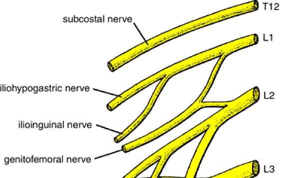
2 Nerves on the Posterior Abdominal Wall Lumbar Plexus The lumbar plexus, which is one of the main nervous pathways supplying the lower limb, is formed in the psoasmuscle from the anterior ramiof the upper four lumbar nerves. The anterior rami receive gray rami communicates from the sympathetic trunk, The upper two only give off white rami communicates to the sympathetic trunk. The branches of the plexus emerge from the lateral and medial borders of the muscle and from its anterior surface. The iliohypogastricnerve, ilioinguinalnerve, lateral cutaneousnerve of the thigh, and femoral nerve emerge from the lateral border of the psoas, in that order from above downward. The iliohypogastricand ilioinguinalnerves (L1) enter the lateral and anterior abdominal walls

3 Nerves on the Posterior Abdominal Wall The iliohypogastricnerve - supplies the skin of the lower part of the anterior abdominal wall, The ilioinguinal nerve - passes through the inguinal canal to supply the skin of the groin and the scrotum or labium majus. The lateral cutaneousnerve of the thig - Crosses the iliac fossain front of the iliacusmuscle and enters the thigh behind the lateral end of the inguinal ligament. -It supplies the skin over the lateral surface of the thigh.
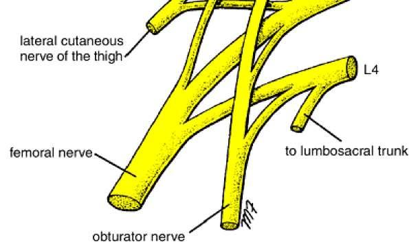
4 Lumber plexus.cont The femoral nerve (L2, 3, and 4) - It is the largest branch of the lumbar plexus. - It runs downward and laterally between the psoas and the iliacusmuscles and enters the thigh behind the inguinal ligament and lateral to the femoral vessels and the femoral sheath. - In the abdomen it supplies the iliacus muscle.
5


6 Lumber plexus cont The Obturatornerve and the fourth lumbar root of the lumbosacral trunk - Emerge from the medial border of the psoasat the brim of the pelvis. - The Obturatornerve (L2, 3, and 4) crosses the pelvic brim in front of the sacroiliac joint and behind the common iliac vessels. - It leaves the pelvis by passing through the Obturatorforamen into the thigh. - The fourth lumbar root of the lumbosacraltrunk takes part in the formation of the sacral plexus. It descends anterior to the ala of the sacrum and joins the first sacral nerve.
7 Lumber plexus.cont The genitofemoral nerve (L1 and 2) - Emerges on the anterior surface of the psoas. - It runs downward in front of the muscle and divides into : 1-A genital branch, which enters the spermatic cord and supplies the Cremasteric muscle 2-A femoral branch, which supplies a small area of the skin of the thigh. Cremasteric reflex - It is the nervous pathway, in which stimulation of the skin of the thigh in the male results in reflex contraction of the cremaster muscle and the drawing upward of the testis within the scrotum. - Cremasteric reflex may be absent with: testicular torsion, upper and lower motor neuron disorders, as well as a spine injury of L1-L2. It can also occur if the ilioinguinal nerve is accidentally cut during a hernia repair


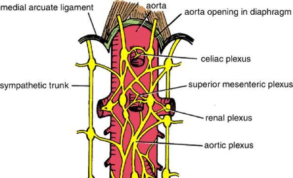
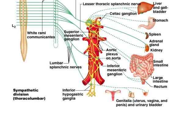
8 Sympathetic Trunk (Abdominal Part) The abdominal part of the sympathetic trunk is continuous above with the thoracic and below with the pelvic parts of the sympathetic trunk. It runs downward along the medial border of the psoasmuscle on the bodies of the lumbar vertebrae. It enters the abdomen from behind the medial arcuateligament and gains entrance to the pelvis below by passing behind the common iliac vessels. The right sympathetic trunk lies behind the right border of the inferior vena cava; the left sympathetic trunk lies close to the left border of the aorta. The sympathetic trunk possesses four or five segmentalyarranged ganglia, the first and second often being fused together.
9 Sympathetic trunk...abdominal part Branches White rami -communicantes join the first two ganglia to the first two lumbar spinal nerves. -A white ramus contains Preganglionic nerve fibers and afferent sensory nerve fibers. Gray rami - communicantes join each ganglion to a corresponding lumbar spinal nerve. - A gray ramus contains postganglionic nerve fibers distributed to blood vessels, sweet gland and skin


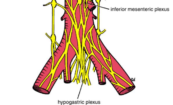
10 Sympathetic.Abdominal part Post gangilionic fibers..cont Distributed through the branches of the spinal nerves to the blood vessels, sweat glands, and arrector pili muscles of the skin. Fibers pass medially to the sympathetic plexuses on the abdominal aorta and its branches. (These plexuses also receive fibers from splanchnic nerves and the vagus.) Fibers pass downward and medially in front of the common iliac vessels into the pelvis, where, together with branches from sympathetic nerves in front of the aorta, they form a large bundle of fibers called the superior hypogastric plexus.


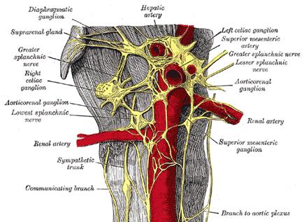
11 Aortic Plexuses Preganglionicand postganglionic sympathetic fibers Preganglionicparasympathetic fibers, and visceral afferent fibers form a plexus of Nerves, the aortic plexus, around the abdominal part of the aorta. Regional concentrations of this plexus around the origins of the celiac, renal arteries Superior mesenteric celiac plexus Inferior mesenteric plexus Renal plexus 1-The celiac plexus consists mainly of two celiac ganglia connected together by a large network of fibers that surrounds the origin of the celiac artery. The ganglia receive the greater and lesser splanchnicnerves (Preganglionicsympathetic fibers). Postganglionic branches accompany the branches of the celiac artery and follow them to their distribution. Parasympathetic vagalfibers also accompany the branches of the artery. 2-The renal plexuses are smaller than the celiac plexus. They are distributed along the branches of the corresponding arteries. The inferior mesenteric plexus is similar but receives parasympathetic fibers from the sacral parasympathetic.
12
13 Sympathetic chain
14 Sympathetic chain

 -L = 4 -S = 4 -Coccygeal=")
15 Sympathetic chain 2 chains extend from level of atlas till coccyx Number of ganglia (in pairs ) -C = 3 -Th. = 10-12(11) -L = 4 -S = 4 -Coccygeal= 1(ganglion impar)
16 Sympathetic chain.cont Pregangilonic fibers: Origin: sympathetic nucleus present in lat. Horn cell of thoracic and upper 2 lumber region of spinal cord = 14 Leave the spinal cord throw the ant. Root and then leave the spinal nerve as white rami to join the symp.chain ( 14 white rami ) Preganglionic fibers when it enters the sympathetic chain may : 1- Synapse with cells in the ganglia it enters (e.g. middle.th.. Segm) 2-Pass up to synapse in higher ganglia (upper Th. Segm 3 cerv. Segm ) 3-Pass down to synapse in lower ganglia ( lower Th & upper 2 lumber go to lumber & sacral ganglia) 4-May not synapse in sympathetic chain & continue as preganglionic fibers to form ( splanchnic nerves)
17 Sympathetic chain


18 Synapse in chain ganglia at same level or different level 18


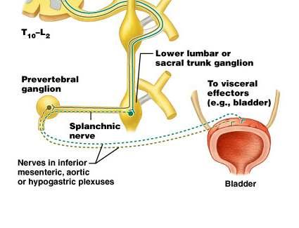
19 Pass through ganglia and synapse in prevertebral ganglion 19
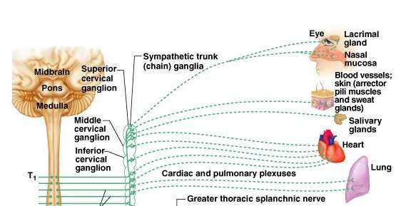
20 Sympathetic chain.cont Nerves which leave the sympathetic chain: A-gray rami ( 31 post ganglionic fibers join spinal nerves to reach sweat glands, errectore papillae & blood vessels - S.C.S.G lower 4 cranial nerves + upper 4 cervical - M.C.S.G 5 th, 6 th cervical nerves - I.C.S.G 7 th, 8 th cervical nerves - Thoracic, lumber, sacral ganglia to corresponding nerves B- visceral nerves 1- Int, & Ext. carotid nerves from S.C.S.G to corresponding arteries 2- pharyngeal branch : from S.C.S.G to pharyngeal plexus 3-pulmonary nerves : 2 nd, 3 rd& 4 th thoracic ganglia 4-cardiac nerves : 2 nd, 3 rd& 4 th thoracic ganglia + 3 cervical ganglia 5- splanchnic nerves : greater, lesser and lowest splanchnic nerves
21 Sympathetic chain

22 Greater splanchnic nerves: Arise from ganglia (5-9 th ) or 10 th Pierce the crussof the diaphragm End in the coeliacganglia Post. ganglia fibers follow the branches of coeliac artery to reach the smooth muscle, gland of stomach
23 Lesser splanchnic nerves: Arise from the 9 th & 10 th Th.ganglia Pierces the crussof diaphragm End in the sup. Mesenteric ganglia Post. Ganglia fibers supply the smooth muscles, glands of small intestine, ascending and transverse parts of colon
24 Lowest splanchnic nerves: May be absent, if present arises from the last one or two th.ganglia Pierces the diaphragm to end in renal plexus
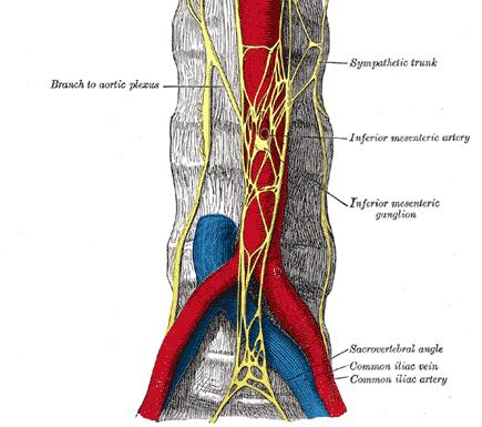
25 Lumber splanchnic branch Arise from L1& L2 ganglia Ends in inferior mesenteric ganglia Post. Gangilionic fibers go to sigmoid and pelvic colon, other post. Gangilionic fibers form the descending hypogastric plexus to supply bladder, rectum and genetalia Branches from sacral part of the chain go to pelvic viscera

 C-The upper five")
26 Thoracic sympathetic chain: Site: enters the thorax in front of neck of 1 st rib and leaves it by passing behind the medial arcuate ligament In the upper part it lies on the necks of the ribs while in the lower part it lies on the side of the bodies of vertebrae Ganglia: (10-12 ),1 st sometimes fuses with the I.C.S.G stellate ganglia Branches: A- Gray & white rami communicants B-2 nd,3 rd & 4 th ganglia ( cardiac & pulmonary ) C-The upper five ganglia give aortic oesophageal branches D- Greater, lesser and lowest splanchnic nerves
27 Sympathetic chain 27
28 Splanchnic nerve

29 Visceral sensory system 29
* *e.g.")
30 Visceral sensory and autonomic neurons participate in visceral reflex arcs Many are spinal reflexes such as defecation and micturition reflexes Some only involve peripheral neurons: spinal cord not involved (not shown)* *e.g. enteric nervous system: 3 neuron reflex arcs entirely within the wall of the gut 30
31 THANK YOU
Group of students. - Rawan almujabili د. محمد المحتسب - 1 P a g e
 - 14 - Group of students - Rawan almujabili د. محمد المحتسب - 1 P a g e Nerves of the posterior abdominal wall The spinal cord gives off spinal nerves between the vertebrae. In the abdomen, through the
[ANATOMY #12] April 28, 2013
![[ANATOMY #12] April 28, 2013](/thumbs/86/93473883.jpg "[ANATOMY #12] April 28, 2013") Sympathetic chain : Sympathetic chain is each of the pair of ganglionated longitudinal cords of the sympathetic nervous system; extend from level of atlas (base of skull) till coccyx. It is paravertebral
Sympathetic chain : Sympathetic chain is each of the pair of ganglionated longitudinal cords of the sympathetic nervous system; extend from level of atlas (base of skull) till coccyx. It is paravertebral
The posterior abdominal wall. Prof. Oluwadiya KS
 The posterior abdominal wall Prof. Oluwadiya KS www.oluwadiya.sitesled.com Posterior Abdominal Wall Lumbar vertebrae and discs. Muscles opsoas, quadratus lumborum, iliacus, transverse, abdominal wall
Sympathetic Nervous System
 Sympathetic Nervous System Lecture Objectives Review the subdivisions of the nervous system. Review the general arrangement and compare the sympathetic and parasympathetic parts. Describe the following
Autonomic Nervous System. Ms. DS Pillay Room 2P24
 Autonomic Nervous System Ms. DS Pillay Room 2P24 OVERVIEW OF THE NERVOUS SYSTEM NERVOUS SYSTEM CNS PNS BRAIN SPINAL CORD SOMATIC ANS SYMPATHEIC PARASYMPATHEIC LOCATION OF GANGLIA IN THE ANS Short post-ganglionic
Autonomic Nervous System DR JAMILA EL MEDANY
 Autonomic Nervous System DR JAMILA EL MEDANY OBJECTIVES At the end of the lecture, students should be able to: Define the autonomic nervous system. Describe the structure of autonomic nervous system Trace
Chapter 15: The Autonomic Nervous System. Copyright 2009, John Wiley & Sons, Inc.
 Chapter 15: The Autonomic Nervous System Comparison of Somatic and Autonomic Nervous Systems Comparison of Somatic and Autonomic Nervous Systems Anatomy of Autonomic Motor Pathways Preganglionic neuron
Organisation of the nervous system
 Chapter1 Organisation of the nervous system 1. Subdivisions of the nervous system The nervous system is divided: i) Structurally The central nervous system (CNS) composed of the brain and spinal cord.
Biology 218 Human Anatomy
 Chapter 20 Adapted form Tortora 10 th ed. LECTURE OUTLINE A. Introduction (p. 632) 1. The autonomic nervous system (ANS) regulates the activity of smooth muscle, cardiac muscle, and certain glands. 2.
The Nervous System: Autonomic Nervous System Pearson Education, Inc.
 17 The Nervous System: Autonomic Nervous System Introduction The autonomic nervous system: Functions outside of our conscious awareness Makes routine adjustments in our body s systems The autonomic nervous
Chapter 16. APR Enhanced Lecture Slides
 Chapter 16 APR Enhanced Lecture Slides See separate PowerPoint slides for all figures and tables pre-inserted into PowerPoint without notes and animations. Copyright The McGraw-Hill Companies, Inc. Permission
Human Anatomy. Autonomic Nervous System
 Human Anatomy Autonomic Nervous System 1 Autonomic Nervous System ANS complex system of nerves controls involuntary actions. Works with the somatic nervous system (SNS) regulates body organs maintains
Tymaa Al-zaben & Amin Al-ajalouni
 Done by: Tymaa Al-zaben & Amin Al-ajalouni ** Hello SERTONIN! SLIDE 3 note:: the slide included within the sheet but make sure back to slide for pictures The Autonomic Nervous System Function : Regulate
cardiac plexus is continuous with the coronary and no named branches pain from the heart and lungs
 Nerves of the Thoracic Region Nerve Source Branches Motor Sensory Notes cardiac plexus cardiac brs. of the vagus n. and cervical ; thoracic l nn. the heart and lungs cardiac, cervical cardiac, vagal vagus
The Nervous System: Autonomic Nervous System
 17 The Nervous System: Autonomic Nervous System PowerPoint Lecture Presentations prepared by Steven Bassett Southeast Community College Lincoln, Nebraska Introduction The autonomic nervous system functions
Fig Glossopharyngeal nerve transmits signals to medulla oblongata. Integrating center. Receptor. Baroreceptors sense increased blood pressure
 Fig. 5. Integrating center Glossopharyngeal nerve transmits signals to medulla oblongata Receptor 3 Vagus nerve transmits inhibitory signals to cardiac pacemaker Baroreceptors sense increased blood pressure
Principles of Anatomy and Physiology
 Principles of Anatomy and Physiology 14 th Edition CHAPTER 15 The Autonomic Nervous System Comparison of Somatic and Autonomic Nervous Systems The somatic nervous system includes both sensory and motor
Part 1. Copyright 2011 Pearson Education, Inc. Copyright 2011 Pearson Education, Inc. Stimulatory
 PowerPoint Lecture Slides prepared by Leslie Hendon University of Alabama, Birmingham C H A P T E R 15 Part 1 The Autonomic Nervous System and Visceral Sensory Neurons The ANS and Visceral Sensory Neurons
Chp. 16: AUTONOMIC N.S. (In Review: Peripheral N. S.)
") Chp. 16: AUTONOMIC N.S. (In Review: Peripheral N. S.) Peripheral nerves contain both motor and sensory neurons Among the motor neurons, some of these are somatic and innervate skeletal muscles while some
Chp. 16: AUTONOMIC N.S. (In Review: Peripheral N. S.) Peripheral nerves contain both motor and sensory neurons Among the motor neurons, some of these are somatic and innervate skeletal muscles while some
ParasymPathetic Nervous system. Done by : Zaid Al-Ghnaneem
 ParasymPathetic Nervous system Done by : Zaid Al-Ghnaneem In this lecture we are going to discuss Parasympathetic, in the last lecture we took sympathetic and one of the objectives of last lecture was
I. Autonomic Nervous System (ANS) A. Dual Innervation B. Autonomic Motor Pathway 1. Preganglionic Neuron a. Preganglionic Fibers (Axons) (1)
 A. Dual Innervation B. Autonomic Motor Pathway 1. Preganglionic Neuron a. Preganglionic Fibers (Axons) (1)") I. Autonomic Nervous System (ANS) A. Dual Innervation B. Autonomic Motor Pathway 1. Preganglionic Neuron a. Preganglionic Fibers (Axons) (1) Acetylcholine - ACh 2. Ganglion (Ganglia) 3. Ganglionic Neuron
I. Autonomic Nervous System (ANS) A. Dual Innervation B. Autonomic Motor Pathway 1. Preganglionic Neuron a. Preganglionic Fibers (Axons) (1) Acetylcholine - ACh 2. Ganglion (Ganglia) 3. Ganglionic Neuron
NERVOUS SYSTEM ANATOMY
 INTRODUCTION to NERVOUS SYSTEM ANATOMY M1 - Gross and Developmental Anatomy Dr. Milton M. Sholley Professor of Anatomy and Neurobiology and Dr. Michael H. Peters Professor of Chemical and Life Science
NERVOUS SYSTEM ANATOMY
 NTRODUCTON to NERVOUS SYSTEM ANATOMY M1 - Gross and Developmental Anatomy Dr. Milton M. Sholley Professor of Anatomy and Neurobiology and Dr. Michael H. Peters Professor of Chemical and Life Science Engineering
musculoskeletal system anatomy nerves of the lower limb 1 done by: dina sawadha & mohammad abukabeer
 musculoskeletal system anatomy nerves of the lower limb 1 done by: dina sawadha & mohammad abukabeer What is the importance of plexuses? plexuses provides us the advantage of a phenomenon called convergence
ANATOMY & PHYSIOLOGY - CLUTCH CH THE AUTONOMIC NERVOUS SYSTEM.
 !! www.clutchprep.com ANATOMY & PHYSIOLOGY - CLUTCH CONCEPT: THE AUTONOMIC NERVOUS SYSTEM: DIVISIONS AND STRUCTURE The Autonomic Nervous System and its Divisions: Autonomic Nervous System (ANS) controls
GI module Lecture: 9 د. عصام طارق. Objectives:
 GI module Lecture: 9 د. عصام طارق Objectives: To list structures forming posterior abdominal wall. To follow aorta & its main branches. To describe IVC & its main tributaries. To list nerves of posterior
Posterior Abdominal wall-
 Structures of posterior abdominal wall: o Bony boundaries: 5 lumber vertebra and their intervertebral disc, iliac fossa and iliac crest. o Muscles: psoas major, quadrates lumborum, transversus abdominis,
Dana Alrafaiah. - Amani Nofal. - Ahmad Alsalman. 1 P a g e
 - 2 - Dana Alrafaiah - Amani Nofal - Ahmad Alsalman 1 P a g e This lecture will discuss five topics as follows: 1- Arrangement of pelvic viscera. 2- Muscles of Pelvis. 3- Blood Supply of pelvis. 4- Nerve
Human Anatomy & Physiology
 PowerPoint Lecture Slides prepared by Barbara Heard, Atlantic Cape Community College Ninth Edition Human Anatomy & Physiology C H A P T E R 14 Annie Leibovitz/Contact Press Images 2013 Pearson Education,
Human Anatomy. Spinal Cord and Spinal Nerves
 Human Anatomy Spinal Cord and Spinal Nerves 1 The Spinal Cord Link between the brain and the body. Exhibits some functional independence from the brain. The spinal cord and spinal nerves serve two functions:
Spinal nerves and cervical plexus Prof. Abdulameer Al Nuaimi. E mail: a.al E. mail:
 Spinal nerves and cervical plexus Prof. Abdulameer Al Nuaimi E mail: a.al nuaimi@sheffield.ac.uk E. mail: abdulameerh@yahoo.com Branches of ophthalmic artery Muscles of face A spinal nerve Spinal
Synapse Homework. Back page last question not counted. 4 pts total, each question worth 0.18pts. 26/34 students answered correctly!
 Synapse Homework Back page last question not counted 26/34 students answered correctly! 4 pts total, each question worth 0.18pts Business TASS hours extended! MWF 1-2pm, Willamette 204 T and Th 9:30-10:30am,
Anatomy of the Large Intestine
 Large intestine Anatomy of the Large Intestine 2 Large Intestine Extends from ileocecal valve to anus Length = 1.5-2.5m = 5 feet Regions Cecum = 2.5-3 inch Appendix= 3-5 inch Colon Ascending= 5 inch Transverse=
Department of Neurology/Division of Anatomical Sciences
 Spinal Cord I Lecture Outline and Objectives CNS/Head and Neck Sequence TOPIC: FACULTY: THE SPINAL CORD AND SPINAL NERVES, Part I Department of Neurology/Division of Anatomical Sciences LECTURE: Monday,
Human Anatomy Biology 351
 nnnnn 1 Human Anatomy Biology 351 Exam #2 Please place your name on the back of the last page of this exam. You must answer all questions on this exam. Because statistics demonstrate that, on average,
THE GOOFY ANATOMIST QUIZZES
 THE GOOFY ANATOMIST QUIZZES 5. NERVES Q1. Which of the following classifications of the nervous systems is correct? A. The autonomic nervous system is composed of the brain, cranial nerves and spinal nerves.
CHAPTER 15 LECTURE OUTLINE
 CHAPTER 15 LECTURE OUTLINE I. INTRODUCTION A. The autonomic nervous system (ANS) regulates the activity of smooth muscle, cardiac muscle, and certain glands. B. Operation of the ANS to maintain homeostasis,
OBJECTIVE: To obtain a fundamental knowledge of the root of the neck with respect to structure and function
 The root of the neck Jeff Dupree, Ph.D. e mail: jldupree@vcu.edu OBJECTIVE: To obtain a fundamental knowledge of the root of the neck with respect to structure and function READING ASSIGNMENT: Moore and
Dr. Weyrich G07: Superior and Posterior Mediastina. Reading: 1. Gray s Anatomy for Students, chapter 3
 Dr. Weyrich G07: Superior and Posterior Mediastina Reading: 1. Gray s Anatomy for Students, chapter 3 Objectives: 1. Subdivisions of mediastinum 2. Structures in Superior mediastinum 3. Structures in Posterior
I. Neural Control of Involuntary Effectors. Chapter 9. Autonomic Motor Nerves. Autonomic Neurons. Autonomic Ganglia. Autonomic Neurons 9/19/11
 Chapter 9 I. Neural Control of Involuntary Effectors The Autonomic Nervous System Lecture PowerPoint Copyright The McGraw-Hill Companies, Inc. Permission required for reproduction or display. Autonomic
Chapter 14 Autonomic Nervous System
 Chapter 14 Autonomic Nervous System Annie Leibovitz/Contact Press Images PowerPoint Lecture Slides prepared by Karen Dunbar Kareiva Ivy Tech Community College Why This Matters Understanding the autonomic
Note: Please refer to handout Spinal Plexuses and Representative Spinal Nerves for
 Chapter 13 Outline Note: Please refer to handout Spinal Plexuses and Representative Spinal Nerves for what you need to know from Exhibits 13.1 13.4 I. INTRODUCTION A. The spinal cord and spinal nerves
Chapter 16. Autonomic nervous system. AP2 Chapter 16: ANS
 Chapter 16 Autonomic nervous system AP2 Chapter 16: ANS 1 Quick Review Nervous System Central Nervous System Peripheral Nervous System Sensory Division Motor Division Somatic Nervous System Autonomic Nervous
Cerebral hemisphere. Parietal Frontal Occipital Temporal
 Cerebral hemisphere Sulcus / Fissure Central Precental gyrus Postcentral gyrus Lateral (cerebral) Parieto-occipital Cerebral cortex Frontal lobe Parietal lobe Temporal lobe Insula Amygdala Hippocampus
Lumbar and Sacral Plexuses. Dr. Heba Kalbouneh Associate Professor of Anatomy and Histology
 Lumbar and Sacral Plexuses Dr. Heba Kalbouneh Associate Professor of Anatomy and Histology Structure of Spinal Nerves: Somatic Pathways dorsal root CNS interneuron spinal nerve dorsal ramus somatic sensory
LAB Notes#1. Ahmad Ar'ar. Eslam
 LAB Notes#1 Ahmad Ar'ar Eslam 1 P a g e Anatomy lab Notes Lower limb bones :- Pelvic girdle: It's the connection between the axial skeleton and the lower limb; it's made up of one bone called the HIP BONE
Introduction to The Autonomic Nervous System. Sympathetic VS Parasympathetic Divisions. Adrenergic and Cholinergic Fibers. ANS Neurotransmitters
 Chapter 15 Introduction to The Autonomic Nervous System Sympathetic VS Parasympathetic Divisions Adrenergic and Cholinergic Fibers ANS Neurotransmitters Autonomic Nervous System Portion of the nervous
Netter's Anatomy Flash Cards Section 4 List 4 th Edition
 Netter's Anatomy Flash Cards Section 4 List 4 th Edition https://www.memrise.com/course/1577335/ Section 4 Abdomen (31 cards) Plate 4-1 Bony Framework of Abdomen 1.1 Costal cartilages 1.2 Iliac crest 1.3
Chapter 15 Lecture Outline
 Chapter 15 Lecture Outline See separate PowerPoint slides for all figures and tables preinserted into PowerPoint without notes. Copyright McGraw-Hill Education. Permission required for reproduction or
Slide Read the tables it is about the difference between male & female pelvis.
 I didn t include the slides, this is only what the doctor read or said because he skipped a lot of things because we took it previously, very important to go back to the slides (*there is an edited version)
Introduction to the Peripheral Nervous System
 Introduction to the Peripheral Nervous System 2 CONTENTS INTRODUCTION PERIPHERAL NERVOUS SYSTEM SPINAL CORD (CENTRAL NERVOUS SYSTEM) OVERVIEW OF THE AUTONOMIC NERVOUS SYSTEM Sympathetic Nervous System
ANATOMY OF PELVICAYCEAL SYSTEM -DR. RAHUL BEVARA
 1 ANATOMY OF PELVICAYCEAL SYSTEM -DR. RAHUL BEVARA 2 KIDNEY:ANATOMY OVERVIEW Kidneys are retroperitoneal, in posterior abdominal region, extending from T12 L3 Bean-shaped Right kidney is lower than left
Abdomen. Retroperitoneal space
 Abdomen. Retroperitoneal space Abdominal cavity The space bounded by: Anterolateral abdominal wall Posterior abdominal wall Diaphragm Pelvic walls and pelvic floor. Subdivided into: True abdominal cavity
Chapter 14. The Nervous System. The Spinal Cord and Spinal Nerves. Lecture Presentation by Steven Bassett Southeast Community College
 Chapter 14 The Nervous System The Spinal Cord and Spinal Nerves Lecture Presentation by Steven Bassett Southeast Community College Introduction The Central Nervous System (CNS) consists of: The spinal
Spinal nerves. Aygul Shafigullina. Department of Morphology and General Pathology
 Spinal nerves Aygul Shafigullina Department of Morphology and General Pathology Spinal nerve a mixed nerve, formed in the vicinity of an intervertebral foramen, where fuse a dorsal root and a ventral root,
The Thoracic wall including the diaphragm. Prof Oluwadiya KS
 The Thoracic wall including the diaphragm Prof Oluwadiya KS www.oluwadiya.com Components of the thoracic wall Skin Superficial fascia Chest wall muscles (see upper limb slides) Skeletal framework Intercostal
4/8/2015. Autonomic Nervous System (ANS) Learn and Understand: Divisions of the ANS. Sympathetic division Parasympathetic division Dual innervation
 Learn and Understand: Divisions of the ANS. Sympathetic division Parasympathetic division Dual innervation") Autonomic Nervous System (ANS) Learn and Understand: Divisions of the ANS Sympathetic division Parasympathetic division Dual innervation ~ All visceral organs served by both divisions, usually cause opposite
Autonomic Nervous System (ANS) Learn and Understand: Divisions of the ANS Sympathetic division Parasympathetic division Dual innervation ~ All visceral organs served by both divisions, usually cause opposite
Faculty of Dental Medicine and Surgery. Sem 4 Peripheral nervous system and nerve plexus Dr. Abbas Garib Alla
 Faculty of Dental Medicine and Surgery Sem 4 Peripheral nervous system and nerve plexus Dr. Abbas Garib Alla PNS Terminology Ganglia neuron cell bodies Peripheral nerves neuronal axons PNS neuroglia Satellite
Human Anatomy and Physiology - Problem Drill 15: The Autonomic Nervous System
 Human Anatomy and Physiology - Problem Drill 15: The Autonomic Nervous System Question No. 1 of 10 Which of the following statements is correct about the component of the autonomic nervous system identified
Lumbar Plexus. Ventral rami L1 L4 Supplies: Major nerves.. Abdominal wall External genitalia Anteromedial thigh
 Lower Limb Nerves Lectures Objectives Describe the structure and relationships of the plexuses of the lower limb. Describe the course, relationships and structures supplied for the major nerves of the
THE AORTA AND IT S MAJOR BRANCHES
 1 THE AORTA AND IT S MAJOR BRANCHES The aorta commences at the aortic valve, above the vestible of the left ventricle and terminates at the level of the fourth lumbar vertebra (L4), where it bifurcates
4/9/2019. Autonomic Nervous System (ANS)
") Autonomic Nervous System (ANS) Learn and Understand: What appears to be two separate systems actually work together to maintain homeostasis under varying conditions. Receptors on the membranes of ANS effectors
Autonomic Nervous System (ANS) Learn and Understand: What appears to be two separate systems actually work together to maintain homeostasis under varying conditions. Receptors on the membranes of ANS effectors
Yes, cranially with ovarian, caudally with vaginal. Yes, with uterine artery (collateral circulation between abdominal +pelvic source)
") Blood supply to internal female genitalia: uterine Internal iliac Sup. large branch: uterus, inf. Small branch: cervix+ sup. Vagina Yes, cranially with ovarian, caudally with vaginal Medially in base of
Blood supply to internal female genitalia: uterine Internal iliac Sup. large branch: uterus, inf. Small branch: cervix+ sup. Vagina Yes, cranially with ovarian, caudally with vaginal Medially in base of
THE SACRAL PARASYMPATHETIC INNERVATION OF THE COLON
 THE SACRAL PARASYMPATHETIC INNERVATION OF THE COLON RUSSELL T. WOODBURNE Department of Anatomy, University of Michigan Hedical School, Ann Arbofi TWO FIGURES Autonomic nerves distribute by a variety of
nerve blocks in the diagnosis and therapy of visceral disease
 Visceral Pain nerve blocks in the diagnosis and therapy of visceral disease Guy Hans, MD, PhD Dept. of Anesthesiology, Multidisciplinary Pain Center Visceral Pain? Type of nociceptive pain (although often
ANATOMY OF SPINAL CORD. Khaleel Alyahya, PhD, MEd King Saud University School of
 ANATOMY OF SPINAL CORD Khaleel Alyahya, PhD, MEd King Saud University School of Medicine @khaleelya OBJECTIVES At the end of the lecture, students should be able to: Describe the external anatomy of the
Autonomic Nervous System
 Autonomic Nervous System Autonomic nervous system organization Sympathetic Nervous System division of the autonomic nervous system that arouses the body, mobilizing its energy in stressful situations
Chapter 14 The Autonomic Nervous System Chapter Outline
 Chapter 14 The Autonomic Nervous System Chapter Outline Module 14.1 Overview of the Autonomic Nervous System (Figures 14.1 14.3) A. The autonomic nervous system (ANS) is the involuntary arm of the peripheral
Introduction. Autonomic means self-governed ; the autonomic nervous system (ANS) is independent of our will
 is independent of our will") Introduction Autonomic means self-governed ; the autonomic nervous system (ANS) is independent of our will It regulates fundamental states and life processes such as heart rate, BP, and body temperature
Introduction Autonomic means self-governed ; the autonomic nervous system (ANS) is independent of our will It regulates fundamental states and life processes such as heart rate, BP, and body temperature
Autonomic Nervous System, Visceral Sensation and Visceral Reflexes Jeff Dupree, Ph.D.
 Autonomic Nervous System, Visceral Sensation and Visceral Reflexes Jeff Dupree, Ph.D. OBJECTIVES After studying the material of this lecture, the student should know the: 1. basic divisions of the autonomic
Composed by Natalia Leonidovna Svintsitskaya, Associate professor of the Chair of Human Anatomy, Candidate of Medicine
 Theoretical background to the study of the autonomic nervous system. Sympathetic and parasympathetic divisions of the autonomic nervous system. Features of the structure, function Composed by Natalia Leonidovna
STRUCTURAL BASIS OF MEDICAL PRACTICE EXAMINATION 3. October 17, 2014
 STRUCTURAL BASIS OF MEDICAL PRACTICE EXAMINATION 3 October 17, 2014 PART l. Answer in the space provided. (12 pts) 1. Identify the structures. (2 pts) A. B. A B C. D. C D 2. Identify the structures. (2
CHAPTER 13 LECTURE OUTLINE
 CHAPTER 13 LECTURE OUTLINE I. INTRODUCTION A. The spinal cord and spinal nerves mediate reactions to environmental changes. B. The spinal cord has several functions. 1. It processes reflexes. 2. It is
Anatomy of the renal system. Professor Nawfal K. Al-Hadithi
 Anatomy of the renal system Professor Nawfal K. Al-Hadithi Objectives To describe the posterior abdominal wall To identify the main anatomical landmarks of the kidneys & ureters To describe the suprarenal
Anatomy & Physiology Pelvic Girdles 10.1 General Information
 Anatomy & Physiology Pelvic Girdles 10.1 General Information ICan2Ed, Inc. In human anatomy, the pelvis (plural pelves or pelvises) is the lower part of. The area of the body that is between the abdomen
Pathogenesis of Chronic Pelvic Pain
 Pathogenesis of Chronic Pelvic Pain Yong-Chul Kim Department of anesthesia and pain medicine, Seoul National University College of Medicine 1 Overview Anatomy Nerve innervation CPP by pathology CPP by
Author(s): Aken Desai, Michael Mathis, 2008
: Aken Desai, Michael Mathis, 2008") Author(s): Aken Desai, Michael Mathis, 2008 License: Unless otherwise noted, this material is made available under the terms of the Creative Commons Attribution Share Alike 3.0 License: http://creativecommons.org/licenses/by-sa/3.0/
Author(s): Aken Desai, Michael Mathis, 2008 License: Unless otherwise noted, this material is made available under the terms of the Creative Commons Attribution Share Alike 3.0 License: http://creativecommons.org/licenses/by-sa/3.0/
3 Circulatory Pathways
 40 Chapter 3 Circulatory Pathways Systemic Arteries -Arteries carry blood away from the heart to the various organs of the body. -The aorta is the longest artery in the body; it branches to give rise to
Divisions of ANS. Divisions of ANS 2 Divisions dualing innervate most organs. Autonomic Nervous System (Chapter 9)
") Autonomic Nervous System (Chapter 9) Autonomic Nervous System (ANS) general properties anatomy Autonomic Effects on Target Organs Subs of Nervous System Central nervous system Brain Spinal cord Peripheral
Autonomic Nervous System (Chapter 9) Autonomic Nervous System (ANS) general properties anatomy Autonomic Effects on Target Organs Subs of Nervous System Central nervous system Brain Spinal cord Peripheral
Mediastinum It is a thick movable partition between the two pleural sacs & lungs. It contains all the structures which lie
 Dr Jamila EL medany OBJECTIVES At the end of the lecture, students should be able to: Define the Mediastinum. Differentiate between the divisions of the mediastinum. List the boundaries and contents of
Gross Anatomy of Lower Spinal Cord
 Chapter 13 Spinal Cord, Spinal Nerves and Somatic Reflexes Spinal cord Spinal nerves Somatic reflexes Gross Anatomy of Lower Spinal Cord Meninges of Vertebra & Spinal Cord Spina Bifida Congenital defect
Classification of the nervous system. Prof. Dr. Nikolai Lazarov 2
 1 1. Formation and general organization 2. Spinal ganglia 3. Zonal and segmental innervation 4. Dorsal rami of the spinal nerves 5. Ventral rami of the spinal nerves 6. Cervical plexus Classification of
AUTONOMIC NERVOUS SYSTEM PART I: SPINAL CORD
 AUTONOMIC NERVOUS SYSTEM PART I: SPINAL CORD How is the organization of the autonomic nervous system different from that of the somatic nervous system? Peripheral Nervous System Divisions Somatic Nervous
Diaphragm and intercostal muscles. Dr. Heba Kalbouneh Associate Professor of Anatomy and Histology
 Diaphragm and intercostal muscles Dr. Heba Kalbouneh Associate Professor of Anatomy and Histology Skeletal System Adult Human contains 206 Bones 2 parts: Axial skeleton (axis): Skull, Vertebral column,
Lab 16: PNS: Nerves and Autonomic NS Hamilton Answers to Pre- Lab Assignments
 Lab 16: PNS: Nerves and Autonomic NS Hamilton Answers to Pre- Lab Assignments Pre-Lab Activity 1: 1. a. olfactory nerve b. optic nerve c. oculomotor nerve d. abducens nerve e. trochlear nerve f. trigeminal
The Spinal Cord, Spinal Nerves, and Spinal Reflexes
 13 The Spinal Cord, Spinal Nerves, and Spinal Reflexes PowerPoint Lecture Presentations prepared by Jason LaPres Lone Star College North Harris An Introduction to the Spinal Cord, Spinal Nerves, and Spinal
The Autonomic Nervous
 Autonomic Nervous System The Autonomic Nervous Assess Prof. Fawzia Al-Rouq System Department of Physiology College of Medicine King Saud University LECTUR (1) Functional Anatomy & Physiology of Autonomic
ANATYOMY OF The thigh
 ANATYOMY OF The thigh 1- Lateral cutaneous nerve of the thigh Ι) Skin of the thigh Anterior view 2- Femoral branch of the genitofemoral nerve 5- Intermediate cutaneous nerve of the thigh 1, 2 and 3 are
Chapter 15 Lecture Outline
 Chapter 15 Lecture Outline See separate PowerPoint slides for all figures and tables preinserted into PowerPoint without notes. Copyright McGraw-Hill Education. Permission required for reproduction or
Benha University. Faculty of Medicine. Anatomy Department Course code (MED 0701) Model answer of Anatomy examination. (Abdomen,Pelvis and Thorax)
 Model answer of Anatomy examination. (Abdomen,Pelvis and Thorax)") 1 Benha University Faculty of Medicine Anatomy Department Course code (MED 0701) Model answer of Anatomy examination (Abdomen,Pelvis and Thorax) 1 st year 2 nd term Date :18 /5 /2013 2 I-Short account
1 Benha University Faculty of Medicine Anatomy Department Course code (MED 0701) Model answer of Anatomy examination (Abdomen,Pelvis and Thorax) 1 st year 2 nd term Date :18 /5 /2013 2 I-Short account
Misc Anatomy. Upper Limb! 2. Lower Limb! 5. Venous Drainage! Head & neck! 8
 Misc Anatomy Upper Limb! 2 Arteries!... 2 Veins!... 2 Spaces!... 4 Lower Limb! 5 Arteries!... 5 Venous Drainage!... 6 Spaces!... 7 Head & neck! 8 Artery!... 8 Ultrasound View for IJ CVL!... 8 Arteries
Autonomic nervous system
 Autonomic nervous system 1. Autonomic nervous system (ANS) nomenclature 2. Topographic organization and structural features of ANS 3. Main subdivisions of the ANS: sympathetic nervous system parasympathetic
Perineum. done by : zaid al-ghnaneem
 Perineum done by : zaid al-ghnaneem Hello everyone, this sheet will talk about 2 nd Lecture which is Perineum but there are some slides and info from 1 st Lecture. Everything included Slides + Pics Let
CARDIOVASCULAR DANIL HAMMOUDI.MD
 CARDIOVASCULAR DANIL HAMMOUDI.MD 18 Systemic Circulation Figure 19.19 Pulmonary Circulation Figure 19.18b 1. Thyroid gland 2. Trachea 3. Brachiocephalic 4. Common carotid 5. Internal jugular 6. Superior
STRUCTURAL BASIS OF MEDICAL PRACTICE EXAMINATION 3. October 16, 2015
 STRUCTURAL BASIS OF MEDICAL PRACTICE EXAMINATION 3 October 16, 2015 PART l. Answer in the space provided. (12 pts) 1. Identify the structures. (2 pts) A. B. A B C. D. C D 2. Identify the structures. (2
Preview from Notesale.co.uk Page 1 of 34
 Abdominal viscera and digestive tract Digestive tract Abdominal viscera comprise majority of the alimentary system o Terminal oesophagus, stomach, pancreas, spleen, liver, gallbladder, kidneys, suprarenal
1 Right & left Hepatic ducts Gastric Impression of spleen
 Pancreatic Model 1 Right & left Hepatic ducts 14 Gastric Impression of spleen 2 Common hepatic duct 15 Renal Impression of spleen 3 Cystic Duct 16 Colic Impression of spleen 4 Common Bile Duct 17 Splenic
Day 5 Respiratory & Cardiovascular: Respiratory System
 Day 5 Respiratory & Cardiovascular: Respiratory System Be very careful not to damage the heart and lungs while separating the ribs! Analysis Questions-Respiratory & Cardiovascular Log into QUIA using your
Chapter 13. The Spinal Cord & Spinal Nerves. Spinal Cord. Spinal Cord Protection. Meninges. Together with brain forms the CNS Functions
 Spinal Cord Chapter 13 The Spinal Cord & Spinal Nerves Together with brain forms the CNS Functions spinal cord reflexes integration (summation of inhibitory and excitatory) nerve impulses highway for upward
The Nervous System PART D. PowerPoint Lecture Slide Presentation by Patty Bostwick-Taylor, Florence-Darlington Technical College
 PowerPoint Lecture Slide Presentation by Patty Bostwick-Taylor, Florence-Darlington Technical College The Nervous System 7 PART D PNS: Spinal Nerves There is a pair of spinal nerves at the level of each