Chief Complaint. Retroperitoneal cystic mass incidentally found at health examination center.
|
|
|
- Willa Burns
- 6 years ago
- Views:
Transcription
1 Personal Information Age: 34 y/o Sex: female Past history: major systemic medical history(-) surgical history(-), family history(-) Denied food or drug allergy
2 Chief Complaint Retroperitoneal cystic mass incidentally found at health examination center.
3 Present Illness Health checkup cystic lesion over adrenal area No specific symptoms Nephrologist -- negative finding Gastro-enterologist further lab and image exam
4 Physical Examination Negative finding.
5 Lab Data CA-199: 2.41 CEA: 0.1 Other data were within normal range.

6 Image CXR Normal lung field. No abnormal defect over bone or soft tissue.

7 Image Abdominal Sonography There was a 8.7x8.3 cm cystic lesion with central septum at pancreatic tail. Head and body were negative.

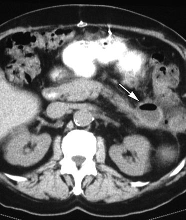
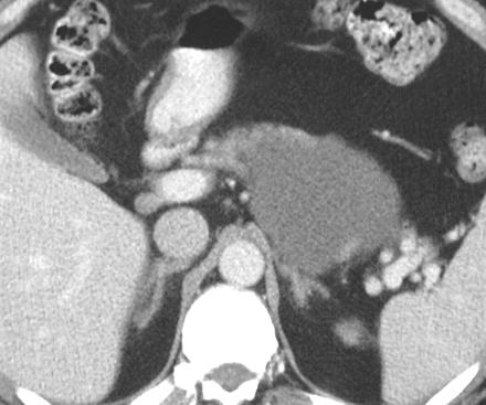
8 Image CT + contrast
9 Image CT + contrast An unenhanced cystic tumor with very thin enhanced capsulation at intraperitoneum. (ant. to pancreatic body/ tail, but post. to stomach)
10 Image CT + contrast 5.8x5.6 cm in largest diameter Mass effect onto spleen and pancreas. No obvious focal infiltration over peri-cystic mesentery. LN (-)
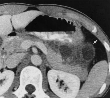
11 Image CT + contrast
12 CT character Solitary. Well capsulation with thin wall. Homogenous fluid content. Located at pancreas or parapancreatic area (near tail). No enhancement with contrast. No inflammation or metastatic sign.
13 D/D for cystic lesion of pancreas or para- pancreatic area by CT
14 D/D by CT 1. Pancreatic pseudocyst 2. Abscess 3. Microcystic Adenoma (Cystadenoma, Serous adenoma, Glycogen-rich adenoma) 4. Lymphangioma 5. Mucinous Adenocarcinoma (Colloid Carcinoma)
15 D/D by CT 6. Mucinous Cystic Tumor (Cystadenocarcinoma, Macrocystic Adenoma) 7. Metastatic tumor 8. Mesenteric cyst 9. Hemangioma 10. Congenital cystic lesion (ADPKD, Von- Hipple-Lindau disease, cystic fibrosis )

16 Pancreatic pseudocyst -- CT
17 Pancreatic pseudocyst CT character Well-defined wall (fibrous tissue, no epithelial lining), may calcified. Uniform, low-attenuation fluid collection with a thin uniform wall that enhances after administration of IV contrast material.
18 Pancreatic pseudocyst CT character Accompanying signs of pancreatitis to a varying degree. (enlarged pancreas, inflammation surrounded)

19 Abscess -- CT
20 Abscess CT character Poorly defined margins and are often suspected when gas is present in a fluid collection. Some rare pancreatic infections, including fungi, tuberculosis, and parasites, can have a cystic appearance.

21 Microcystic Adenoma -- CT
22 Microcystic Adenoma CT character Most are < 2cm. Central stellate scar, with or without calcification. Contains intracellular glycogen but no mucin. (aspiration D/D)
23 Microcystic Adenoma CT character Can be water, soft-tissue, or mixed density. Margin ranging from poorly defined to a thin well-defined capsule. Enhancement of cyst walls and septa ranges from moderate to marked.
24 Lymphangioma -- CT
25 Lymphangioma -- CT
26 Lymphangioma CT character Most often homogeneous, thinwalled, fluid-filled cysts. May have septa, thick walls, calcification, and internal debris. Have epithelial lining but do not contain keratin. (D/D from lymphoepithelial cysts)
27 Lymphangioma CT character The lesion displaces solid organs, has uniform septa which slightly enhance and has contents of attenuation near that of water.

28 Mucinous Adenocarcinoma -- CT
29 Mucinous Adenocarcinoma -- CT Well-defined cystic lesion. Enlarged pancreas and destruction of normal shape may noted.


30 Mucinous Cystic Tumor -- CT
31 Mucinous Cystic Tumor -- CT Most lesions are located in the pancreatic body or tail. Most are larger than 2 cm. Near-water-density unilocular or multilocular cystic lesion with enhancing walls. Peripheral or curvilinear calcifications.

32 Metastatic tumor -- CT
33 Metastatic tumor -- CT Breast, lung, melanoma, and gastrointestinal tract were the most common primary sites. Mostly multiple lesions. Cystic metastases can be the result of central necrosis or cystic degeneration.

34 Mesenteric cyst -- CT
35 Mesenteric cyst -- CT Cysts can be unilocular or multilocular, and may contain chylous, serous or infrequently hemorrhagic fluid. Calcification and reactive, chronic inflammatory changes may be present within their fibrous walls.
36 Mesenteric cyst -- CT May be quite large with several complications : torsion, infarction, volvulus formation, perforation, infection, anemia from intracystic hemorrhage, intestinal obstruction and obstructive uropa
37 Hemangioma -- CT
38 Hemangioma -- CT Enhanced with contrast media. Heterogenous content.

39 Congenital Cyst (ADPKD)
40 Congenital Cyst (ADPKD) Accompanied with other cystic lesions over different organs (Kidney, liver..). Family history.
41 CT Impression Lymphangioma Mesenteric cyst Mucinous cystic tumor

42 Image MRI, T1 W
43 Image MRI, T1 W

44 Image MRI, T2 W
45 Image MRI, T2 W Cystic lesion enhanced in T2 phase With septum
46 Clinical Impression Pancreatic cyst r/o lymphangioma. Mesenteric cyst Mucinous cystic tumor
47 Final Diagnosis Surgical resection Pathology: --grossly measured 6x6x1.5 cm in size. --multiloculated cystic spaces with thin and transparent wall and clear fluid.

48 Pathology histologic picture Enlarged cysticlike space lined by endothelial cell. Numerous lymphocytes are present in fibrous stroma.
49 Pathology character Multiple cysts lined by endothelial cells. Irregularly distributed smooth muscle cells. Lymphoid aggregates in the wall of the cyst.
50 Final Diagnosis --Cystically dilated lymphatics lined by flattened lining cells and lymphlike fluid, focal lymphocytes infiltration, and fibrous stroma with scattered smooth muscle fibers. Pancreas, tail, excision, lymphangioma
51 Discussion Pancreatic Lymphangioma
52 Background Neoplasms, hamartomas, or lymphangiectasias?? Malformations arising from sequestered lymphatic channels or Acquired lesions due to obstruction caused by fibrosis of lymph channels.
53 Background Developmental anomaly, distension of sequestered lymphatic channels within primitive mesenchyme rather than a true neoplasm.
54 Background Most common sites: head, neck, and axilla. Only 5 % at mesentery, omentum, mesocolon, and retroperitoneum. Pancreatic lymphangiomas (1%) occur predominantly in women. (F/M: 2:1). Average age : 25.6 years.
55 Classification Cystic, capillary, and cavernous. Only cystic and cavernous types have been reported in the pancreas Considered to be of pancreatic origin : in the pancreatic parenchyma, adjacent to the pancreas, connected to the organ by a pedicle
56 Clinical Presentations Abdominal pain Nausea Vomiting Palpable mass Silent, incidental finding.
57 Morbidity/ Mortality No risk of malignant transformation. May local invasion. Strong tendency for local recurrence unless they are completely excised.
58 Gross Soft, multiloculated cystic masses. Content: serous, serosanguinous, or lymphatic fluid.
59 Histology Dilated lymphatic channels, separated by thin septa. Cystic spaces lining: flattened or cuboidal endothelial cells. Aggregates of lymphocytes. The septa and walls: smooth muscle fascicles and collagenous connective tissue.
60 Image -- Sonogrphy character Anechoic or hypoechoic Fluid-filled Multiseptated mass in the pancreatic region.
61 Image -- CT character Well-circumscribed Homogeneous cystic masses in or adjacent to the pancreas. Septums and thin walls may enhance after IV contrast injection.
62 Image -- MRI character Hypointense on T1-weighted image Hyperintense on T2-weighted image. Not provide new information.
63 Diagnosis History Imaging: sono, CT, MRI Fine-needle aspiration cytology. Definite diagnosis: pathology report.
64 Treatment No proven medical care for lymphangiomas exists. Treatment of choice: complete surgical excision
65 Prognosis Lymphangiomas are benign hamartomatous malformations instead of true neoplasms. Locally invasion may occur. The prognosis is excellent.
66 Thanks for your attention~
67 Reference T. C. Demos, H. V. Posniak, C. Harmath, M. C. Olson, and G. Aranha Cystic Lesions of the Pancreas Am. J. Roentgenol., December 1, 2002; 179(6): M. Nishino, K. Hayakawa, M. Minami, A. Yamamoto, H. Ueda, and K. Takasu Primary Retroperitoneal Neoplasms: CT and MR Imaging Findings with Anatomic and Pathologic Diagnostic Clues RadioGraphics, January 1, 2003; 23(1): Liron Pantanowitz, Maria Botero: Giant Mesenteric Cyst: A Case Report And Review Of The Literature. The Internet Journal of Pathology Volume 1 Number.
68 Reference From the Department of Traumatology and Critical Care Medicine, Kyorin University School of Medicine, Shinkawa Mitaka-shi, Tokyo , Japan. Received July 19, 2003; revision requested October 3; final revision received April 22, 2004; accepted June 17. Acute presentation of lymphangioma of the retropritoneum 129 The Ulster Medical Journal, Volume 73, No. 2, pp , November DH Taylor, C Loughrey. Cystic Lymphangioma of the Pancreas American Journal of Roentgenology Received March 13, 2001; accepted after revision April 4, 2001.
Name : 黃 XX Age : 52 Sex : 女 Occupation : 廚房阿姨 Marital status : 已婚
 Name : 黃 XX Age : 52 Sex : 女 Occupation : 廚房阿姨 Marital status : 已婚 Chief Complaint Mild postprandial fullness for 2 months Present Illness This 52 year-old female suffered from intermittent post-prandial
Sex: 女 Age: 51 Occupation: 無 Admission date:92/07/22
 Sex: 女 Age: 51 Occupation: 無 Admission date:92/07/22 Chief complaint Unknown fever for one month Hand tremor and left huge renal tumor was noted Present illness Suffered from fever for one month, hand
Evaluation and Management of Cystic Lesions of the Pancreas: When to Resect, When to Follow and When to Forget
 Evaluation and Management of Cystic Lesions of the Pancreas: When to Resect, When to Follow and When to Forget Randall Brand, MD Professor of Medicine Division of Gastroenterology, Hepatology and Nutrition
CT 101 :Pancreas and Spleen
 CT 101 :Pancreas and Spleen Shikha Khullar,, MD, MPH Division of Radiology University of South Alabama The Pancreas Normal Pancreas 3 Phase Pancreatic CT Non contrast Arterial phase : 30-35 35 second
Cystic Pancreatic Lesions: Approach to Diagnosis
 Cystic Pancreatic Lesions: Approach to Diagnosis Poster No.: R-0130 Congress: RANZCR-AOCR 2012 Type: Educational Exhibit Authors: A. AGARWAL, R. M. Mendelson; Perth/AU Keywords: Cysts, Biopsy, Endoscopy,
Morphologic features in cystic lesions of pancreas-a retrospective analysis
 International Journal of Advances in Medicine Cicy PJ et al. Int J Adv Med. 2018 Feb;5(1):192-196 http://www.ijmedicine.com pissn 2349-3925 eissn 2349-3933 Original Research Article DOI: http://dx.doi.org/10.18203/2349-3933.ijam20180083
X-ray Corner. Imaging of The Pancreas. Pantongrag-Brown L
 X-ray Corner 125 Imaging of The Pancreas Modern imaging modalities commonly used in pancreas include ultrasound (US), CT, and MRI. Pancreas is a retroperitoneal organ which makes it difficult to visualize
Disorders of Cell Growth & Neoplasia. Histopathology Lab
 Disorders of Cell Growth & Neoplasia Histopathology Lab Paul Hanna April 2010 Case #84 Clinical History: 5 yr-old, West Highland White terrier. skin mass from axillary region. has been present for the
Role of Imaging Methods in Diagnosis of Acute Pancreatitis. Válek V. Radiologická klinika, FN Brno a LF MU v Brně
 Role of Imaging Methods in Diagnosis of Acute Pancreatitis Válek V. Radiologická klinika, FN Brno a LF MU v Brně New Classification: Acute Pancreatitis 2007 revision of Atlanta classification and definitions
Matthew McCollough, M.D. April 9, 2009 University of Louisville
 Matthew McCollough, M.D. April 9, 2009 University of Louisville List the differential diagnosis for pancreatic cysts Review the epidemiology Illustrate the types of cysts through case discussions Discuss
X-Ray Corner. Imaging Approach to Cystic Liver Lesions. Pantongrag-Brown L. Solitary cystic liver lesions. Hepatic simple cyst (Figure 1)
") THAI J 136 Imaging Approach to Cystic Liver Lesions GASTROENTEROL 2013 X-Ray Corner Imaging Approach to Cystic Liver Lesions Pantongrag-Brown L Cystic liver lesions are common findings in daily practice
THAI J 136 Imaging Approach to Cystic Liver Lesions GASTROENTEROL 2013 X-Ray Corner Imaging Approach to Cystic Liver Lesions Pantongrag-Brown L Cystic liver lesions are common findings in daily practice
Neoplasias Quisticas del Páncreas
 SEAP -Aproximación Práctica a la Patología Gastrointestinal- Madrid, 26 de mayo, 2006 Neoplasias Quisticas del Páncreas Gregory Y. Lauwers, M.D. Director, Service Massachusetts General Hospital Harvard
Pediatric Retroperitoneal Masses Radiologic-Pathologic Correlation
 Acta Radiológica Portuguesa, Vol.XVIII, nº 70, pág. 61-70, Abr.-Jun., 2006 Pediatric Retroperitoneal Masses Radiologic-Pathologic Correlation Marilyn J. Siegel Mallinckrodt Institute of Radiology, Washington
Case Discussion Splenic Abscess
 Case Discussion Splenic Abscess Personal Data Gender: male Birth Date: 1928/Mar/06th Allergy: Mefenamic Smoking: 0.5 PPD for 55 years Alcohol: negative (?) 4 Months Ago Abdominal pain: epigastric area
The role of endoscopy in the diagnosis and treatment of cystic pancreatic neoplasms
 The role of endoscopy in the diagnosis and treatment of cystic pancreatic neoplasms CYSTIC LESIONS AND FLUID COLLECTIONS OF THE PANCREAS Their pathology ranges from pseudocysts and pancreatic necrosis
HEPATO-BILIARY IMAGING
 HEPATO-BILIARY IMAGING BY MAMDOUH MAHFOUZ MD PROF.OF RADIOLOGY CAIRO UNIVERSITY mamdouh.m5@gmail.com www.ssregypt.com CT ABDOMEN Indications Patient preparation Patient position Scanogram Fasting 4-6 hours
Cystic Lymphangioma of the Adrenal Gland: a rare case report
 J Radiol Sci 2013; 38: 59-64 Cystic Lymphangioma of the Adrenal Gland: a rare case report Xiang-Jun Lin Chun-Chao Huang She-Meng Cheng Department of Radiology, Mackay Memorial Hospital and Mackay Medical
A Case of Giant Mesenteric Cyst Originating from the Small Intestine
 Showa Univ J Med Sci 27 2, 125 129, June 2015 Case Report A Case of Giant Mesenteric Cyst Originating from the Small Intestine Takahiro UMEMOTO 1, Tetsuji WAKABAYASHI 1, Nobuyuki OHIKE 2, Ryuichi SEKINE
Pancreatic Cysts. Darius C. Desai, MD FACS St. Luke s University Health Network
 Pancreatic Cysts Darius C. Desai, MD FACS St. Luke s University Health Network None Disclosures Incidence Widespread use of cross sectional imaging Seen in over 2% of patients having abdominal imaging
40th European Congress of Cytology Liverpool, UK, 2-5 th October 2016
 40th European Congress of Cytology Liverpool, UK, 2-5 th October 2016 EUS FNA of abdominal organs: An approach to reporting and triage for ancillary testing Date and time: Sunday 2 nd October 2016 15.00-16.30
Malignant Focal Liver Lesions
 Malignant Focal Liver Lesions Other Than HCC Pablo R. Ros, MD, MPH, PhD Departments of Radiology and Pathology University Hospitals Cleveland Medical Center Case Western Reserve University Pablo.Ros@UHhospitals.org
Select problems in cystic pancreatic lesions
 Disclosure Select problems in cystic pancreatic lesions Five Prime Therapeutics shareholder Adicet Bio shareholder Bristol-Meyer Squibb advisory board grace.kim@ucsf.edu Pancreatic cystic lesions Intraductal
Video Microscopy Tutorial 19
 Video Microscopy Tutorial 19 EUS FNA of Pancreatic Cysts Martha Pitman, MD There are no disclosures necessary. EUS-FNA of Pancreatic Cysts Martha Bishop Pitman, M.D. Massachusetts General Hospital Harvard
Pancreatic Cystic Lesions 원자력병원
 Pancreatic Cystic Lesions 원자력병원 박선 후 Lines of cellular differentiation Ductal Acinar Undetermined Ductal adenocarcinoma Serous/ mucinous tumor Intraductal papillary mucinous neoplasm Acinar cell carcinoma
Kenneth D. Chi, MD Medical Director GI Lab Advocate Lutheran General Hospital
 Kenneth D. Chi, MD Medical Director GI Lab Advocate Lutheran General Hospital Advances in Digestive Health for the Primary Care Physician Symposium May 2, 2015 None Case Presentation Types of Pancreatic
Diagnostic imaging of lymphatic malformations
 Diagnostic imaging of lymphatic malformations Poster No.: C-1440 Congress: ECR 2016 Type: Educational Exhibit Authors: M. M. Coman 1, M. T. A. Buzan 2, S. Manole 3, S. M. Dudea 3 ; 1 2 3 Campia Turzii/RO,
CYSTIC TUMORS OF THE KIDNEY JOHN N. EBLE, M.D. CYSTIC NEPHROMA
 Page 1 CYSTIC TUMORS OF THE KIDNEY JOHN N. EBLE, M.D. Department of Pathology & Laboratory Medicine Phone (317) 274-4806 Medical Science A-128 FAX: (317) 278-2018 635 Barnhill Drive jeble @iupui.edu Indianapolis,
Brief History. Identification : Past History : HTN without regular treatment.
 Brief History Identification : Name : 陳 x - Admission : 94/10/06 Gender : male Age : 75 y/o Chief Complaint : Urinary difficulty for months. Past History : HTN without regular treatment. Brief History
Sonography of soft-tissue vascular lesions
 Sonography of soft-tissue vascular lesions Oscar M. Navarro Associate Professor, University of Toronto Dept. of Diagnostic Imaging, The Hospital for Sick Children Toronto, Canada Declaration of Disclosure
GASTROINTESTINAL IMAGING STUDY GUIDE
 GASTROINTESTINAL IMAGING STUDY GUIDE Pharynx Diverticula Foreign bodies Trauma o Motility Disorders Esophagus Diverticula Trauma Esophagitis Barrett esophagus Rings, webs, and strictures Varices Benign
Cystic Lesions of the Pancreas
 Residents Section Pattern of the Month w668 04.29.11 Khan et al. Residents Section Pattern of the Month Residents inradiology tif Khan 1 Faisal Khosa Ronald L. Eisenberg Khan, Khosa F, Eisenberg RL Keywords:
CTA/MRA of Pediatric Hepatic Masses Radiology-Pathology Correlation
 Acta Radiológica Portuguesa, Vol.XVIII, nº70, pág. 41-50, Abr.-Jun., 2006 CTA/MRA of Pediatric Hepatic Masses Radiology-Pathology Correlation Marilyn J. Siegel Mallinckrodt Institute of Radiology, Washington
CT & MRI of Benign Liver Neoplasms Srinivasa R Prasad
 CT & MRI of Benign Liver Neoplasms Srinivasa R Prasad No financial disclosures Acknowledgements Many thanks to Drs. Heiken, Narra & Menias (MIR) Dr. Sahani (MGH) for sharing images Benign Liver Tumors:
Chief complaint. A mass at right chest
 Chief complaint A mass at right chest Present illness This 1-year-5-month-old girl had a mass at right side chest since one month ago. flat and not tender at first In the recent 2 days, the mass enlarged
Female Genital Tract Lab. Dr. Nisreen Abu Shahin Assistant Professor of Pathology University of Jordan
 Female Genital Tract Lab Dr. Nisreen Abu Shahin Assistant Professor of Pathology University of Jordan Ovarian Pathology A 20-year-old female presented with vague left pelvic pain. Pelvic exam revealed
Essentials of Clinical MR, 2 nd edition. 65. Benign Hepatic Masses
 65. Benign Hepatic Masses Pulse sequences acquired for abdominal MRI typically consist of fast acquisition schemes such as single-shot turbo spin echo (i.e. HASTE) and gradient echo schemes such as FLASH
Renal masses - the role of diagnostic imaging
 Renal masses - the role of diagnostic imaging Poster No.: C-2471 Congress: ECR 2015 Type: Educational Exhibit Authors: V. Rai#; Bjelovar/HR Keywords: Cysts, Cancer, Structured reporting, Ultrasound, MR,
General history. Basic Data : Age :62y/o Date of admitted: Married status : Married
 General history Basic Data : Age :62y/o Date of admitted:940510 Married status : Married General history Chief Complain : bilateral ovarian cyst incidentally being found out during pap smear. Present Illness
Chronic pancreatitis mimicking intraductal papillary mucinous neoplasm of the pancreas; Report of tow cases
 Jichi Medical University Journal Chronic pancreatitis mimicking intraductal papillary mucinous neoplasm of the pancreas; Report of tow cases Noritoshi Mizuta, Hiroshi Noda, Nao Kakizawa, Nobuyuki Toyama,
Anatomy of the biliary tract
 Harvard-MIT Division of Health Sciences and Technology HST.121: Gastroenterology, Fall 2005 Instructors: Dr. Jonathan Glickman Anatomy of the biliary tract Figure removed due to copyright reasons. Biliary
Intracystic papillary carcinoma of the breast
 Intracystic papillary carcinoma of the breast Poster No.: C-1932 Congress: ECR 2011 Type: Educational Exhibit Authors: V. Dimarelos, F. TZIKOS, N. Kotziamani, G. Rodokalakis, 1 2 3 1 1 1 2 T. MALKOTSI
Salivary Glands 3/7/2017
 Salivary Glands 3/7/2017 Goals and objectives Focus on the entities unique to H&N Common board type facts Information for your future practice Salivary Glands Salivary Glands Major gland. Paratid. Submandibular.
Cystic Disease of the Liver Work Up and Management. Louis Ferrari MD, PGY 3 6/9/16 SUNY Downstate Medical Center
 Cystic Disease of the Liver Work Up and Management Louis Ferrari MD, PGY 3 6/9/16 SUNY Downstate Medical Center The Case 73F presents to clinic after diagnostic laparoscopy at OSH. Known liver mass for
Evaluation of Liver Mass Lesions. American College of Gastroenterology 2013 Regional Postgraduate Course
 Evaluation of Liver Mass Lesions American College of Gastroenterology 2013 Regional Postgraduate Course Lewis R. Roberts, MB ChB, PhD Division of Gastroenterology and Hepatology Mayo Clinic College of
Intraperitoneal cysts in infancy and childhood An overview and sonographic differentiation
 Intraperitoneal cysts in infancy and childhood An overview and sonographic differentiation M. Mearadji International Foundation for Pediatric Imaging Aid Rotterdam, The Netherlands Intraperitoneal cysts
Solid pseudopapillary tumour of the pancreas: Report of five cases
 ISPUB.COM The Internet Journal of Pathology Volume 8 Number 2 Solid pseudopapillary tumour of the pancreas: Report of five cases P Srilatha, V Manna, P Kanthilatha Citation P Srilatha, V Manna, P Kanthilatha..
ACG Clinical Guideline: Diagnosis and Management of Pancreatic Cysts
 ACG Clinical Guideline: Diagnosis and Management of Pancreatic Cysts Grace H. Elta, MD, FACG 1, Brintha K. Enestvedt, MD, MBA 2, Bryan G. Sauer, MD, MSc, FACG (GRADE Methodologist) 3 and Anne Marie Lennon,
TUMOR AND TUMOR-LIKE CONDITIONS OF THE PERITONEUM AND OMENTUM/MESENTERY 40 th. Annual Meeting SCBTMR September 9-13, 2017, Nashville, Tennessee
 TUMOR AND TUMOR-LIKE CONDITIONS OF THE PERITONEUM AND OMENTUM/MESENTERY 40 th. Annual Meeting SCBTMR September 9-13, 2017, Nashville, Tennessee Isaac R Francis University of Michigan Department of Radiology
Workup of a Solid Liver Lesion
 Workup of a Solid Liver Lesion Joseph B. Cofer MD FACS Chief Quality Officer Erlanger Health System Affiliate Professor of Surgery UTHSC-Chattanooga I have no financial or other relationships with any
the urinary system pathology Dr. Fairoz A Eltorgman
 the urinary system pathology Dr. Fairoz A Eltorgman Tumors of the renal pelvis & kidney Benign tumors of the renal pelvis: Hemangioma Leiomyoma Malignant tumors: Transitional cell carcinoma Squamous cell
Financial Disclosure
 Benign Liver Masses Adil Abdalla, MBBS Creighton University-CHI Health August 25, 2018 Financial Disclosure Nothing to disclose Financial Disclosure 1 Objectives To assess patients with benign liver tumors
performed to help sway the clinician in what the appropriate diagnosis is, which can substantially alter the treatment of management.
 Hello, I am Maura Polansky at the University of Texas MD Anderson Cancer Center. I am a Physician Assistant in the Department of Gastrointestinal Medical Oncology and the Program Director for Physician
Radiology Pathology Conference
 Radiology Pathology Conference Nadia F. Yusaf, M.D. PGY-3 1/29/2010 Presentation material is for education purposes only. All rights reserved. 2010 URMC Radiology Page 1 of 90 Case 1 60 year- old man presents
ABDOMINAL LYMPHANGIECTASIA
 wjpmr, 2016,2(6), 123-129. SJIF Impact Factor: 3.535 Case Report WORLD JOURNAL OF PHARMACEUTICAL AND MEDICAL RESEARCH ISSN 2455-3301 WJPMR ABDOMINAL LYMPHANGIECTASIA 1 Dr. Kishor Rajpal, 2 Dr. Ashwini
Characterization of adrenal lesions on CT and MRI: all that a radiologist must know
 Characterization of adrenal lesions on CT and MRI: all that a radiologist must know Poster No.: C-2476 Congress: ECR 2013 Type: Educational Exhibit Authors: N. Benzina, S. MAJDOUB, C. H. ZARRAD, H. Zaghouani,
Interesting Cases from Liver Tumor Board. Jeffrey C. Weinreb, M.D.,FACR Yale University School of Medicine
 Interesting Cases from Liver Tumor Board Jeffrey C. Weinreb, M.D.,FACR Yale University School of Medicine jeffrey.weinreb@yale.edu Common Liver Diseases Hemangioma Cyst FNH Focal Fat/Sparing THID Non-Cirrhotic
Common and unusual CT and MRI manifestations of pancreatic adenocarcinoma: a pictorial review
 Review Article Common and unusual CT and MRI manifestations of pancreatic adenocarcinoma: a pictorial review Min-Jie Yang, Su Li, Yong-Guang Liu, Na Jiao, Jing-Shan Gong Department of Radiology, Shenzhen
EDUCATIONAL CASES E1 & E2. Natasha Inglis 20/03/15
 EDUCATIONAL CASES E1 & E2 Natasha Inglis 20/03/15 CASE E1 79 year old female Rectum. Altemeier operation Histology Superficial erosions and mucosal congestion volcano lesion and pseudomembrane formation
04/10/2018. Intraductal Papillary Neoplasms Of Breast INTRADUCTAL PAPILLOMA
 Intraductal Papillary Neoplasms Of Breast Savitri Krishnamurthy MD Professor of Pathology Deputy Division Head The University of Texas MD Anderson Cancer Center 25 th Annual Seminar in Pathology Pittsburgh,
PAPILLARY THYROID CARCINOMA PRESENTING AS A LATERAL NECK MASS MASS. Dr. Pamela Hanson DO PGY3
 PAPILLARY THYROID CARCINOMA PRESENTING AS A LATERAL NECK MASS MASS Dr. Pamela Hanson DO PGY3 MK CASE PRESENTATION 28 yo Female presented to the ENT Clinic in October 2016, with the complaint of chronic
Outline 11/2/2017. Pancreatic EUS-FNA general aspects. Cytomorphologic features of solid neoplasms/lesions of the pancreas
 ENDOSCOPIC ULTRASOUND GUIDED-FINE NEEDLE ASPIRATION CYTOLOGY OF PANCREAS Khalid Amin M.D. Assistant Professor Department of Laboratory Medicine and Pathology University of Minnesota Outline Pancreatic
Case 1307 Mesothelial cysts
 Case 1307 Mesothelial cysts Vinhais S, Monteiro M, Cunha TM INSTITUTO PORTUGUÊS DE ONCOLOGIA de Francisco Gentil de LISBOA Section: Gastro-Intestinal Imaging Published: 2001, Nov. 23 Patient: 44 year(s),
Radiologic overview of primary pancreatic neoplasms beyond adenocarcinoma
 Radiologic overview of primary pancreatic neoplasms beyond adenocarcinoma Poster No.: C-0663 Congress: ECR 2014 Type: Educational Exhibit Authors: L. M. Klein, J. A. Pérez Retortillo, L. Gijón de la Santa,
Gross appearance of peritoneal cysts. They have a thin, translucent wall and contain a clear fluid.
 Gross appearance of peritoneal cysts. They have a thin, translucent wall and contain a clear fluid. So-called multicystic benign mesothelioma. A, Gross appearance. So-called multicystic benign mesothelioma.
Pitfalls in thyroid tumor pathology. Prof.Valdi Pešutić-Pisac MD, PhD
 Pitfalls in thyroid tumor pathology Prof.Valdi Pešutić-Pisac MD, PhD Too many or... Tumour herniation through a torn capsule simulating capsular invasion fibrous capsule with a sharp discontinuity, suggestive
CELL AND TISSUE INJURY COURSE-II PATHOLOGY LABORATORY. PATHOLOGY of MASS LESIONS and TISSUE DEFECTS -MACROSCOPY Assoc. Professor Rengin Ahıskalı
 CELL AND TISSUE INJURY COURSE-II PATHOLOGY LABORATORY PATHOLOGY of MASS LESIONS and TISSUE DEFECTS -MACROSCOPY Assoc. Professor Rengin Ahıskalı M1 - RENAL TUBERCULOSIS cavitary areas caseous necrosis fibrous
Biliary tract tumors
 Short Course 2010 Annual Fall Meeting of the Korean Society for Pathologists Biliary tract tumors Joon Hyuk Choi, M.D., Ph.D. Professor, Department of Pathology, Yeungnam Univ. College of Medicine, Daegu,
Radiological Analysis of Cystic lesions of the Pancreas
 September 2002 Radiological Analysis of Cystic lesions of the Pancreas Shruthi Mahalingaiah, Harvard Medical School Year III, Agenda Background Anatomy and histology Radiological workup of a cyst in the
Case Presentation 主治醫師 : 宋文鑫日期 :
 Case Presentation 主治醫師 : 宋文鑫日期 : 2015-2-28 General Data Name:OOO Chart Number:OOOOOOO Date of Admission:2014 年 08 月 04 日 Age: 33 y/o Sex:female Occupation : 會計 Chief Complaint Palpable soft tissue mass
Contents. Basic Ultrasound Principles and Terminology. Ultrasound Nodule Characteristics
 Contents Basic Ultrasound Principles and Terminology Basic Ultrasound Principles... 1 Ultrasound System... 2 Linear Transducer for Superficial Images and Ultrasound-Guided FNA... 3 Scanning Planes... 4
GENERAL ABDOMINAL IMAGING PERITONEAL SPACE, PANCREAS, & SPLEEN. VMB 960 March 25, 2013
 GENERAL ABDOMINAL IMAGING PERITONEAL SPACE, PANCREAS, & SPLEEN VMB 960 March 25, 2013 REFERENCE Chapters 35-36 Pages 650-678 Chapter 37 Pages 694-701 Chapter 3 Pages 38-49 OBJECTIVES Radiography and Ultrasound
Ó Journal of Krishna Institute of Medical Sciences University 112
 ISSN 2231-4261 CASE REPORT Large Pseudocyst of the Adrenal Gland: A Radiological Pathological Correlation 1* 1 2 1 1 Abhishek Dwivedi, Satveer Choudhary, Vikram Trehan, Narender Yadav, K. Leela Kanth 1
Diseases of the breast (1 of 2)
") Diseases of the breast (1 of 2) Introduction A histology introduction Normal ducts and lobules of the breast are lined by two layers of cells a layer of luminal cells overlying a second layer of myoepithelial
Diseases of the breast (1 of 2) Introduction A histology introduction Normal ducts and lobules of the breast are lined by two layers of cells a layer of luminal cells overlying a second layer of myoepithelial
objectives Pitfalls and Pearls in PET/CT imaging Kevin Robinson, DO Assistant Professor Department of Radiology Michigan State University
 objectives Pitfalls and Pearls in PET/CT imaging Kevin Robinson, DO Assistant Professor Department of Radiology Michigan State University To determine the regions of physiologic activity To understand
Case Scenario 1: Thyroid
 Case Scenario 1: Thyroid History and Physical Patient is an otherwise healthy 80 year old female with the complaint of a neck mass first noticed two weeks ago. The mass has increased in size and is palpable.
Malignant Cardiac Tumors Rad-Path Correlation
 Malignant Cardiac Tumors Rad-Path Correlation Vincent B. Ho, M.D., M.B.A. 1 Jean Jeudy, M.D. 2 Aletta Ann Frazier, M.D. 2 1 Uniformed Services University of the Health Sciences 2 University of Maryland
Imaging of Gastrointestinal Stromal Tumors (GIST) Amir Reza Radmard, MD Assistant Professor Shariati hospital Tehran University of Medical Sciences
 Amir Reza Radmard, MD Assistant Professor Shariati hospital Tehran University of Medical Sciences") Imaging of Gastrointestinal Stromal Tumors (GIST) Amir Reza Radmard, MD Assistant Professor Shariati hospital Tehran University of Medical Sciences Describe the typical imaging findings of GIST at initial
Imaging of Gastrointestinal Stromal Tumors (GIST) Amir Reza Radmard, MD Assistant Professor Shariati hospital Tehran University of Medical Sciences Describe the typical imaging findings of GIST at initial
Primary Retroperitoneal Myxofibrosarcoma: a case report and review of the literature
 J Radiol Sci 2014; 39: 57-62 Primary Retroperitoneal Myxofibrosarcoma: a case report and review of the literature Chih-Yu Chen 1 Yueh-Min Lin 2 Shang-Yun Ho 1 Kwo-Whei Lee 1 Ching Hsueh 1 Department of
Imaging in breast cancer. Mammography and Ultrasound Donya Farrokh.MD Radiologist Mashhad University of Medical Since
 Imaging in breast cancer Mammography and Ultrasound Donya Farrokh.MD Radiologist Mashhad University of Medical Since A mammogram report is a key component of the breast cancer diagnostic process. A mammogram
Recently role of non-invasive diagnostics methods
 CERTAIN ASPECTS OF NLS-DIAGNOSTICS OF LIVER FOCAL PATHOLOGY A.Y. Shvack, V.I. Nesterov, N.L. Ogluzdina This article contains information about NLS-graphy application in diagnostics of liver focal affections:
What s Your Diagnosis??? Renée Fahrenholz, Class of 2012
 Renée Fahrenholz, Class of 2012 What s Your Diagnosis??? Signalment Emma, a 9 year old, Female, Spayed, Domestic Short Haired Feline Presenting Complaint Weight loss, vomited the morning of her visit,
Role of imaging in RCC. Ultrasonography. Solid lesion. Cystic RCC. Solid RCC 31/08/60. From Diagnosis to Treatment: the Radiologist Perspective
 Role of imaging in RCC From Diagnosis to Treatment: the Radiologist Perspective Diagnosis Staging Follow up Imaging modalities Limitations and pitfalls Duangkamon Prapruttam, MD Department of Therapeutic
Unusual Pancreatic Neoplasms RTC 2/11/2011
 Unusual Pancreatic Neoplasms RTC 2/11/2011 Objectives Intraductal Papillary Mucinous Neoplasm (IPMN) Mucinous Cystic Neoplasm (MCN) Islet Cell Tumors Insulinoma Glucagonoma VIPoma Somatostatinoma Gastrinoma
Retroperitoneal tumors: Computed Tomography (CT) and Magnetic Resonance (MR) patterns
 and Magnetic Resonance (MR) patterns") Retroperitoneal tumors: Computed Tomography (CT) and Magnetic Resonance (MR) patterns Poster No.: C-1369 Congress: ECR 2011 Type: Educational Exhibit Authors: G. Cardone, A. Messina, D. Vergnaghi, P. Mangili,
Retroperitoneal tumors: Computed Tomography (CT) and Magnetic Resonance (MR) patterns Poster No.: C-1369 Congress: ECR 2011 Type: Educational Exhibit Authors: G. Cardone, A. Messina, D. Vergnaghi, P. Mangili,
Retroperitoneal tumors: Computed Tomography (CT) and Magnetic Resonance (MR) patterns
 and Magnetic Resonance (MR) patterns") Retroperitoneal tumors: Computed Tomography (CT) and Magnetic Resonance (MR) patterns Poster No.: C-1369 Congress: ECR 2011 Type: Educational Exhibit Authors: G. Cardone, A. Messina, D. VERGNAGHI, P. Mangili,
Retroperitoneal tumors: Computed Tomography (CT) and Magnetic Resonance (MR) patterns Poster No.: C-1369 Congress: ECR 2011 Type: Educational Exhibit Authors: G. Cardone, A. Messina, D. VERGNAGHI, P. Mangili,
Case 1. Case 1: EUS Report 5/1/2017. Interesting Cases of Pancreatic Masses
 Interesting Cases of Pancreatic Masses Martha Bishop Pitman, MD Professor of Pathology Harvard Medical School Director of Cytopathology Massachusetts General Hospital Boston, MA MASSACHUSETTS GENERAL PHYSICIANS
Imaging Guided Biopsy. Edited & Presented by ; Hussien A.B ALI DINAR. Msc Lecturer,Reporting Sonographer
 Imaging Guided Biopsy Edited & Presented by ; Hussien A.B ALI DINAR. Msc Lecturer,Reporting Sonographer Objective By the End of this lessons you should : Define what biopsy Justify Aim to perform biopsy
Pre-operative assessment of patients for cytoreduction and HIPEC
 Pre-operative assessment of patients for cytoreduction and HIPEC Washington Hospital Center Washington, DC, USA Ovarian Cancer Surgery New Strategies Bergamo, Italy May 5, 2011 Background Cytoreductive
Cystic lesions of the liver
 Cystic lesions of the liver Poster No.: C-0408 Congress: ECR 2014 Type: Educational Exhibit Authors: E. Rosado, J. Pereira, S. El Bouchaibi, M. A. A. Bali ; 1 1 2 2 3 3 4 4 Amadora/PT, Lisboa/PT, Bruxelles/BE,
Concurrent Multilocular Cystic Renal Cell Carcinoma and Leiomyoma in the Same Kidney: Previously Unreported Association
 218 Concurrent Multilocular Cystic Renal Cell Carcinoma and Leiomyoma in the Same Kidney: Previously Unreported Association Min Su Cheong a Dong Hun Koo a In-Sung Kim a Kyung Chul Moon b Ja Hyeon Ku a
Case Scenario 1. Pathology report Specimen from mediastinoscopy Final Diagnosis : Metastatic small cell carcinoma with residual lymphatic tissue
 Case Scenario 1 Oncology Consult: Patient is a 51-year-old male with history of T4N3 squamous cell carcinoma of tonsil status post concurrent chemoradiation finished in October two years ago. He was hospitalized
Patient History. A 58 year old man presents with a 16 mm cyst in the pancreatic tail. The cyst is unilocular with a thick wall and no mural nodule.
 Case 1 Martha Bishop Pitman, MD Director of Cytopathology Massachusetts General Hospital Associate Professor of Pathology Harvard Medical School Boston, MA Patient History A 58 year old man presents with
Images In Gastroenterology
 Images In Gastroenterology Thong-Ngam D, et al. THAI J GASTROENTEROL 2005 Vol. 6 No. 2 May - Aug. 2005 105 Imaging of Gastrointestinal Stromal Tumors Pornpim Fuangtharnthip, M.D. Narumol Hargroove, M.D.
RADIOFREQUENCY ABLATION
 RADIOFREQUENCY ABLATION ELIZABETH DAVID M D FRCPC VASCULAR A ND INTERVENTIONAL RADIOLOGIST SUNNYBROOK HEALTH SCIENCES CENTRE GIST GASTROINTESTINAL STROMAL TUMORS Stromal or mesenchymal neoplasms affecting
Abdominal Cystic Lymphangioma in Children
 Annals of Pediatric Surgery Vol 5, No 2, April 2009, PP 132-136 Original Article Abdominal Cystic Lymphangioma in Children Hisham Fayad Aly Department of Pediatric Surgery, Faculty of Medicine, Tanta University,
Radiology of hepatobiliary diseases
 GI cycle - Lecture 14 436 Teams Radiology of hepatobiliary diseases Objectives 1. To Interpret plan x-ray radiograph of abdomen with common pathologies. 2. To know the common pathologies presentation.
Neuro-endocrine and pancreatic non-adenocarcinomas. Marc Engelbrecht, AMC, Amsterdam
 Neuro-endocrine and pancreatic non-adenocarcinomas Marc Engelbrecht, AMC, Amsterdam Pancreatic Tumors q Epithelial Exocrine q Mesenchymal Ductal Adenocarcinoma (85-95%) Metastasis Lymfoma Acinar Cell Carcinoma
SESSION 1: GENERAL (BASIC) PATHOLOGY CONCEPTS Thursday, October 16, :30am - 11:30am FACULTY COPY
 PATHOLOGY CONCEPTS Thursday, October 16, :30am - 11:30am FACULTY COPY") SESSION 1: GENERAL (BASIC) PATHOLOGY CONCEPTS Thursday, October 16, 2008 9:30am - 11:30am FACULTY COPY GOAL: Describe the basic morphologic (structural) changes which occur in various pathologic conditions.
SESSION 1: GENERAL (BASIC) PATHOLOGY CONCEPTS Thursday, October 16, 2008 9:30am - 11:30am FACULTY COPY GOAL: Describe the basic morphologic (structural) changes which occur in various pathologic conditions.
CT EVALUATION OF GASTRIC LESIONS:
 CT EVALUATION OF GASTRIC LESIONS: Pictural essay Hasni Bouraoui I, Kahloun A, Jemni H, Elouni F, Moulahi H, Daadoucha A, Ben Ali A, Sriha B, Tlili Graies K Departments of Radiology, Gastro enterology,
Update on Thyroid FNA The Bethesda System. Shikha Bose M.D. Associate Professor Cedars Sinai Medical Center
 Update on Thyroid FNA The Bethesda System Shikha Bose M.D. Associate Professor Cedars Sinai Medical Center Thyroid Nodules Frequent occurrence Palpable: 4-7% of adults Ultrasound: 10-31% Majority benign